Article

Noble Pen Shell (Pinna nobilis) Mortalities along the Eastern Adriatic Coast with a Study of the Spreading Velocity

1
Department of Pathology, Croatian Veterinary Institute, 10000 Zagreb, Croatia
2
Mljet National Park, Goveđari, 20226 Mljet, Croatia
3
Department of Bacteriology and Parasitology, Croatian Veterinary Institute, 10000 Zagreb, Croatia
*
Author to whom correspondence should be addressed.
J. Mar. Sci. Eng. 2021, 9(7), 764; https://doi.org/10.3390/jmse9070764
Submission received: 14 June 2021 / Revised: 8 July 2021 / Accepted: 9 July 2021 / Published: 12 July 2021
(This article belongs to the Special Issue Microbial Diseases of Marine Organisms)

Abstract

:
Noble pen shells (Pinna nobilis) along the Eastern Adriatic coast were affected by mass mortalities similarly to the populations across the Mediterranean basin. Samples of live animals and organs originating from sites on Mljet Island on the south and the Istrian peninsula on the north of the Croatian Adriatic coast were analyzed using histology and molecular techniques to detect the presence of the previously described Haplosporidium pinnae and Mycobacterium spp. as possible causes of these mortalities. To obtain more information on the pattern of the spread of the mortalities, a study was undertaken in Mljet National Park, an area with a dense population of noble pen shells. The results of the diagnostic analysis and the velocity of the spread of the mortalities showed a significant correlation between increases in water temperature and the onset of mortality. Moderate to heavy lesions of the digestive glands were observed in specimens infected with H. pinnae. A phylogenetic analysis of the detected Haplosporidium pinnae showed an identity of 99.7 to 99.8% with isolates from other Mediterranean areas, while isolated Mycobacterium spp. showed a higher heterogeneity among isolates across the Mediterranean. The presence of Mycobacterium spp. in clinically healthy animals a few months before the onset of mortality imposes the need for further clarification of its role in mortality events.

1. Introduction

As in many parts of the Mediterranean area, the noble pen shell (Pinna nobilis) is the largest bivalve (60–120 cm) and an endemic inhabitant of shallow waters along the Croatian Adriatic coastline [1]. Over the last few decades, its numbers have drastically declined [2], and the species is now protected under Annex IV of the Habitats Directive, Annex II of the Barcelona Convention, and national legislation in Croatia and most Mediterranean countries. The decline has been attributed to uncontrolled trawling [3], illegal collection for food or souvenirs by amateur divers, and devastation of their natural habitats due to anthropogenic inputs [1,4]. Some coastal waters in the Mediterranean basin that are known as natural habitats of this protected species have been designated as marine parks, such as the Mljet Island National Park in Croatia or the Parque Natural de Cabo de Gata-Nijar [5] and the Parque Nacional Marítimo-Terrestre del Archipiélago de Cabrera in Spain [6].
Unfortunately, in early autumn of 2016, mass mortalities that affected the noble pen shell at several different points along the Spanish Mediterranean coast were reported [7], indicating the presence of a haplosporidian-like parasite within the digestive gland as a possible cause of the mortalities. Shortly thereafter, severe mortalities spread to most of the Spanish coast, Corsica in France [8], several sites in Italy [9,10], and the Aegean Sea [11,12]. Recently, they were also reported in the Adriatic Sea, affecting the populations in Albania and Croatia [13,14,15], as well as in Turkey [16], Tunisia, and Morocco [17]. These mass mortality outbreaks are widespread and worrying. Consequently, P. nobilis was included in the red list of critically endangered species [18].
Studies on the etiology described the presence of Haplosporidium species that parasitized the digestive epithelium. As a result of the morphology and molecular phylogeny, the new species Haplosporidium pinnae was designated [8]. The same parasite (morphologically and phylogenetically) was reported to be associated with inflammation and heavy lesions in the digestive tubules that were a cause of a mass mortality event (MME) in the Gulf of Taranto (Ionian Sea) in southern Italy [9]. Nonetheless, Carella et al. [10] observed strong inflammatory lesions in the connective tissue surrounding the digestive system and gonads and linked these to the presence of Mycobacterium spp. In the same animals, Haplosporidium pinnae was observed. Hence, the MMEs were attributed to co-infection of the parasite and bacterium. This statement was supported by the findings of co-infections of the same pathogens in moribund pen shells collected from MME sites in Greece [11,12]. However, some other pathogens, such as bacteria from the different genera (e.g., Vibrio, Rhodococcus sp., and Mycoplasma) and parasites of the genus Perkinsus [16,19,20,21,22,23,24], were also detected in noble pen shells.
One of the most important habitats of P. nobilis in the southern Eastern Adriatic Sea is in Mljet National Park [25], which was two lake-like inlets: The Small Lake and Big Lake. Šiletić and Peharda [26] found that the density of the individuals in this area appeared to be higher than in other parts of the Adriatic and Mediterranean areas, and that the population comprised primarily of adults and older individuals that were potentially aged 8–15 years [5,27]. To monitor the health status of the P. nobilis in the Adriatic Sea, we conducted an inspection of the P. nobilis populations, primarily in the Mljet National Park and in habitats along the Istrian peninsula, from April 2019 until May 2020.
The main objectives of this study are (1) to present the results of laboratory analyses of pen shells collected from habitats along the Istrian peninsula and the Mljet National Park; (2) to report the observations of a small-scale study carried out in the Mljet National Park to elucidate the rate of the spread of MMEs within dense populations; and (3) to implement and assess a new non-lethal method of mantle tissue sampling for diagnostic purposes.

2. Materials and Methods

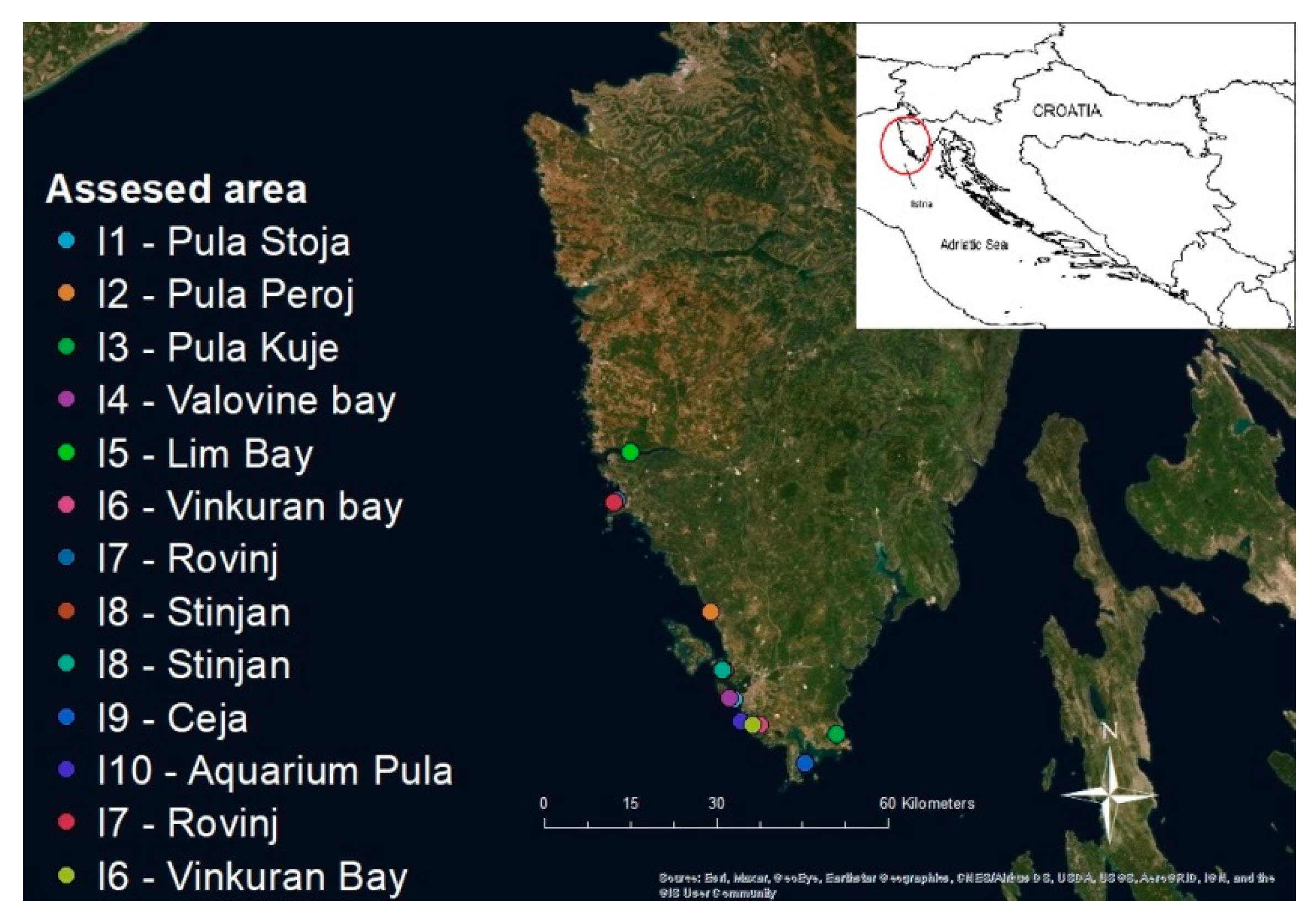
2.1. Sampling Sites and Sample Collection

In this study, we collected noble pen shells from two habitats along the Eastern Adriatic coast: The Mljet National Park (MNP) and the Istrian peninsula. The sampling sites were determined according to the previously collected information on the natural habitats of P. nobilis (Figure 1 and Figure 2; Table 1). Samples of live animals were collected in the MNP by scuba diving during April 2019, and they were delivered to the laboratory under cooling conditions with temperatures not exceeding 8 °C. The other samplings—both along the Istrian peninsula and in the MNP—were also carried out by scuba diving. All the samplings were carried out with the permission of the Croatian Ministry of Environmental Protection and Energy (CLASS UP/1-612-17/18-48/172; No. 517-05-1-1-18-4 of 21 December 2018 and CLASS UP/1-612-07/19-48/193; No. 517-05-1-1-19-3 of 11 September 2019).
During the sampling, the appearance of pen shells in their habitat was evaluated, and portions of digestive glands, gills, mantles, gonads, and muscles were collected separately and preserved in Davidson solution [28] for histological examination and in either EtOH or RNAlater (Thermo Fisher, Waltham, MA, USA) for molecular analysis. Exceptionally, 20 mantle tissue biopsies were taken during September 2019 with a non-lethal sampling method and were preserved in EtOH.

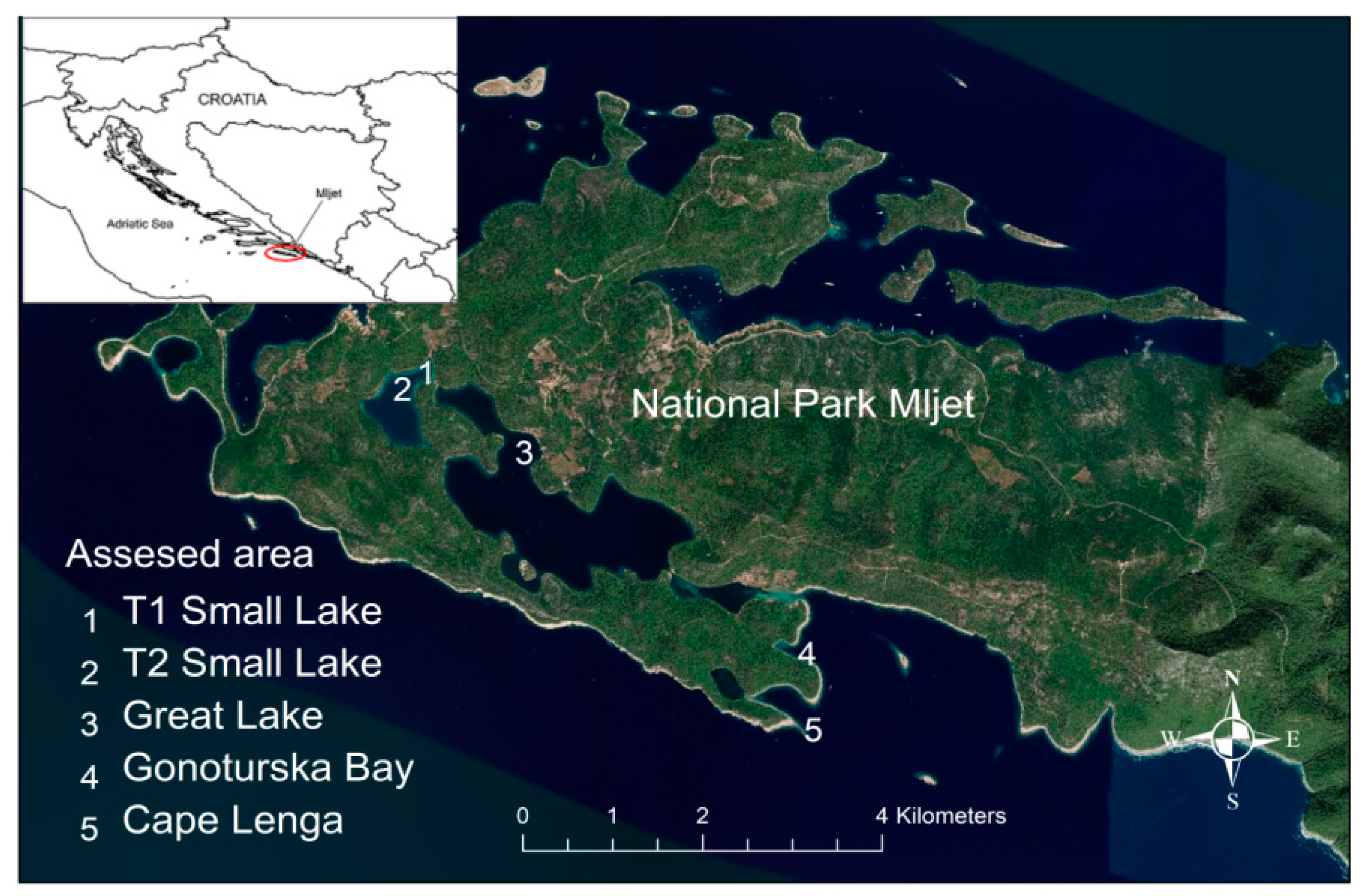
2.2. Site and Design of the Small-Scale Study within the MNP

The study was conducted from April to November of 2019 in the MNP, which is in the south-east Adriatic (Figure 2), a highly productive and biodiverse marine ecosystem within the Natura 2000 European Network of Protected Areas (code HR5000037). Five sampling points were selected according to the most abundant pen shell populations. Two sampling points (1 and 2) were situated in the Small Lake, a lake-like inlet connected by a shallow, narrow channel, while sampling point 3 was in the Big Lake, which was connected to the open sea by a slightly deeper, wider channel. Another two sampling points in addition to these lake-like inlets were Gonoturska Bay (sampling point 4) and Cape Lenga (sampling point 5). The distance between the most external sampling point (5) and the most internal sampling points (1 and 2) was about four kilometers. Based on the discovery of Haplosporidium pinnae at the outer sites in April, a field experiment was set up to determine the pattern and rate of the spread of mortality within this dense population. As it was obvious that MMEs started to spread quickly through the inner part of the MNP, we set up the study with the aim of learning more about the pattern of the spread of the disease.
Two 100 m surveillance sentinel transects—transect 1 (T1) and transect 2 (T2)—were placed in the Small Lake (Figure 2) in September 2019, perpendicularly to the shoreline and extending towards a deeper part of the inlet (Figure 3). The deepest points along the transects were 3.8 m for T1 and 7.3 m for T2. All the clinically healthy pen shells were tagged, and their positions along the transects were recorded within a 2 m corridor on each side of the transect line, covering approximately 400 m2 along each transect. A caliper was used to determine the maximum unburied length (UL) and minimum dorsoventral height or minimum width (w), and the maximum height was determined using the formula Ht = 1.79 w + 0.5 + UL, as described by García-March and Vicente [29]. The data were recorded underwater on a plastic slate, while the depth was determined using a diving computer. Along transect T1, there were 75 individuals with the density of 20.8 per 100 m2, while along T2, there were 106 individuals with a density of 37.83 individuals per 100 m2. Every 2 weeks from September to December 2019, two divers clinically examined all the pen shells along both transects and noted the results of the clinical examinations and the numbers of live specimens. The temperature and the salinity of the sea were measured and noted daily.

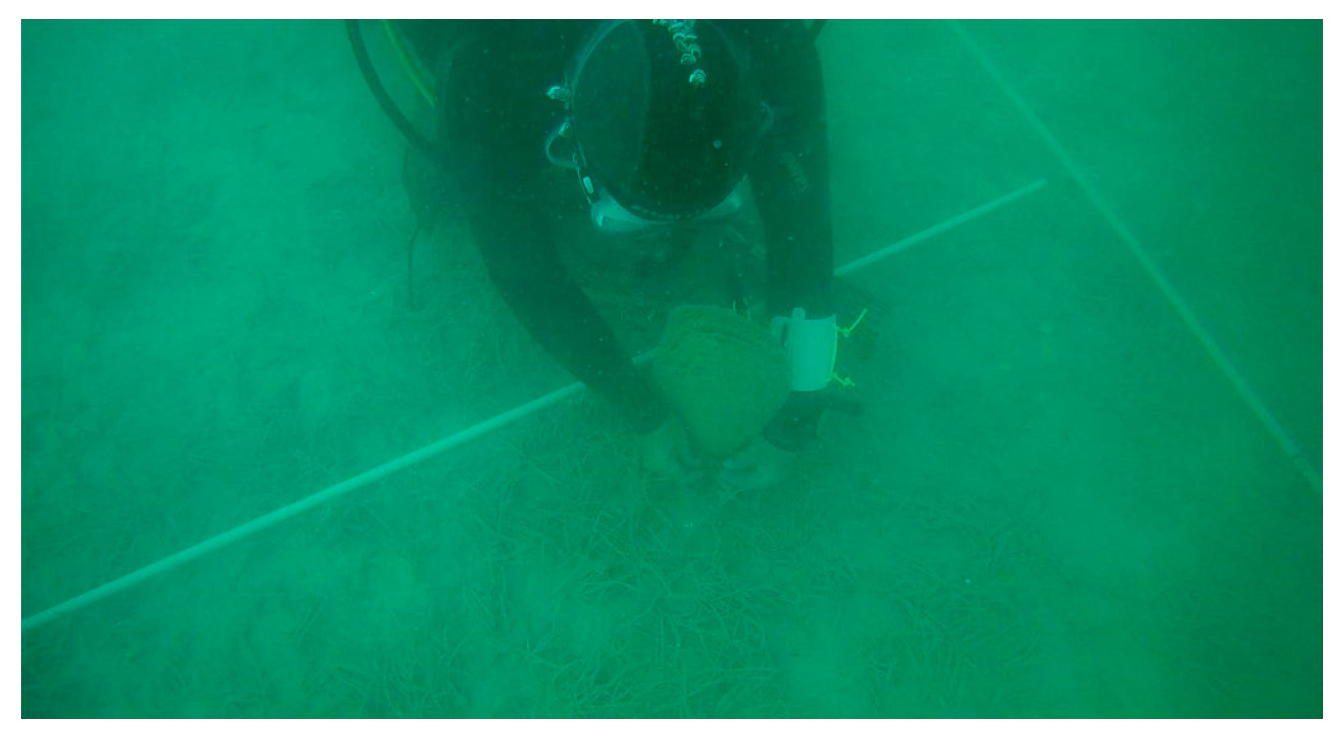
2.3. Non-Lethal Sampling

As the mortality rate within the study area rose to 20% in September, biopsies of the mantle tissue from 20 randomly selected individuals along both transects were taken to assess the non-lethal sampling methodology and to obtain information on the prevalence of infections. The biopsies of the mantles were collected according to the method described by Sanna et al. [30]. Obtaining biopsies did not cause significant damage since the individuals’ valves were held open with a wooden stick (diameter = 0.5 cm), which was placed in proximity (4–5 cm) to the hinge ligament, and about 50 mg of mantle tissue samples were removed using forceps. The stick was then removed, and the tissue sample was stored in a 5 mL tube. Once the diver returned to the surface, the tissue samples were preserved in EtOH and transferred to the laboratory in a refrigerated box.

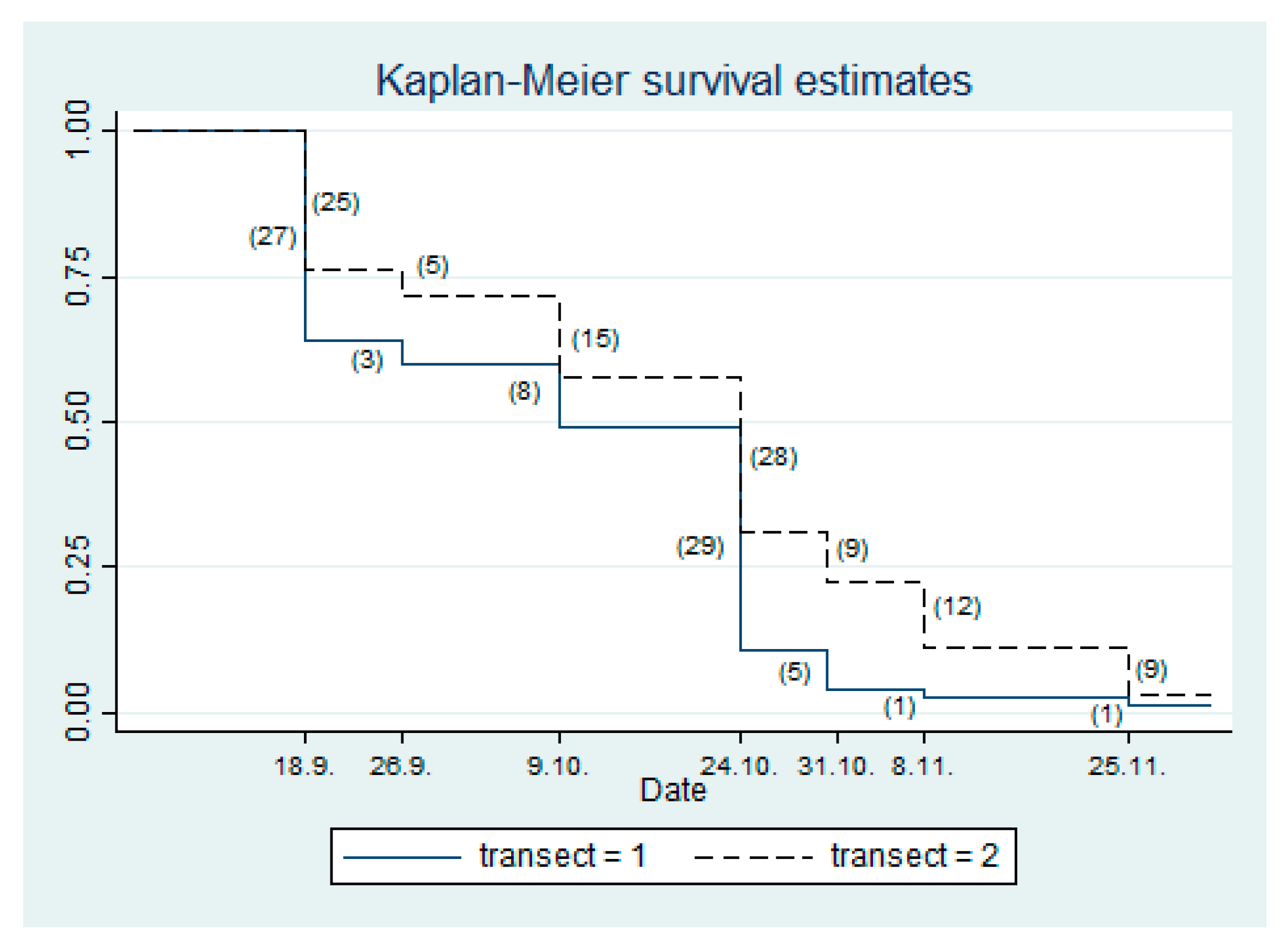
2.4. Statistical Analysis

Differences in the morphometric data between transects were compared with a multivariant regression analysis using STATA 13.1 (Stata, College Station, TX, USA). Survival curves were calculated according to the Kaplan–Meier method [31].

2.5. Laboratory Procedures

The live mollusks that were sent to the laboratory were opened by cutting the adductor muscle according to the methods described by Morton and Puljas [32]. The in situ appearance of the organs was evaluated. Samples of the gills, mantle, digestive gland, gonads, and muscles were collected from each individual for both molecular and histological purposes and were immediately preserved in ethanol or Davidson fixative, respectively [28].

2.6. Histological Analysis

Samples of the organs fixed in Davidson solution were processed for histological examination. Tissues were dehydrated with an ethanol series, cleared in xylene, embedded in paraffin, sectioned at 3 μm, and mounted on Microme EC 350-2 slides (Thermo Scientific, Waltham, MA, USA). Mounted slides were heated to 60 °C, deparaffinized in xylene, and rehydrated in an ethanol series and distilled water, followed by staining with hematoxylin and eosin (H&E, Harris).

2.7. DNA Extraction

DNA was extracted from approximately 25 mg of the gill, mantle, digestive gland, gonads, and muscle tissues, respectively, which were preserved in either EtOH or RNAlater, using a MagMAX CORE Nucleic Acid Purification Kit (Applied Biosystems, Foster City, CA, USA) according to the manufacturer’s instructions on a KingFisher Duo Prime Purification System (Thermo Scientific, USA). The extracted DNA was stored at −20 °C until the analysis.

2.8. PCR for Detection of Haplosporidium spp.

For the detection of Haplosporidium spp., the Haplosporidium-specific primers described by Renault et al. [33] were used. We used 2 µL of extracted DNA and 0.5 µM of the primers HAP-F1 (5′-GTTCTTTCWTGATTCTATGMA-3′) and HAP-R2 (5′-GATGAAYAATTGCAATCAYCT-3′). PCR reactions were performed using the GoTaq G2 Hot Start Colorless Master Mix (Promega, Madison, WI, USA) on a ProFlex PCR System (Applied Biosystems, USA) with a final volume of 20 µL. The temperature protocol involved enzyme activation at 95 °C for 2 min, followed by 40 cycles of denaturation at 94 °C for 30 s, primer annealing at 48 °C for 30 s, and elongation at 72 °C for 60 s, and the process was completed with a final elongation step at 72 °C for 5 min. The results of the PCRs were checked through electrophoresis on a QIAxcel system (Qiagen, Hilden, Germany).

2.9. PCR for Detection of Mycobacterium spp.

For the detection of Mycobacterium spp., we used two protocols. For the amplification of a segment of the hsp65 gene, we used the primers Tb11 (5′-ACCAACGATGGTGTGTCCAT-3′) and Tb12 (5′-CTTGTCGAACCGCATACCCT-3′), which were first described by Telenti et al. [34]. The thermal protocol involved enzyme activation at 95 °C for 2 min, followed by 45 cycles of denaturation at 94 °C for 60 s, primer annealing at 60 °C for 60 s, and elongation at 72 °C for 60 s, and the process was completed with a final elongation step at 72 °C for 5 min. For the amplification of a segment of the mycobacterial 16S rRNA gene, we used the primers 246 (5′-AGAGTTTGATCCTGGCTCAG-3′) and 264 (5′-TGCACACAGGCCACAAGGGA-3′), which were first described by Böddinghaus et al. [35]. The thermal protocol was the same as that with the first set of primers, except for the annealing temperature, which was 55 °C. All the PCR reactions were performed using the GoTaq G2 Hot Start Colorless Master Mix (Promega) on a ProFlex PCR System (Applied Biosystems). For each reaction, we used 2 µL of extracted DNA and 0.5 µM primers with a final volume of 20 µL.

2.10. Sequencing and Phylogeny

To obtain longer sequences of the haplosporidian 18S rRNA gene for phylogenetic comparisons, for all the samples that tested positive for haplosporidian DNA, we performed PCR with the primers HPN-F1, HPN-F3, HPN-R3, and 16SB, as described by Catanese et al. [7]. All the PCR products obtained were sequenced, and the resulting sequences were aligned and assembled in continuous reads using MAFFT version 7 [36]. For a phylogenetic comparison of Mycobacterium spp., the PCR products obtained using 246–264 primer pairs were sequenced. Direct Sanger sequencing was performed by Macrogen Europe (Amsterdam, The Netherlands). The sequences obtained [37] were identified using BLAST [38]. The sequences were aligned with MAFFT v7.388 [37] with the default parameters. The phylogenetic analysis was performed in MEGA 10.0.5 [39]. The best model was selected based on the lowest BIC score. Maximum likelihood trees were calculated with the following settings: Five discrete gamma categories, use of all sites, SPR level 5 heuristic method with a very strong branch-swap filter, the K2+G substitution model for the haplosporidian tree, and the HKY+G+I substitution model for the Mycobacterium spp. tree. Phylogeny was tested with the bootstrap method with 1000 replications. The obtained tree was visualized in iTOL [40].

3. Results

During the study, the health checks of noble pen shells (Pina nobilis) collected on 15 sampling sites were included, five in the MNP and 10 along the Istrian coast during 2019 and the first half of 2020 (Figure 1 and Figure 2; Table 1).

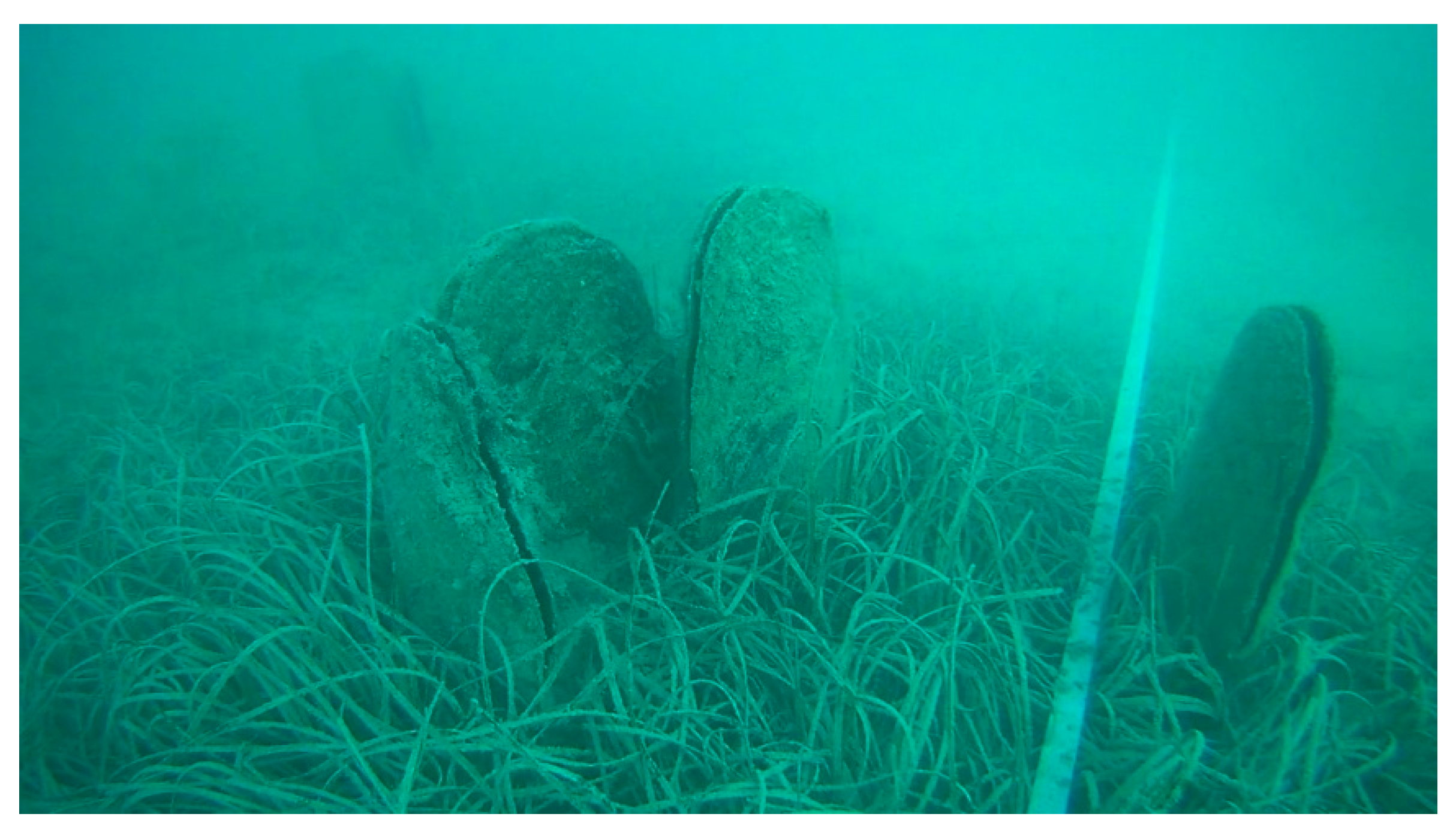
3.1. Description of the Pen Shells Collected in Situ

During the monitoring of the sampling sites included in this study, we observed the different appearances of the present noble pen shells. Healthy individuals, individuals with disease symptoms, and even empty shells were observed (Figure 4). The external symptoms were very scarce, and the affected animals displayed weakened reactions to stimuli, such as gaping (slower closing of shells) after touching or increased drifting and obvious shrinkage of the mantle, causing the illusion of a greater space between the shells than there was. At the sites with high mortalities of pen shells, many empty shells remained in an upward position, causing the illusion that the animals were still alive. In this situation, soft tissues were never found. When the sick animals were dissected, a certain amount of liquid was observed within the shells, and the mantle was darker than usual (Figure 5).

3.2. Results of the Pilot Study (Morphometrics, Environmental Conditions, and Mortality Patterns)

During the first health screening in the MNP, which was carried out in mid-April 2019, a total of six specimens were collected in the sampling points shown in Figure 2. Three of them were collected in the Small Lake (sampling points 1 and 2), and no changes in appearance were observed. However, two out of the three samples tested positive for Mycobacterium spp. The others were collected outside the lake-like inlets, one in Gonoturska (sampling point 4) and two around Cape Lenga (sampling point 5). In Gonoturska (sampling point 4), there were no signs of unusual behavior, and a single sample tested negative for both pathogens. Around Cape Lenga (sampling point 5), a high mortality rate of up to 80% was noticed, which was based on the presence of unresponsive specimens and empty shells. In the other four sampling points, the mortality rate was about 9%, which is considered normal. Laboratory tests revealed the positive discovery of Haplosporidium pinnae in both individuals from sampling point 5—with both histology (Figure 7a–d) and PCR—while both specimens tested negative for Mycobacterium spp. (Table 1). At the beginning of September, a mortality rate of almost 100% was observed in the Big Lake, and a rate of about 20% was observed in the Small Lake. Following the spread of mortality in the Big Lake and when setting up the pilot study, we found that all the individuals were adults, and those along T1 had a mean height of 37.1 cm (±0,65), mean width of 15.28 cm (±0.35), and calculated mean maximum height of 64.56 cm (±1.11). The mean height of the individuals along T2 was 39.19 cm (±0.56), they had a width of 14.77 cm (±0.28), and the calculated maximum height was 65.64 cm (±0.76). The detailed morphometric properties of the individuals marked along both transects did not differ significantly (Supplement 1). At the beginning of the surveillance in April, the temperature of the sea was 15 °C, and it increased over the following months, reaching 27 °C in August, and slowly decreased over the study period from 25 °C at the beginning of September to 10 °C in December. During the whole study period, the salinity had an average value of 37.98%—ranging from 35.30 to 39.20%—on the surface or 38.04%—ranging from 35.30 to 39.00%—at a depth of 2 m.
The mortality rate during April 2019 at sampling point 5 was estimated to be 80%, and in the rest of the study area, it was estimated to be about 9% based on the number of empty shells (Figure 2). During the control diving on August 1, the mortality in the Big Lake was about 20%, and at the beginning of September, 100% mortality was observed in the Great Lake, also in addition to the increased mortality in the Small Lake (Table 2). At the time of setting up the transects, along T1, there were 62 dead individuals out of a total of 137 (Mt = 45.26%), and along T2, there were 28 dead individuals out of 134 (Mt = 20.90%). Over the study period, the mortality rate increased, resulting in the final survival of one individual from the T1 and three individuals from T2.
The Kaplan–Meier survival curves indicate two mass mortality events during the study period: The first was between 4 and 18 September, when 52 animals died, and the second was between 9 and 24 October 2019, when 57 animals died (Figure 6 and Table 2). By 25 November 2019, one of the initial 75 pen shells were alive along T1, corresponding to a survival rate of 1.33%, while three of the initial 106 pen shells were alive along T2, corresponding to a survival rate of 2.83%. The overall survival rate was 2.21%.
During the clinical checks, we observed that the mortalities affected certain individuals and spread diffusely to others in the close vicinity, affecting primarily those in the shallower area of the Small Lake and, later, those in the deeper parts.

3.3. Necropsy and Histological Findings

Six live animals were sacrificed for health evaluation, and organs from 37 additional animals were collected for molecular analysis (Table 1). Live animals were collected before the first notification of the MME, while the organs were collected prior, during, and after the events. On dissection, two of the animals collected in April showed a darker mantle and reduced soft tissues (Figure 5). The results of the histological analyses in the infected animals disclosed moderate to heavy infiltration of hemocytes into the connective tissues of the digestive gland and gonads, as well as light necrosis in the connective tissues of the gills, digestive gland, and muscle (Figure 7a). Uni-nucleated Haplosporidium-like parasite stages were observed in the connective tissues of the mantle (Figure 7b) and musculature (Figure 7c). Different Haplosporidium-like parasite stages, such as uni- or binucleate stages and plasmodia, were present in the connective tissues and epithelia of the digestive glands (Figure 7d,e). Intra-hemocytic stages of uninucleate cells were observed in the heavily infected specimens, in which sporogonia were observed in the epithelia of the digestive gland (Figure 7e,f). However, very few specimens were analyzed using histology, and sporulation was observed in the digestive gland in the sample collected in May 2020 in Vinkuranska vala in the Istrian peninsula.

3.4. Molecular Analysis for the Presence of Haplosporidium spp. and Mycobacterium spp.

Eleven out of the 43 animals tested positive for Haplosporidium spp. (Table 1). Since the DNA was isolated from the digestive glands, mantles, gonads, and muscles of the infected animals, mostly the digestive glands and gonads, followed by muscles and, in a few animals, mantles were positive for Haplosporidium spp. Here, it should be emphasized that four biopsies of mantles tested positive for Haplosporidium spp., as well. Furthermore, eight out of 43 samples tested positive for Mycobacterium spp. As in the case of Haplosporidium spp., two of the positive cases were found with biopsies.

3.4.1. Sequencing of Haplosporidium spp. Isolates

All the Haplosporidium-positive PCR products (1451 bp) were successfully sequenced, and since the obtained sequences did not differ, only one haplotype was deposited in the GenBank (accession number MT367896). The sequences of the Croatian isolates (Figure 8) showed 99.8% identity with the Italian isolates deposited by Scarpa et al. [23] and Tiscar et al. (unpublished data) and 99.7% with the Spanish isolates [8], confirming the presence of Haplosporidium pinnae in all the positive noble shells.

3.4.2. Sequencing of Mycobacterium spp. Isolates

In addition, all the PCR products that tested positive for Mycobacterium spp. (985 bp) were sequenced, and they were identical. For this reason, only one haplotype was deposited in the GenBank (accession number MT367873). The obtained sequence was identical to the Italian one (Figure 9) obtained by Carella et al. [10].

4. Discussion

The results of this study are in line with the results obtained by other Croatian researchers [14,15] and clearly show that MMEs have spread from the south to the middle parts of the Eastern Adriatic regions, as well as, ultimately, to the north. We first detected Haplosporidium pinnae in association with mortalities on the Croatian Adriatic coast in individuals collected in mid-April 2019 on the outer side of the Mljet Island, situated on the most southern point. At the same time, there were no mortality events among populations in the inner parts of the Mljet Island, and all the sampled animals tested negative for H. pinnae, but two individuals from the Small Lake tested positive for Mycobacterium spp., as shown in Table 1. Then, in May, mortalities and detection of samples from Elafiti Island (which is a bit more to the north) that were positive for H. pinnae and Mycobacterium spp. were reported [14]. During the summer period, mortalities were reported at different sites along the coast and close to the islands, mostly in the southern and middle parts of the Eastern Adriatic coast [15]. Interestingly, the data from the mentioned reports, as well as notifications from fishermen, divers, and other parties, indicated that the mortalities primarily affected habitats on the outer parts of the islands and were spreading towards sites that were closer to the mainland—from southern sites to northern sites. In 2019, there were no reports of mortalities in the northern part of the Eastern Adriatic coast, around the Istrian peninsula. The results of the laboratory analysis of the sampled animals supported the absence of both pathogens in the northern regions. Unfortunately, less than half a year later, during May 2020, the pathogens reached the northern sites along the Istrian peninsula and caused high mortalities of the natural populations (Table 1), as indicated by the diagnosis with H. pinnae and Mycobacterium spp.
Generally, it seems that the mortalities were triggered by an increase in the temperature in the spring, and they reached a high intensity when the sea had a high temperature during the summer. The results of the pilot study undertaken in the Small Lake of the Mljet National Park (Figure 2) support the hypothesis that the increase in mortalities was influenced by high temperatures. The MMEs did not affect populations in the inner parts of the lakes of the National Park from April until the beginning of August, when high mortalities were observed in the Big Lake. This was strange since the distance from the site of the first record of H. pinnae at Cape Lenga (Figure 2, sampling point 5) is about 4 km, and the water enters into the lakes from the open sea. At the same time, the MMEs spread to more distant sites in the Middle Adriatic. It seems that the inlet into the lakes does not allow the entry of large quantities of water, which also creates a barrier to the entry of pathogens and causes them to proliferate slowly. Most likely, the high temperature of the sea, which reached its maximum in August, strongly supported the extremely quick spread of the pathogen in the Big Lake and also affected the more isolated population in the Small Lake. The observations from the pilot study indicate that some regularity exists in the spread of the disease. The mortalities of individuals along T1, which was closer to the inlet of the water from the Big Lake, occurred earlier than those along T2. It was also observed that the spread of the disease was not linear, but rather diffuse, affecting primarily weaker individuals and spreading to others in close proximity in the dense population. A higher mortality rate was also observed in the shallower parts of the studied area. The difference related to the depth might be attributed to the higher water temperature in the shallower areas and the depletion of oxygen, which could additionally worsen the environmental conditions for the host. Additionally, it seems that higher water temperatures favor the propagation of both of the detected pathogens. Mycobacterium spp. require a higher water temperature for growth, as it is known that a representative of this genus, Mycobacterium marinum, a pathogen of marine fish, usually causes disease during warmer parts of the year with water temperatures above 25 °C [41]. Moreover, Mycobacterium spp. are opportunistic bacteria that cause chronic diseases, and it should be possible to detect their presence in healthy individuals long before they cause granulomatous lesions, as observed by Carella et al. [10] and Latos et al. [12]. Unfortunately, we analyzed very few samples using histology, and in our scarce histological slides, we did not observe the lesions described by the aforementioned authors [10,12]. Interestingly, in the samples of mantle biopsies collected during the pilot study at the end of August, we detected H. pinnae in four out of the 20 tested animals and Mycobacterium spp. in two out the 20 animals, which could suggest that the parasite was more representative and more responsible for the MMEs compared to Mycobacterium spp. It should be emphasized that the pen shells in the Small Lake of the Mljet National Park did not show increased mortality for 6 months, although it was confirmed that they were infected with Mycobacterium spp. in April. Infection with Mycobacterium spp. does not appear to be sufficient to trigger a mass mortality event among pen shells, but its co-occurrence with H. pinnae contributes to high mortality. Similarly, Box et al. [42] concluded that high mortalities occur in noble pen shells that have been infected with H. pinnae and additionally aggravated by co-infection with Mycobacterium spp. or other Gram-negative bacteria.
From the results of morphometry, it was obvious that all the marked animals were adults, with the smallest reaching a maximum height of 37.9 cm. It seems that the older individuals were more susceptible to infection with H. pinnae, as previously reported by Vázquez-Luis et al. [7], as well as to cases of infection with other genera of Haplosporidium, where mortalities occurred in older individuals concurrently with the highest levels of infection [43].
The peak prevalence of the mortalities of Eastern oyster (Crassostrea virginica) caused by Haplosporidium nelsoni in Delaware Bay, USA [43] was reported in autumn. The mortalities observed in the pilot study with the pen shells peaked during the autumn and slowly decreased during the winter, similarly to those caused by H. nelsoni. From the Kaplan–Meier survival estimates (Figure 6), it was visible that the mortalities increased gradually and that the most numerous mortalities occurred at the end of September and again at the end of October. Unfortunately, only four animals survived until the end of November. Undoubtedly, the pathogens identified in our research were those detected as the cause of mortalities throughout the Mediterranean region. The phylogenetic analysis of our isolates of H. pinnae showed 99.8% similarity (Figure 8) with those described in the Ionian Sea [9] and 99.7% similarity with Spanish isolates [8]. The phylogenetic analysis showed that the 16S sequences of mycobacteria detected in P. nobilis were more distant from each other than some sequences of mycobacteria belonging to different species. For example, M. sherrisii, M. stomatepiae, M. florentinum, M. triplex, and Mycobaterium spp. from P. nobilis from Croatia (MT367873) and Italy (MH569646) are more similar to each other than the two strains of Mycobaterium spp. from P. nobilis from Italy (MH569647 and MH569649) (Figure 9). This suggests that the detected mycobacteria from P. nobilis belong to a heterogeneous group and are not members of the same species.
There are still doubts about what enabled the quick spread of the pathogen through the Mediterranean basin. It was undoubtable that the pathogen entered into the Adriatic Sea and spread from the south to the north by following the main flows of the currents [44]. It is hard to believe that only water currents carry the pathogen, and the pathogen could probably spread through pelagic larval stages of other intermediate hosts. Nevertheless, the histological finding of all the stages of H. pinnae in the same animal—uninucleate cells observed in the mantle, gills, gonads, digestive gland, and connective tissue, followed by plasmodia in the connective tissue of the digestive gland, and sporocysts in the epithelium of the digestive tubules (Figure 7a–f)—contributed to the direct transmission from one to another individual, as postulated by Catanese et al. [8]. The quick spread of mortalities within the dense population in the pilot study in the Mljet National Park suggests the direct transmission of H. pinnae. Since it is known that spores of Haplosporidium are persistent in the environment [45], the involvement of an intermediate host that is abundant in the marine environment and is capable of quick movement through water should be considered, and future studies should aim to prove this hypothesis.
Further joint efforts of marine biologists and invertebrate health experts should be engaged in aiming to understand why only adult specimens were present in particular marine areas, and the reasons were for the increased susceptibility of the noble pen shells to pathogens.

Supplementary Materials

The following are available online at https://www.mdpi.com/article/10.3390/jmse9070764/s1. Table S1: Morphometric characteristics of noble pen shells along the transect 1 (T1); Table S2: Morphometric characteristics of noble pen shells along the transect 2 (T2).

Author Contributions

Conceptualization, Ž.M.; data curation, M.B.; formal analysis, Ž.P. and I.G.Z.; funding acquisition, B.H.; investigation, A.P., O.P., and I.S.; methodology, D.O.; writing—original draft, S.Z. All authors have read and agreed to the published version of the manuscript.

Funding

This research received no external funding.

Institutional Review Board Statement

All the sampling was carried out with the permission of the Ministry of Environmental Protection and Energy (CLASS: UP/1-612-17/18-48/172, No. 517-05-1-1-18-4 issued on 21 December 2018 and CLASS: UP/1-612-07/19-48/193, No. 517-05-1-1-19-3 issued on 11 September 2019).

Data Availability Statement

All the data are available upon reasonable request from the corresponding author.

Acknowledgments

The authors kindly acknowledge Angela Bradarić for the technical support in the field and the laboratory work, Pavel Ankon for the instructions on non-lethal sampling, and Ana Car and Iris Duplić Radić for sharing the salinity data in the Small Lake during the pilot study. Special thanks go to Isabelle Arzul and the EURL for mollusk diseases (IFREMER, La Tremblade) for their confirmation of the detection of Haplosporidium pinnae.

Conflicts of Interest

The authors declare no conflict of interest.

References

  1. Zavodnik, D.; Hrs-Brenko, M.; Legas, M. Synopsis on the fan shell Pinna nobilis L. in the eastern Adriatic Sea. Les Espèces Marines à protegér en Méditerranée. Gis Posidonie. Publ. Marseille 1991, 169–178. [Google Scholar]
  2. Cabanellas-Reboredo, M.; Vazquez-Luis, M.; Mourre, B.; Álvarez, E.; Deudero, S.; Amores, Á.; Addis, P.; Bal-lesteros, E.; Barrajon, A.; Coppa, S.; et al. Tracking a mass mortality outbreak of pen shell Pinna nobilis populations: A collaborative effort of scientists and citizens. Sci. Rep. 2019, 9, 1–11. [Google Scholar] [CrossRef] [Green Version]
  3. Tsatiris, A.; Papadopoulos, V.; Makri, D.; Topouzelis, K.; Manoutsoglou, E.; Hasiotis, T.; Katsanevakis, S. Spatial distribution, abundance and habitat use of the endemic Mediterranean fan mussel Pinna nobilis in Gera Gulf, Lesvos (Greece): Comparison of design-based and model-based approaches. Mediterr. Mar. Sci. 2018, 19, 642–655. [Google Scholar] [CrossRef] [Green Version]
  4. Katsanevakis, S. Growth and mortality rates of the fan mussel Pinna nobilis in Lake Vouliagmeni (Korin-thiakos Gulf, Greece): A generalized additive modelling approach. Mar. Biol. 2007, 152, 1319–1331. [Google Scholar] [CrossRef]
  5. Richardson, C.A.; Peharda, M.; Kennedy, H.; Kennedy, P.; Onofri, V. Age, growth rate and season of recruitment of Pinna nobilis (L) in the Croatian Adriatic determined from Mg:Ca and Sr:Ca shell profiles. J. Exp. Mar. Biol. Ecol. 2004, 299, 1–16. [Google Scholar] [CrossRef]
  6. Deudero, S.; Grau, A.; Vázquez-Luis, M.; Álvarez, E.; AlOmar, C.; Hendriks, I.E. Reproductive investment of the pen shell Pinna nobilis Linnaeus, 1758 in Cabrera National Park (Spain). Mediterr. Mar. Sci. 2017, 18, 271. [Google Scholar] [CrossRef] [Green Version]
  7. Vázquez-Luis, M.; Álvarez, E.; Barrajón, A.; García-March, J.R.; Grau, A.; Hendriks, I.E.; Jiménez, S.; Kersting, D.; Moreno, D.; Pérez, M.; et al. S.O.S. Pinna nobilis: A Mass Mortality Event in Western Mediterranean Sea. Front. Mar. Sci. 2017, 4, 220. [Google Scholar] [CrossRef] [Green Version]
  8. Catanese, G.; Grau, A.; Valencia, J.M.; March, J.R.G.; Vázquez-Luis, M.; Alvarez, E.; Deudero, S.; Darriba, S.; Carballal, M.J.; Villalba, A. Haplosporidium pinnae sp. Nov., a haplosporidan parasite associated with mass mortalities of the fan mussel, Pinna nobilis, in the Western Mediterranean Sea. J. Invertebr. Pathol. 2018, 157, 9–24. [Google Scholar] [CrossRef]
  9. Panarese, R.; Tedesco, P.; Chimienti, G.; Latrofa, M.S.; Quaglio, F.; Passantino, G.; Buonavoglia, C.; Gustinelli, A.; Tursi, A.; Otranto, D. Haplosporidium pinnae associated with mass mortality in endangered Pinna nobilis (Linnaeus 1758) fan mussels. J. Invertebr. Pathol. 2019, 164, 32–37. [Google Scholar] [CrossRef]
  10. Carella, F.; Aceto, S.; Pollaro, F.; Miccio, A.; Iaria, C.; Carrasco, N.; Prado, P.; De Vico, G. A mycobacterial disease is associated with the silent mass mortality of the pen shell Pinna nobilis along the Tyrrhenian coast-line of Italy. Sci. Rep. 2019, 9, 2725. [Google Scholar] [CrossRef]
  11. Katsanevakis, S. The cryptogenic parasite Haplosporidium pinnae invades the Aegean Sea and causes the collapse of Pinna nobilis populations. Aquat. Invasions 2019, 14, 150–164. [Google Scholar] [CrossRef]
  12. Lattos, A.; Giantsis, I.A.; Karagiannis, D.; Michaelidis, B. First detection of the invasive Haplosporidian and Mycobacteria parasites hosting the endangered bivalve Pinna nobilis in Thermaikos Gulf, North Greece. Mar. Environ. Res. 2020, 155, 104889. [Google Scholar] [CrossRef] [PubMed]
  13. Tiscar, P.G.; Rubino, F.; Fanelli, G.; Paoletti, B.; Della Salda, L. Mass Mortality of the Fan Mussel Pinna nobilis in Apulia (Ionian Sea) Caused by Haplosporidium pinnae. In Proceedings of the 42nd CIESM Congress, Cascais, Portugal, 7–11 October 2019. [Google Scholar]
  14. Čižmek, H.; Čolić, B.; Gračan, R.; Grau, A.; Catanese, G. An emergency situation for pen shells in the Mediterranean: The Adriatic Sea, one of the last Pinna nobilis shelters, is now affected by a mass mortality event. J. Invertebr. Pathol. 2020, 173, 107388. [Google Scholar] [CrossRef] [PubMed]
  15. Šarić, T.; Župan, I.; Aceto, S.; Villari, G.; Palić, D.; De Vico, G.; Carella, F. Epidemiology of Noble Pen Shell (Pinna nobilis L. 1758) Mass Mortality Events in Adriatic Sea Is Characterised with Rapid Spreading and Acute Disease Progression. Pathogens 2020, 9, 776. [Google Scholar] [CrossRef] [PubMed]
  16. Künili, I.E.; Gürkan, S.E.; Aksu, A.; Turgay, E.; Çakir, F.; Gürkan, M.; Altinağaç, U. Mass mortality in endangered fan mussels Pinna nobilis (Linnaeus 1758) caused by co-infection of Haplosporidium pinnae and multiple Vibrio infection in Çanakkale Strait, Turkey. Biomarkers 2021, 26, 450–461. [Google Scholar] [CrossRef]
  17. International Union for Conservation of Nature (IUCN). Mediterranean Noble Pen Shell Crisis (Pinna nobilis)-January 2020 Update. Available online: https://www.iucn.org/news/mediterranean/202001/mediterranean-noble-pen-shell-crisis-pinna-nobilis-january-2020-update (accessed on 10 March 2021).
  18. Kersting, D.; Benabdi, M.; Čižmek, H.; Grau, A.; Jimenez, C.; Katsanevakis, S.; Öztürk, B.; Tuncer, S.; Tunesi, L.; Vázquez-Luis, M.; et al. Pinna Nobilis. The IUCN Red List of Threatened Species 2019; IUCN Red List: London, UK, 2019. [Google Scholar] [CrossRef]
  19. Pavlinec, Ž.; Zupičić, I.G.; Oraić, D.; Petani, B.; Mustać, B.; Mihaljević, Ž.; Beck, R.; Zrnčić, S. Assessment of predominant bacteria in noble pen shell (Pinna nobilis) collected in the Eastern Adriatic Sea. Environ. Monit. Assess. 2020, 192, 1–10. [Google Scholar] [CrossRef]
  20. Carella, F.; Antuofermo, E.; Farina, S.; Salati, F.; Mandas, D.; Prado, P.; Panarese, R.; Marino, F.; Fiocchi, E.; Pretto, T.; et al. In the wake of the ongoing mass mortality events: Co-occurrence of Mycobacterium, Haplosporidium and other pathogens in Pinna nobilis collected in Italy and Spain (Mediterranean Sea). Front. Mar. Sci. 2020, 7, 48. [Google Scholar] [CrossRef] [Green Version]
  21. Prado, P.; Carrasco, N.; Catanese, G.; Grau, A.; Cabanes, P. Presence of Vibrio mediterranei associated to major mortality in stabled individuals of Pinna nobilis L. Aquaculture 2020, 519, 734899. [Google Scholar] [CrossRef]
  22. Lattos, A.; Bitchava, K.; Giantsis, I.; Theodorou, J.; Batargias, C.; Michaelidis, B. The Implication of Vibrio Bacteria in the Winter Mortalities of the Critically Endangered Pinna nobilis. Microorganisms 2021, 9, 922. [Google Scholar] [CrossRef]
  23. Scarpa, F.; Sanna, D.; Azzena, I.; Mugetti, D.; Cerruti, F.; Hosseini, S.; Cossu, P.; Pinna, S.; Grech, D.; Cabana, D.; et al. Multiple Non-Species-Specific Pathogens Possibly Triggered the Mass Mortality in Pinna nobilis. Life 2020, 10, 238. [Google Scholar] [CrossRef]
  24. Lattos, A.; Giantsis, I.A.; Karagiannis, D.; Theodorou, J.A.; Michaelidis, B. Gut Symbiotic Microbial Communities in the IUCN Critically Endangered Pinna nobilis Suffering from Mass Mortalities, Revealed by 16S rRNA Amplicon NGS. Pathogens 2020, 9, 1002. [Google Scholar] [CrossRef]
  25. Orepić, N.; Vidmar, J.; Zahtila, E.; Zavodnik, D. A marine benthos survey in the lakes of the National park Mljet (Adriatic Sea). Period. Biol. 1997, 99, 229–245. [Google Scholar]
  26. Šiletić, T.; Peharda, M. Population study of the fan shell Pinna nobilis L. in Malo and Veliko Jezero of the Mljet National Park (Adriatic Sea). Sci. Mar. 2003, 67, 91–98. [Google Scholar] [CrossRef] [Green Version]
  27. Peharda, M.; Hrs-Brenko, M.; Bogner, D.; Onofri, V.; Benović, A. Spatial distribution of live and dead bivalves in saltwater lake Malo Jezero (Mljet National Park). Period. Biol. 2002, 104, 115–122. [Google Scholar]
  28. Howard, D.; Lewis, E.; Keller, J.; Smith, C. Histological Techniques for Marine Bivalve Mollusks and Crustaceans, 2nd ed.; NOAA (National Ocean Service): Oxford, MD, USA, 2004; 60p. [Google Scholar]
  29. Garcia-March, J.R.; Vicente, N. Protocol to Study and Monitor Pinna nobilis Populations within Marine Protected Areas. Report of the Malta Environment and Planning Authority (MEPA) and MedPAN-Interreg IIIC-Project. 2006. Available online: https://drive.google.com/file/d/131bRQrcQQ62NUep1V1J3f_W33JAmANbO/view (accessed on 5 September 2019).
  30. Sanna, D.; Cossu, P.; Dedola, G.L.; Scarpa, F.; Maltagliati, F.; Castelli, A.; Franzoi, P.; Lai, T.; Cristo, B.; Curini-Galletti, M.; et al. Mitochondrial DNA Reveals Genetic Structuring of Pinna nobilis across the Mediterranean Sea. PLoS ONE 2013, 8, e67372. [Google Scholar] [CrossRef] [PubMed] [Green Version]
  31. Dudley, W.N.; Wickham, R.; Coombs, M.N. An Introduction to Survival Statistics: Kaplan-Meier Analysis. J. Adv. Pr. Oncol. 2016, 7, 91–100. [Google Scholar] [CrossRef] [Green Version]
  32. Morton, B.; Puljas, S. An improbable opportunistic predator: The functional morphology of Pinna nobilis (Bivalvia: Pterioida: Pinnidae). J. Mar. Biol. Assoc. UK 2019, 99, 359–373. [Google Scholar] [CrossRef]
  33. Renault, T.; Stokes, N.; Chollet, B.; Cochennec, N.; Berthe, F.; Gerard, A.; Burreson, E. Haplosporidiosis in the Pacific oyster Crassostrea gigas from the French Atlantic coast. Dis. Aquat. Org. 2000, 42, 207–214. [Google Scholar] [CrossRef]
  34. Telenti, A.; Marchesi, F.; Balz, M.; Bally, F.; Böttger, E.C.; Bodmer, T. Rapid identification of mycobacteria to the species level by polymerase chain reaction and restriction enzyme analysis. J. Clin. Microbiol. 1993, 31, 175–178. [Google Scholar] [CrossRef] [PubMed] [Green Version]
  35. Böddinghaus, B.; Rogall, T.; Flohr, T.; Blöcker, H.; Böttger, E.C. Detection and identification of mycobacteria by amplification of rRNA. J. Clin. Microbiol. 1990, 28, 1751–1759. [Google Scholar] [CrossRef] [PubMed] [Green Version]
  36. Katoh, K.; Standley, D.M. MAFFT Multiple Sequence Alignment Software Version 7: Improvements in Performance and Usability. Mol. Biol. Evol. 2013, 30, 772–780. [Google Scholar] [CrossRef] [Green Version]
  37. Benson, D.A.; Cavanaugh, M.; Clark, K.; Mizrachi, I.K.; Lipman, D.J.; Ostell, J.; Sayers, E.W. GenBank. Nucleic Acids Res. 2012, 41, D36–D42. [Google Scholar] [CrossRef] [PubMed] [Green Version]
  38. Altschul, S.F.; Gish, W.; Miller, W.; Myers, E.W.; Lipman, D.J. Basic local alignment search tool. J. Mol. Biol. 1990, 215, 403–410. [Google Scholar] [CrossRef]
  39. Kumar, S.; Stecher, G.; Li, M.; Knyaz, C.; Tamura, K. MEGA X: Molecular evolutionary genetics analysis across computing platforms. Mol. Biol. Evol. 2018, 35, 1547–1549. [Google Scholar] [CrossRef] [PubMed]
  40. Letunic, I.; Bork, P. Interactive Tree of Life (iTOL) v4: Recent updates and new developments. Nucleic Acids Res. 2019, 47, W256–W259. [Google Scholar] [CrossRef] [PubMed] [Green Version]
  41. Hashish, E.; Merwad, A.-R.; Elgaml, S.; Amer, A.; Kamal, H.; Elsadek, A.; Marei, A.; Sitohy, M. Mycobacterium marinum infection in fish and man: Epidemiology, pathophysiology and management; a review. VetIQ Petcare 2018, 38, 35–46. [Google Scholar] [CrossRef] [PubMed] [Green Version]
  42. Box, A.; Capó, X.; Tejada, S.; Catanese, G.; Grau, A.; Deudero, S.; Sureda, A.; Valencia, J.M. Reduced Antioxidant Response of the Fan Mussel Pinna nobilis Related to the Presence of Haplosporidium pinnae. Pathogens 2020, 9, 932. [Google Scholar] [CrossRef]
  43. Arzul, I.; Carnegie, R.B. New perspective on the haplosporidian parasites of molluscs. J. Invertebr. Pathol. 2015, 131, 32–42. [Google Scholar] [CrossRef] [Green Version]
  44. Mihanović, H.; Cosoli, S.; Vilibić, I.; Ivanković, D.; Dadić, V.; Gačić, M. Surface current patterns in the northern Adriatic extracted from high-frequency radar data using self-organizing map analysis. J. Geophys. Res. Space Phys. 2011, 116, 08033. [Google Scholar] [CrossRef] [Green Version]
  45. Haskin, H.H.; Andrews, J.D. Uncertainties and Speculations about the Life Cycle of the Eastern Oyster Pathogen Haplosporidium nelsoni (MSX). In Disease Processes in Marine Bivalve Molluscs; American Fisheries Society: Bethesda, MD, USA, 1988; Available online: https://scholarworks.wm.edu/cgi/viewcontent.cgi?article=1117&context=vimsbooks (accessed on 5 March 2021).
Figure 1. Sampling sites along the Istrian peninsula in the northern part of the Eastern Adriatic coast.
Jmse 09 00764 g001
Figure 2. Study site within the Mljet National Park with five sampling points and two transect areas.
Jmse 09 00764 g002
Figure 3. Labeling healthy individuals along the transect.
Jmse 09 00764 g003
Figure 4. Clinical inspection of the live noble pen shells in situ, where both healthy and sick animals were in an upward position. Differentiation between them was made by checking their reactions to stimuli. The healthy specimens quickly closed their shells.
Jmse 09 00764 g004
Figure 5. Darker mantle and shrunken soft tissues in a noble pen shell infected by Haplosporidium pinnae.
Jmse 09 00764 g005
Figure 6. Kaplan–Meier survival estimates.
Jmse 09 00764 g006
Figure 7. Histological findings in the organs of noble pen shells: Moderate infiltration of hemocytes into the connective tissue of the digestive gland (a); uni- and binucleate cells of H. pinnae in the connective tissue of the mantle (b); uninucleate cells of H. pinnae in the muscle (c); uninucleate cells and plasmodia in the connective tissue of the digestive gland (d); intra-hemocytic stages of uninucleate cells in the connective tissue of the digestive gland (e); sporulation stages of H. pinnae in the epithelium of the digestive gland with a disruption of the digestive tubules (f).
Jmse 09 00764 g007
Figure 8. Maximum likelihood phylogenetic tree for haplosporidians. The numbers on the branches represent bootstrap values.
Jmse 09 00764 g008
Figure 9. Maximum likelihood phylogenetic tree for Mycobacterium spp. The numbers on the branches represent bootstrap values.
Jmse 09 00764 g009
Table 1. List of sampling points and samples collected during 2019 and 2020, which were analyzed using PCR for detection of Haplosporidium spp. (Haplo) and Mycobacterium spp. (Myco) and using histology.
Date of Sampling
(D/M/Y)
Name and Number of the SiteSea Temp.
°C
SampleResults of Laboratory Analysis
PCR HaploPCR MycoHistology
19/04/19MNP1—Small Lake 17.2Whole animal0/32/3neg
19/04/19MNP5—Cape Lenga 15.9Whole animal2/20/2Haplo +
19/04/19MNP4—Gonoturska 15.9Whole animal0/10/1neg
04/09/19I1—Pula-Stoja 24.5Organs in EtOH *0/30/3n/d
04/09/19I2—Pula-Peroj 24.8Organs in EtOH0/10/1n/d
04/09/19I3—Pula-Kuje 25.2Organs in EtOH0/10/1n/d
24/09/19MNP1 Mljet-Small Lake 28Mantles in EtOH2/41/4n/d
30/09/19MNP1 Mljet-Small Lake 26Mantles in EtOH2/161/16n/d
13/01/20I4—Uvala Valovine 12.3Organs in EtOH0/10/1n/d
13/01/20I5—Limski Bay12.8Organs in RNAlater0/10/1n/d
18/02/20I6—Vinkuran Bay12.2Organs in EtOH0/10/1n/d
19/02/20I7—Rovinj12.2Organs in EtOH0/20/2n/d
20/02/20I8—Štinjan11.9Organs in EtOH0/10/1n/d
19/05/20I8—Štinjan21.5Organs in EtOH2/22/2n/d
19/05/20I9—Ceja22.7Organs in EtOH1/10/1n/d
28/05/20I10—Aquarium Pula18.0Organs in EtOH and HCHO **0/11/1n/d
28/05/20I7—Rovinj18.5Organs in EtOH and HCHO1/11/1Haplo +
28/05/20I6—Vinkuranska vala17.8Organs in EtOH and HCHO1/10/1Haplo +
Legend: MNP: Mljet National Park; I: Istrian peninsula; 2/2: Two positive animals out of two tested; n/d: Analysis not performed due to the absence of appropriate samples; * EtOH: Ethanol; ** HCHO: Formaldehyde.
Table 2. Number of dead noble pen shells found along the transects from the setup of the pilot study (4 September 2019) until its completion (25 November 2019).
DateNumber of Dead Noble Pen Shells
found in the Small Lake
Transect 1 (T1)Transect 2 (T2)
4 September 6228
18 September 2725
26 September 35
9 October 815
24 October2928
31 October59
8 November 112
25 November 19
Total74/75103/106
Publisher’s Note: MDPI stays neutral with regard to jurisdictional claims in published maps and institutional affiliations.

