Article

Combining Subtractive Genomics with Computer-Aided Drug Discovery Techniques to Effectively Target S. sputigena in Periodontitis

by
Mallari Praveen
1,
Chendruru Geya Sree
2,
Simone Brogi
3,4,*,
Vincenzo Calderone
3 and
Kamakshya Prasad Kanchan Prava Dalei
5
1
Department of Research and Development, Academy of Bioelectric Meridian Massage Australia (ABMMA), Noosaville, QLD 4566, Australia
2
Department of Biotechnology, Sri Padmavati Mahila Visvavidyalayam, Tirupati, Andhra Pradesh 517502, India
3
Department of Pharmacy, University of Pisa, Via Bonanno 6, 56126 Pisa, Italy
4
Bioinformatics Research Center, School of Pharmacy and Pharmaceutical Sciences, Isfahan University of Medical Sciences, Isfahan 81746-73461, Iran
5
Department of Commerce, Khaira College, Balasore, Odhissa 756019, India
*
Author to whom correspondence should be addressed.
Computation 2025, 13(2), 34; https://doi.org/10.3390/computation13020034 (registering DOI)
Submission received: 11 December 2024 / Revised: 19 January 2025 / Accepted: 29 January 2025 / Published: 1 February 2025
(This article belongs to the Section Computational Biology)

Abstract

:
This study aimed to provide an inclusive in silico investigation for the identification of novel drug targets that can be exploited to develop drug candidates for treating oral infections caused by S. sputigena. By coupling subtractive genomics with an in silico drug discovery approach, we identified dTDP-4-dehydrorhamnose 3,5-epimerase (UniProt ID: C9LUR0), UTP-glucose-1-phosphate uridyltransferase (UniProt ID: C9LRH1), and imidazole glycerol phosphate synthase (UniProt ID: C9LTU7) as three unique proteins crucial for the S. sputigena life cycle with no substantial similarity to human proteins. These potential drug targets served as the starting point for screening bioactive phytochemicals (1090 compounds) from the Indian Medicinal Plants, Phytochemistry and Therapeutics (IMPPAT) database. Among the screened natural products, cubebin (IMPHY001912) showed a higher affinity for two of the three selected targets, as evidenced by molecular docking and molecular dynamics studies. Given its favorable drug-like profile and possible multitargeting behavior, cubebin could be further exploited as an antibacterial agent for treating S. sputigena-mediated oral infections. It is worth nothing that cubebin could be the active ingredient of appropriate formulations such as mouthwash and/or toothpaste to treat S. sputigena-induced periodontitis, with the advantage of limiting the adverse effects that could affect the use of current drugs.

1. Introduction

Selenomonas sputigena is an anaerobic Gram-negative bacterial pathogen belonging to the genus Selenomonas [1]. S. sputigena has garnered significant attention in the field of microbiology because of its unique characteristics and role in various ecological and physiological processes. S. sputigena falls under Firmicutes, indicating that its habitat is the host (human) oral cavity [2]. Traces have also been found in the gastrointestinal tract, where they play a role in the complex microbial communities that reside [3]. Typically, oral bacteria form a biofilm that adheres to tooth surfaces, serving as a protective shield against pathogenic bacteria in the oral cavity, making it harder for the host’s immune system and antimicrobial treatments to eradicate them [4,5].
S. sputigena is found in consort with other periodontal pathogens, such as Porphyromonas gingivalis and Tannerella forsythia [6]. Thus, understanding S. sputigena’s role in periodontitis is essential for understanding the intricate microbiome dynamics of oral health. S. sputigena actively serves as a pathogen in the context of periodontal health, particularly in the development and progression of periodontal disease [7], which is considered a common oral health disorder characterized by inflammation leading to the destruction of the supporting tissues around teeth, causing the characteristic symptoms of periodontitis, including gum recession, bleeding, and tooth mobility [8].
Previous studies highlighted the role of S. sputigena in periodontal diseases. It has been observed that individuals who brush their teeth had 10-fold lower amounts of S. sputigena, S. salivarius, A. naeslundii, and S. oralis bacteria in their tooth plaque. However, those using the toothbrush had significantly higher counts of other bacteria, such as S. intermedius, A. actinomycetemcomitans, Veillonella parvula, Actinomyces israelii, Capnocytophaga gingivalis, and other unidentified bacteria. These findings suggest that S. sputigena can be used to diagnose gum disease and that the use of a brush can reduce the levels of several types of bacteria in the mouth [9].
Some of these bacteria include S. sputigena, S. salivarius, A. naeslandii, and S. oralis. Interestingly, the mentioned microorganism showed limited pathogenicity, but they are often isolated from plague below the gum line. Accordingly, it has been suggested that they are necessary to develop favorable conditions that promote the growth of pathogenic microbes, leading to their survival. In fact, although they are harmless when present individually, these bacteria facilitate the development of oral diseases by changing conditions that enhance the growth and acceleration of oral diseases in the presence of other pathogenic agents [10,11]. For example, citrate-soluble bacteria species such as S. intermedius, A. actinomycetemcomitans, V. parvula, A. israelii, and C. gingivalis have been found to have a positive correlation with periodontal disease progression [12]. S. intermedius has virulence factors that allow it to invade tissues, form abscesses, and disrupt the normal wound healing process. A. actinomycetemcomitans affects white blood cells and remains undetected by the immune system. Accordingly, these bacteria can be considered highly pathogenic because of these conditions cause chronic inflammation, a hallmark of aggressive periodontitis. Some other bacteria associated with this form of gingivitis include V. parvula, A. israelii, and C. gingivalis, and these bacteria have also been implicated in increasing infection and tissue destruction [13,14]. Antibiotic management plans are, therefore, designed to target pathogenic organisms and avoid non-pathogenic beneficial bacteria such as S. salivarius from competing with these pathogens. Effectively prescribed and administered antimicrobial therapy can also have beneficial effects in cases of significant overgrowth of pathogenic microbes in the oral cavity. Further investigation into the relationships among these species will help expand our understanding of the etiology of oral infections.
On the other hand, plant-based medicines could offer several benefits in maintaining oral hygiene and preventing dental issues because of their interesting pharmacological properties [15]. These potential therapeutics often have anti-inflammatory, immunomodulatory, and antimicrobial properties and significant effects on the oral microbiome, potentially affecting the abundance of S. sputigena and other oral bacteria. Furthermore, many herbal remedies, such as those used in traditional medicine and natural oral care products, are often used to reduce dental problems [16,17]. Research studies on plant-based therapeutics for oral inflammation and their potential to modulate the mechanism of infection by S. sputigena could shed light on its role in periodontitis treatment.
Based on previous studies, the prominent involvement of S. sputigena as a causative agent of periodontitis disease leads to poor dental health. There are requirements for identifying potential biomarkers and drug targets of S. sputigena, which will pave the way for the design of small molecules and discovery of natural products as potential therapeutic agents for periodontitis. To the best of our knowledge, this is the primary attempt to employ integrated bioinformatic approaches such as (i) subtractive genomics methodology to identify the targets to design drugs and (ii) in silico structure-based drug discovery using plant-based drugs to control S. sputigena infection in periodontitis. In fact, subtractive genomics is a widely applied strategy that involves eliminating the sequences found in both the host and pathogen proteomes and metabolic pathways to identify a subset of proteins essential for the microorganism but absent in the host. Subtractive genomics is instrumental in pinpointing key, potential drug targets unique to the pathogen, enabling it to thrive without disrupting the host’s systematic metabolic pathways. When potential drug targets are found, they can be used to identify molecules by using structure-based approaches, mainly based on molecular docking and molecular dynamics (MD) to identify potential compounds able to interact with the selected targets [18,19]. The detailed methodology of this study is presented in Figure 1.

2. Materials and Methods

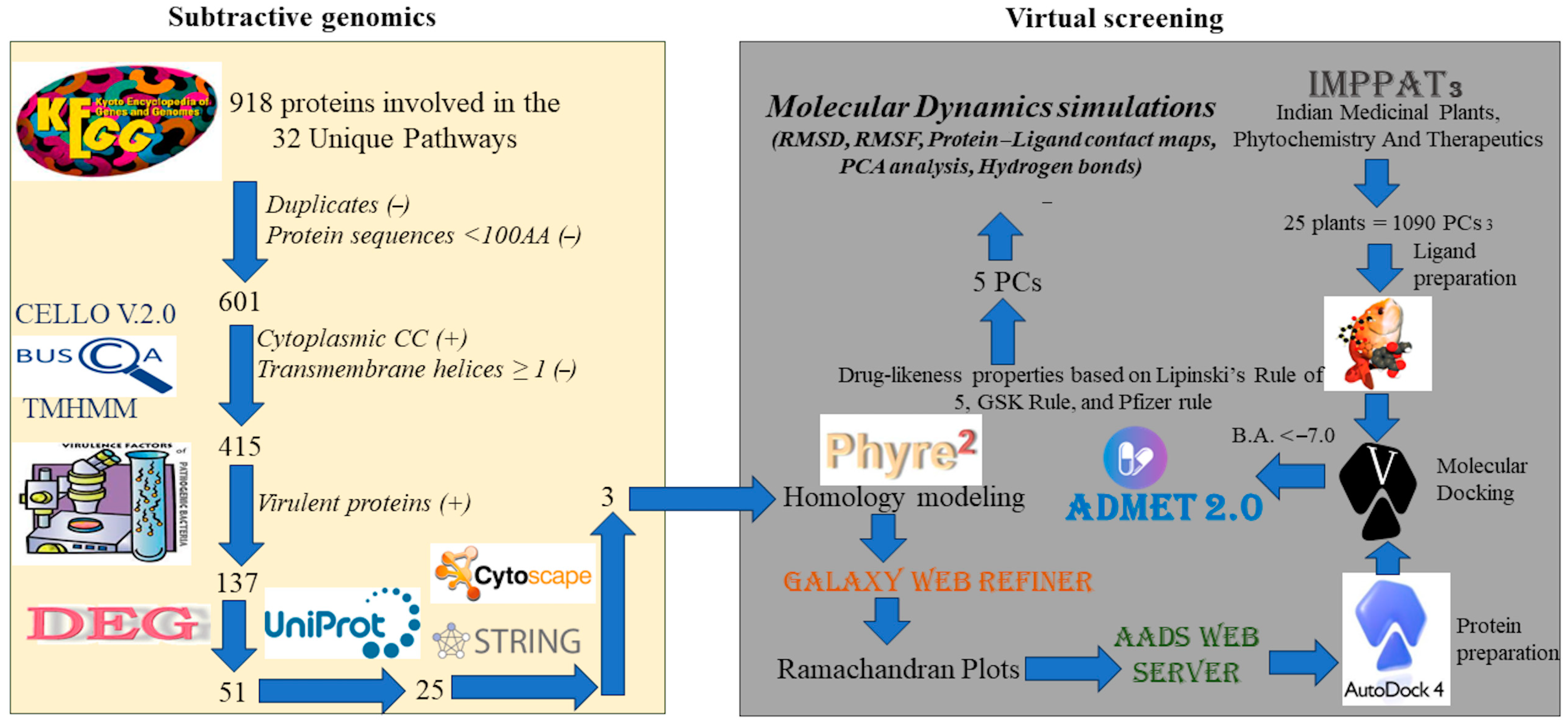
2.1. Subtractive Genomics

2.1.1. Identification of Unique Pathways

S. sputigena was actively involved in 2456 pathways, whereas H. sapiens had 36,591 pathways. Both species pathway details and identifiers were retrieved from the Kyoto Encyclopedia of Genes and Genomes (KEGG) (https://www.genome.jp/kegg/pathway.html, accessed on 10 August 2024) [20]. The recurrent pathways were eliminated in both organisms. The unique pathways in S. sputigena were identified in BioTools (https://www.biotools.fr/misc/venny, accessed on 10 August 2024) [21]. Proteins of S. sputigena involved in unique pathways were considered for further analysis.

2.1.2. Identification of Protein Subcellular Localization

Protein localization plays a crucial role in identifying and ranking drug targets in the drug discovery process [22]. We chose two web servers, Cello (http://cello.life.nctu.edu.tw/, accessed on 10 August 2024) [23,24] and Bologna Unified Subcellular Component Annotator (BUSCA) [25] (https://busca.biocomp.unibo.it/, accessed on 10 August 2024), to identify the localization of the examined proteins. Proteins localized in the cytoplasm were selected for further analysis.

2.1.3. Identification of Protein Transmembrane Helices and Virulence Factors

For the cytoplasmic proteins predicted by the above method, again, transmembrane protein sequences were subjected to identification through TransMembrane prediction using Hidden Markov Models (TMHMM) (https://services.healthtech.dtu.dk/services/TMHMM-2.0/, accessed on 10 August 2024) [26]. The outer membrane proteins were excluded because they are highly conserved and immunogenic, contribute to bacterial pathogenesis, and have epitopes essential for binding to B and T cells [27].
Bacterial pathogenic virulence factors play pivotal roles in altering host defenses and host–pathogen interactions [28]. The virulence factor database (VFDB) (http://www.mgc.ac.cn/VFs/main.htm, accessed on 10 August 2024) is an online web server developed to predict the virulence of bacterial protein sequences against the core (R1) dataset [29]. It functions via Blastp analysis on a BLOSUM62 matrix with an E-value < 0.01. The query sequence was aligned with the deposited pathogenic sequences, and the results were displayed based on the given criteria.

2.1.4. Essential Gene Identification (DEG)

Essential genes necessary for cellular function were identified using the Database of Essential Genes (DEG) (http://origin.tubic.org/deg/public/index.php/blast/bacteria, accessed on 10 August 2024) [30]. The results of the VFDB were given as input, and the results were analyzed on the basis of an E-value ≤ 0.01, percentage identity ≥ 50, and bit score ≥ 40. For DEG, E value ≤ 0.01, percent identity ≥ 50, and bit score ≥ 40 to 51 were considered.

2.1.5. Identification of Non-Homologous Proteins

Bacterial proteins with the host proteome might alter the host’s molecular function when a druggable target in the bacterial matches a higher homology with host proteins [31]. The essential genes identified in the DEG were subjected to sequence alignment with the Homo sapiens proteome in UniProt (https://www.uniprot.org/blast, accessed on 10 August 2024) with an E value of ≤0.01 and percentage identity of ≤35.

2.1.6. Protein–Protein Interaction (PPI) Network

A protein–protein interaction (PPI) network of S. sputigena was constructed with non-homologous proteins identified in UniProt using StringDB (https://string-db.org/, accessed on 10 August 2024) [32]. A full-string network with evidence network edges and an interaction score of 0.7 was used to construct a PPI. The PPI network was analyzed using Cytoscape 3.10.1 [33]. A weighted network was represented based on edge-combined scores between 0.1 and 1.0. To better understand the weighted graph, we further analyzed the network topological parameters, such as degree centrality, average shortest path, betweenness centrality, closeness centrality, and clustering coefficient, to identify potential target proteins for drug discovery and design.

2.2. In Silico Structure-Based Drug Discovery

2.2.1. Homology Modeling

To the three targets identified in the PPI network—dTDP-4-dehydrorhamnose-3,5-epimerase (UniProt ID: C9LUR0; Gene: rfbC), UTP-glucose-1-phosphate uridyltransferase (UniProt ID: C9LRH1; Gene: galU), and imidazole glycerol phosphate synthase subunit (UniProt ID: C9LTU7; Gene: hisF)—their protein sequences were used to generate a 3D structure through homology modeling technique using Phyre2 (http://www.sbg.bio.ic.ac.uk/phyre2/, accessed on 30 August 2024) because the protein data bank (PDB) does not contain wild-type proteins of S. sputigena [34]. Phyre2 was used to build models by finding homologous proteins with psi-blast, building a hidden Markov sequence model, and checking the transmembrane helices, followed by loop modeling.
The generated structures were subjected to the Saves server v6.0 (https://saves.mbi.ucla.edu/, accessed on 30 August 2024) [35] for validation, and the Ramachandran plot was used to examine the percentage of residue placements in the favored regions. The modeled proteins contained approximately 90% of the residues in the allowed regions. The protein structures were further analyzed using the GalaxyRefine web server (https://galaxy.seoklab.org/, accessed on 30 August 2024) [36]. The resulting structures attained > 90% of the residues in the allowed regions, as assessed by the Ramachandran plot.

2.2.2. Target-Based Virtual Screening

Protein preparation was carried out in AutoDock [37,38] by removing the extra bound elements, followed by adding the missing atoms, polar hydrogens, and Kollman charges, and finally saving in .pdbqt format. Indian Medicinal Plants, Phytochemistry and Therapeutics (IMPPAT) (https://cb.imsc.res.in/imppat/, accessed on 30 August 2024) [39] was used to retrieve phytocompounds based on the disease name “periodontitis”. IMPPAT is an online database comprising 2074 phytochemicals with 11,514 therapeutic applications. The phytochemicals obtained were subjected to open babel for ligand preparation [40]. Ligands were optimized under physiological conditions (pH 7.4) to obtain a reliable protonation state, and energy minimization was performed using the MMFF94 force field.
The Automated Active Site Identification, Docking and Scoring (AADS) web server (http://www.scfbio-iitd.res.in/AADS/, accessed on 30 August 2024) [41] was used to identify potential active sites of the generated 3D proteins. Active site regions contain residues that are actively involved in the binding of molecules/substrates and protein functions [42,43]. Using the prepared proteins, the ligand library was docked (high throughput screening) using AutoDock vina [44] to determine the docking score of each complex. To perform molecular docking calculations, the following grid parameters were used: size x = 40, y = 40, z = 40, for all proteins; coordinates C9LUR0, x = 19.08, y = 30.36, z = 16.65; C9LRH1, x = 21.86, y = −11.91, z = −29.84; C9LTU7, x = 65.89; y = 23.81, z = 69.04. The interactions between the protein–ligand complexes were visualized in the Discovery Studio.
Computational screening was performed on a library of 1090 pharmacologically active compounds to identify potential ligands for predictive targets in S. sputigena. Binding affinities between phytochemicals and S. sputigena protein targets were estimated computationally, and phytochemicals exhibiting binding affinities lower than −7.0 kcal/mol were selected for further analysis. This resulted in an initial set of 403 potential hit compounds, which were further sorted to identify compounds adhering to criteria consistent with Lipinski’s rule of five (RO5) to improve compound drug-likeness. Compounds were filtered to have molecular weights ≤ 500 Da, hydrogen bond acceptors ≤ 10, hydrogen bond donors ≤ 5, and logP ≤ 5 through ADMETLab 2.0 [45].

2.2.3. Molecular Dynamic Simulations

MD simulation studies using CUDA API technology with two NVIDIA graphics processing units (GPUs) were conducted using Desmond software through the graphical interface of Maestro (Desmond Molecular Dynamics System 6.4 academic version, D. E. Shaw Research (“DESRES”), New York, NY, USA, 2020. Maestro-Desmond Interoperability Tools, Schrödinger, New York, NY, USA, 2020). The complexes derived from the molecular docking studies were saved in pdb and imported into Maestro. The complexes were treated by using protein preparation wizard protocol implemented in Maestro (Protein Preparation Wizard workflow 2020). This protocol, through a series of computational steps, allowed us to obtain a reasonable starting structure of the proteins for MD simulation by a series of in silico steps to: (1) add hydrogens; (2) optimize the orientation of hydroxyl groups, Asn, and Gln, as well as the protonation state of His (the assignment of the protonation state was performed by using PROPKA implemented in the protein preparation wizard at physiological pH (7.4)); and (3) perform a constrained refinement with the impref utility, setting the max RMSD of 0.30. The impref utility consists of cycles of energy minimization based on the impact molecular mechanics engine and on the OPLS_2005 force field [46]. To generate three-dimensional ligand–protein complexes (C9LRH1/cubebin, C9LTU7/cubebin, C9LUR0/cubebin) embedded into an orthorhombic box filled with water molecules (TIP3P), the system builder provided by Desmond software was utilized [47]. The box size was 455,376 Å3 for C9LRH1/cubebin, 288,771 Å3 for C9LTU7/cubebin, and 277,764 Å3 for C9LUR0/cubebin. Na+ and Cl ions were added to the biological system to reach a physiological concentration of monovalent ions of 0.15 M. The OPLS3 force field was used for MD simulation [48]. The NPT ensemble class (constant number of particles, 300 K for the temperature, and 1.01325 bar for the pressure) was used for the simulation. The RESPA integrator (inner time step of 2.0 fs) was utilized to estimate the motion for bonded and non-bonded interactions within the short-range cutoff [49]. To maintain a constant temperature during the simulation, the Nosé–Hoover thermostat technique was employed [50], whereas the pressure was kept constant utilizing the Martyna–Tobias–Klein method [51]. To compute long-range electrostatic interactions, the particle mesh Ewald technique (PME) was applied (van der Waals and short-range electrostatic interactions were fixed at 9.0 Å) [52]. Using the default procedure, which involved several constrained minimizations and MD simulations, the selected system was gradually relaxed and brought to equilibrium. For each complex, we performed two independent runs; however, considering that the results for the complex C9LUR0/IMPHY001912 were controversial, we confirmed the lack of the affinity of IMPHY001912 by conducting an additional MD run for a total of three independent MD runs. The Desmond package’s simulation event analysis tools were employed to investigate the MD outputs generated during the MD simulation experiments, as previously reported [53]. The thermal MM-GBSA script available in Desmond (thermal_mmgbsa.py) was used to evaluate the ΔGbind for the selected complexes as reported [54,55]. This tool used the Desmond MD trajectory, splitting it into individual frame snapshots, and it runs each one through MM-GBSA analysis. During the MM-GBSA calculation, 1000 snapshots from the 100 ns MD simulation were used as input to compute the average binding free energy. The evaluated ΔGbind is reported as an average value in the Results section along with the energy components.

3. Results

3.1. Subtractive Genomics

Subtractive genomics was used to identify potential targets involved in unique pathways necessary for the life cycle of S. sputigena. These pathways could be cytoplasmic and virulent, and special attention was paid to proteins unique and non-homologous to Homo sapiens. This approach allowed the selection of possible drug targets that, if modulated, could offer novel insights into the development of effective antibacterial agents against the identified microorganism responsible for periodontitis without interfering with human proteins, thus, reducing the possibility of undesired effects.

3.1.1. Unique Pathways Identifications and Proteins Involved

After removing the recurring pathways, we found 102 and 356 pathways for S. sputigena and H. sapiens, respectively. Both species shared 70 common pathways, with S. sputigena having 32 pathways and H. sapiens having 286 pathways. Only the unique pathways that were present in S. sputigena, the 32 pathways in Figure 2, common pathways between the organisms, and H. sapiens pathways were excluded. Again, 918 proteins were involved in these 32 pathways after removing duplicates and proteins with less than 100 AA sequences, resulting in 601 proteins.

3.1.2. Subcellular Localization

The Cello web server was used to predict subcellular localization of the proteins. The results indicated that most of the proteins were cytoplasmic proteins (82.56%), and the rest of the protein’s localization was distributed among InnerMembrane (9.66%), periplasmic (5.32%), extracellular (1.49%), and OuterMembrane (0.99%) in Figure 3A. To provide more reliable results, we used BUSCA, another web server, to further characterize the proteins. The results suggested that higher protein localization was observed in the cytoplasm (73.21%), with fewer plasma membranes (24.12%) and extracellular space (2.66%) (Figure 3B). Proteins predicted to be subcellularly localized in the cytoplasm of both Cello and BUSCA servers were considered for further analysis.

3.1.3. Identification of Protein Transmembrane Helices and Virulent Factors

Considering the objective of the study, of the 415 cytoplasmic proteins subjected to identifying the TM protein sequences in TMHMM, only three proteins with less than one TM helices sequence were excluded. The remaining 412 proteins did not exhibit any TM helices, representing purely cytoplasmic proteins. In the VFDB, 137 of the 412 cytoplasmic proteins were identified as virulent. These virulent proteins were aligned with the bacterial virulent sequence dataset deposited in the VFDB.

3.1.4. Essential Genes and Non-Homologous Protein Identification

S. sputigena cytoplasmic virulent proteins were sent as queries to identify essential genes in the DEG database. The essential bacterial gene dataset was obtained using Blastp, which contains 51 essential genes. Blastp identified 26 homologous proteins out of 51 query sequences in S. sputigena compared with H. sapiens. Accordingly, 25 non-homologous proteins were identified as prominent proteins of S. sputigena Table 1.

3.1.5. Protein–Protein Interaction Analysis

The PPI network of non-homologous proteins generated in StringDB includes 18 nodes (proteins) and 42 edges (interactions) with a diameter of 5. The characteristic features of the network include an average no. of neighbors of 4.667, a path length of 2.176, a clustering coefficient of 0.464, and network density and centrality of 0.275 and 0.221. The colors connecting each node represent the edges, and the colors are designed based on the edge-weighted scores, which results in a weighted PPI network. Scores ranging from 0.1 to 1.0 were classified (0.1–0.7, yellow; 0.8–0.87, green; and 0.88–1.0, red in Figure 4).
The PPI network was analyzed using the network topological parameters, and three proteins were identified as potential drug targets: dTDP-4-dehydrorhamnose-3,5-epimerase (UniProt ID: C9LUR0; Gene: rfbC), UTP-glucose-1-phosphate uridyltransferase (UniProt ID: C9LRH1; Gene: galU), and the imidazole glycerol phosphate synthase subunit (UniProt ID: C9LTU7; Gene: hisF). Descriptors such as degree centrality, average shortest path, betweenness centrality, closeness centrality, and clustering coefficient values were considered when selecting potential targeting proteins. High-degree centrality-valued nodes and the remaining descriptors with low values were selected for subsequent investigations.

3.2. Structure-Based Drug Discovery

The subtractive genomics approach identified three potential drug targets that were exploited to identify chemical entities from a library of bioactive phytochemicals that can interfere with protein functions and potentially reduce the bacterial load of S. sputigena in periodontitis, providing potential novel therapeutics to treat this disorder. Accordingly, a series of computer-based techniques were integrated to screen the chemical library described above.

3.2.1. Homology Modeling and Protein Optimization

The models generated by Phyre2 were selected based on query coverage greater than 90% and sequence identity greater than 55%. C9LUR0 was aligned with 179 residues of RmlC-like dTDP-sugar isomerase (PDB ID: 1DZR) of 56% identity, C9LRH1 3D structure was generated with 63% identity aligning 276 residues of UTP-glucose-1-phosphate uridylyltransferase (PDB ID: 7B1R), and C9LTU7 tertiary model constructed by aligning 249 residues of ribulose-phosphate binding barrel (PDB ID: 1KA9) with 63% identity.
Tertiary proteins generated by Phyre2 were validated in the Saves v6.0 server using Ramachandran plots. Proteins containing >90% of the residues in the allowed regions may be considered structurally validated and used for further analysis. Because the homology-modeled protein residues were below 90%, all structures were subjected to the GalaxyRefine server. The refined structures were validated using Saves v6.0 to obtain Ramachandran plots. The resulting structures were well-refined and resulted in >90% residues in the allowed regions, as shown in Table 2 (details regarding the homology modeling approach, including the alignment and validation step by the Ramachandran plot, are reported in Figures S1–S9).

3.2.2. Phytochemical Screening

After the identification of the potential drug targets of S. sputigena, we conducted a virtual screening of a natural product database to identify the most promising compounds that could potentially interact with the selected drug targets. In this chapter, we discuss the most promising hit compounds selected based on the binding affinity and the drug-like profile. The results of the screening are reported in Table S1. The phytochemical (4bS,8aR)-2,4b,8,8-tetramethyl-7,10-dioxo-5,6,8a,9-tetrahydrophenanthrene-3-carboxylic acid (IMPHY005303) showed an interesting pattern of interaction, establishing polar contacts within the selected binding site of the protein typified by the code C9LUR0. In particular, IMPHY005303 formed four H-bonds with residues Asn48, Ser52, and Phe51 (Figure 5A). This binding mode accounted for a calculated binding affinity of −7.2 kcal/mol (Table 3).
Considering the other phytochemicals, pluviatilol (IMPHY006624) was able to interact with the binding site of the protein typified by the code C9LUR0, showing a docking score of −7.0 kcal/mol, which was slightly higher than that of the previously discussed compound (Table 3). In particular, the compound could target residues Asn48, Arg59, Tyr138, Ala166, and Lys167 via H-bonds. In addition, we observed further contacts indicated as carbon–H-bonds with residue Ser165. Hydrophobic interactions with Tyr138 (π–π stacking interaction and π–alkyl contact) were detected. Alkyl interactions with Ala166 and Lys167 stabilized the retrieved binding mode (Figure 5B). The interaction colored in red between the OH group and the Gln46 residue in the image indicates an unfavorable interaction, likely due to steric hindrance or unfavorable electrostatic interactions.
The next compound, 5-[3-(1,3-Benzodioxol-5-yl)-1,3,3a,4,6,6a-hexahydrofuro [3,4-c]furan-6-yl]-1,3-benzodioxole (IMPHY014895), showed a docking score of −7.6 kcal/mol within the C9LUR0 binding site (Table 3). Polar interactions were detected with residues Ala53, Glu54, Tyr138, Lys167 (H-bonds), and Leu58 (carbon–H-bond). Various hydrophobic contacts were detected with Ala53, Thr57, Leu58, Arg59, Tyr138, and Lys167 (Figure 5C).
Figure 5D shows the computational assessment of gadain (IMPHY004244) within the C9LUR0 binding site. The compound formed H-bonds with Ser52 and Arg59, whereas hydrophobic interactions were found with residues Arg59 (π–sigma and π–alkyl) and Tyr138 (π–alkyl). This binding mode resulted in a docking score of −7.1 kcal/mol (Table 3).
The molecular docking output of the last compound (cubebin, IMPHY001912) is shown in Figure 5E. This compound was able to establish polar and hydrophobic contacts within the selected binding site of the protein typified with the code C9LUR0. A canonical H-bond was detected with Ser52, whereas a carbon–H-bond was evident with residue Phe51. A strong network of hydrophobic interactions was established by IMPHY001912 with Arg59 and Tyr138. The docking score for this binding mode was −7.1 kcal/mol (Table 3). The 3D models of molecular docking output regarding the protein typified with the code C9LUR0 are provided in Figure S10.
Compound IMPHY005303 showed a docking score of −8.6 kcal/mol when docked into the binding site of the protein C9LRH1 (Table 3). In particular, the compound was able to establish H-bonds with residues Thr253, Leu206, and Arg208 (Figure 6A). Further hydrophobic interactions were detected with residues Leu132, Val205, Tyr209, and Leu232.
IMPHY006624 within the C9LRH1 binding site established a H-bond with Arg171 and carbon–H-bonds with Asp134 and Leu206. Hydrophobic interactions with Tyr172 (π–alkyl), and further contacts with residues Leu132 and Asp134 were detected (Figure 6B). This binding mode resulted in a docking score of −8.3 kcal/mol (Table 3).
IMPHY014895 established polar and non-polar contacts within the C9LRH1 binding site. In particular, it established a H-bond with Lys30 and a carbon–H-bond with residue Gly258. Different hydrophobic contacts were detected using Val205. Furthermore, additional electrostatic interactions were observed with residues Arg20 and Asp134 (Figure 6C). The docking score value of −8.7 kcal/mol was related to the described binding mode (Table 3).
IMPHY004244 exhibited a docking score of −8.6 kcal/mol within the C9LRH1 binding site (Table 3). IMPHY004244 established several hydrophobic interactions: π–π stacking with Tyr172 and Tyr209, two alkyl interactions with Leu132 and Leu232 residues. Four π–alkyl contacts with Tyr172, Val205, Tyr209, and Leu232 (Figure 6D).
Finally, compound IMPHY001912 was found to be the best performing compound considering the C9LRH1 enzyme. IMPHY001912 showed a docking score of −9.0 kcal/mol (Table 3). Several contacts were found between the selected binding site and IMPHY001912. Two H-bonds and a carbon–H-bond were observed with Glu192, Thr233, and Asp135, respectively. Numerous hydrophobic contacts were established from IMPHY001912 with the following residues: Tyr172 and Tyr209 (π–π stacking and π–alkyl contacts), Leu132 (π–alkyl and alkyl contacts), Leu232, and Ile236 (alkyl contacts) (Figure 6E). The 3D models of molecular docking output regarding the protein typified with the code C9LRH1 are provided in Figure S11.
The last examined target was the protein typified by the code C9LTU7. The compound IMPHY005303 showed a docking score of −8.3 kcal/mol within the binding site of the protein (Table 3). H-bonds were observed among the compound and residues Ile83, Asn103, Thr104, and Ala105. A π-donor–H-bond was established with Gly82, and a π–alkyl hydrophobic contact with His56 was detected (Figure 7A). The interaction colored in red between the OH group and the Gly81 residue in the image indicates an unfavorable interaction, likely due to steric hindrance or unfavorable electrostatic interactions.
Figure 7B illustrates the docking output of IMPHY006624 within the binding site of C9LTU7. H-bonds were observed with residues Asp130 and Ala205, and a carbon–H-bond was evident with Ala205. IMPHY006624 was further able to establish two π–anion electrostatic interactions with residues Asp130 and Asp177. Furthermore, three alkyl hydrophobic bonds were formed with Leu50, Ala130, and Leu170, and a π–alkyl bond with Ala226 (Figure 7B). This binding mode accounted for a docking score of −8.0 kcal/mol (Table 3).
IMPHY014895 showed a docking score of −9.0 kcal/mol with the C9LTU7 enzyme (Table 3). H-bonds were observed with residue Ser202, and a carbon–H-bond was evident with Asp130. A π-donor–H-bond was established with Gly82. Moreover, a π–anion electrostatic bond with Asp130 was observed. Further hydrophobic contacts were found with residues Leu50 and Ala105 (Figure 7C).
Next, the compound IMPHY004244 established a carbon–H-bond with Thr172, a π–anion electrostatic bond with Asp130. Three alkyl bonds with residues Leu50, Arg84, and Ala128. Further π–alkyl bonds with Leu50, His56, and Ala128 were observed (Figure 7D). The described binding mode accounted for a docking score of −8.2 kcal/mol (Table 3).
Finally, Figure 7E shows the docking output of IMPHY001912. Considering this target, the mentioned compound achieved the best computational score in terms of the prediction of affinity within C9LTU7 (Table 3). In fact, a docking score of −9.1 kcal/mol was related to the following binding mode. IMPHY001912 established H-bonds with Asp11 and Ala205. Two π–anion electrostatic interactions were observed with residues Asp130 and Asp177. Furthermore, several hydrophobic contacts were detected with residues Leu50, Ala128, Ala205, and Ala226, indicating a significant predicted affinity of IMPHY001912 for the selected target. The 3D models of molecular docking output regarding the protein typified with the code C9LRH1 are provided in Figure S12.

3.2.3. Drug Likeness Properties

Of the total 1090 phytochemicals, 403 exhibited docking scores lower than −7.0 kcal/mol for the predicted targets of S. sputigena. These compounds were evaluated for their drug-like profile by applying different rule descriptors. First, the rule of five (RO5) identifiers, molecular weight (MW) ≤ 500 and HBA ≤ 10, resulted in 347 phytochemicals, HBD ≤ 5 showed 324 phytochemicals, and 310 phytochemicals with logP ≤ 5. The Pfizer 3/75 rule exhibited 63 phytochemicals in the range of logP ≤ 3 and TPSA > 75. The GSK rule sorted five phytochemicals as final ligands for further analysis (Table 4). Furthermore, we assessed the capability of the selected compounds to cross biological membranes, which is a necessary property for their use as antibacterial agents. According to the analysis, the chosen compounds demonstrated a significant ability to cross biological membranes, except for IMPHY005303, whose parameters were less favorable, suggesting their potential as antibacterial agents.

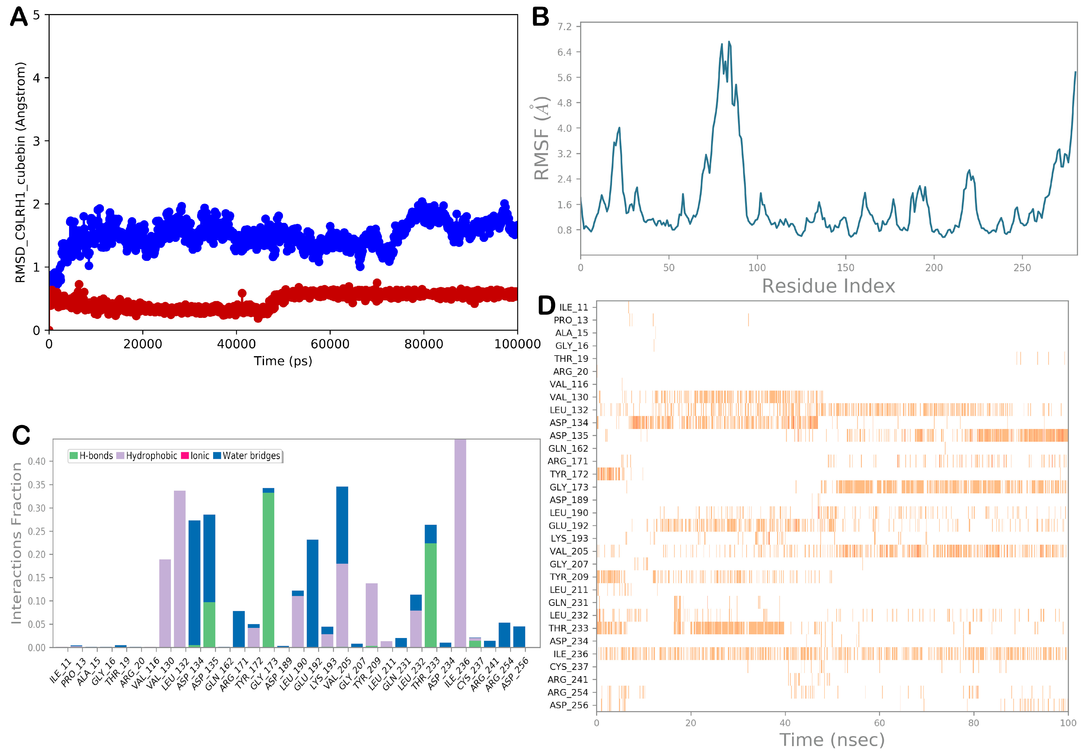
3.2.4. Molecular Dynamic Simulations

To provide a comprehensive overview of the binding mode of IMPHY001912, which was found to be the most promising ligand for the selected proteins, we conducted MD simulation studies. Apart from visually examining the generated trajectories, the trajectories were evaluated by computing the RMSD and RMSF values and conducting analysis of the dynamic ligand interaction graphs. Overall, the MD simulation considering the complexes C9LRH1/IMPHY001912 and C9LTU7/IMPHY001912 showed satisfactory stability of the binding mode of IMPHY001912 within the mentioned proteins, exhibiting small RMSD values and slight variations in the proteins, as shown by the RMSF. In contrast, when the complex C9LUR0/IMPHY001912 was examined, MD simulation analysis provided a relevant uncertainty concerning the binding mode of IMPHY001912, with limited stability and high RMSD, indicating a very weak possibility of interaction with the selected protein. Accordingly, IMPHY001912 may function as an inhibitor of the proteins C9LRH1 and C9LTU7, but not for the protein C9LUR0, based on the stability of the biological systems and the analysis of the pattern of interaction within the chosen binding sites throughout the course of the 100 ns MD simulations.
A detailed analysis of complex C9LRH1/IMPHY001912 is presented in Figure 8 and in Figure S13A. In particular, IMPHY001912 established a strong hydrophobic contact network with Val130, Leu132, Leu190, Val205, Thr209, Leu232, and Ile236. Furthermore, significant polar contacts, mainly H-bonds, with the side chains of Asp135 and Thr233 and with the backbone of Gly173 were observed. Overall, the MD simulation output supported the docking results, indicating significant interactions within the selected binding site, suggesting the potential of IMPHY001912 to inhibit the protein typified with the code C9LRH1. The region with higher RMSF (Figure 8B) comprises approximately twenty residues structured in a loop that is not close to the binding site.
The MD simulation output for the complex C9LTU7/IMPHY001912 is shown in Figure 9 and in Figure S13B. Hydrophobic contacts with Val48, Leu50, Ala128, Leu170, and Leu224 were particularly evident during the simulation. In addition, a network of polar contacts (H-bonds) with Cys9 and Asp11 and an ionic bond with Asp130 were observed. Accordingly, the MD simulation output confirmed the docking results, indicating relevant interactions within the selected binding site, suggesting the potential of IMPHY001912 to act as an inhibitor of the protein typified with the code C9LTU7.
The MD simulation output for the complex C9LUR0/IMPHY001912 is shown in Figure 10 and Figure S13C. In contrast to the two previously discussed complexes, IMPHY001912 appeared to have not a significant affinity for this selected protein. In particular, as indicated by the graphs regarding the MD trajectory, the ligand showed high instability and was not able to reach a stable bioactive conformation; instead, several conformational changes were observed, precluding strong binding with the selected binding site (no contacts that occurred more than 30.0% of the simulation time were observed). Furthermore, after approximately half of the simulation time, IMPHY001912 lacked the capacity to interact with the selected binding site and disappeared from the selected binding site without any contact within the protein. These results were also obtained by considering two additional independent MD runs. Accordingly, IMPHY001912 showed limited affinity for the protein typified by the code C9LUR0, suggesting that it could not inhibit the aforementioned protein.
To further corroborate the MD simulation results, we determined the free binding energy (ΔGbind) considering the entire MD trajectory of the selected complexes, providing a comprehensive picture of the binding affinity. Accordingly, by employing the MM/GBSA techniques, we obtained the ΔGbind values listed in Table 5. The main energy contributions to the overall binding free energy, taking into account various energy sources, are included in Table 5. The van der Waals energy contribution was found to be the main source of the ligand binding energy considering the targets C9LRH1 and C9LTU7. This result emphasizes the critical role that hydrophobic interactions play in the stability of ligand/protein complexes, given the hydrophobic nature of the binding sites. On the contrary, as expected by analyzing the trajectory of the complex C9LUR0/IMPHY001912, we observed a dramatic decrease in the affinity for the compound IMPHY001912, indicating a very weak affinity for the considered target. In summary, based on the MD analysis and the calculated ΔGbind, we conclude that IMPHY001912 (cubebin) could have a multitarget behavior because of the strong binding for the targets C9LRH1 and C9LTU7, whereas the affinity for the target C9LUR0 was limited.

4. Discussion

The objective of the present study was to identify possible targets for screening plant-based phytochemicals using subtractive genomics and in silico structure-based approaches. In the subtractive genomics step, we identified three predicted proteins as putative drug targets. Each protein plays a prominent role in the life cycle of S. sputigena. In fact, dTDP-4-dehydrorhamnose 3,5-epimerase (UniProt ID: C9LUR0) is involved in streptomycin biosynthesis, polyketide sugar unit biosynthesis, O-antigen nucleotide sugar biosynthesis, and the biosynthesis of secondary metabolites pathway. UTP-glucose-1-phosphate uridyltransferase is involved in the O-antigen nucleotide sugar biosynthesis and the biosynthesis of secondary metabolites pathway. The imidazole glycerol phosphate synthase subunit hisF is involved in the biosynthesis of secondary metabolites. Among the four pathways between proteins, the biosynthesis of the secondary metabolite pathway is a common pathway.
dTDP-4-dehydrorhamnose 3,5-epimerase converts dTDP-4-dehydrorhamnose to dTDP-L-rhamnose via an epimerization reaction through a change in the central chiral carbon atoms, leading to different series of reactions that result in the final product streptomycin. Usually, this sugar component is found in various cell surface molecules, including the bacterial cell wall [56]. O-antigens are important components of bacterial lipopolysaccharides (LPS) that contribute to the structural diversity of the bacterial outer membrane. O-antigen nucleotide biosynthesis is involved in the synthesis of nucleotide sugars, where the dTDP-4-dehydrorhamnose 3,5-epimerase product, dTDP-L-rhamnose, undergoes a series of modifications, particularly during the conversion of dTDP-4-oxo-6-deoxy D-glucose to dTDP-4-dehydro-6-deoxy-L-mannose. dTDP-L-rhamnose, which is produced by the action of dTDP-4-dehydrorhamnose 3,5-epimerase, serves as a substrate for glycosyltransferase enzymes. These enzymes transfer the rhamnose moiety onto the growing O-antigen polysaccharide chain, contributing to the unique structure of the O-antigen [57].
The role of UTP-glucose-1-phosphate uridyltransferase (UniProt ID: C9LRH1) in O-antigen nucleotide sugar biosynthesis is UDP-glucose formation, a key nucleotide sugar. It converts glucose-1-phosphate and UTP (uridine triphosphate) into UDP-glucose and pyrophosphate via a hydrolyzing reaction. This reaction represents the activation of glucose for its subsequent incorporation into the growing O-antigen chain [57]. The product UDP-glucose is used in various cellular activities, especially in the glycosylation of secondary metabolites [58].
Imidazole glycerol phosphate synthase (UniProt ID: C9LTU7), a cyclase subunit, is a major enzyme in this pathway that acts as a precursor for secondary metabolites and is involved in histidine metabolism. The enzyme catalyzes two reactions: N-1-(5′-phosphoribosyl)-ATP pyrophosphorylase and imidazole glycerol phosphate cyclase. The cyclase subunit of imidazole glycerol phosphate synthase is present in the second step of this process, the cyclization of N-[(5′-phosphoribulosyl)formimino]-5-aminoimidazole-4-carboxamide-ribonucleotide (PRFAR), to give the product of imidazole glycerol phosphate synthase, which is an important intermediate in the biosynthesis of an essential amino acid, i.e., histidine [59]. Histidine biosynthesis is a critical process in bacterial growth and survival [60]. Some histidine derivatives, such as histidine-containing peptides, play roles in cellular signaling pathways, provide virulence and adaptation to specific host environments, and are precursors of bioactive compounds and protein synthesis [61]. In the second step of our computational protocol, putative drug targets, characterized by their unicity with respect to proteins related to H. sapiens, were used in a structure-based approach to identify possible phytochemicals that could be used as novel therapeutic agents against oral infections caused by S. sputigena. By applying several computational steps (molecular docking, drug-likeness, and MD simulation), we screened a library containing phytochemicals. Results provided interesting hints concerning a few natural products that were found to be the top-ranked compounds. Among them, the compound cubebin (IMPHY001912) was the most interesting. Compound IMPHY001912 has the potential to inhibit two of the three targeted enzymes, namely UTP-glucose-1-phosphate uridyltransferase (UniProt ID: C9LRH1) and imidazole glycerol phosphate synthase (UniProt ID: C9LTU7) and could be utilized as an antibacterial agent to treat S. sputigena oral infections due to its promising drug-like profile.
Cubebin, an alkaloid derived from the Piper genus, can be useful to treat and control different oral diseases owing to its fungicidal, anti-inflammatory, and analgesic properties [62]. Previous studies have shown that cubebin possesses antibacterial properties against different bacteria that cause dental caries and periodontal diseases including S. mutans and P. gingivalis. They also blocked S. mutans adhesion and displaced mature P. gingivalis biofilms. Cubebin has anti-fungal activity against Candida albicans, the main causative agent of oral thrush [63]. Therefore, it is reasonable to consider that cubebin may help relieve the symptoms of thrush. Furthermore, cubebin decreased CCL2 levels in experimental models suggesting its potential as an anti-inflammatory agent against oral diseases such as pulpitis, gingivitis, and periodontitis [64]. This anti-inflammatory effect also points to the fact that cubebin can be used for pain associated with several oral diseases, for which pain management is important for the patient’s comfort. Cubebin exhibits multiple activities against infections and symptoms of most common oral diseases and conditions. Further studies are required to elucidate the effectiveness and side effects of cubebin when used to treat oral ailments [65,66,67]. The compound holds great potential for the treatment of oral diseases based on its potential pharmacological activities, as evidenced by the experiments conducted to date.

5. Conclusions

Overall, our research integrates multiple computational methods to conduct a thorough analysis for identifying potential drug targets of S. sputigena that could be further investigated to develop molecules for treating oral infections caused by S. sputigena. We combined subtractive genomics, which allowed the identification of enzymes characterized by their unicity with respect to proteins related to H. sapiens, with computer-based drug discovery techniques to screen a chemical library containing 1090 bioactive phytochemicals. Our computational analysis enabled us to identify three unique proteins that are crucial for the life cycle of S. sputigena. dTDP-4-dehydrorhamnose 3,5-epimerase (UniProt ID: C9LUR0), UTP-glucose-1-phosphate uridyltransferase (UniProt ID: C9LRH1), and imidazole glycerol phosphate synthase (UniProt ID: C9LTU7) could represent significant targets for the development of effective therapeutic agents against S. sputigena-mediated infections. Furthermore, our in silico analysis indicated that few natural products could interact with the selected drug targets. Among them, cubebin (IMPHY001912) was the most promising, being potentially able to efficiently target two out of three retrieved bacterial proteins (C9LRH1 and C9LTU7), as demonstrated by molecular docking and MD simulation experiments. Accordingly, cubebin (IMPHY001912) could be further exploited as an antibacterial agent for treating oral infections because of its multitargeting capability and satisfactory drug-like properties. Notably, its effectiveness could be explored further by researching suitable formulations such as mouthwash and/or toothpaste for use in this context, thus, minimizing the negative side effects associated with current treatments for S. sputigena-induced periodontitis.

Supplementary Materials

The following supporting information can be downloaded at: https://www.mdpi.com/article/10.3390/computation13020034/s1, Figures S1–S9: Homology modeling results. Figures S10–S12: Molecular docking outputs. Figure S13: Ligand-interaction diagrams for MD simulation. Table S1: List of the screened compounds with related calculated parameters.

Author Contributions

Conceptualization, M.P., S.B., and K.P.K.P.D.; methodology, M.P., C.G.S., S.B., V.C., and K.P.K.P.D.; software, M.P., C.G.S., and S.B.; validation, M.P., C.G.S., S.B., V.C., and K.P.K.P.D.; formal analysis, M.P., C.G.S., S.B., V.C., and K.P.K.P.D.; investigation, M.P., C.G.S., and S.B.; data curation, M.P., C.G.S., S.B., V.C., and K.P.K.P.D.; writing—original draft preparation, M.P., S.B., and K.P.K.P.D.; writing—review and editing, M.P., C.G.S., S.B., V.C., and K.P.K.P.D.; supervision, S.B. All authors have read and agreed to the published version of the manuscript.

Funding

This research received no external funding.

Data Availability Statement

Data are contained within the article.

Conflicts of Interest

The authors declare no conflicts of interest.

References

  1. Rath, C.B.; Schirmeister, F.; Figl, R.; Seeberger, P.H.; Schaffer, C.; Kolarich, D. Flagellin Glycoproteomics of the Periodontitis Associated Pathogen Selenomonas sputigena Reveals Previously Not Described O-glycans and Rhamnose Fragment Rearrangement Occurring on the Glycopeptides. Mol. Cell. Proteom. 2018, 17, 721–736. [Google Scholar] [CrossRef] [PubMed]
  2. Hiranmayi, K.V.; Sirisha, K.; Ramoji Rao, M.V.; Sudhakar, P. Novel Pathogens in Periodontal Microbiology. J. Pharm. Bioallied Sci. 2017, 9, 155–163. [Google Scholar] [CrossRef] [PubMed]
  3. Mok, S.F.; Karuthan, C.; Cheah, Y.K.; Ngeow, W.C.; Rosnah, Z.; Yap, S.F.; Ong, H.K.A. The oral microbiome community variations associated with normal, potentially malignant disorders and malignant lesions of the oral cavity. Malays. J. Pathol. 2017, 39, 1–15. [Google Scholar]
  4. Marsh, P.D.; Martin, M.V.; Lewis, M.A.O.; Williams, D. Oral Microbiology E-Book; Elsevier Health Sciences: Amsterdam, The Netherlands, 2009. [Google Scholar]
  5. Praveen, M. Multi-epitope-based vaccine designing against Junín virus glycoprotein: Immunoinformatics approach. Futur. J. Pharm. Sci. 2024, 10, 29. [Google Scholar] [CrossRef]
  6. Lukic, D.; Karygianni, L.; Flury, M.; Attin, T.; Thurnheer, T. Endodontic-Like Oral Biofilms as Models for Multispecies Interactions in Endodontic Diseases. Microorganisms 2020, 8, 674. [Google Scholar] [CrossRef] [PubMed]
  7. Goncalves, L.F.; Fermiano, D.; Feres, M.; Figueiredo, L.C.; Teles, F.R.; Mayer, M.P.; Faveri, M. Levels of Selenomonas species in generalized aggressive periodontitis. J. Periodontal Res. 2012, 47, 711–718. [Google Scholar] [CrossRef] [PubMed]
  8. Kononen, E.; Gursoy, M.; Gursoy, U.K. Periodontitis: A Multifaceted Disease of Tooth-Supporting Tissues. J. Clin. Med. 2019, 8, 1135. [Google Scholar] [CrossRef] [PubMed]
  9. Al-Otaibi, M.; Al-Harthy, M.; Gustafsson, A.; Johansson, A.; Claesson, R.; Angmar-Mansson, B. Subgingival plaque microbiota in Saudi Arabians after use of miswak chewing stick and toothbrush. J. Clin. Periodontol. 2004, 31, 1048–1053. [Google Scholar] [CrossRef]
  10. Teughels, W.; Van Essche, M.; Sliepen, I.; Quirynen, M. Probiotics and oral healthcare. Periodontology 2000 2008, 48, 111–147. [Google Scholar] [CrossRef]
  11. Allaker, R.P.; Ian Douglas, C.W. Non-conventional therapeutics for oral infections. Virulence 2015, 6, 196–207. [Google Scholar] [CrossRef] [PubMed]
  12. Abranches, J.; Zeng, L.; Kajfasz, J.K.; Palmer, S.R.; Chakraborty, B.; Wen, Z.T.; Richards, V.P.; Brady, L.J.; Lemos, J.A. Biology of Oral Streptococci. Microbiol. Spectr. 2018, 6. [Google Scholar] [CrossRef]
  13. Norskov-Lauritsen, N.; Claesson, R.; Birkeholm Jensen, A.; Aberg, C.H.; Haubek, D. Aggregatibacter Actinomycetemcomitans: Clinical Significance of a Pathobiont Subjected to Ample Changes in Classification and Nomenclature. Pathogens 2019, 8, 243. [Google Scholar] [CrossRef] [PubMed]
  14. Socransky, S.S.; Haffajee, A.D. The bacterial etiology of destructive periodontal disease: Current concepts. J. Periodontol. 1992, 63, 322–331. [Google Scholar] [CrossRef]
  15. Jantan, I.; Ahmad, W.; Bukhari, S.N. Plant-derived immunomodulators: An insight on their preclinical evaluation and clinical trials. Front. Plant Sci. 2015, 6, 655. [Google Scholar] [CrossRef]
  16. Azad, A.K.; Praveen, M.; Sulaiman, W.M.A.B.W. Assessment of Anticancer Properties of Plumbago zeylanica. In Harnessing Medicinal Plants in Cancer Prevention and Treatment; IGI Global Scientific Publishing: Hershey, PA, USA, 2024; pp. 91–121. [Google Scholar] [CrossRef]
  17. Kumar, R.; Mirza, M.A.; Naseef, P.P.; Kuruniyan, M.S.; Zakir, F.; Aggarwal, G. Exploring the Potential of Natural Product-Based Nanomedicine for Maintaining Oral Health. Molecules 2022, 27, 1725. [Google Scholar] [CrossRef]
  18. Ashraf, B.; Atiq, N.; Khan, K.; Wadood, A.; Uddin, R. Subtractive genomics profiling for potential drug targets identification against Moraxella catarrhalis. PLoS ONE 2022, 17, e0273252. [Google Scholar] [CrossRef] [PubMed]
  19. Uddin, R.; Siddiqui, Q.N.; Sufian, M.; Azam, S.S.; Wadood, A. Proteome-wide subtractive approach to prioritize a hypothetical protein of XDR-Mycobacterium tuberculosis as potential drug target. Genes. Genom. 2019, 41, 1281–1292. [Google Scholar] [CrossRef] [PubMed]
  20. Kanehisa, M.; Goto, S. KEGG: Kyoto encyclopedia of genes and genomes. Nucleic Acids Res. 2000, 28, 27–30. [Google Scholar] [CrossRef] [PubMed]
  21. Pérez, M.A.O.; Knapp, R.B. BioTools: A Biosignal Toolbox for Composers and Performers. In Computer Music Modeling and Retrieval. Sense of Sounds. CMMR 2007; Lecture Notes in Computer Science, Volume 4969; Springer: Berlin/Heidelberg, Germany, 2008; pp. 441–452. [Google Scholar] [CrossRef]
  22. Javed, F.; Hayat, M. Predicting subcellular localization of multi-label proteins by incorporating the sequence features into Chou’s PseAAC. Genomics 2019, 111, 1325–1332. [Google Scholar] [CrossRef]
  23. Yu, C.S.; Lin, C.J.; Hwang, J.K. Predicting subcellular localization of proteins for Gram-negative bacteria by support vector machines based on n-peptide compositions. Protein Sci. 2004, 13, 1402–1406. [Google Scholar] [CrossRef] [PubMed]
  24. Yu, C.S.; Chen, Y.C.; Lu, C.H.; Hwang, J.K. Prediction of protein subcellular localization. Proteins 2006, 64, 643–651. [Google Scholar] [CrossRef] [PubMed]
  25. Savojardo, C.; Martelli, P.L.; Fariselli, P.; Profiti, G.; Casadio, R. BUSCA: An integrative web server to predict subcellular localization of proteins. Nucleic Acids Res. 2018, 46, W459–W466. [Google Scholar] [CrossRef]
  26. Krogh, A.; Larsson, B.; von Heijne, G.; Sonnhammer, E.L. Predicting transmembrane protein topology with a hidden Markov model: Application to complete genomes. J. Mol. Biol. 2001, 305, 567–580. [Google Scholar] [CrossRef] [PubMed]
  27. Maiti, B.; Dubey, S.; Munang’andu, H.M.; Karunasagar, I.; Karunasagar, I.; Evensen, O. Application of Outer Membrane Protein-Based Vaccines Against Major Bacterial Fish Pathogens in India. Front. Immunol. 2020, 11, 1362. [Google Scholar] [CrossRef]
  28. Ahmad, S.; Ranaghan, K.E.; Azam, S.S. Combating tigecycline resistant Acinetobacter baumannii: A leap forward towards multi-epitope based vaccine discovery. Eur. J. Pharm. Sci. 2019, 132, 1–17. [Google Scholar] [CrossRef] [PubMed]
  29. Chen, L.; Yang, J.; Yu, J.; Yao, Z.; Sun, L.; Shen, Y.; Jin, Q. VFDB: A reference database for bacterial virulence factors. Nucleic Acids Res. 2005, 33, D325–D328. [Google Scholar] [CrossRef] [PubMed]
  30. Zhang, R.; Ou, H.Y.; Zhang, C.T. DEG: A database of essential genes. Nucleic Acids Res. 2004, 32, D271–D272. [Google Scholar] [CrossRef]
  31. Sarkar, M.; Maganti, L.; Ghoshal, N.; Dutta, C. In silico quest for putative drug targets in Helicobacter pylori HPAG1: Molecular modeling of candidate enzymes from lipopolysaccharide biosynthesis pathway. J. Mol. Model. 2012, 18, 1855–1866. [Google Scholar] [CrossRef]
  32. Szklarczyk, D.; Kirsch, R.; Koutrouli, M.; Nastou, K.; Mehryary, F.; Hachilif, R.; Gable, A.L.; Fang, T.; Doncheva, N.T.; Pyysalo, S.; et al. The STRING database in 2023: Protein-protein association networks and functional enrichment analyses for any sequenced genome of interest. Nucleic Acids Res. 2023, 51, D638–D646. [Google Scholar] [CrossRef] [PubMed]
  33. Shannon, P.; Markiel, A.; Ozier, O.; Baliga, N.S.; Wang, J.T.; Ramage, D.; Amin, N.; Schwikowski, B.; Ideker, T. Cytoscape: A software environment for integrated models of biomolecular interaction networks. Genome Res. 2003, 13, 2498–2504. [Google Scholar] [CrossRef] [PubMed]
  34. Kelley, L.A.; Mezulis, S.; Yates, C.M.; Wass, M.N.; Sternberg, M.J. The Phyre2 web portal for protein modeling, prediction and analysis. Nat. Protoc. 2015, 10, 845–858. [Google Scholar] [CrossRef]
  35. Laskowski, R.A.; MacArthur, M.W.; Thornton, J.M. PROCHECK: Validation of protein-structure coordinates. Int. Tables Crystallogr. 2012, F, 684–687. [Google Scholar] [CrossRef]
  36. Heo, L.; Park, H.; Seok, C. GalaxyRefine: Protein structure refinement driven by side-chain repacking. Nucleic Acids Res. 2013, 41, W384–W388. [Google Scholar] [CrossRef] [PubMed]
  37. El-Hachem, N.; Haibe-Kains, B.; Khalil, A.; Kobeissy, F.H.; Nemer, G. AutoDock and AutoDockTools for Protein-Ligand Docking: Beta-Site Amyloid Precursor Protein Cleaving Enzyme 1(BACE1) as a Case Study. Methods Mol. Biol. 2017, 1598, 391–403. [Google Scholar] [CrossRef] [PubMed]
  38. Praveen, M.; Ullah, I.; Buendia, R.; Khan, I.A.; Sayed, M.G.; Kabir, R.; Bhat, M.A.; Yaseen, M. Exploring Potentilla nepalensis Phytoconstituents: Integrated Strategies of Network Pharmacology, Molecular Docking, Dynamic Simulations, and MMGBSA Analysis for Cancer Therapeutic Targets Discovery. Pharmaceuticals 2024, 17, 134. [Google Scholar] [CrossRef]
  39. Mohanraj, K.; Karthikeyan, B.S.; Vivek-Ananth, R.P.; Chand, R.P.B.; Aparna, S.R.; Mangalapandi, P.; Samal, A. IMPPAT: A curated database of Indian Medicinal Plants, Phytochemistry And Therapeutics. Sci. Rep. 2018, 8, 4329. [Google Scholar] [CrossRef]
  40. O’Boyle, N.M.; Banck, M.; James, C.A.; Morley, C.; Vandermeersch, T.; Hutchison, G.R. Open Babel: An open chemical toolbox. J. Cheminform. 2011, 3, 33. [Google Scholar] [CrossRef] [PubMed]
  41. Singh, T.; Biswas, D.; Jayaram, B. AADS--an automated active site identification, docking, and scoring protocol for protein targets based on physicochemical descriptors. J. Chem. Inf. Model. 2011, 51, 2515–2527. [Google Scholar] [CrossRef]
  42. Tsou, C.L. Active site flexibility in enzyme catalysis. Ann. N. Y. Acad. Sci. 1998, 864, 1–8. [Google Scholar] [CrossRef]
  43. Praveen, M.; Morales-Bayuelo, A. Drug Designing against VP4, VP7 and NSP4 of Rotavirus Proteins –Insilico studies. Mor. J. Chem. 2023, 11, 729–741. [Google Scholar]
  44. Trott, O.; Olson, A.J. AutoDock Vina: Improving the speed and accuracy of docking with a new scoring function, efficient optimization, and multithreading. J. Comput. Chem. 2010, 31, 455–461. [Google Scholar] [CrossRef] [PubMed]
  45. Xiong, G.; Wu, Z.; Yi, J.; Fu, L.; Yang, Z.; Hsieh, C.; Yin, M.; Zeng, X.; Wu, C.; Lu, A.; et al. ADMETlab 2.0: An integrated online platform for accurate and comprehensive predictions of ADMET properties. Nucleic Acids Res. 2021, 49, W5–W14. [Google Scholar] [CrossRef]
  46. Brogi, S.; Sirous, H.; Calderone, V.; Chemi, G. Amyloid beta fibril disruption by oleuropein aglycone: Long-time molecular dynamics simulation to gain insight into the mechanism of action of this polyphenol from extra virgin olive oil. Food Funct. 2020, 11, 8122–8132. [Google Scholar] [CrossRef]
  47. Brogi, S.; Guarino, I.; Flori, L.; Sirous, H.; Calderone, V. In Silico Identification of Natural Products and World-Approved Drugs Targeting the KEAP1/NRF2 Pathway Endowed with Potential Antioxidant Profile. Computation 2023, 11, 255. [Google Scholar] [CrossRef]
  48. Jorgensen, W.L.; Maxwell, D.S.; Tirado-Rives, J. Development and Testing of the OPLS All-Atom Force Field on Conformational Energetics and Properties of Organic Liquids. J. Am. Chem. Soc. 1996, 118, 11225–11236. [Google Scholar] [CrossRef]
  49. Humphreys, D.D.; Friesner, R.A.; Berne, B.J. A Multiple-Time-Step Molecular Dynamics Algorithm for Macromolecules. J. Phys. Chem. 2002, 98, 6885–6892. [Google Scholar] [CrossRef]
  50. Hoover, W.G. Canonical dynamics: Equilibrium phase-space distributions. Phys. Rev. A Gen. Phys. 1985, 31, 1695–1697. [Google Scholar] [CrossRef]
  51. Martyna, G.J.; Tobias, D.J.; Klein, M.L. Constant pressure molecular dynamics algorithms. J. Chem. Phys. 1994, 101, 4177–4189. [Google Scholar] [CrossRef]
  52. Essmann, U.; Perera, L.; Berkowitz, M.L.; Darden, T.; Lee, H.; Pedersen, L.G. A smooth particle mesh Ewald method. J. Chem. Phys. 1995, 103, 8577–8593. [Google Scholar] [CrossRef]
  53. da Silva, E.R.; Brogi, S.; Lucon-Junior, J.F.; Campiani, G.; Gemma, S.; Maquiaveli, C.D.C. Dietary polyphenols rutin, taxifolin and quercetin related compounds target Leishmania amazonensis arginase. Food Funct. 2019, 10, 3172–3180. [Google Scholar] [CrossRef] [PubMed]
  54. Yazdani, B.; Sirous, H.; Brogi, S.; Calderone, V. Structure-Based High-Throughput Virtual Screening and Molecular Dynamics Simulation for the Discovery of Novel SARS-CoV-2 NSP3 Mac1 Domain Inhibitors. Viruses 2023, 15, 2291. [Google Scholar] [CrossRef]
  55. Yazdani, B.; Sirous, H.; Enguita, F.J.; Brogi, S.; Wing, P.A.C.; Fassihi, A. Discovery of novel direct small-molecule inhibitors targeting HIF-2alpha using structure-based virtual screening, molecular dynamics simulation, and MM-GBSA calculations. Mol. Divers. 2024, 28, 1203–1224. [Google Scholar] [CrossRef]
  56. Wehmeier, U.F.; Piepersberg, W. Enzymology of aminoglycoside biosynthesis-deduction from gene clusters. Methods Enzymol. 2009, 459, 459–491. [Google Scholar] [CrossRef] [PubMed]
  57. Samuel, G.; Reeves, P. Biosynthesis of O-antigens: Genes and pathways involved in nucleotide sugar precursor synthesis and O-antigen assembly. Carbohydr. Res. 2003, 338, 2503–2519. [Google Scholar] [CrossRef] [PubMed]
  58. De Bruyn, F.; Maertens, J.; Beauprez, J.; Soetaert, W.; De Mey, M. Biotechnological advances in UDP-sugar based glycosylation of small molecules. Biotechnol. Adv. 2015, 33, 288–302. [Google Scholar] [CrossRef] [PubMed]
  59. Ahangar, M.S.; Vyas, R.; Nasir, N.; Biswal, B.K. Structures of native, substrate-bound and inhibited forms of Mycobacterium tuberculosis imidazoleglycerol-phosphate dehydratase. Acta Crystallogr. D Biol. Crystallogr. 2013, 69, 2461–2467. [Google Scholar] [CrossRef]
  60. Lonergan, Z.R.; Palmer, L.D.; Skaar, E.P. Histidine Utilization Is a Critical Determinant of Acinetobacter Pathogenesis. Infect. Immun. 2020, 88. [Google Scholar] [CrossRef] [PubMed]
  61. Alkatheri, A.H.; Yap, P.S.; Abushelaibi, A.; Lai, K.S.; Cheng, W.H.; Erin Lim, S.H. Microbial Genomics: Innovative Targets and Mechanisms. Antibiotics 2023, 12, 190. [Google Scholar] [CrossRef] [PubMed]
  62. Silva, M.L.; Coimbra, H.S.; Pereira, A.C.; Almeida, V.A.; Lima, T.C.; Costa, E.S.; Vinholis, A.H.; Royo, V.A.; Silva, R.; Filho, A.A.; et al. Evaluation of piper cubeba extract, (-)-cubebin and its semi-synthetic derivatives against oral pathogens. Phytother. Res. 2007, 21, 420–422. [Google Scholar] [CrossRef]
  63. Rajalekshmi, D.S.; Kabeer, F.A.; Madhusoodhanan, A.R.; Bahulayan, A.K.; Prathapan, R.; Prakasan, N.; Varughese, S.; Nair, M.S. Anticancer activity studies of cubebin isolated from Piper cubeba and its synthetic derivatives. Bioorg. Med. Chem. Lett. 2016, 26, 1767–1771. [Google Scholar] [CrossRef] [PubMed]
  64. Chen, X.; Daliri, E.B.; Kim, N.; Kim, J.R.; Yoo, D.; Oh, D.H. Microbial Etiology and Prevention of Dental Caries: Exploiting Natural Products to Inhibit Cariogenic Biofilms. Pathogens 2020, 9, 569. [Google Scholar] [CrossRef] [PubMed]
  65. Lee, G.I.; Ha, J.Y.; Min, K.R.; Nakagawa, H.; Tsurufuji, S.; Chang, I.M.; Kim, Y. Inhibitory effects of Oriental herbal medicines on IL-8 induction in lipopolysaccharide-activated rat macrophages. Planta Med. 1995, 61, 26–30. [Google Scholar] [CrossRef]
  66. Ware, I.; Franke, K.; Dube, M.; Ali El Enshasy, H.; Wessjohann, L.A. Characterization and Bioactive Potential of Secondary Metabolites Isolated from Piper sarmentosum Roxb. Int. J. Mol. Sci. 2023, 24, 1328. [Google Scholar] [CrossRef] [PubMed]
  67. Zoric, N.; Kosalec, I.; Tomic, S.; Bobnjaric, I.; Jug, M.; Vlainic, T.; Vlainic, J. Membrane of Candida albicans as a target of berberine. BMC Complement. Altern. Med. 2017, 17, 268. [Google Scholar] [CrossRef] [PubMed]
Figure 1. Methodology followed in this study. Abbreviation: (+): considered; (−): removed; AA: amino acid; CC: cellular component; PCs: phytochemicals; RMSD: root mean square deviation; RMSF: root mean square fluctuation; PCA: principal component analysis; BA: binding affinity.
Computation 13 00034 g001
Figure 2. Representation of the number of pathways distributed between the S. sputigena (ssg) and H. sapiens (hsa).
Computation 13 00034 g002
Figure 3. Subcellular localization results of the S. sputigena in Cello (A) and BUSCA (B) web servers.
Computation 13 00034 g003
Figure 4. Protein–protein interaction network of the non-homologous essential virulent cytoplasmic S. sputigena proteins. Red lines indicate strong or significant interactions and direct or important connections; green lines represent moderate interactions, indicating indirect or less significant relationships; and yellow lines denote weaker interactions, representing peripheral or less direct connections.
Computation 13 00034 g004
Figure 5. Interaction of the ligands with C9LUR0. (A) (4bS,8aR)-2,4b,8,8-tetramethyl-7,10-dioxo-5,6,8a,9-tetrahydrophenanthrene-3-carboxylic acid (IMPHY005303); (B) pluviatilol (IMPHY006624); (C) 5-[3-(1,3-benzodioxol-5-yl)-1,3,3a,4,6,6a-hexahydrofuro [3,4-c]furan-6-yl]-1,3-benzodioxole (IMPHY014895); (D) gadain (IMPHY004244); (E) cubebin (IMPHY001912).
Computation 13 00034 g005
Figure 6. Interaction of the ligands with C9LRH1. (A) (4bS,8aR)-2,4b,8,8-tetramethyl-7,10-dioxo-5,6,8a,9-tetrahydrophenanthrene-3-carboxylic acid (IMPHY005303); (B) pluviatilol (IMPHY006624); (C) 5-[3-(1,3-benzodioxol-5-yl)-1,3,3a,4,6,6a-hexahydrofuro [3,4-c]furan-6-yl]-1,3-benzodioxole (IMPHY014895); (D) gadain (IMPHY004244); (E) cubebin (IMPHY001912).
Computation 13 00034 g006
Figure 7. Interactions of the ligands with C9LTU7. (A) (4bS,8aR)-2,4b,8,8-tetramethyl-7,10-dioxo-5,6,8a,9-tetrahydrophenanthrene-3-carboxylic acid (IMPHY005303); (B) pluviatilol (IMPHY006624); (C) 5-[3-(1,3-benzodioxol-5-yl)-1,3,3a,4,6,6a-hexahydrofuro [3,4-c]furan-6-yl]-1,3-benzodioxole (IMPHY014895); (D) gadain (IMPHY004244); (E) cubebin (IMPHY001912).
Computation 13 00034 g007
Figure 8. (A) RMSD evaluation (protein: blue line, C-α atoms; and ligand: red line). (B) RMSF assessment for the complex C9LRH1/IMPHY001912, obtained by docking studies, following a 100 ns MD simulation. (C,D) IMPHY001912 was observed throughout the MD run. Four types of interactions can be distinguished: water bridges (blue), ionic (magenta), hydrophobic (gray), and H-bonds (green). Over the trajectory, the stacked bar charts are normalized. For instance, a value of 0.7 indicates that a particular contact is maintained 70% of the time during the simulation. Values greater than 1.0 could occur because a protein residue could interact with the ligand more than once using the same subtype. A timeline explanation of the primary interactions is shown in the following diagram in the figure. Those residues that interact with the ligand in each trajectory frame are displayed in the output. A darker orange hue denotes several contacts that some residues have with the ligand. Maestro and Desmond software tools were utilized to generate the pictures (Maestro, Schrödinger LLC, release 2020-3).
Computation 13 00034 g008
Figure 9. (A) RMSD evaluation (protein: blue line, C-α atoms; and ligand: red line). (B) RMSF assessment for the complex C9LTU7/IMPHY001912, obtained by docking studies, following a 100 ns MD simulation. (C,D) IMPHY001912 was observed throughout the MD run. Four types of interactions can be distinguished: water bridges (blue), ionic (magenta), hydrophobic (gray), and H-bonds (green). Over the trajectory, the stacked bar charts are normalized. For instance, a value of 0.7 indicates that a particular contact is maintained 70% of the time during the simulation. Values greater than 1.0 could occur because a protein residue could interact with the ligand more than once using the same subtype. A timeline explanation of the primary interactions is shown in the following diagram in the figure. Those residues that interact with the ligand in each trajectory frame are displayed in the output. A darker orange hue denotes several contacts that some residues have with the ligand. Maestro and Desmond software tools were utilized to generate the pictures (Maestro, Schrödinger LLC, release 2020-3).
Computation 13 00034 g009
Figure 10. (A) RMSD evaluation (protein: blue line, C-α atoms; and ligand: red line). (B) RMSF assessment for the complex C9LUR0/IMPHY001912, obtained by docking studies, following a 100 ns MD simulation. (C,D) IMPHY001912 was observed throughout the MD run. Four types of interactions can be distinguished: water bridges (blue), ionic (magenta), hydrophobic (gray), and H-bonds (green). Over the trajectory, the stacked bar charts are normalized. For instance, a value of 0.7 indicates that a particular contact is maintained 70% of the time during the simulation. Values greater than 1.0 could occur because a protein residue could interact with the ligand more than once using the same subtype. A timeline explanation of the primary interactions is shown in the following diagram in the figure. Those residues that interact with the ligand in each trajectory frame are displayed in the output. A darker orange hue denotes several contacts that some residues have with the ligand. Maestro and Desmond software tools were utilized to generate the pictures (Maestro, Schrödinger LLC, release 2020-3).
Computation 13 00034 g010
Table 1. List of the cytoplasmic, transmembrane, virulent, essential and non-homologous proteins of S. sputigena.
UniProt IDDescriptionLocalization 1,2TM Helices 3Virulent 4Essential 5Non-Homologous 6
C9LY48Histidinol-phosphate aminotransferaseCytoplasmic0YesYesYes
C9LUR0dTDP-4-dehydrorhamnose 3,5-epimeraseCytoplasmic0YesYesYes
F4EVW5KpsF/GutQ family proteinCytoplasmic0YesYesYes
F4EYF8ADP-L-glycero-D-manno-heptose-6-epimeraseCytoplasmic0YesYesYes
C9LRN83-deoxy-manno-octulosonate cytidylyltransferaseCytoplasmic0YesYesYes
F4EW902-dehydro-3-deoxyphosphooctonate aldolaseCytoplasmic0YesYesYes
C9LVT5Nucleotide sugar dehydrogenaseCytoplasmic0YesYesYes
C9LXZ9DegT/DnrJ/EryC1/StrS aminotransferaseCytoplasmic0YesYesYes
F4EYU7Glucose-1-phosphate cytidylyltransferaseCytoplasmic0YesYesYes
F4EVY9Bifunctional protein GlmUCytoplasmic0YesYesYes
C9LRH0Phosphoglucomutase/phosphomannomutase alpha/beta/alpha domain ICytoplasmic0YesYesYes
C9LRH1UTP-glucose-1-phosphate uridylyltransferaseCytoplasmic0YesYesYes
C9LS71UDP-N-acetylglucosamine 2-epimeraseCytoplasmic0YesYesYes
C9LXV5Aspartate 1-decarboxylaseCytoplasmic0YesYesYes
C9LTU81-(5-phosphoribosyl)-5-[(5-phosphoribosylamino) methylideneamino]imidazole-4-carboxamide isomeraseCytoplasmic0YesYesYes
C9LTU7Imidazole glycerol phosphate synthase Cytoplasmic0YesYesYes
C9LT30RelA/SpoT domain proteinCytoplasmic0YesYesYes
C9LRN03-hydroxyacyl-[acyl-carrier-protein] dehydrataseCytoplasmic0YesYesYes
C9LSI7Anthranilate synthase glutamine amidotransferaseCytoplasmic0YesYesYes
C9LSQ0Response regulator receiver domain proteinCytoplasmic0YesYesYes
C9LYT4CheB methylesteraseCytoplasmic0YesYesYes
F4EXR6Methyl-accepting chemotaxis sensory transducerCytoplasmic0YesYesYes
C9LTV8Signal recognition particle proteinCytoplasmic0YesYesYes
C9LW81Signal recognition particle receptor FtsYCytoplasmic0YesYesYes
C9LTA9RNA polymerase sigma factor SigACytoplasmic0YesYesYes
1,2 Cello, BUSCA; 3 TMHMM; 4 VFDB; 5 DEG; 6 UniProt.
Table 2. Results of homology modeling approach regarding the potential selected drug targets of S. sputigena.
UniProt IDCoverage (%)Identity (%)Residues in Favored Regions (%, Ramachandran Plot)
Before OptimizationAfter Optimization
C9LUR0945689.895.3
C9LRH1926384.693.1
C9LTU7996389.395.8
Table 3. Binding affinity (kcal/mol) of phytochemicals considering the selected protein targets.
Plant NamePlant PartPhytochemical NameC9LUR0C9LRH1C9LTU7
Azadirachta indicaBark(4bS,8aR)-2,4b,8,8-tetramethyl-7,10-dioxo-5,6,8a,9-tetrahydrophenanthrene-3-carboxylic acid (IMPHY005303)−7.2−8.6−8.3
Commiphora wightiiplant exudatePluviatilol (IMPHY006624)−7.0−8.3−8.0
5-[3-(1,3-Benzodioxol-5-yl)-1,3,3a,4,6,6a-hexahydrofuro [3,4-c]furan-6-yl]-1,3-benzodioxole (IMPHY014895)−7.6−8.7−9.0
Jatropha gossypiifoliastemGadain (IMPHY004244)−7.1−8.6−8.2
Mimusops elengibarkCubebin (IMPHY001912)−7.1−9.0−9.1
Table 4. Drug likeness determination based on the RO5, Pfizer, and GSK rule descriptor values considering the best predicted phytochemicals.
Phytochemical NameMWHBAHBDLogPTPSAQPPCacoQPPMDCK
(4bS,8aR)-2,4b,8,8-tetramethyl-7,10-dioxo-5,6,8a,9-tetrahydrophenanthrene-3-carboxylic acid (IMPHY005303)314.15413.3371.444924
Pluviatilol (IMPHY006624)356.13613.2166.3832041741
5-[3-(1,3-Benzodioxol-5-yl)-1,3,3a,4,6,6a-hexahydrofuro [3,4-c]furan-6-yl]-1,3-benzodioxole (IMPHY014895)354.11603.0655.3899065899
Gadain (IMPHY004244)352.09603.9963.2237562068
Cubebin (IMPHY001912)356.13613.6066.3846812623
Abbreviation: MW: molecular weight; HBA: hydrogen bond acceptors; HBD: hydrogen bond donors; LogP: logarithm of partition coefficient; TPSA: topological polar surface area; QPPCaco predicted apparent Caco-2 cell permeability in nm/sec (range or recommended value for 95% of known drugs > 500 great); QPPMDCK predicted apparent MDCK cell permeability in nm/sec (range or recommended value for 95% of known drugs > 500 great).
Table 5. Predicted binding free energy (ΔGbind) as determined by MM/GBSA calculations, and the energy components of the binding considering IMPHY001912 with three potential targets.
ComplexΔGvdw a
(kcal/mol)
ΔGcoul b
(kcal/mol)
ΔGHbond c
(kcal/mol)
ΔGLipo d
(kcal/mol)
ΔGPack e
(kcal/mol)
ΔGSolGB f
(kcal/mol)
ΔGbind g
(kcal/mol)
C9LRH1/IMPHY001912−50.10−37.66−1.91−28.16−1.5626.04−68.89
C9LTU7/IMPHY001912−49.12−26.19−2.89−22.34−2.1729.73−64.73
C9LUR0/IMPHY001912−22.87−10.38−1.03−11.27−1.0225.88−27.18
a Contribution of van der Waals interaction energy to binding free energy; b contribution of Coulomb energy to binding free energy; c H-bonding contributions to binding free energy; d lipophilic energy contribution to binding free energy; e π−π packing energy contribution to binding free energy; f generalized Born electrostatic solvation energy contribution to binding free energy; g total binding free energy.
Disclaimer/Publisher’s Note: The statements, opinions and data contained in all publications are solely those of the individual author(s) and contributor(s) and not of MDPI and/or the editor(s). MDPI and/or the editor(s) disclaim responsibility for any injury to people or property resulting from any ideas, methods, instructions or products referred to in the content.

