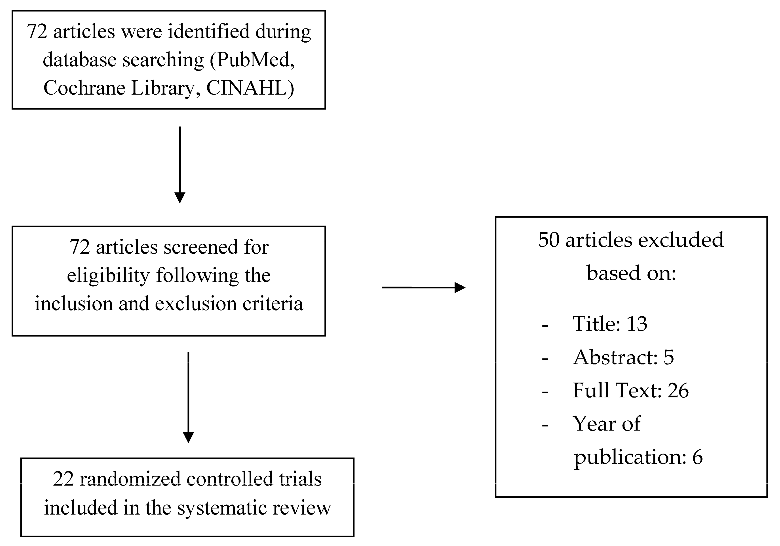
The results of individual studies are presented in
Table 2,
Table 3,
Table 4,
Table 5 and
Table 6. In order to evaluate the efficacy of chitosan–based scaffolds during bone regeneration, data about cell proliferation and viability were recorded from 10 of the included articles: five of them used the MTT assay of cementoblasts [
18,
19,
20], periodontal ligament cells [
18,
28,
37], one used the Cell-Counting Kit test (CCK-8) [
23,
26], four applied the AlamarBlue assay [
25,
29,
30,
32] and one the PrestoBlue test [
22] (
Table 2,
Table 3 and
Table 4). ALP activity of periodontal ligament cells [
21], osteoblasts [
34,
35], and mesenchymal stem or stromal cells [
24] was assessed in four studies (
Table 5). Three items analyzed the mineralization level of the newly formed bone with the Masson’s trichrome staining method [
27,
33,
38], two assessed it with the ARS staining [
31,
36] and one with the immunofluorescent staining technique for osteocalcin (OCN) [
39] (
Table 6). Twelve of the selected papers performed their experiment in vivo, creating bone defects, which were later covered by CS-based scaffolds: Ge et al. [
21] created bilateral parietal bone defects (with a diameter of 5 mm) in eighteen eight-week-old rats (weight = 180–220 g), scoring the anesthetized cranial skin, exposing calvaria; parietal cranial 15 mm oval-shaped defects were obtained by Jayash et al. [
25] using a bone trephine drill. A 5 mm diameter parietal defect was created in thirty-two five-month-old male rats by Li et al. [
27] thanks to a trephine drill under copious saline irrigation. Guo et al. [
23], after making an intraperitoneal injection with 10% chloral hydrate to 30 rats, performed a “V” type incision on the skull with a blade and drew with a drill, a 5 mm-diameter defect reaching the dura mater. Shah et al. [
32] tested the CS-based scaffold on a subcutaneous pouch of eight-week healthy adult rats weighing 140–180 g. Xue et al. [
36] anesthetize intramuscularly 3 white rabbits (4–6 months, weight 2–3 kg), exposing the lower edge of the mandible and creating a bone defect in the molar area of the mandibular body. Zang et al (2016) [
38], used one-wall, box-shaped, infrabony defects (4 mm width, 7 mm depth) at the distal and mesial aspects of the third premolars and at the mesial aspects of the first molars. In the study of 2019 by the same author, bilateral class III furcation defects (4 mm wide and 5 mm high) were created on the third and fourth mandibular premolars [
39]. The proliferation of cementoblasts (CB) and periodontal ligament cells (PDLCs) on pure chitosan scaffolds resulted in lower than those on the chitosan-based scaffolds combined with other molecules and biomaterials. The study by Akman et al. [
18] compared the proliferation of these two cellular type on chitosan- based scaffold with the addition of HA and bFGF with those on pure chitosan one, showing that the absorbance values (at 570 nm) of the cells on day 7 and 8 were equal to 1.7 (CB) and 1 (PDLCs) and 0.6 (CB and PDLCs), respectively. Pure chitosan scaffolds were investigated in comparison with IGF-1, and BMP-6 added CS/AL/PLGA and β-tricalcium phosphate/CS scaffolds in the research by Duruel et al. [
20] and Liao et al. [
28] respectively, recording a higher absorbance value of CB and PDLCs in the second groups (2/2.6 and 0.9/1 on day 12 and 6, respectively). The MTT assay of PDLCs showed no significant differences between an autoclaved chitosan powder/β-glycerophosphate thermosensitive hydrogel (CS-PA/GP) and an autoclaved chitosan solution/GP hydrogel: absorbance values at 490 nm were equal to 0.7 and 0.6, respectively [
37]. Dental pulp stem cells (DPSCs) were seeded on two scaffold types by Bakopoulou et al. [
19]: CS combined with gelatine (CS/Gel) fabricated using 0.1% and 1% of the crosslinker glutaraldehyde (GTA). The OD values (545–630 mm) recorded in the MTT assay of DPSCs seeded on the two scaffolds after seven days were 1.6 for CS/Gel-0.1 and 1.3 for CS/Gel-1 (
p < 0.01), but this statistical difference was compensated to non-significant at day 14. The trial by Miranda et al. [
29] cultured osteoblast- and fibroblast-like cells on CS-Ha hydrogel, Ha hydrogel, and pure CS scaffolds; a quantitative evaluation of cell viability was conducted for 24, 48, and 72 h, using Alamar Blue, which was also added to a phosphate buffer solution without cells: the test showed increased cellular viability (20%) in both cellular groups compared with the control one; however, none of them were statistically different. The metabolic activity of hPDLCs and human bone marrow stromal cells (hBMSCs) recorded by the AlamarBlue assay in the trial by Mota et al. [
30] presented higher values in the CS/bioactive glass nanoparticles (BG-NPs) membranes compared with CS one. The same test was performed by Jayash et al. [
25] in order to assess the cell viability in a new osteoprotegerin-chitosan gel. After 24 h, the viability of the OPG-CS and CS gels (25 and 50 kDa) was significantly higher than those of the controls (OPG-CS and CS = 140%, controls = 110%). The cell proliferation and viability (assessed with AlamarBlue) of MC3T3-E1 cells on the trilayered functionally-graded CS membrane (FGM) with bioactive glass gradient (50%, 25%, 0% wt.) resulted in being higher than those in the control group: the relative percentage AB reduction after seven days was equal to 150% for tge FGM group and 90% for the control one [
32]. In the in vitro study by Gümüşderelioğlu et al. [
22], CS-based multifunctional and double-faced barrier membrane was realized: hard tissue was put in contact with the porous side of the membrane coated with HA, in which BMP-6 was also embedded. The nonporous surface of the membrane was in contact with the inflammatory soft tissue, and it was coated with electrospun PCL fibers. PrestoBlue assay on day 21 assessed that mitochondrial activities of MC3T3-E1 cells seeded on different membranes showed no statistical differences (CS = 0.58, HA/CS = 0.63, HA/CS + BMP-6 = 0.64, HA/CS/PCL = 0.62). Data demonstrated that these cells grew on all CS-based scaffolds, recording higher cellular activity in HA/CS membrane. The CCK-8 test of PDLCs in the article by Li et al. [
26] demonstrated higher OD values (0.7) in CS-based hydrogel/α β –GP scaffold loaded with BMP2 plasmid DNA (pDNA-BMP2) than in those without pDNA-BMP2 (0.6). The same test used by Guo et al. [
23] highlighted better cell viability on the electrospun collage-chitosan composite membrane than in the electrospun collagen one (OD values were 0.7 and 0.4 respectively). Sundaram et al. [
34] analyzed the ability of a bilayered construct composed by PCL multiscale electrospun membrane and a chitosan/2 wt% CaSO
4 scaffold to regenerate periodontal ligament and alveolar bone simultaneously. The authors of this study found a higher level of alkaline phosphatase activity of hDFCs on day 7 in the latter group (ALP protein concentration = 8 ng/mg) than in the control one (ALP protein concentration = 3.5 ng/mg). Multitissue simultaneous regeneration was also studied by Varoni et al. [
35], who recorded no significant differences between the ALP activity of osteoblasts (OB) on day 7 provided by a CS-based genipin-cross-linked trilayered scaffold and the control group (460 and 480 pNpp/nmol min respectively). The RCT by Ge et al. [
21] measured the ALP activity of PDLSCs up to 14 days in two different scaffold types: nanohydroxyapatite-coated –genipin-CS conjuction and genipin-CS-framework, showing higher values in the first group on day 7 (30 u/gprot and 25 u/gprot respectively), but registering similar values in both groups on day 14. The ALP activity of hMSCs seeded on HCG membrane recorded by Hunter et al. [
24] showed a peak at 14 days of cultures, demonstrating that this type of membrane enhances hMSCs proliferation and osteogenic differentiation. Masson’s trichrome staining performed by Sukpaita et al. [
33] found an increased amount of collagen and bone matrix in CS/Dicarboxylic acid scaffold with and without PDLCs seeding. In the study by Zang et al. [
38], the same test showed more dense and well-organized PDLCs in the chitosan scaffold with hJBMMSCSs than the chitosan/anorganic bovine bone and pure chitosan groups. ARS staining of hPDLCs performed by Xue et al. [
36] recorded more mineralized nodules on the nPLGA/nCS/nAG complex than in negative control group, showing the that this type of membrane may promote cell mineralization. Human mesenchymal stem cells (MSCs) were cultured by Rammal et al. [
31] on a bone-mimetic material (B-MM) made from inorganic calcium phosphate combined with CS and hyaluronic acid biopolymers, which acted as a framework for the osteogenic potential of MSCs. ARS staining detected the formation by MMSCs of the mineralized matrix on B-MM, contrary to the control glass coverslip, on which no morphological changes and no nodules were found. In the study by Zang et al. [
39], the number of OCN-positive cells in beagles mandibular class III furcation defects resulted in being higher on CS/β-GP/BMP-7/ORN and on CS/β-GP/BMP-7 membranes (45 and 43, respectively) than those on CS/β-GP/ORN and control group (22 and 19, respectively). Finally, Li et al. [
27] used the Masson’s trichrome staining to compare the mineralization level of the newly formed bone (NB) in an injectable CS-based thermosensitive hydrogel scaffold with and without the incorporation of pDNA-BMP2 (CS/CSn(pDNA-BMP2)-GP). The study found out that the width of the NB was 500 µm for the first group and 300 µm for the second one, showing that CS/CSn-GP has greater capacity for alveolar bone regeneration when combined with pDNA-BMP2.

 ,
, 


