Article

Amino-Functionalized Fe3O4@SiO2 Core-Shell Magnetic Nanoparticles for Dye Adsorption

1
Department of Applied Physics, National Pingtung University, Pingtung City 90003, Taiwan
2
Kirensky Institute of Physics, FRC KSC SB RAS, 660036 Krasnoyarsk, Russia
3
Institute of Engineering Physics and Radio Electronics, Siberian Federal University, 660041 Krasnoyarsk, Russia
*
Authors to whom correspondence should be addressed.
Nanomaterials 2021, 11(9), 2371; https://doi.org/10.3390/nano11092371
Submission received: 22 August 2021 / Revised: 9 September 2021 / Accepted: 9 September 2021 / Published: 12 September 2021
(This article belongs to the Section Nanocomposite Materials)

Abstract

:
Fe3O4@SiO2 core-shell nanoparticles (NPs) were synthesized with the co-precipitation method and functionalized with NH2 amino-groups. The nanoparticles were characterized by X-ray, FT-IR spectroscopy, transmission electron microscopy, selected area electron diffraction, and vibrating sample magnetometry. The magnetic core of all the nanoparticles was shown to be nanocrystalline with the crystal parameters corresponding only to the Fe3O4 phase covered with a homogeneous amorphous silica (SiO2) shell of about 6 nm in thickness. The FT-IR spectra confirmed the appearance of chemical bonds at amino functionalization. The magnetic measurements revealed unusually high saturation magnetization of the initial Fe3O4 nanoparticles, which was presumably associated with the deviations in the Fe ion distribution between the tetrahedral and octahedral positions in the nanocrystals as compared to the bulk stoichiometric magnetite. The fluorescent spectrum of eosin Y-doped NPs dispersed in water solution was obtained and a red shift and line broadening (in comparison with the dye molecules being free in water) were revealed and explained. Most attention was paid to the adsorption properties of the nanoparticles with respect to three dyes: methylene blue, Congo red, and eosin Y. The kinetic data showed that the adsorption processes were associated with the pseudo-second order mechanism for all three dyes. The equilibrium data were more compatible with the Langmuir isotherm and the maximum adsorption capacity was reached for Congo red.

1. Introduction

Silica (SiO2) is frequently used as a support-material in core-shell structures; it does not only help nanoparticles (NPs) to become stable at certain conditions, but also provides them an opportunity to be easily modified with other functional groups and, additionally, to be environmentally compatible. Furthermore, silica coated magnetic NPs can be dispersed in water without adding other surfactants due to the negative charges on the silica shells. The fields of application of Fe3O4@SiO2 NPs functionalized with different surfactants, and more frequently with amines, are different. A number of authors have demonstrated their effective applications in biology and medicine [1,2], in catalysis [3,4], and, especially, as effective adsorbents for the removal of pollutants from wastewaters [5,6,7,8,9,10,11,12]. In ref. [1], the amino modifications of the silica surface of Fe3O4@SiO2 NPs reduced the detrimental interactions with cellular membranes and prolonged the blood circulation time after in vivo administration. In ref. [12], the magnetic core-shell Fe3O4@SiO2 NPs synthesized by the modified Stöber method and functionalized with amino and carboxyl groups were used as a nano-adsorbent for scandium ions from aqueous solutions. In Refs. [7,9], the synthesized Fe3O4@SiO2-NH2 nanocomposites were embedded into the polyether-sulfone membranes with different concentrations via the phase inversion method. Due to the nanocomposite adsorption properties, the significant enhancement in efficiency of the modified membranes for the removal of Cd(II) ions and methyl red dye was achieved. A new type of magnetic fluorescent nanocomposite (Fe3O4@SiO2-NH2/CQDs) was prepared by bonding of carbon quantum dots (CQDs) with Fe3O4@SiO2-NH2 nanocomposites through amine-carbonyl interactions and used as a fluorescent probe to detect Cu2+ [13]. The authors of ref. [11] concluded that the strategy of coating with silica and amino group functionalization of Fe3O4 nanoparticles is useful for increasing the adsorption capacity. In particular, they obtained adsorption capacities of 29.3 and 28.6 mg/g for light green and brilliant yellow anionic dyes, respectively, using Fe3O4@SiO2@NH2 NPs, while 12.1 and 9.4 mg/g only for Fe3O4 NPs. Since this line of research is rather new, and the properties and application possibilities of NPs strongly depend on the details of their synthesis, the search for optimal synthesis conditions and the study of the properties of the functionalized particles by various methods can be considered as an urgent task.
The present work is devoted to the study of the morphology, magnetic and adsorption properties of Fe3O4 NPs obtained by the co-precipitation method and then coated with amorphous silica to produce Fe3O4@SiO2 core-shell NPs, which was functionalized by an amine group to fabricate Fe3O4@SiO2-NH2 nanocomposites. The methylene blue (MB), Congo red (CR) and eosin Y (EY) were selected as the typical organic cation and anion pollutants to test the ability of the prepared composite for the adsorptive removal of organic pollutants from water. The study of the fluorescent properties of eosin Y-doped NPs was also carried out.

2. Materials and Methods

2.1. Synthesis Procedure

Ferrous sulfate heptahydrate (FeSO4·7H2O) (>99%) was obtained from Sigma-Aldrich, ethanol (CH3CH2OH) (>95%) was obtained from Fullin Nihon Shiyaku Biochemical Ltd., tetraethyl orthosilicate (Si(OC2H5)4, TEOS) (>99%) and ammonia solution (28–30 wt.% NH3) were purchased from Acros Organics. Potassium nitrate (KNO3) (>99%) was purchased from Wako Pure Chemical Industries, Ltd. Sodium hydroxide pellets was obtained from PanReac AppliChem. Poly(sodium 4-styrene-sulfonate) solution (30 wt.%) in water was purchased from Aldrich. All the chemicals were used without further purification. Several stages were used to synthesize nanocomposites.
Magnetic NPs, Fe3O4, were produced by co-precipitation, hydrolysis of iron sulfate under Ar flow. In the typical synthesis, a mixture of the basic solution is 0.02 mol KNO3 and 0.2 mol NaOH in 50 mL of ultrapure water, previously deoxygenated, was prepared under Ar and, then was added dropwise to the Fe (II) solution (one is 0.25 M, the other is 0.75 M, and mixed together), under Ar and at 90 °C. The black precipitate formed was stirred over 1 h at 90 °C, then, washed thoroughly with ethanol. Silica coating of the magnetite nanoparticles was performed via the Stöber process. First, Fe3O4 NPs (200 mg) were dispersed in ethanol (150 mL) and kept immersed in a water bath over 15 min under sonication. Then, an ammonia solution (6 mL) and TEOS (200 μL) were slowly added to the Fe3O4 suspension. Finally, the SiO2 coated particles were collected magnetically using an NdFeB magnet, and the isolated powders were washed thoroughly with ethanol (sample Fe3O4@SiO2). The amino functionalization of silica coated NPs was made in the same manner as is mentioned above, replacing TEOS to (3-aminopropyl)-triethoxysilane (APTES) (sample Fe3O4@SiO2-NH2). At last, eosin Y was attached to the amino silanized magnetic nanoparticles covalently. First, Fe3O4@SiO2-NH2 NPs (200 mg) were dispersed in ultrapure water (50 mL) and over 15 min were exposed to sonication. The 0.2 mmol eosin Y and catalyst were added to the NPs suspension under stirring during 1 h, then, washed thoroughly with ethanol (sample Fe3O4@SiO2-NH2-EY).

2.2. Characteristic Methods

The crystal structure of the NPs was characterized by X-ray diffraction measurements using a Bruker D8 Advance diffractometer (Cu Kα radiation, 40 kV, 25 mA, λ = 1.5418 Å) (Bruker Optik GmbH, Ettlingen, Germany).
The morphology and microstructure of the NPs were investigated using transmission electron microscopy (TEM). TEM experiments were performed with a JEM-2100 (JEOL Ltd., Tokyo, Japan) microscope operating at the accelerating voltage of 200 kV (Siberian Federal University) and JEOL JEM-1230 microscope (JEOL Ltd., Tokyo, Japan) operated at an accelerating voltage of 80 kV (Precision Instruments Center of NPUST). Selected-area electron diffraction (SAED) was used to determine the structure of the NPs.
Fourier transform infrared absorption (FT-IR) spectra were recorded with a VERTEX 70 (Bruker Optik GmbH, Ettlingen, Germany) spectrometer in the spectral region of 400 ÷ 4000 cm−1 with the resolution 4 cm−1. The spectrometer was equipped with a Globar as the light source and a wide band KBr beam splitter and RT-DLaTG as the detector (Bruker Optik GmbH). For the measurements, round tablet samples of about 0.5 mm thick and of 13 mm in diameter containing NPs were prepared as follows: nanopowders in amount lower than 0.001 g were thoroughly ground with 0.14 g of KBr; the mixtures were formed into tablets which were subjected to cold pressing at 10,000 kg.
The magnetic properties were measured with the vibrating sample magnetometer Lakeshore 7400 series VSM (Lake Shore Cryotronics, Inc., Westerville, OH, USA).
The excitation and fluorescence spectra were measured on a Fluorolog 3–22 spectrofluorometer (Horiba Jobin Yvon, Edison, NJ, USA). The obtained spectra were corrected for sensitivity of PMT, reabsorption effects and background. The quartz cells with the cross sections of 10 × 10 mm2 were used to investigate the solutions for L-geometry of excitation. All the measurements were carried out at room temperature.
The absorption spectra were recorded with a UV/VIS circular dichroism spectrometer SKD-2MUF (OEP ISAN, Moscow, Russia). Quartz cells with the optical path length of 5 mm were used.

3. Results and Discursion

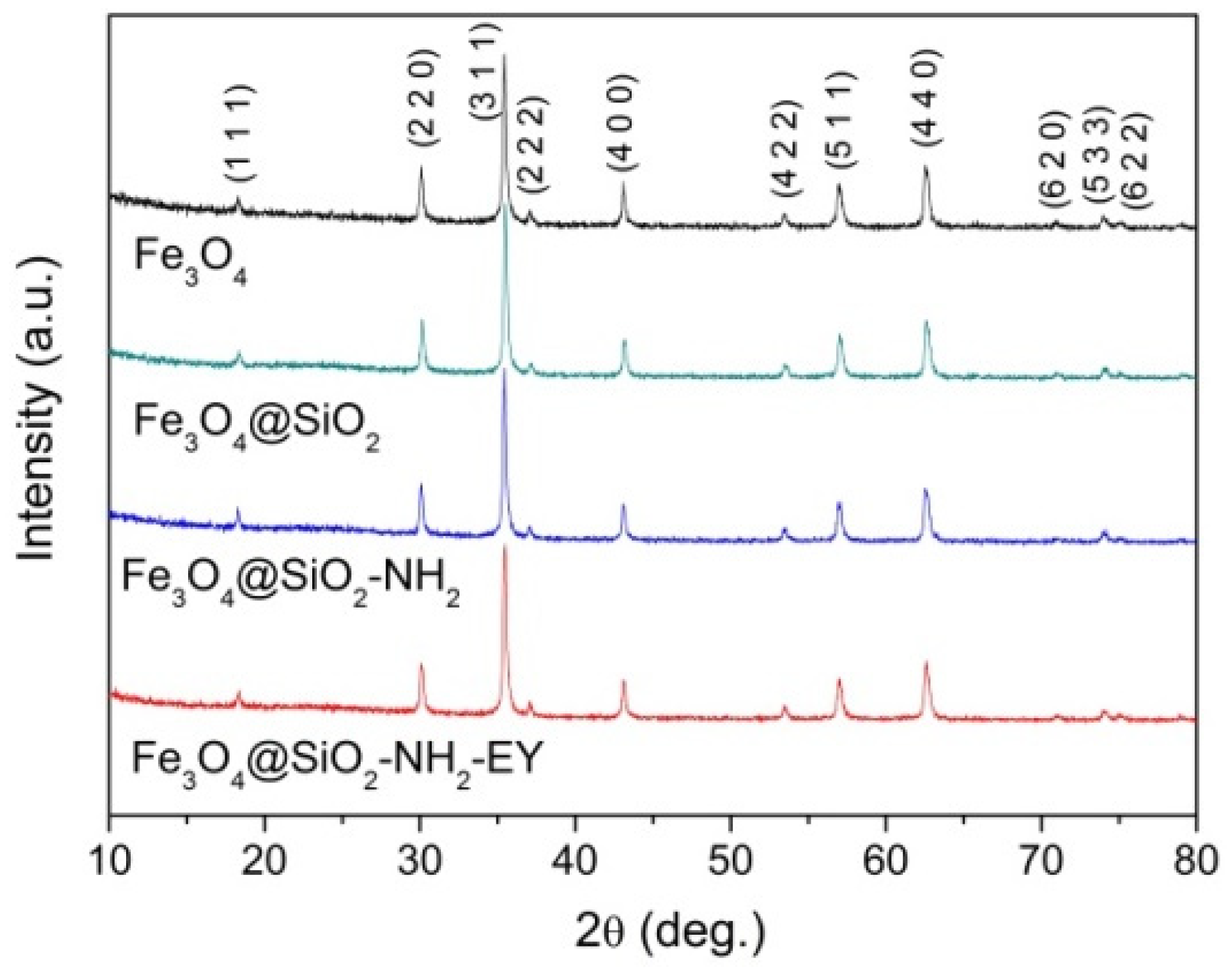
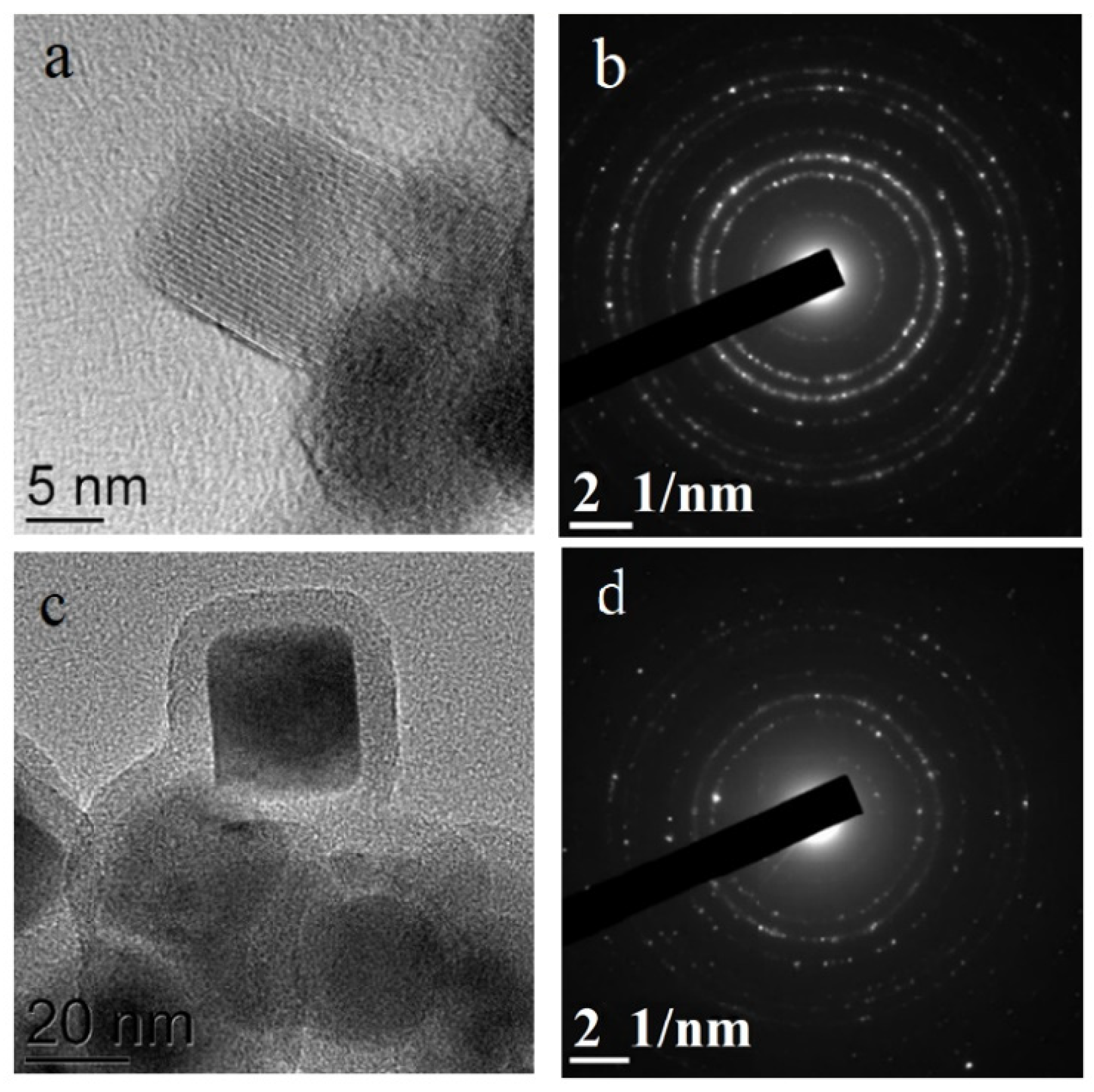
3.1. NPs Structure and Morphology

The XRD patterns (Figure 1) revealed that the parent NPs and magnetic cores of all the nanocomposites were of the spinel ferrite crystal structure with the parameters of the most intense peaks corresponding to the Fe3O4 phase (PDF Card # 04-005-4319).
The TEM images (Figure 2a–d) revealed NPs of predominantly rectangular shape with the average size of 25 ± 5 nm (Figure 2e). At the same time, a small amount of ellipsoidal NPs with the sizes of about 10 nm and rectangular NPs of a larger size can be noticed. The almost ideal crystal structure of the initial Fe3O4 NPs is seen very well in the HRTEM image in Figure 3a. As a result of the silica coating, the initial NPs became covered with a homogeneous amorphous silica shell, 6–7 nm thick seen especially well in the HRTEM image (Figure 3b). Functionalization with NH2 and further doping with eosin Y led to some blurring of the HRTEM image (Figure 3c,d). The selected area electron diffraction (SAED) patterns also shown in Figure 3c,d confirm the presence of the Fe3O4 crystalline core in all NPs.
The FT-IR spectra (Figure 4) show the appearance of new bands upon the transition Fe3O4 → Fe3O4@SiO2 → Fe3O4@SiO2-NH2 → Fe3O4@SiO2-NH2-EY evidencing the appearance of chemical bonds. In the spectrum of pure Fe3O4 NPs, the strong band at 580 cm−1 is due to Fe-O stretching vibrations in accordance with other authors [9,11,13]. This band is observed in the spectra of all the samples. The wide asymmetric band at 1094 cm−1 appeared in the spectrum of the Fe3O4@SiO2 sample and is seen in the spectra of two next samples having a SiO2 shell. This band can be related to the asymmetric stretching vibrations of Si-O-Si. An analogous band was observed and interpreted equally by all the authors studying Fe3O4@SiO2 NPs [9,11,13]. Symmetric Si-O-Si stretching vibrations were associated in [9] with the weak band near 808 cm−1. The same weak band is seen at 800 cm−1 in Figure 4. At the same time, the authors of ref. [13] associated the band at 797 cm−1 with the Si-O-Fe stretching vibrations proving the presence of a chemical bond between the magnetic core and silica shell [9]. Only minor changes appear in the spectra of the Fe3O4@SiO2-NH2 and Fe3O4@SiO2-NH2-EY samples. The appearance of a wide band near 3430 cm−1 is a more noticeable feature characteristic of the NH2 amino group [14]. A small amount of covalently attached eosin Y in comparison with Fe3O4@SiO2-NH2 NPs does not allow one to clearly see changes in the bending and stretching vibrations of the amino groups.

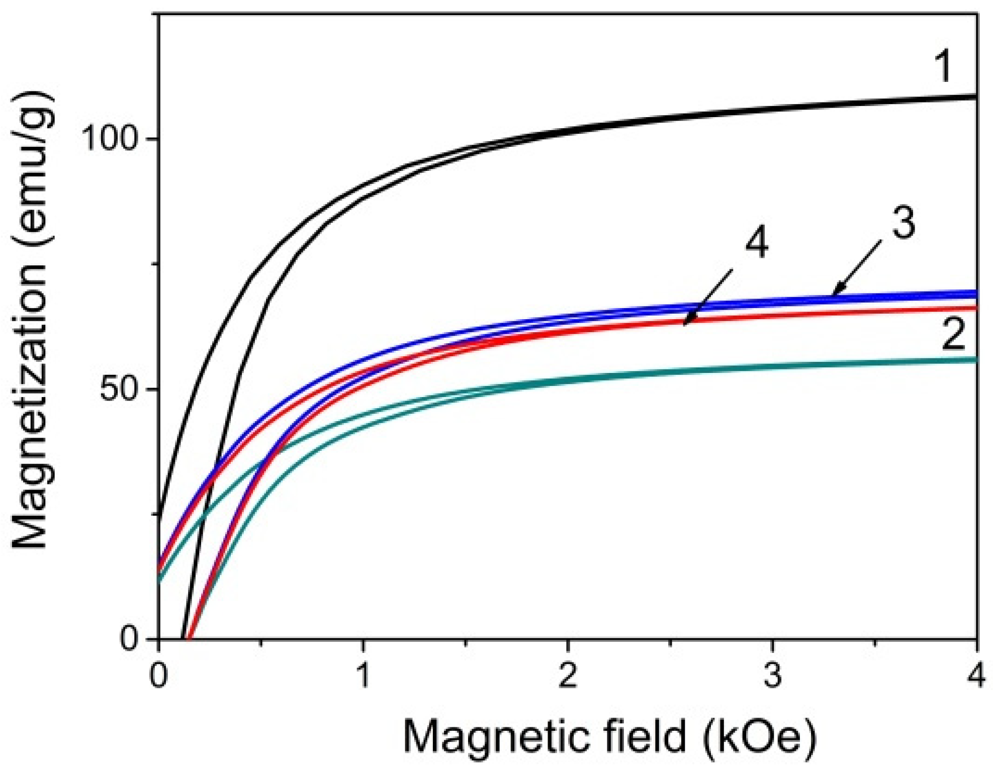
3.2. NPs Magnetic Properties

The magnetic measurements (Figure 5) show very narrow hysteresis loops with magnetic saturation in the external magnetic field near 3 kOe and coercive force of about 100 Oe. The saturation magnetization Ms value of the initial Fe3O4 NPs is exceptionally high, significantly higher than the one presented by other authors for Fe3O4 NPs, for example [11] and even higher than in bulk Fe3O4 samples, 92 emu/g at room temperature [15]. The redistribution of Fe ions between the oppositely magnetized tetrahedral and octahedral sublattices in magnetite NPs caused, for example, by the technological conditions can be one of the reasons of the observed Ms increase. Since the resulting magnetization of the sample is due to the difference between the magnetic moments of Fe ions occupying octahedral and tetrahedral positions, the Fe ion deficiency in tetrahedral position can be responsible for an increase of the sample magnetization.
The surface modifications lead to the Ms decrease. However, it remains quite high. A similar decrease in the saturation magnetization of Fe3O4 NPs coated with silica compared to the initial Fe3O4 nanoparticles was noted by a number of authors. For example, in ref. [11] Ms was equal to 38, 23, and 11 emu/g for Fe3O4, Fe3O4@SiO2 and Fe3O4@SiO2@NH2 NPs, correspondingly; in ref. [16]—80, 31 and 20 emu/g for Fe3O4, Fe3O4@SiO2 and Fe3O4@SiO2-NH2 NPs respectively.
The decrease in magnetization of NPs after coating with silicon oxide(curve 2 in Figure 5) can be caused by a number of factors. First of all, when determining the magnetization value, the whole particle mass was taken into account including the silica shell. In addition, the shell can affect the spin state of the magnetic core surface layers. However, the addition of the amino groups and then eosin Y resulted in an increase in magnetization. Such a behavior of the magnetization was not previously observed in the above-cited and other works. It is possible that, in the process of amino functionalization, cation redistribution between the sublattices continued. This question needs special consideration.

3.3. Application of Synthesized NPs as Fluorescent Probes

The development of hybrid nanoparticle technology, synthesized fluorescent nanoparticles with encapsulated quantum dots [17], or dyes [18] have attracted great interest in recent years. Despite the excellent brightness and photostability of quantum dots for imaging applications, the risk of systemic toxicity remains high due to the incorporation of heavy metals. Thus, dye-doped nanoparticles still appear very promising. The simultaneous combination of fluorescent and magnetic properties of NP imaging would greatly benefit in the diagnostics and monitoring of living cells and organisms [19,20].
The spectral properties of the synthesized EY-doped Fe3O4@SiO2@NH2 dispersed in water solution are presented in Figure 6a. The fluorescence spectrum turned out to be independent of the excitation wavelength and NPs give the green emission with the maximum at 542 nm with the excitation maximum at 515 nm. The fluorescent spectrum of EY-doped NPs dispersed in solution displays a red shift of 6 nm and 40 % broadening as compared with the dye molecules free in water in Figure 6b. On the contrary, the maximum of the excitation spectrum (measured at 580 nm) shows a slight blue shift of 1 nm and 30% broadening. The reasons for the observed spectral changes could include the change in the ionic form of the EY molecules and polarity decrease in the microenvironment. In the water solution, pH 6, most of the EY molecules are in the dianionic form. When attached to the amino groups in NPs, the dye changes its ionic form to the anionic or even neutral one and the spectral properties of these forms are different, on the one hand [21]. On other hand, as the solvent polarity decreases, the red shift in the emission of EY is observed [22].

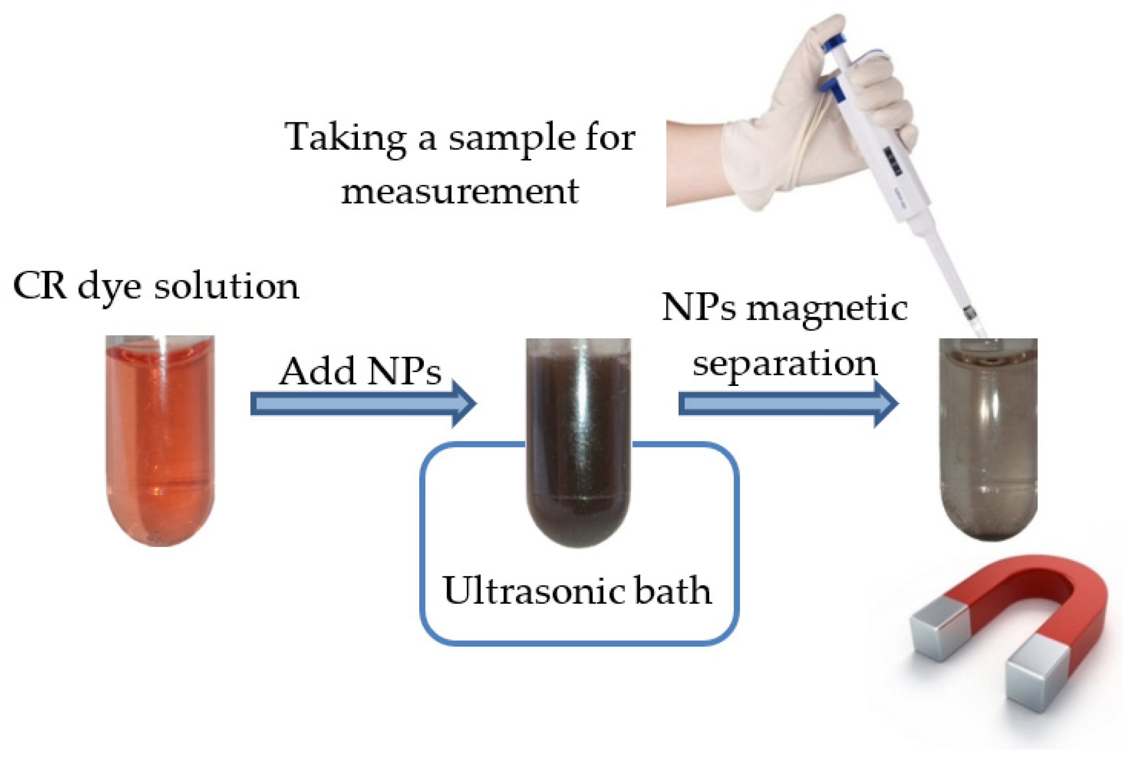
3.4. Application of Synthesized NPs for Dye Adsorption

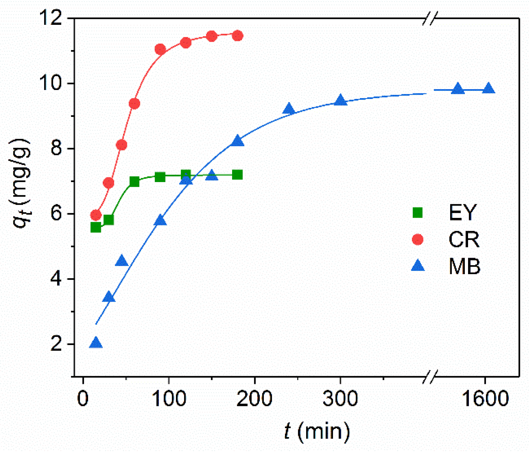
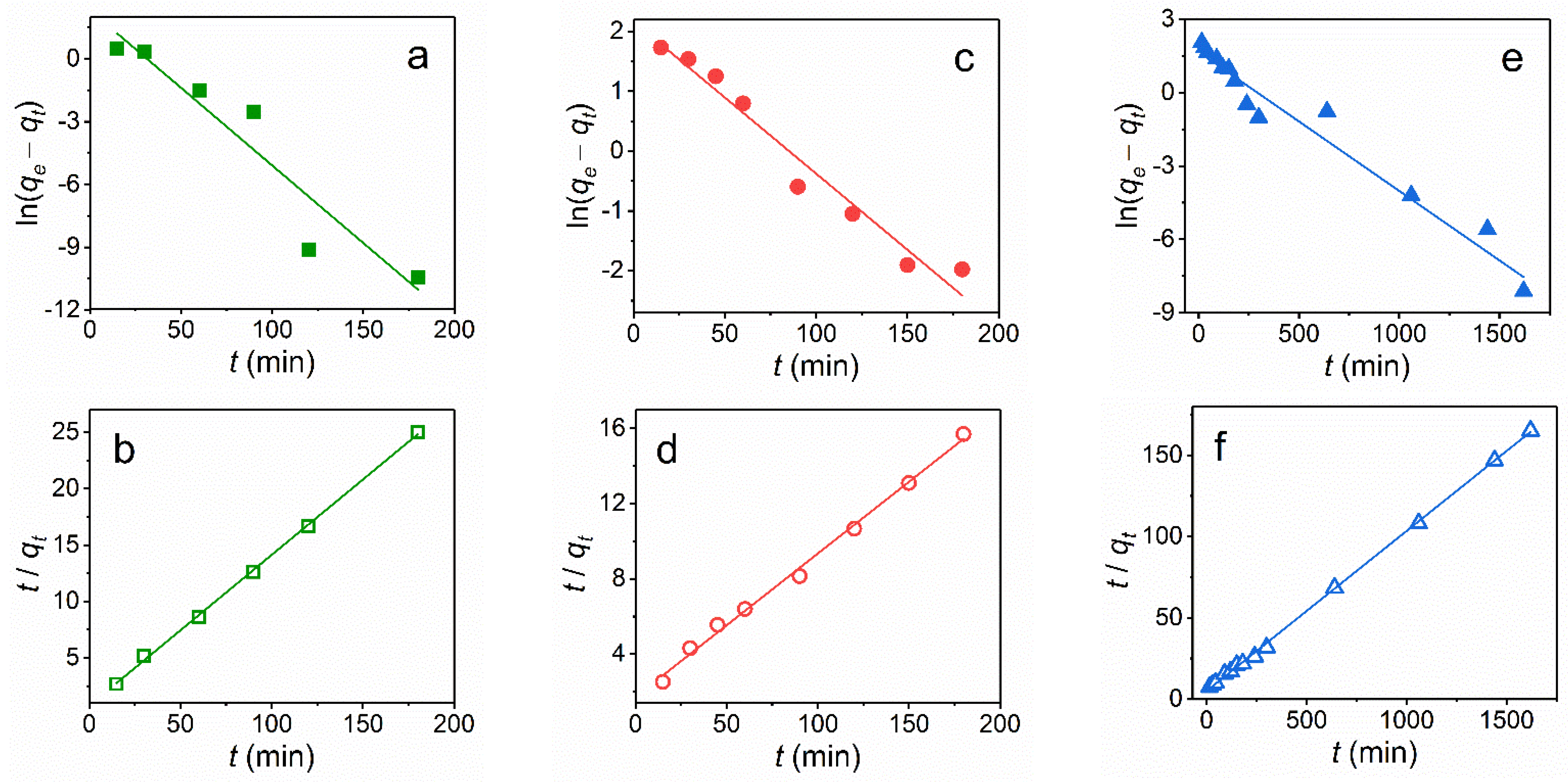
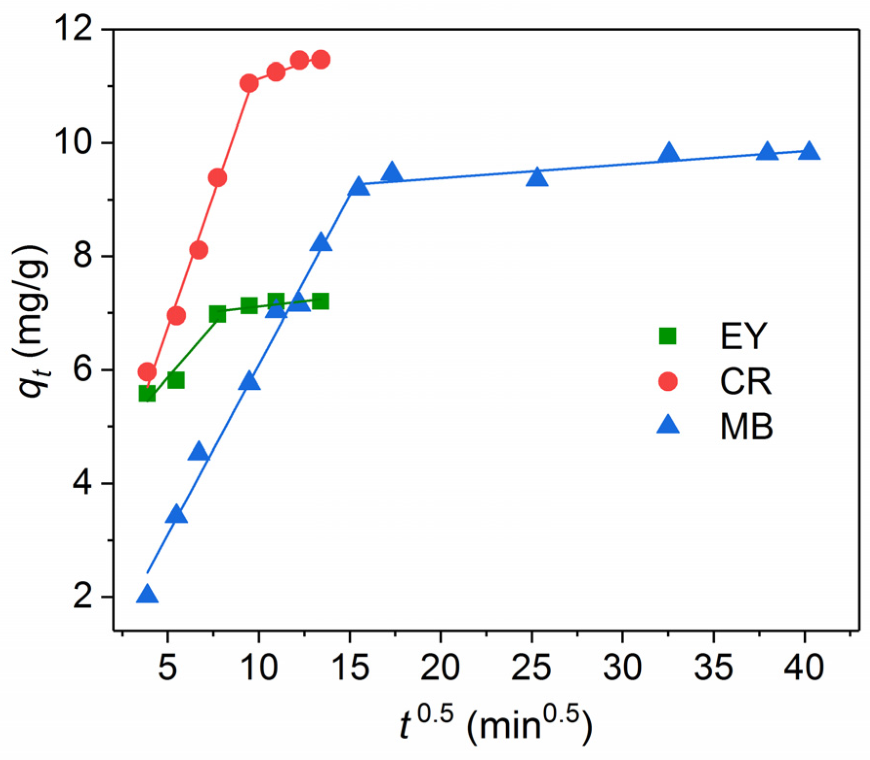
3.4.1. Adsorption Kinetics

The spectral properties of two anionic (eosin Y (EY) and Congo red (CR), and one cationic) methylene blue (MB) dyes were used to find out the adsorption capacity of amino-functionalized Fe3O4@SiO2 core-shell magnetic NPs in distilled water (measured pH 5.5) at 25 °C. The dye concentration was determined by absorbance at the wavelength corresponding to the maxima in the spectra of 490 nm for eosin Y, 505 nm for CR, and 664 nm for MB.
For a typical experiment shown schematically in Figure 7, 3 mg of adsorbent was dispersed in 1.5 mL of the dye aqueous solution at the initial concentration of the dye C0 = 30 mg/L. The solution was placed in an ultrasonic bath for 10 min for intensive mixing. Then, a magnetic nanoadsorbent was separated from the solution by applying a magnetic field and the absorption spectra of the solution were measured. The shaking and magnetic separation was repeated multiple times to obtain kinetic curves. The value of the adsorption capacity qt (mg/g) of NPs was calculated as follows
q t = ( C 0 C t ) V m ,
where C0 and Ct (mg/L) are the initial concentration and concentration of the dye at the contact time t, V (L) is the volume of the solution; and m (g) is the mass of the adsorbent (NPs).
The effect of the contact time on the adsorption of Fe3O4@SiO2-NH2 NPs is shown in Figure 8 for the dyes at the same initial concentration of C0 = 30 mg/L. At the initial stage of adsorption kinetics, the amount of the adsorbed dye onto the magnetic NPs increases rapidly due to a large number of vacant active sites on the surface of the amino-functionalized silica coated magnetic NPs. After that, these surface sites are gradually occupied, so the adsorption rate decreases until the adsorption equilibrium is established. The time to reach the equilibrium adsorption for the anionic dyes on Fe3O4@SiO2-NH2 NPs is much faster than for the cationic dye: 60 min for EY, 100 min for CR, and more than 300 min for MB. The other adsorbent Fe3O4@SiO2@NH2@Zn−TDPA NPs [TDPAT = 2,4,6-tris(3,5-dicarboxyl phenylamino)-1,3,5-triazine] showed the similar time of 120 min both for the cationic MB and anionic CR dye [23]. The adsorption of CR by the Fe3O4@SiO2 nanospheres achieved the equilibrium only after 5 h [24]. The equilibrium adsorption for initial concentration of C0 = 30 mg/L was measured after 24 h of contact of NPs with the dye solution. The equilibrium adsorption capacity qe of CR (11.6 mg/g) is higher than that of EY (7.2 mg/g) and MB (9.8 mg/g).
A quantitative understanding of the adsorption is possible with the help of kinetic models. The adsorption kinetics of the dyes on the magnetic NPs was described by the kinetic models of the pseudo-first order:
ln ( q e q t ) = ln q e k 1 t
and of the pseudo-second order:
t q t = 1 k 2 q e 2 + t q e ,
where qe and qt (mg/g) are the amounts of the dye at equilibrium and at contact time t, respectively; k1 (1/min) and k2 (g/(mg min)) are the adsorption rate constants of the reaction of the pseudo-first and pseudo-second orders, respectively.
The curve-fitting plots of the adsorption ability of the dyes by Fe3O4@SiO2-NH2 NPs using the two kinetic models are shown in Figure 9, and the fitted parameters with the correlation coefficients R2 are summarized in Table 1. The R2 values for the pseudo-second order kinetic model (R2 = 0.995–0.999) were higher than those of the pseudo-first order model (R2 = 0.903–0.970). In addition, the calculated values of qe (Table 1) determined by the pseudo-second order model are more consistent with the measured values of qe than that of the pseudo-first-order model. These results prove that the adsorption process of these dyes on Fe3O4@SiO2-NH2 NPs completely followed a the pseudo-second order kinetic model, suggesting that adsorption is dependent on the amount of the solute adsorbed on the surface of the adsorbent and the amount of active sites. It should be noted that the dye adsorption kinetics on magnetic nanoparticles is most often described in terms of the pseudo-second order model [23,24,25,26,27,28,29,30].
The intraparticle diffusion model is often used to identify diffusion mechanisms [23,25,26,27,28,30,31]. In this model, the rate of intraparticle diffusion is a function of t0.5 and can be determined as follows:
q t = k i t 0.5 + C ,
where ki is the intraparticle diffusion rate constant, (mg/g.min0.5). C is the intercept of the linear curve. According to the model proposed by Weber and Morris [32], the adsorption process is controlled only by intraparticle diffusion if the plot is a straight line and passes through the origin. Otherwise, if the plot is multilinear or does not pass the origin, more than one diffusion mechanism might determine the adsorption process and adsorption is related to diffusion within the particles.
As shown in Figure 10, at least two stages are observed for anionic CR, EY as well as for the cationic MB dye, and thus, two or more diffusion mechanisms can affect the adsorption. The first stage refers to the transport of the dye molecules from the solution to the external surface of NPs. This stage is completed after up to 60 min for EY, 90 min for CR, and 240 min for MB. The second stage corresponds to the diffusion of the dye molecules within the micropores of Fe3O4@SiO2-NH2 NPs. The high initial absorption rates ki1 of the first stage (Table 1) are observed for all the dyes, indicating a fast initial dye removal process and the predominant role of external surface diffusion, especially, for CR. Extremely low adsorption rates ki2 (8–25 fold less than for the first stage) shows a negligible proportion of intraparticle diffusion of the dye molecules within the micropores of NPs. A similar two-stage adsorption process was observed for MB onto B-Fe3O4@C NPs [28], for CR and MB dyes onto Fe3O4@SiO2@NH2@Zn−TDPA NPs [23], and for EY onto Fe3O4/polypyrrole composites [30].

3.4.2. Adsorption Isotherms

Two adsorption isotherm models (Langmuir and Freundlich) were applied to understand the adsorbate–adsorbent interaction. The Langmuir equation was defined as follows
q e = q max K L C e K L C e + 1 ,  
where qe (mg/g) is the amount of the dye adsorbed at the equilibrium, qmax represents the maximum adsorption capacity (mg/g), KL is the Langmuir adsorption constant (L/mg), and Ce is the equilibrium concentration of the adsorbed dye (mg/L).
The Freundlich equation is expressed as:
q e = K F C e 1 / n ,
where KF is the Freundlich adsorption constant (L/mg); the dimensionless constant 1/n is an empirical parameter related to the isotherm shape. Based on the 1/n values, the adsorption process can be classified as irreversible (1/n = 0), favorable (0 < 1/n < 1), or unfavorable (1/n > 1) [33].
The Langmuir model illustrates the formation of a homogeneous adsorbed monolayer, while the adsorbed molecules do not interact with each other. The Freundlich model considers the existence of a more complicated multilayered structure.
Based on the results presented above, we have chosen the anionic CR dye for further investigation owing to its highest capacity and shortest time of the adsorption process. The adsorption isotherm models were used to describe the equilibrium between the adsorbed CR on the surface of Fe3O4@SiO2-NH2 NPs (Figure 11). The obtained parameters are presented in Table 2. According to the correlation coefficients, the Langmuir model (R2 = 0.967) coincided with the experimental data much better than the Freundlich model (R2 = 0.789), which indicates that the homogeneous and monolayer adsorption is the dominant adsorption in the case of CR. Although the 1/n value is lower than 1 and indicates that the adsorption of CR is favorable, the R2 values of the Freundlich isotherm do not fall within the acceptable range. In contrast to our results, the adsorption isotherm of CR on Fe3O4@SiO2@Zn-TDPAT NPs was satisfactorily described both by the Langmuir and Freundlich models [23].
The adsorbents based on Fe3O4@SiO2 nanoparticles for the removal of the anionic CR and cationic MB dyes are summarized in Table 3. The maximum adsorption capacity qmax of the synthesized amino-functionalized Fe3O4@SiO2 core-shell magnetic nanoparticles is comparable to other analogous adsorbents [10,23,24,29,34].
As is seen from Table 3, the adsorption efficiency can further be improved by the surface modification of NPs. It should be emphasized that the pH and ionic strength of the aqueous solution [23] as well as the temperature [24] could also significantly influence the efficiency of the dye adsorption on NPs due to the involvement of different adsorption mechanisms including electrostatic and hydrophobic interactions, hydrogen bonding, van der Waals forces, etc. The measured zeta potential of the Fe3O4@mSiO2-NH2 nanoparticles vs. pH showed [35] that the isoelectric point was at 5.9 due to the amino-functionalization, denoting that the modified nanoparticles would be positively charged at pH < 5.9 and negatively charged at pH > 5.9. Under the conditions of our experiment (the initial pH 5.5), the surface of the magnetic NPs had a slightly positive charge. Table 3 indicates that the maximum adsorption capacity qmax is close for both anionic CR and cationic MB dyes, so the electrostatic interactions are not only responsible for the adsorption of the dyes on NPs.

4. Conclusions

In this study, core-shell magnetic nanoparticles, Fe3O4@SiO2, were synthesized by silica coating of the initial Fe3O4 NPs via the Stöber process and then functionalized with amino groups NH2. X-ray and electron diffraction data showed the magnetic core of the particles to have the magnetite Fe3O4 crystal structure without the presence of any other phases. Transmission electron microscopy showed predominantly rectangular NPs with the average size of 25 ± 5 nm in the initial Fe3O4 powder sample. The homogeneous amorphous silica shells with the thickness of about 7 nm were formed around each initial NP. The FT-IR spectra confirmed the appearance of the chemical bonds between the silica shell and magnetic core of NPs as well between the silica and amino group. The magnetic measurements revealed unusually high saturation magnetization Ms of the initial Fe3O4 NPs even higher than this value of the bulk magnetite crystal. Ms of the functionalized samples also significantly exceeded Ms of similar samples presented in literature. The high Ms value can be considered as an advantage of the studied nanomaterials since higher magnetization requires the use of weaker magnetic fields to control the processes involving these materials.
The fluorescence spectrum of EY-doped Fe3O4@SiO2@NH2 NPs dispersed in water solution was studied. NPs gave the green emission with the maximum at 542 nm with the excitation maximum at 515 nm. The spectrum displayed a red shift of about 6 nm and 40% broadening as compared to the dye molecules free in water while the maximum of the excitation spectrum (measured at 580 nm) showed a slight blue shift of 1 nm and 30% broadening. The observed spectral changes were associated with the change in the ionic form of the EY molecules due to the attachment to the amino groups in NPs.
The dye adsorption capacity and kinetics of the amino-functionalized Fe3O4@SiO2 core-shell magnetic NPs were studied in application to two anionic (eosin Y (EY) and Congo red (CR), and one cationic) methylene blue (MB) dyes. It was shown that the adsorption process of these dyes on the studied NPs followed the pseudo-second order kinetic model, suggesting that sorption is dependent on the amount of the solute adsorbed on the surface of the adsorbent and the amount of active sites. At least two stages were revealed in the adsorption time dependence for all three dyes. In the first stage, the transport of the dye molecules from the solution to the external surface of NPs occurs. The second stage corresponds to the diffusion of the dye molecules within the micropores of NPs. The high initial absorption rates of the first stage were observed for all the dyes, indicating a fast initial dye removal process and the predominant role of the external surface diffusion, especially, for CR. It was shown that the experimental data were fitted to the Langmuir model of the adsorption processes indicating that the homogeneous and monolayer adsorption was the dominant adsorption in the considered cases. The maximum adsorption capacity of the synthesized amino-functionalized Fe3O4@SiO2 core-shell magnetic nanoparticles is comparable to other analogous adsorbents presented in literature.

Author Contributions

Conceptualization, C.-R.L. and I.S.E.; data curation, O.S.I., A.E.S., and D.A.P.; formal analysis, M.A.G.; funding acquisition, C.-R.L. and I.S.E.; investigation, M.A.G., O.S.I., D.A.P., A.E.S., S.M.Z., Y.-Z.C., and N.P.S.; methodology, M.A.G., O.S.I., and S.M.Z., Y.-T.T.; project administration, C.-R.L. and I.S.E.; resources, A.E.S. and Y.-Z.C.; supervision, C.-R.L. and I.S.E.; validation, I.S.E.; visualization M.A.G., Y.-T.T., and O.S.I.; writing—original draft, I.S.E.; writing—review and editing, M.A.G. and I.S.E. All authors have read and agreed to the published version of the manuscript.

Funding

The authors are thankful for the financial support the Russian Foundation for Basic Research, Grant № 19-52-52002, Ministry of Science and Technology of Taiwan, Grants MOST № 108-2923-M-153-001-MY3 and № 109-2112-M-153-003-, the Russian Foundation for Basic Research with Government of Krasnoyarsk Territory, Krasnoyarsk Regional Fund of Science, the research project number 19-42-240005: “Features of the electronic structure, magnetic properties and optical excitations in nanocrystals of the multifunctional magnetic chalcogenides Fe3S4 and FeSe”. We thank also the SFU Joint Scientific Center supported by the State assignment (#FSRZ-2020-0011) of the Ministry of Science and Higher Education of the Russian Federation, where the Transmission Electron Microscopy studies were carried out.

Institutional Review Board Statement

The study was conducted according to the guidelines of the Declaration of Helsinki, and approved by the Institutional Review Board of Kirensky Institute of Physics (b/n 20.08.2021).

Informed Consent Statement

Informed consent was obtained from all subjects involved in the study.

Data Availability Statement

The data presented in this study are available on request from the corresponding author. The data are not publicly available since they are a part of ongoing research.

Conflicts of Interest

The authors declare no conflict of interest.

References

  1. Li, J.; Yuan, Z.; Liu, H.; Feng, J.; Chen, Z. Size-dependent tissue-specific biological effects of core–shell struc-tured Fe3O4@SiO2–NH2 nanoparticles. J. Nanobiotechnol. 2019, 17, 124. [Google Scholar] [CrossRef] [Green Version]
  2. Aslani, E.; Abri, A.; Pazhang, M. Immobilization of trypsin onto Fe3O4@SiO2–NH2 and study of its activity and stability. Colloids Surf. B Biointerfaces 2018, 170, 553–562. [Google Scholar] [CrossRef]
  3. Izgi, M.S.; Ece, M.Ş.; Kazici, H.Ç.; Şahin, Ö.; Onat, E. Hydrogen production by using Ru nanoparticle decorated with Fe3O4@SiO2–NH2 core-shell microspheres. Int. J. Hydrog. Energy 2020, 45, 30415–30430. [Google Scholar] [CrossRef]
  4. Veisi, H.; Ozturk, T.; Karmakar, B.; Tamoradi, T.; Hemmati, S. In situ decorated Pd NPs on chitosan-encapsulated Fe3O4/SiO2-NH2 as magnetic catalyst in Suzuki-Miyaura coupling and 4-nitrophenol reduction. Carbohydr. Polym. 2020, 235, 115966. [Google Scholar] [CrossRef]
  5. Salman, M.; Jahan, S.; Kanwal, S.; Mansoor, F. Recent advances in the application of silica nanostructures for highly improved water treatment: A review. Environ. Sci. Pollut. Res. 2019, 26, 21065–21084. [Google Scholar] [CrossRef] [PubMed]
  6. Xie, H.; Wu, Z.; Wang, Z.; Lu, J.; Li, Y.; Cao, Y.; Cheng, H. Facile fabrication of acid-resistant and hydrophobic Fe3O4@SiO2@C magnetic particles for valid oil-water separation application. Surf. Interfaces 2020, 21, 100651. [Google Scholar] [CrossRef]
  7. Zhang, S.; Zhang, Y.; Liu, J.; Xu, Q.; Xiao, H.; Wang, X.; Xu, H.; Zhou, J. Thiol modified Fe3O4@SiO2 as a robust, high effective, and recycling magnetic sorbent for mercury removal. Chem. Eng. J. 2013, 226, 30–38. [Google Scholar] [CrossRef] [Green Version]
  8. Ghorbani, F.; Kamari, S. Core–shell magnetic nanocomposite of Fe3O4@SiO2@NH2 as an efficient and highly re-cyclable adsorbent of methyl red dye from aqueous environments. Environ. Technol. Innov. 2019, 14, 100333. [Google Scholar] [CrossRef]
  9. Kamari, S.; Shahbazi, A. Biocompatible Fe3O4@SiO2-NH2 nanocomposite as a green nanofiller embedded in PES-nanofiltration membrane matrix for salts, heavy metal ion and dye removal: Long-term operation and re-usability tests. Chemosphere 2020, 243, 125282. [Google Scholar] [CrossRef]
  10. Alizadeh, A.; Fakhari, M.; Safaei, Z.; Khodeai, M.; Repo, E.; Asadi, A. Ionic liquid-decorated Fe3O4@SiO2 nanocomposite coated on talc sheets: An efficient adsorbent for methylene blue in aqueous solution. Inorg. Chem. Commun. 2020, 121, 108204. [Google Scholar] [CrossRef]
  11. Erdem, B.; Avşar, S.B.; Erdem, S.; Tekin, N. Adsorption of light green and brilliant yellow anionic dyes using amino functionalized magnetic silica (Fe3O4@SiO2@NH2) nanocomposite. J. Dispers. Sci. Technol. 2018, 40, 1227–1235. [Google Scholar] [CrossRef]
  12. Salman, A.D.; Juzsakova, T.; Ákos, R.; Ibrahim, R.I.; Al-Mayyahi, M.; Mohsen, S.; Abdullah, T.A.; Domokos, E. Synthesis and surface modification of magnetic Fe3O4@SiO2 core-shell nanoparticles and its application in up-take of scandium (III) ions from aqueous media. Environ. Sci. Pollut. Res. 2021, 28, 28428–28443. [Google Scholar] [CrossRef]
  13. Dong, S.; Wang, S.; Wang, X.; Zhai, L. Superparamagnetic nanocomposite Fe3O4@SiO2-NH2/CQDs as fluorescent probe for copper (II) detection. Mater. Lett. 2020, 278, 128404. [Google Scholar] [CrossRef]
  14. Alenazi, N.A.; Hussein, M.A.; Alamry, K.A.; Asiri, A.M. Nanocomposite-Based Aminated Polyethersulfone and Carboxylate Activated Carbon for Environmental Application. A Real Sample Analysis. C 2018, 4, 30. [Google Scholar] [CrossRef] [Green Version]
  15. Smit, J.; Wijn, H.P.J. Ferrites: Physical Properties of Ferrimagnetic Oxides in Relation to their Technical Applications; Philips Gloeilampenfabrieken: Eindhoven, The Netherlands, 1959. [Google Scholar]
  16. Mahdavi, M.; Ahmad, M.B.; Haron, M.J.; Gharayebi, Y.; Shameli, K.; Nadi, B. Fabrication and characterization of SiO2/(3-aminopropyl) triethoxysilane-coated magnetite nanoparticles for lead(II) removal from aqueous solution. J. Inorg. Organomet. Polym. 2013, 23, 599–607. [Google Scholar] [CrossRef]
  17. Rizvi, S.B.; Ghaderi, S.; Keshtgar, M.; Seifalian, A.M. Semiconductor quantum dots as fluorescent probes for in vitro and in vivo bio-molecular and cellular imaging. Nano Rev. 2010, 1, 5161. [Google Scholar] [CrossRef] [PubMed]
  18. Zhang, W.-H.; Hu, X.-X.; Zhang, X.-B. Dye-doped fluorescent silica nanoparticles for live cell and in vivo bioimaging. Nanomaterials 2016, 6, 81. [Google Scholar] [CrossRef] [PubMed] [Green Version]
  19. Chekina, N.; Horák, D.; Jendelova, P.; Trchova, M.; Beneš, M.J.; Hruby, M.; Herynek, V.; Turnovcová, K.; Syková, E. Fluorescent magnetic nanoparticles for biomedical applications. J. Mater. Chem. 2011, 21, 7630–7639. [Google Scholar] [CrossRef]
  20. Heitsch, A.T.; Smith, D.K.; Patel, R.N.; Ress, D.; Korgel, B.A. Multifunctional particles: Magnetic nanocrystals and gold nanorods coated with fluorescent dye-doped silica shells. J. Solid State Chem. 2008, 181, 1590–1599. [Google Scholar] [CrossRef] [Green Version]
  21. Slyusareva, E.A.; Gerasimova, M.A. pH-dependence of the absorption and fluorescent properties of fluorone dyes in aqueous solutions. Russ. Phys. J. 2014, 56, 1370–1377. [Google Scholar] [CrossRef]
  22. Chakraborty, M.; Panda, A.K. Spectral behaviour of eosin y in different solvents and aqueous surfactant media. Spectrochim. Acta A 2011, 82, 458–465. [Google Scholar] [CrossRef] [PubMed]
  23. Wo, R.; Li, Q.-L.; Zhu, C.; Zhang, Y.; Qiao, G.-F.; Lei, K.-Y.; Du, P.; Jiang, W. Preparation and Characterization of Functionalized Metal–Organic Frameworks with Core/Shell Magnetic Particles (Fe3O4@SiO2@MOFs) for Removal of Congo Red and Methylene Blue from Water Solution. J. Chem. Eng. Data 2019, 64, 2455–2463. [Google Scholar] [CrossRef]
  24. Wang, P.; Wang, X.; Yu, S.; Zou, Y.; Wang, J.; Chen, Z.; Alharbi, N.S.; Alsaedi, A.; Hayat, T.; Chen, Y.; et al. Silica coated Fe3O4 magnetic nanospheres for high removal of organic pollutants from wastewater. Chem. Eng. J. 2016, 306, 280–288. [Google Scholar] [CrossRef]
  25. Hamedi, A.; Trotta, F.; Zarandi, M.B.; Zanetti, M.; Caldera, F.; Anceschi, A.; Nateghi, M.R. In Situ Synthesis of MIL-100(Fe) at the Surface of Fe3O4@AC as Highly Efficient Dye Adsorbing Nanocomposite. Int. J. Mol. Sci. 2019, 20, 5612. [Google Scholar] [CrossRef] [Green Version]
  26. Huang, P.; Xia, D.; Kazlauciunas, A.; Thornton, P.; Lin, L.; Menzel, R. Dye-mediated interactions in chitosan-based polyelectrolyte/organoclay hybrids for enhanced adsorption of industrial dyes. ACS Appl. Mater. Interfaces 2019, 11, 11961–11969. [Google Scholar] [CrossRef] [PubMed] [Green Version]
  27. Gouamid, M.; Ouahrani, M.R.; Bensaci, M.B. Adsorption equilibrium, kinetics and thermodynamics of methylene blue from aqueous solutions using Date palm Leaves. Energy Procedia 2013, 36, 898–907. [Google Scholar] [CrossRef] [Green Version]
  28. Qu, L.; Han, T.; Luo, Z.; Liu, C.; Mei, Y.; Zhu, T. One-step fabricated Fe3O4@C core–shell composites for dye removal: Kinetics, equilibrium and thermodynamics. J. Phys. Chem. Solids 2015, 78, 20–27. [Google Scholar] [CrossRef]
  29. Harsini, N.N.; Ansari, M.; Kazemipour, M. Synthesis of Molecularly Imprinted Polymer on Magnetic Core-Shell Silica Nanoparticles for Recognition of Congo Red. Eurasian J. Anal. Chem. 2018, 13, 20. [Google Scholar] [CrossRef]
  30. Zhang, M.; Yu, Z.; Yu, H. Adsorption of eosin Y, methyl orange and brilliant green from aqueous solution using ferroferric oxide/polypyrrole magnetic composite. Polym. Bull. 2020, 77, 1049–1066. [Google Scholar] [CrossRef]
  31. Sadeghi, M.; Irandoust, M.; Khorshidi, F.; Feyzi, M.; Jafari, F.; Shojaeimehr, T.; Shamsipur, M. Removal of arsenic (III) from natural contaminated water using magnetic nanocomposite: Kinetics and isotherm studies. J. Iran Chem. Soc. 2016, 13, 1175–1188. [Google Scholar] [CrossRef]
  32. Weber, W.J.; Morris, J.C. Kinetics of adsorption on carbon from solutions. J. Sanit. Eng. Div. Am. Soc. Civ. Eng. 1963, 89, 31–60. [Google Scholar] [CrossRef]
  33. Allen, S.; Mckay, G.; Porter, J. Adsorption isotherm models for basic dye adsorption by peat in single and binary component systems. J. Colloid Interface Sci. 2004, 280, 322–333. [Google Scholar] [CrossRef] [PubMed]
  34. Yimin, D.; Jiaqi, Z.; Danyang, L.; Lanli, N.; Liling, Z.; Yi, Z.; Xiaohong, Z. Preparation of Congo red functionalized Fe3O4@SiO2 nanoparticle and its application for the removal of methylene blue. Colloids Surf. A 2018, 550, 90–98. [Google Scholar] [CrossRef]
  35. Meng, C.; Zhikun, W.; Qiang, L.; Chunling, L.; Shuangqing, S.; Songqing, H. Preparation of ami-no-functionalized Fe3O4@mSiO2 core-shell magnetic nanoparticles and their application for aqueous Fe3+ removal. J. Hazard. Mater. 2018, 341, 198–206. [Google Scholar] [CrossRef] [PubMed]
Figure 1. XRD patterns of synthesized NPs. The reflexes corresponding to PDF Card # 04-005-4319 are indicated in parenthesis.
Nanomaterials 11 02371 g001
Figure 2. TEM images of Fe3O4 (a), Fe3O4@SiO2 (b), Fe3O4@SiO2-NH2 (c) and Fe3O4@SiO2-NH2-EY (d) NPs. Size distribution of Fe3O4 NPs (e).
Nanomaterials 11 02371 g002
Figure 3. HRTEM image and selected area electron diffraction (SAED) of Fe3O4 (a,b) and Fe3O4@SiO2-NH2 (c,d) NPs, respectively.
Nanomaterials 11 02371 g003
Figure 4. FT-IR spectra for Fe3O4, Fe3O4@SiO2, Fe3O4@SiO2-NH2, and Fe3O4@SiO2-NH2-EY.
Nanomaterials 11 02371 g004
Figure 5. Room temperature magnetization curves for all NPs: Fe3O4 (1), Fe3O4@SiO2 (2), Fe3O4@SiO2-NH2 (3), and Fe3O4@SiO2-NH2-EY (4).
Nanomaterials 11 02371 g005
Figure 6. (a) Excitation spectrum and fluorescence spectrum of EY-doped Fe3O4@SiO2@NH2 dispersed in water under different excitation wavelengths. (b) Spectral changes of EY-doped NPs in comparison with EY free in distilled water (measured pH 5.5).
Nanomaterials 11 02371 g006
Figure 7. Schematic picture showing the preparation of a sample to measure the adsorption capacity, using the CR (C0 = 30 mg/L) water solution as an example.
Nanomaterials 11 02371 g007
Figure 8. The effect of contact time on the dye adsorption for Fe3O4@SiO2@NH2 NPs at 25 °C. Experimental conditions: C0 = 30 mg/L, m(NPs) = 3 mg in V = 1.5 mL.
Nanomaterials 11 02371 g008
Figure 9. The pseudo-first-order (a,c,e) and pseudo-second-order (b,d,f) kinetics of EY (a,b), CR (c,d) and MB (e,f) adsorption on amino-functionalized Fe3O4@SiO2 core-shell magnetic NPs at 25 °C.
Nanomaterials 11 02371 g009
Figure 10. Intraparticle diffusion kinetic model of the dye adsorption on the amino-functionalized Fe3O4@SiO2 core-shell magnetic NPs at 25 °C.
Nanomaterials 11 02371 g010
Figure 11. Adsorption isotherms of CR on magnetic NPs at 25 °C.
Nanomaterials 11 02371 g011
Table 1. Kinetic parameters (Equations (2)–(4)) for the adsorption of the dyes (initial C0 = 30 mg/L) on Fe3O4@SiO2-NH2 NPs at 25 °C.
KineticsParametersEYCRMB
Pseudo-first order modelk1 (1/min)0.074 ± 0.0120.025 ± 0.0020.0057 ± 0.0003
qe (mg/g)10.2 ± 3.48.7 ± 1.45.3 ± 1.2
R20.9030.9640.970
Pseudo-second order modelk2 (g/(mg min))0.023 ± 0.0050.0034 ± 0.00050.0019 ± 0.0002
qe (mg/g)7.5 ± 0.612.6 ± 0.910.2 ± 0.8
R20.9990.9950.999
Intraparticle diffusion modelki1 (mg/(g min0.5))0.37 ± 0.070.93 ± 0.060.60 ± 0.03
C1 (mg/g)4.0 ± 0.62.1 ± 0.30.11 ± 0.03
R20.9380.9880.986
ki2 (mg/g min0.5)0.04 ± 0.010.11 ± 0.020.024 ± 0.006
C2 (mg/g)6.7 ± 0.210.0 ± 0.38.9 ± 0.2
R20.8730.9290.902
Table 2. Adsorption isotherm parameters (Equations (5) and (6)) for Congo red at 25 °C.
Langmuir ModelFreundlich Model
qmax, mg/gKL, L/mgR21/nKF, L/mgR2
24 ± 10.21 ± 0.040.9670.18 ± 0.040.010 ± 0.0020.789
Table 3. Comparison of the adsorption capacity of the Fe3O4@SiO2 core-shell magnetic nanoparticles for the anionic CR and cationic MB dyes.
Anionic DyeAdsorbentqmax (mg/g)Cationic DyeAdsorbentqmax (mg/g)
CRFe3O4@SiO2-NH224.0 [This work]MBFe3O4@SiO2-NH220 a [This work]
Fe3O4@SiO2-NH218.0 [29]Fe3O4@SiO231.8 [24]
Fe3O4@SiO2-NH2-MIP b35.3 [29]Fe3O4@SiO232.3 [23]
Fe3O4@SiO236.2 [24]Fe3O4@SiO2-CR c31.4 [34]
Fe3O4@SiO214.8 [23]Fe3O4@SiO2@Zn−TDPAT d58.7 [23]
Fe3O4@SiO2@Zn-TDPAT d17.7 [23]Fe3O4@SiO2-(CH2)3-IL/Talc e6.2 [10]
a qmax = qe at C0 = 60 mg/L. b MIP is the molecularly imprinted polymer. c CR is Congo red. d TDPAT is 2,4,6-tris(3,5-dicarboxyl phenylamino)-1,3,5-triazine. e IL is the ionic liquid.
Publisher’s Note: MDPI stays neutral with regard to jurisdictional claims in published maps and institutional affiliations.

