
A Review of Microbial Mediated Iron Nanoparticles (IONPs) and Its Biomedical Applications

 ,
,  ,
,  ,
,  ,
,
Abstract
:1. Introduction
2. Bacterial Mediated Synthesis
3. Fungus Mediated Synthesis

| S.no | Species | Location of Synthesis | Characterization | Functional Group | Shape | Size (nm) | Ref. |
|---|---|---|---|---|---|---|---|
| 1 | Alternaria alternata | Extracellular | SEM, TEM, and EDX | NR | Cubic shape | 3–9 | [4] |
| 2 | Pochonia chlamydosporium | Both Extracellular and Intracellular | TEM and FTIR | NR | NR | 20–40 | [10] |
| Aspergillus fumigatus | Both Extracellular and Intracellular | TEM and FTR | NR | NR | 20–40 | [10] | |
| 3 | Fusarium Oxysporum | Extracellular | TEM and FTIR | NR | Spherical | 20–40 | [44] |
| Actinomycetes specie | Extracellular | TEM and FTR | NR | Spherical | 20–40 | [44] | |
| 4 | Aspergillus oryzae | NR | TEM and FTIR | NR | ---- | 10 and 24.6 | [51] |
| 5 | Pochonia chlamydosporium | Intracellular | TEM and FTR | NR | Spherical | 4–80 | [10] |
| 6 | Pleurotus specie | Intracellular | TEM and FTIR | OH, NH2, and COOH | NR | ---- | [52] |
| 7 | Fusarium oxysporum | Extracellular | TEM and FTR | Amide I and II | Cube | 10–40 | [53] |
| Verticillium specie | Extracellular | TEM and FTIR | Amide I and II | Cube | 10–40 | [53] | |
| 10 | Aspergillus specie | Extracellular | TEM, Atomic Absorption Spectrophotometry | NR | NR | 50–20 | [19] |
| 11 | Aspergillus japonicus | Extracellular | XRD, SEM, and EDS | NR | Cubic | 60–70 | [54] |
| 12 | Neurospora crassa | NR | SEM, XRD, EDX, and FTIR | OH, C–H, and Fe–O | Coralline appearance, | 50 | [55] |
| 13 | Trichoderma specie | UV-Vis and FTIR | C–H, C=O, C≡N, C=H, and OH | NR | ---- | [56] | |
| Yeast | |||||||
| 14 | Cryptococcus humicola | NR | TEM and X-rays | NR | Spherical | 8–9 | [49] |
| 15 | Candida bombicola | Extracellular | TEM, FTIR, and XRD | COOH | 8.5–4.5 | [50] |
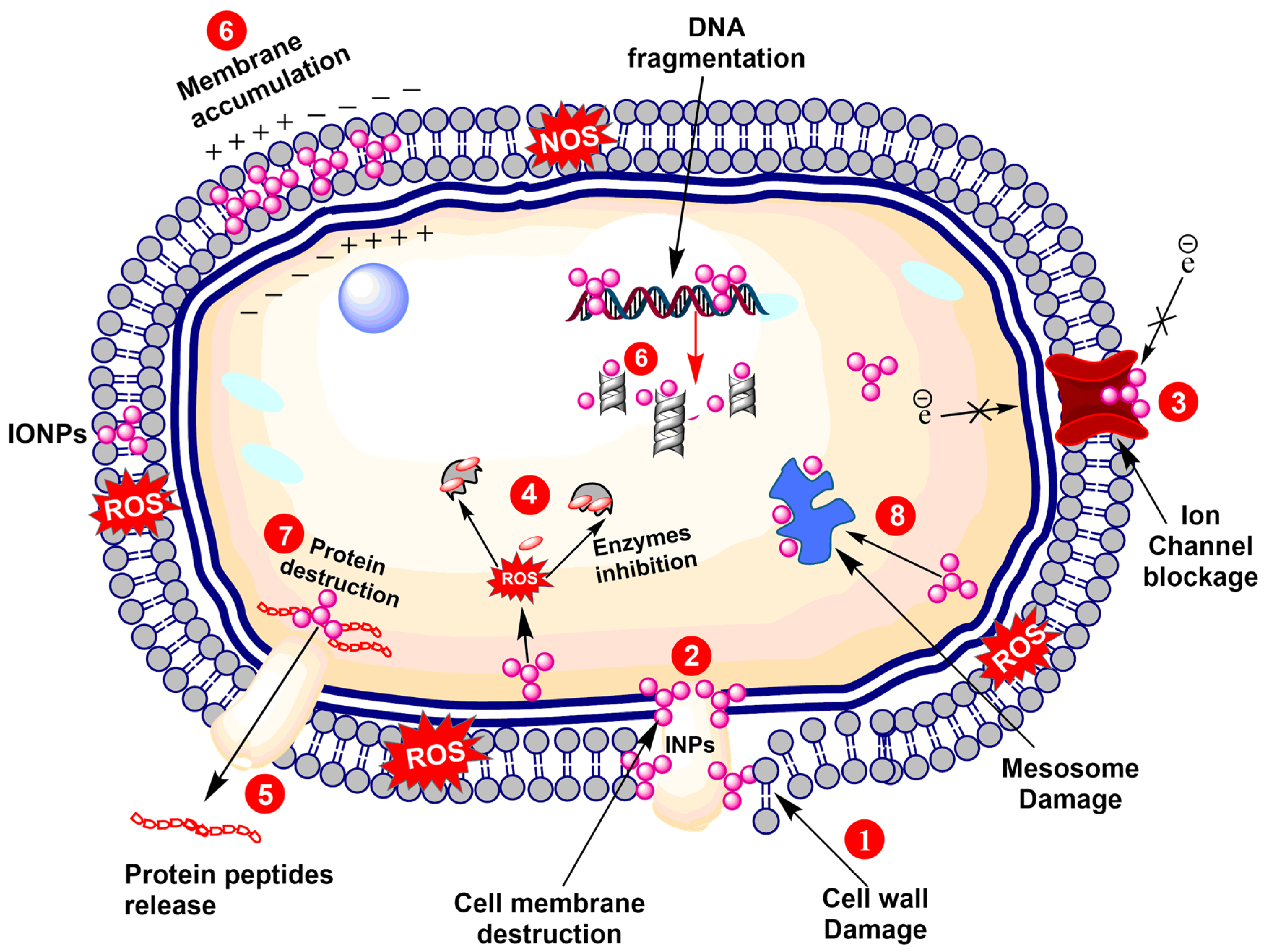
4. Antimicrobial Potential of IONPs
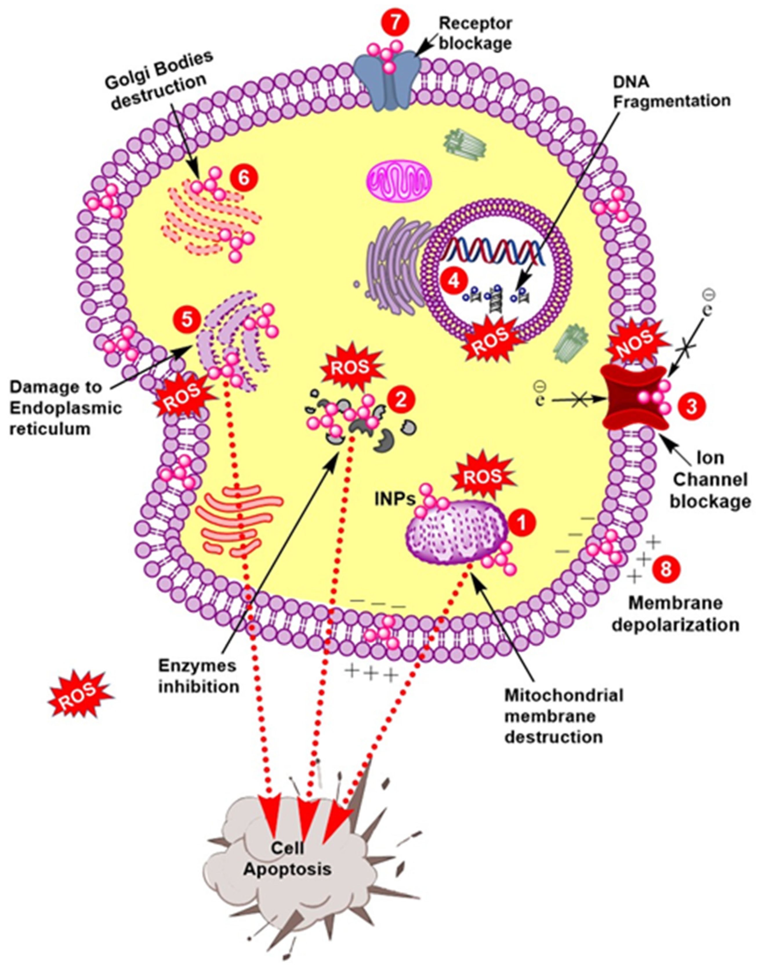
5. Anticancer Activity
6. Other Potential Applications
7. Conclusions
Author Contributions
Funding
Institutional Review Board Statement
Informed Consent Statement
Data Availability Statement
Conflicts of Interest
References
- Nadeem, M.; Khan, R.; Afridi, K.; Nadhman, A.; Ullah, S.; Faisal, S.; Mabood, Z.U.; Hano, C.; Abbasi, B.H. Green synthesis of cerium oxide nanoparticles (CeO2 NPs) and their antimicrobial applications: A review. Int. J. Nanomed. 2020, 15, 5951. [Google Scholar] [CrossRef] [PubMed]
- Nadeem, M.; Abbasi, B.H.; Younas, M.; Ahmad, W.; Khan, T. A review of the green syntheses and anti-microbial applications of gold nanoparticles. Green Chem. Lett. Rev. 2017, 10, 216–227. [Google Scholar] [CrossRef] [Green Version]
- Hashmi, S.S.; Shah, M.; Muhammad, W.; Ahmad, A.; Ullah, M.A.; Nadeem, M.; Abbasi, B.H. Potentials of Phyto-Fabricated nanoparticles as ecofriendly agents for Photocatalytic degradation of toxic dyes and waste water treatment, risk assessment and probable mechanism. J. Indian Chem. Soc. 2021, 98, 100019. [Google Scholar] [CrossRef]
- Mohamed, Y.; Azzam, A.; Amin, B.; Safwat, N.A. Mycosynthesis of iron nanoparticles by Alternaria alternata and its antibacterial activity. Afr. J. Biotechnol. 2015, 14, 1234–1241. [Google Scholar] [CrossRef] [Green Version]
- Asha, A.; Sivaranjani, T.; Thirunavukkarasu, P.; Asha, S. Green synthesis of silver nanoparticle from different plants—A review. Int. J. Pure Appl. Biosci. 2016, 4, 118–124. [Google Scholar] [CrossRef]
- Khan, I.; Saeed, K.; Khan, I. Nanoparticles: Properties, applications and toxicities. Arab. J. Chem. 2019, 12, 908–931. [Google Scholar] [CrossRef]
- Nadeem, M.; Tungmunnithum, D.; Hano, C.; Abbasi, B.H.; Hashmi, S.S.; Ahmad, W.; Zahir, A. The current trends in the green syntheses of titanium oxide nanoparticles and their applications. Green Chem. Lett. Rev. 2018, 11, 492–502. [Google Scholar] [CrossRef] [Green Version]
- Patil, R.M.; Thorat, N.D.; Shete, P.B.; Bedge, P.A.; Gavde, S.; Joshi, M.G.; Tofail, S.A.; Bohara, R.A. Comprehensive cytotoxicity studies of superparamagnetic iron oxide nanoparticles. Biochem. Biophys. Rep. 2018, 13, 63–72. [Google Scholar] [CrossRef]
- Sangaiya, P.; Jayaprakash, R.; Magnetism, N. A review on iron oxide nanoparticles and their biomedical applications. J. Supercond. Nov. Magn. 2018, 31, 3397–3413. [Google Scholar] [CrossRef]
- Kaul, R.; Kumar, P.; Burman, U.; Joshi, P.; Agrawal, A.; Raliya, R.; Tarafdar, J.C. Magnesium and iron nanoparticles production using microorganisms and various salts. Mater. Sci.-Pol. 2012, 30, 254–258. [Google Scholar] [CrossRef]
- Bose, S.; Hochella, M.F., Jr.; Gorby, Y.A.; Kennedy, D.W.; McCready, D.E.; Madden, A.S.; Lower, B.H. Bioreduction of hematite nanoparticles by the dissimilatory iron reducing bacterium Shewanella oneidensis MR-1. Geochim. Cosmochim. Acta 2009, 73, 962–976. [Google Scholar] [CrossRef]
- Ali, A.; Hira Zafar, M.Z.; ul Haq, I.; Phull, A.R.; Ali, J.S.; Hussain, A. Synthesis, characterization, applications, and challenges of iron oxide nanoparticles. Nanotechnol. Sci. Appl. 2016, 9, 49–67. [Google Scholar] [CrossRef] [Green Version]
- Wu, W.; Dey, D.; Memis, O.; Katnelson, A.; Mohseni, H.J.L. A Novel self-aligned and maskless process for formation of highly uniform arrays of nanoholes and nanopillars. Nanoscale Res. 2008, 3, 123. [Google Scholar] [CrossRef] [Green Version]
- Cheng, Z.; Tan, A.L.K.; Tao, Y.; Shan, D.; Ting, K.E.; Yin, X.J. Synthesis and characterization of iron oxide nanoparticles and applications in the removal of heavy metals from industrial wastewater. Int. J. Photoenergy 2012, 2012, 1–5. [Google Scholar] [CrossRef]
- Agarwal, H.; Kumar, S.V.; Rajeshkumar, S. A review on green synthesis of zinc oxide nanoparticles—An eco-friendly approach. Resour.-Effic. Technol. 2017, 3, 406–413. [Google Scholar] [CrossRef]
- Arias, S.L.; Shetty, A.R.; Senpan, A.; Echeverry-Rendón, M.; Reece, L.M.; Allain, J.P. Fabrication of a functionalized magnetic bacterial nanocellulose with iron oxide nanoparticles. J. Vis. Exp. 2016, 111, e52951. [Google Scholar] [CrossRef] [Green Version]
- Rajeswaran, S.; Thirugnanasambandan, S.S.; Dewangan, N.K.; Moorthy, R.K.; Kandasamy, S.; Vilwanathan, R. Multifarious pharmacological applications of green routed eco-friendly iron nanoparticles synthesized by Streptomyces Sp. (SRT12). Biol. Trace Elem. Res. 2020, 194, 273–283. [Google Scholar] [CrossRef]
- Majeed, S.; Danish, M.; Ibrahim, M.N.M.; Sekeri, S.H.; Ansari, M.T.; Nanda, A.; Ahmad, G. Bacteria mediated synthesis of iron oxide nanoparticles and their antibacterial, antioxidant, cytocompatibility properties. J. Clust. Sci. 2021, 32, 1083–1094. [Google Scholar] [CrossRef]
- Mathur, P.; Saini, S.; Paul, E.; Sharma, C.; Mehtani, P. Endophytic fungi mediated synthesis of iron nanoparticles: Characterization and application in methylene blue decolorization. Curr. Res. Green Sustain. Chem. 2021, 4, 100053. [Google Scholar] [CrossRef]
- Ghosh, S.; Ahmad, R.; Banerjee, K.; AlAjmi, M.F.; Rahman, S. Mechanistic Aspects of Microbe-Mediated Nanoparticle Synthesis. Front. Microbiol. 2021, 12, 867. [Google Scholar] [CrossRef] [PubMed]
- Vetchinkina, E.; Loshchinina, E.; Kupryashina, M.; Burov, A.; Pylaev, T.; Nikitina, V. Green synthesis of nanoparticles with extracellular and intracellular extracts of basidiomycetes. PeerJ 2018, 6, e5237. [Google Scholar] [CrossRef]
- Torabian, P.; Ghandehari, F.; Fatemi, M. Biosynthesis of iron oxide nanoparticles by cytoplasmic extracts of bacteria lactobacillus casei. Asian J. Green Chem. 2018, 2, 181–188. [Google Scholar]
- Kianpour, S.; Ebrahiminezhad, A.; Deyhimi, M.; Negahdaripour, M.; Raee, M.J.; Mohkam, M.; Rezaee, H.; Irajie, C.; Berenjian, A.; Ghasemi, Y. Structural characterization of polysaccharide-coated iron oxide nanoparticles produced by Staphylococcus warneri, isolated from a thermal spring. J. Basic Microbiol. 2019, 59, 569–578. [Google Scholar] [CrossRef]
- Sundaram, P.A.; Augustine, R.; Kannan, M. Extracellular biosynthesis of iron oxide nanoparticles by Bacillus subtilis strains isolated from rhizosphere soil. Biotechnol. Bioprocess Eng. 2012, 17, 835–840. [Google Scholar] [CrossRef]
- Bharde, A.A.; Parikh, R.Y.; Baidakova, M.; Jouen, S.; Hannoyer, B.; Enoki, T.; Prasad, B.; Shouche, Y.S.; Ogale, S.; Sastry, M.J.L. Bacteria-Mediated precursor-dependent biosynthesis of superparamagnetic iron oxide and iron sulfide nanoparticles. Langmuir 2008, 24, 5787–5794. [Google Scholar] [CrossRef] [PubMed]
- Rosenfeldt, S.; Mickoleit, F.; Jörke, C.; Clement, J.H.; Markert, S.; Jérôme, V.; Schwarzinger, S.; Freitag, R.; Schüler, D.; Uebe, R.; et al. Towards standardized purification of bacterial magnetic nanoparticles for future in vivo applications. Acta Biomater. 2021, 120, 293–303. [Google Scholar] [CrossRef] [PubMed]
- Alphandéry, E. Applications of magnetosomes synthesized by magnetotactic bacteria in medicine. Front. Bioeng. Biotechnol. 2014, 2, 5. [Google Scholar] [PubMed]
- De França Bettencourt, G.M.; Degenhardt, J.; Torres, L.A.Z.; de Andrade Tanobe, V.O.; Soccol, C.R. Green biosynthesis of single and bimetallic nanoparticles of iron and manganese using bacterial auxin complex to act as plant bio-fertilizer. Biocatal. Agric. Biotechnol. 2020, 30, 101822. [Google Scholar] [CrossRef]
- Byrne, J.; Telling, N.; Coker, V.; Pattrick, R.; Van Der Laan, G.; Arenholz, E.; Tuna, F.; Lloyd, J.R. Control of nanoparticle size, reactivity and magnetic properties during the bioproduction of magnetite by Geobacter sulfurreducens. Nanotechnology 2011, 22, 455709. [Google Scholar] [CrossRef] [PubMed] [Green Version]
- Raĭkher, Y.L.; Stepanov, V.; Stolyar, S.; Ladygina, V.; Balaev, D.; Ishchenko, L.; Balasoiu, M. Magnetic properties of biomineral particles produced by bacteria Klebsiella oxytoca. Phys. Solid State 2010, 52, 298–305. [Google Scholar] [CrossRef]
- Fani, M.; Ghandehari, F.; Rezaee, M.; Sciences, C. Biosynthesis of iron oxide nanoparticles by cytoplasmic extract of bacteria Lactobacillus Fermentum. J. Med. Chem. Sci. 2018, 1, 28–30. [Google Scholar]
- Zaki, S.A.; Eltarahony, M.M.; Abd-El-Haleem, D.A. Disinfection of water and wastewater by biosynthesized magnetite and zerovalent iron nanoparticles via NAP-NAR enzymes of Proteus mirabilis 10B. Environ. Sci. Pollut. Res. 2019, 26, 23661–23678. [Google Scholar] [CrossRef]
- Crespo, K.A.; Baronetti, J.L.; Quinteros, M.A.; Páez, P.L.; Paraje, M.G. Intra-and extracellular biosynthesis and characterization of iron nanoparticles from prokaryotic microorganisms with anticoagulant activity. Pharm. Res. 2017, 34, 591–598. [Google Scholar] [CrossRef]
- Das, K.R.; Kerkar, S. Biosynthesis of iron nanoparticles by sulphate reducing bacteria and its application in remediating chromium from water. Int. J. Pharma Bio Sci. 2017, 8, 538–546. [Google Scholar] [CrossRef]
- Desai, M.P.; Pawar, K.D. Immobilization of cellulase on iron tolerant Pseudomonas stutzeri biosynthesized photocatalytically active magnetic nanoparticles for increased thermal stability. Mater. Sci. Eng. C 2020, 106, 110169. [Google Scholar] [CrossRef]
- Daneshvar, M.; Hosseini, M.R. From the iron boring scraps to superparamagnetic nanoparticles through an aerobic biological route. J. Hazard. Mater. 2018, 357, 393–400. [Google Scholar] [CrossRef] [PubMed]
- Hashimoto, H.; Yokoyama, S.; Asaoka, H.; Kusano, Y.; Ikeda, Y.; Seno, M.; Takada, J.; Fujii, T.; Nakanishi, M.; Murakami, R. Characteristics of hollow microtubes consisting of amorphous iron oxide nanoparticles produced by iron oxidizing bacteria, Leptothrix ochracea. J. Magn. Magn. Mater. 2007, 310, 2405–2407. [Google Scholar] [CrossRef]
- Habibi, N. Immobilization of bacterial S-layer proteins from Caulobacter crescentus on iron oxide-based nanocomposite: Synthesis and spectroscopic characterization of zincite-coated Fe2O3 nanoparticles. Spectrochim. Acta Part A 2014, 125, 359–362. [Google Scholar] [CrossRef] [PubMed]
- Kim, Y.; Lee, Y.; Roh, Y. Microbial synthesis of iron sulfide (FeS) and iron carbonate (FeCO3) nanoparticles. J. Nanosci. Nanotechnol. 2015, 15, 5794–5797. [Google Scholar] [CrossRef]
- Haikarainen, T.; Paturi, P.; Lindén, J.; Haataja, S.; Meyer-Klaucke, W.; Finne, J.; Papageorgiou, A.C. Magnetic properties and structural characterization of iron oxide nanoparticles formed by Streptococcus suis Dpr and four mutants. J. Biol. Inorg. Chem. 2011, 16, 799–807. [Google Scholar] [CrossRef] [PubMed]
- Jajan, L.H.-G.; Hosseini, S.N.; Ghorbani, M.; Mousavi, S.F.; Ghareyazie, B.; Abolhassani, M. Effects of environmental conditions on high-yield magnetosome production by Magnetospirillum gryphiswaldense MSR-1. Iran. Biomed. J. 2019, 23, 209. [Google Scholar]
- Elcey, C.; Kuruvilla, A.T.; Thomas, D. Synthesis of magnetite nanoparticles from optimized iron reducing bacteria isolated from iron ore mining sites. Int. J. Curr. Microbiol. Appl. Sci. 2014, 3, 408–417. [Google Scholar]
- Dlamini, N.G.; Basson, A.K.; Pullabhotla, R.V. Wastewater treatment by a polymeric bioflocculant and iron nanoparticles synthesized from a bioflocculant. Polymers 2020, 12, 1618. [Google Scholar] [CrossRef]
- Abdeen, M.; Sabry, S.; Ghozlan, H.; El-Gendy, A.A.; Carpenter, E.E. Microbial-physical synthesis of Fe and Fe3O4 magnetic nanoparticles using Aspergillus niger YESM1 and supercritical condition of ethanol. J. Nanomater. 2016, 2016, 9174891. [Google Scholar] [CrossRef] [Green Version]
- Sidkey, N. Biosynthesis, Characterization And Antimicrobial Activity Of Iron Oxide Nanoparticles Synthesized By Fungi. Al-Azhar J. Pharm. Sci. 2020, 62, 164–179. [Google Scholar] [CrossRef]
- Baskar, G.; Chandhuru, J.; Praveen, A.; Fahad, K.S. Technology. Anticancer activity of iron oxide nanobiocomposite of fungal asparaginase. Int. J. Mod. Sci. Technol. 2017, 2, 98–104. [Google Scholar]
- Mahanty, S.; Bakshi, M.; Ghosh, S.; Chatterjee, S.; Bhattacharyya, S.; Das, P.; Das, S.; Chaudhuri, P.J. Green synthesis of iron oxide nanoparticles mediated by filamentous fungi isolated from Sundarban mangrove ecosystem, India. BioNanoScience 2019, 9, 637–651. [Google Scholar] [CrossRef]
- Adeleye, T.; Kareem, S.; Kekere-Ekun, A. Optimization Studies on Biosynthesis of Iron Nanoparticles using Rhizopus Stolonifer. In Proceedings of the IOP Conference Series: Materials Science and Engineering, Ogbomoso, Nigeria, 22–24 October 2019; IOP Publishing: Bristol, UK, 2020; Volume 805, p. 012037. [Google Scholar]
- Vainshtein, M.; Belova, N.; Kulakovskaya, T.; Suzina, N.; Sorokin, V. Synthesis of magneto-sensitive iron-containing nanoparticles by yeasts. J. Ind. Microbiol. Biotechnol. 2014, 41, 657–663. [Google Scholar] [CrossRef]
- Baccile, N.; Noiville, R.; Stievano, L.; Van Bogaert, I. Sophorolipids-Functionalized iron oxide nanoparticles. Phys. Chem. Chem. Phys. 2013, 15, 1606–1620. [Google Scholar] [CrossRef] [Green Version]
- Tarafdar, J.C.; Raliya, R. Rapid, low-cost, and ecofriendly approach for iron nanoparticle synthesis using Aspergillus oryzae TFR9. J. Nanoparticles 2013, 2013, 141274. [Google Scholar] [CrossRef] [Green Version]
- Mazumdar, H.; Haloi, N. A study on biosynthesis of iron nanoparticles by Pleurotus sp. J. Microbiol. Biotechnol. Res. 2011, 1, 39–49. [Google Scholar]
- Bharde, A.; Rautaray, D.; Bansal, V.; Ahmad, A.; Sarkar, I.; Yusuf, S.M.; Sanyal, M.; Sastry, M.J.S. Extracellular biosynthesis of magnetite using fungi. Small 2006, 2, 135–141. [Google Scholar] [CrossRef]
- Bhargava, A.; Jain, N.; Barathi, M.; Akhtar, M.S.; Yun, Y.-S.; Panwar, J. Synthesis, Characterization and Mechanistic Insights of Mycogenic Iron Oxide Nanoparticles. In Nanotechnology for Sustainable Development; Springer: Berlin/Heidelberg, Germany, 2013; pp. 337–348. [Google Scholar]
- Li, Q.; Liu, D.; Wang, T.; Chen, C.; Gadd, G.M. Iron coral: Novel fungal biomineralization of nanoscale zerovalent iron composites for treatment of chlorinated pollutants. Chem. Eng. J. 2020, 402, 126263. [Google Scholar] [CrossRef]
- Kareem, S.; Adeleye, T.; Ojo, R. Effects of pH, Temperature and Agitation on the Biosynthesis of Iron Nanoparticles Produced by Trichoderma Species. In Proceedings of IOP Conference Series: Materials Science and Engineering, Ogbomoso, Nigeria, 22–24 October 2019; IOP Publishing: Bristol, UK, 2020; p. 012036. [Google Scholar]
- Jeevanandam, J.; Barhoum, A.; Chan, Y.S.; Dufresne, A.; Danquah, M.K. Review on nanoparticles and nanostructured materials: History, sources, toxicity and regulations. Beilstein J. Nanotechnol. 2018, 9, 1050–1074. [Google Scholar] [CrossRef] [Green Version]
- Arora, S.K.; Porter, A.L.; Youtie, J.; Shapira, P. Capturing new developments in an emerging technology: An updated search strategy for identifying nanotechnology research outputs. Scientometrics 2013, 95, 351–370. [Google Scholar] [CrossRef]
- Samrot, A.V.; Sahithya, C.S.; Selvarani, J.; Purayil, S.K.; Ponnaiah, P. A review on synthesis, characterization and potential biological applications of superparamagnetic iron oxide nanoparticles. Curr. Res. Green Sustain. Chem. 2021, 4, 100042. [Google Scholar] [CrossRef]
- Ahmed, V.; Kumar, J.; Kumar, M.; Chauhan, M.B.; Dahiya, P.; Chauhan, N.S. Functionalised iron nanoparticle–penicillin G conjugates: A novel strategy to combat the rapid emergence of β-lactamase resistance among infectious micro-organism. J. Exp. Nanosci. 2015, 10, 718–728. [Google Scholar] [CrossRef] [Green Version]
| S.no | Species | Location of Synthesis | Characterization | Functional Group Involved in Reduction | Shape | Size (nm) | Ref |
|---|---|---|---|---|---|---|---|
| 1 | Actinobacter sp. | Extracellular | TEM, XRD, and FTIR | Fe–O bond | Crystal | 50 | [25] |
| 2 | Shewanella oneidensis | NR | TEM, XRD, and AFM | NR | Pseudo-hexagonal shape | 11, 30, 99 | [11] |
| 3 | Magnetospirillum gryphiswaldense | Extracellular | DLS, TEM, SAXS, and FTIR | NR | Polydispersed | 25–55 | [26] |
| 4 | Magnetotactic bacteria | Intracellular | TEM | NR | Spherical | 25–50 | [27] |
| 5 | Paenibacillus polymyxa | NR | TEM, FTIR, and UV-Vis | O–H, C–H, CO2NH3, C=O, C=C, and N–H | Spherical | 26.65 | [28] |
| 6 | Geobacter sulfurreducens | Extracellular | PXRD and TEM | NR | NR | 10–50 | [29] |
| 7 | Klebsiella Oxytoca | NR | ---- | NR | NR | 2–5 | [30] |
| 8 | Lactobacillus Fermentum | Intracellular | XRD and TEM | NR | Spherical | 10–15 | [31] |
| 9 | Gluconacetobacter xylinus | Intracellular | SEM | NR | NR | 50 | [16] |
| 10 | Proteus mirabilis | NR | XRD, EDX, TEM, UV-Vis, and Zeta sizer | NR | Spherical | 1.44–1.92 | [32] |
| 11 | Escherichia coli | Extracellular | FESEM, EDX, TEM, and UV-Vis | NR | Spherical | 23 | [33] |
| Pseudomonas aeruginosa | Extracellular | FESEM, EDX, TEM, and UV-Vis | NR | Spherical | 23 | [33] | |
| 12 | Desulfotomacculum acetoxidans | NR | SEM-EDS and XRD | NR | NR | 21 | [34] |
| 13 | Pseudomonas stutzeri | NR | XRD, FTIRUV-Vis, SEM, and TEM | O–H, C–H, Fe–O, C=C, and N–H | NR | 10–20 | [35] |
| 14 | Desulfovibrio | NR | TEM, XRD, and FTIR | NR | NR | 19 | [34] |
| 15 | Bacillus subtilis | Extracellular | FE-SEM, TEM, XRD, FTIR, DLS, and VSM | O–H, C–H, Fe–O, C=C, and N–H | Rhombohedral | 37–97 | [36] |
| Bacillus pasteurii | NR | FE-SEM, TEM, XRD, FTIR, DLS, and VSM | O–H, C–H, Fe–O, C=C, N–H | Rhombohedral | 37–97 | [36] | |
| Bacillus licheniformis | NR | FE-SEM, TEM, XRD, FTIR, DLS, and VSM | O–H, C–H, Fe–O, C=C, and N–H | Rhombohedral | 37–97 | [36] | |
| 16 | Leptothrix ochracea | Extracellular | SEM, EDX, and XRD | NR | hollow tube | 100 | [37] |
| 17 | Caulobacter crescentus | NR | FE-SEM, XRD, AFM, and EDAX | NR | Spherical | 50 | [38] |
| 18 | Geobacterspecie | NR | XRD, SEM-EDX, TEM-EDX, and ICP-AES | NR | NR | 50–60 | [39] |
| 19 | Streptococcus suis | NR | EXAFS and XRD | NR | NR | ---- | [40] |
| 20 | Magnetospirillum gryphiswaldense | Extracellular | TEM | NR | NR | ---- | [41] |
| 21 | Thiobacillus thioparus | NR | SDS PAGE Gel | NR | NR | ---- | [42] |
| 22 | Alcaligenes faecalis | Extracellular | SEM, EDX, and FTIR | HO–NH3 | NR | [43] |
| S.no | Species | Inhibition Method | Activity Against | Ref. |
|---|---|---|---|---|
| 1 | Proteus vulgaris | Disc Diffusion method | Salmonella enterica, Escherichia coli, Vibrio cholera, Salmonella typhi, and Staphylococcus epidermidis | [18] |
| 2 | Streptomyces (SRT12) | Disc Diffusion method | Bacillus subtilis, Staphylococcus aureus, Klebsiella pneumoniae, Shigella flexneri, and Escherichia coli. | [17] |
| 3 | Proteus mirabilis | Well-diffusion method | E. coli, Salmonella typhi, P. aeruginosa, Clostridium perfringens, Aspergillus Brasiliensis, and Candida Albicans | [32] |
| 4 | Alternaria alternata | Well-diffusion method | Bacillus subtilis | [4] |
| 5 | Fusarium oxysporum | Disc diffusion method | Staphylococcusaureus, Klebsiella pneumoniae, Proteus vulgaris, Pseudomonas aeruginosa, and Escherichia coli | [24] |
| 6 | Aspergillus flavus | Diffusion agar technique | Staphylococcus aureus, Escherichia coli, Candida albicans, and Aspergillus Fumigatus | [45] |
| 7 | NPs-penicillin G conjugates | Disc Diffusion method | Staphylococcus aureus | [60] |
Publisher’s Note: MDPI stays neutral with regard to jurisdictional claims in published maps and institutional affiliations. |
© 2021 by the authors. Licensee MDPI, Basel, Switzerland. This article is an open access article distributed under the terms and conditions of the Creative Commons Attribution (CC BY) license (https://creativecommons.org/licenses/by/4.0/).
Share and Cite
Nadeem, M.; Khan, R.; Shah, N.; Bangash, I.R.; Abbasi, B.H.; Hano, C.; Liu, C.; Ullah, S.; Hashmi, S.S.; Nadhman, A.; et al. A Review of Microbial Mediated Iron Nanoparticles (IONPs) and Its Biomedical Applications. Nanomaterials 2022, 12, 130. https://doi.org/10.3390/nano12010130
Nadeem M, Khan R, Shah N, Bangash IR, Abbasi BH, Hano C, Liu C, Ullah S, Hashmi SS, Nadhman A, et al. A Review of Microbial Mediated Iron Nanoparticles (IONPs) and Its Biomedical Applications. Nanomaterials. 2022; 12(1):130. https://doi.org/10.3390/nano12010130
Chicago/Turabian StyleNadeem, Muhammad, Rijma Khan, Nausheen Shah, Ishrat Rehman Bangash, Bilal Haider Abbasi, Christophe Hano, Chunzhao Liu, Sana Ullah, Syed Salman Hashmi, Akhtar Nadhman, and et al. 2022. "A Review of Microbial Mediated Iron Nanoparticles (IONPs) and Its Biomedical Applications" Nanomaterials 12, no. 1: 130. https://doi.org/10.3390/nano12010130