Article

Deposition Mechanism and Characterization of Plasma-Enhanced Atomic Layer-Deposited SnOx Films at Different Substrate Temperatures

1
School of Ocean Information Engineering, Jimei University, Jimei District, Xiamen 361021, China
2
School of Opto-Electronic and Communication Engineering, Xiamen University of Technology, Xiamen 361024, China
3
Department of Materials Science and Engineering, National United University, Miaoli 36063, Taiwan
4
Department of Biomedical Engineering, Da-Yeh University, Changhua 51591, Taiwan
5
Department of Applied Physics, National University of Kaohsiung, Kaohsiung University Road, Kaohsiung 81148, Taiwan
6
Department of Applied Materials and Optoelectronic Engineering, National Chi Nan University, Nantou 54561, Taiwan
7
Department of Materials Science and Engineering, Da-Yeh University, Changhua 51591, Taiwan
*
Author to whom correspondence should be addressed.
Nanomaterials 2022, 12(16), 2859; https://doi.org/10.3390/nano12162859
Submission received: 16 July 2022 / Revised: 15 August 2022 / Accepted: 15 August 2022 / Published: 19 August 2022
(This article belongs to the Special Issue Nanotechnologies and Nanomaterials: Selected Papers from CCMR)

Abstract

:
The promising functional tin oxide (SnOx) has attracted tremendous attention due to its transparent and conductive properties. The stoichiometric composition of SnOx can be described as common n-type SnO2 and p-type Sn3O4. In this study, the functional SnOx films were prepared successfully by plasma-enhanced atomic layer deposition (PEALD) at different substrate temperatures from 100 to 400 °C. The experimental results involving optical, structural, chemical, and electrical properties and morphologies are discussed. The SnO2 and oxygen-deficient Sn3O4 phases coexisting in PEALD SnOx films were found. The PEALD SnOx films are composed of intrinsic oxygen vacancies with O-Sn4+ bonds and then transformed into a crystalline SnO2 phase with increased substrate temperature, revealing a direct 3.5–4.0 eV band gap and 1.9–2.1 refractive index. Lower (<150 °C) and higher (>300 °C) substrate temperatures can cause precursor condensation and desorption, respectively, resulting in reduced film qualities. The proper composition ratio of O to Sn in PEALD SnOx films near an estimated 1.74 suggests the highest mobility of 12.89 cm2 V−1 s−1 at 300 °C.

1. Introduction

Transparent conductive oxide (TCO) materials have been widely used and intensively researched in a wide range of industries during the last half-century [1,2,3,4]. Because of its increasing use in many instruments, this large area of constantly expanding research has focused on the preparations and properties of TCO films such as tin oxide (SnO2), indium tin oxide (ITO), zinc oxide (ZnO), aluminum-doped ZnO (AZO), and titanium oxide (TiO2) [5,6,7,8,9]. Non-stoichiometric SnO2 (SnOx), in particular, has recently gained substantial interest as a potential functional oxide semiconductor for use in a wide range of optoelectronics due to its specific features in its stoichiometry [10]. For instance, the SnOx films are prepared with nanocomposite porous silicon for application and used in gas microsensors [11]. Due to their superior chemical and mechanical stability over other known oxide films, SnOx films are also employed as electron selective film candidates for solar cells and light-emitting diodes based on perovskite, quantum dots, and organic materials [12,13,14]. Furthermore, various experiments have been conducted to examine tin oxides with different oxygen stoichiometry, such as Sn2O3 [15], Sn3O4 [16], and Sn5O6 [17]. Because of its oxygen-deficient property, p-type Sn3O4 has gained a significant amount of interest. However, the impact of this Sn3O4 phase on the optical, electrical, physical, and chemical characteristics of the film, which usually coexists with the SnO2 phase, is sometimes underestimated.
In the literature, various deposition processes such as chemical vapor deposition (CVD), low-pressure chemical vapor deposition, plasma-enhanced chemical vapor deposition (PECVD), physical vapor deposition (PVD), and so on have been employed to prepare multifunctional SnOx films [18,19,20,21]. Currently, atomic layer deposition (ALD), as an appealing deposition process with low deposition temperature, atomic-scale thickness controllability, and remarkable conformity, permits the considerable scaling-down and 3D structuring of devices as compared to CVD and PVD [22,23]. In ALD, two self-limiting surface reactions are used, in which two reactant gases are pulsed into the chamber in two different dosages, resulting in the formation of individual mono-layers per reaction cycle. Furthermore, as a better approach, plasma-enhanced ALD (PEALD) employs plasma-generated oxidizing agents to effectively augment the reactivity between plasma species and precursors, allowing for lower deposition temperatures without affecting film quality [24,25,26,27]. Film properties are affected by different deposition modes driven by lower or higher substrate temperatures [28,29,30], perhaps due to precursor condensation/adsorption within an incomplete reaction or decomposition/desorption. Thus, it is important to focus on the impact of various substrate temperatures on the PEALD SnOx films and validate which deposition mode will occur with the various substrate temperatures in order to acquire the optimal stoichiometry of oxygen and tin.
PEALD SnOx films deposited at substrate temperatures ranging from 100 to 400 °C are investigated in this study. The metal precursor is tetrakis(dimethylamino)tin (TDMA-Sn), which reacts with oxygen and argon plasma reactants. The optical, electrical, physical, and chemical characteristics are analyzed and discussed to determine the optimal stoichiometric ratio of O to Sn.

2. Materials and Methods

2.1. Materials and PEALD Process

The SnOx films were deposited on silicon wafers (4 inches with 450 μm and a resistivity of 50 Ω-cm) by the PEALD system (R-200, Picosun, Finland) with six source channels, where the TDMA-Sn (purity: 99.9999%, Aimou Yuan, Nanjing, China) was used as the Sn metal precursor. Each experimental variable was used for preparing five samples at different substrate temperatures, and silicon wafers were cleaned by a standard procedure, including deionized water (DI-water) for 10 s, hydrofluoric acid for 1 min, and DI water for 10 s. Before being transferred to the vacuum chamber, the silicon wafer was blow-dried with nitrogen (N2, 99.99%). We operated the Ar and O2 (both of them with an ultra-high purity of 99.999%) plasma in a quartz cavity by the inductive coupling of RF power. The SnOx deposition was performed with a total of 300 ALD cycles. Table 1 shows the preparation parameters of the PEALD SnOx films, and the substrate temperature was varied from 100 to 400 °C.

2.2. Characteristic Measurements

The ellipsometer (M-2000, J. A. Woollan Co., Lincoln, NE, USA) was used to determine the thickness, refractive index (n), and deposition rate (nm/cycle). The estimated thickness value had an error of less than ±2% to show satisfying reproducibility. The model of “air, air/SnOx, SnOx, SnOx/silicon” was used to complete the fitting ellipsometric data for the PEALD SnOx films by the Drude-Lorentz model. For the optical properties of films, all samples were measured by ultraviolet-visible spectroscopy (MFS-630, Hong-Ming Technology, New Taipei City, Taiwan) in the wavelength range from 350 to 850 nm. For the structural properties of films, the grazing incidence X-ray diffraction (XRD, Rigaku TTRAXIII, Ibaraki, Japan) with a selected 0.5° incident angle and a wavelength of 0.15418 nm was used at 50 kV and 300 mA to obtain the orientation in diffraction patterns within a 2θ range of 20° to 70°. Field emission scanning electron microscopy (FESEM, JSM-7800F, JEOL, Tokyo, Japan) at 9.6 × 10⁻⁵ Pa and atomic force microscopy (AFM, XE7, Park, Korea) at ambient conditions were used to obtain the top-view surface morphologies. Further microstructure characteristics were shown in the cross-sectional transmission electron microscopy (TEM) images. For the chemical properties of the films, the X-ray photoelectron spectroscopy (XPS, ESCALAB, 250Xi, Thermo Fisher, Waltham, MA, USA) spectra were performed and calibrated by C 1s (284.5 eV). Before XPS measurement, the surface contamination was removed by sputtering. For the electrical properties of films, the resistivity, carrier concentration, and mobility were conducted by Hall-effect measurements (HMS-5000, Side Semiconductor Technology, Ecopia, Anyang, Korea) at room temperature. Both XRD and XPS results were further analyzed by peak-differentiated and imitating methods to demonstrate the phase and bonding characteristics of the films, respectively.

3. Results and Discussion

3.1. Deposition Mechanism

The schematic deposition mechanism of the PEALD SnOx films is shown in Figure 1. Three growth modes concerning the first (steps 1 and 3) and second self-limiting surface reactions (steps 2 and 4) are described as (a) precursor condensation (<150 °C), (b) saturation reaction (250–300 °C), and (c) thermal desorption (350–400 °C), where the reaction can be represented via the following equations [28,29]:
S^-(OH)3 + 2 Sn(N(CH3)2)4  S^-OH-Sn(N(CH3)2)4 + S^-O2Sn(N(CH3)2)2 + 2 NC2H7
S^-(OH)2 + Sn(N(CH3)2)4  S^-O2Sn(N(CH3)2)2 + 2 NC2H7
S^-(OH)2 + Sn(N(CH3)2)4  S^-O2Sn(N(CH3)2)2 + NC2H7+ Sn(NC2H7)2
S^-O2Sn(N(CH3)2)2 + Plasma (O*/Ar*/e)  S^-SnO2-H + (COX + NOX + H2O)
S^-SnO2-2H + Sn(N(CH3)2)4  S^-SnO2-Sn(N(CH3)2)2 + NC2H7
S^-SnO2-Sn(N(CH3)2)2 + Plasma (O*/Ar*/e)  S^-SnO2-SnO2-2H + (COX + NOX + H2O)
where the S^ and symbols represent the substrate surface and by-product with volatile gaseous phase, respectively. In Equation (1), the TDMA-Sn molecules will react with the hydroxyl (OH) groups on the substrate surface. Equation (1a) reveals that the low substrate temperature (<150 °C) causes the condensation of the TDMA-Sn precursor mainly due to the physisorption, where it is quite mobile and oscillating on the surface. This result is similar to some other studies [28]. With the increasing substrate temperatures (200–400 °C), the physisorption becomes a minor factor and the film growth gradually turns into chemisorption as a significant factor. As shown in Equations (1b) and (1c), the thermal activation induces the irreversible break of chemical bonding and the electron transfer between the deposited surface and adsorbed molecules [31,32]. Notably, when the substrate temperature is in the range of 250–300 °C, a self-limiting PEALD process emerges as Equation (1b) due to enough heat energy, leading to the saturation reaction of precursors and oxygen radicals. However, these adsorbed precursor molecules will further desorb, as in Equation (1c), when the surface possesses excess heat energy at higher substrate temperatures of 350–400 °C. In the second self-limiting surface reaction, the plasma reaction is shown as the following formula to generate oxygen (O2) radicals: Ar + O2 + e2O* + Ar* + e, where the asterisk mark describes the excited state. The Sn-O bonds and initial hydroxyl ligands are formed, and then the released by-products (COX, NOX, and H2O gas), as described in Equation (2), are purged. So far, one PEALD cycle has finished, and we continuously used more than one cycle to complete the film growth by repeating Equations (3) and (4).
Figure 2a shows the substrate temperature-dependent growth per cycle (GPC) of PEALD SnOx films on the Si wafer from 100 to 400 °C. The trend line of corresponding thickness at each GPC is plotted in Figure 2b. We calculate the GPC value by dividing the film thickness by the number of cycles. Three reaction regions are obviously demonstrated with respect to the substrate temperature. The GPC of 0.117 nm/cycle at 100 °C is mainly induced by the precursor physisorption and condensation [28,33]; however, at 150–200 °C, the GPC decreases to 0.117–0.087 nm/cycle, inferring that the surface reaction changes from physisorption to chemisorption-dominated. These low GPC values are likely due to the low chemical reaction rates at low temperatures [34,35]. The high GPC values of 0.138 nm/cycle at 250 °C and 0.131 nm/cycle at 300 °C are ascribed to the saturation of chemical-adsorbed precursors. However, the GPC rapidly drops to 0.094 nm/cycle at 350 °C and 0.082 nm/cycle at 400 °C due to the severe thermal desorption between the precursors and surface [34]. In other words, the self-limiting process as a unique feature of PEALD is verified by observing the saturation reaction of the GPC value as a function of the substrate temperature. Compared to some studies contrary to our results [28], these observations indicate that the higher substrate temperature causes the low GPC owing to the precursor’s desorption [34]. In the ALD process, the saturation reaction should lead to a relatively high GPC value and simultaneously a small change in GPC, which were observed in the range of 250 to 300 °C in this study. This temperature range is reasonable as compared to the literature [36].

3.2. Chemical and Electronic State of the Sn and O

Figure 3a shows the XPS full-side spectra for the films deposited at different temperatures. All the peaks are labeled and hydrogen is not detectable in XPS, while nitrogen and carbon may be contained in the films, but only in low amounts. The nitrogen content of around 2.5 at.% at 100 and 150 °C results from the unreacted ligands of TDMA-Sn, possibly due to the low reactivity at low substrate temperatures. At higher substrate temperatures (>200 °C), the nitrogen content is as low as around 0.5 at.%. In particular, Sn 3d3/2 and 3d5/2 peak at ~495.6 and~487.0 eV [37], respectively, and the O 1s peak at ~530.7 eV is commonly used for further analysis. In Figure 3b, showing the high-resolution Sn 3d peaks, the peak position is slightly different among the samples with different substrate temperatures. This is related to the Sn4+ and Sn2+ components, e.g., at respectively 487.5 eV and at 486.4 eV for the Sn5/2 peaks [38]. The Sn4+ and Sn2+ components indicate the coexistence of the SnO2 and the metastable Sn oxide (such as Sn3O4). This is also supported by the O 1s spectra illustrated in Figure 3c. The spectra are deconvoluted into three peaks at 530.0 eV, associated with the lattice oxygen bonded to Sn2+ (OL–Sn2+); 531 ± 0.1 eV, to the lattice oxygen bonded to Sn4+ (OL–Sn4+); and 532 ± 0.1 eV to oxygen-deficient regions in oxides [24,39,40]. The ratio of each oxygen component to the total is calculated and shown in Figure 3d. At low temperatures (100–200 °C) the OL–Sn2+ area ratio decreases from 20.07% to the lowest of 15.23%, and the oxygen vacancy (OV) defects proportion increases from 10.25% to the highest of 12.62%, primarily due to the precursor chemisorption dominating at 200 °C.
The maximum 22.57% OL–Sn2+ area ratio and the minimum 3.15% OV proportion at 300 °C are observed. This suppression of OV defects is mainly due to the best decomposition of the precursor at 300°C. Besides, the OV defects proportion increases to 7.61% at 400 °C due to the out-diffusion of the oxygen atoms from SnO2 films. It is deduced that at higher substrate temperatures, the SnO2 decomposes thermally and oxygen breaks bonds between itself and metal and diffuses towards the film surface. The oxygen then leaves the film as O2, and it is possible that a small amount of oxygen leaves the film as CO2. The atomic ratios of elemental compositions, including O, Sn, and nitrogen (N), as a function of substrate temperature, are shown in Figure 3e. Notably, the high N ratio of ~2.5% at 100 °C and 150 °C dramatically decreases to ~0.5% in the range of 200–400 °C, demonstrating that the precursors are decomposed above 200°C. To analyze the stoichiometric SnOx films, the O to Sn ratio values (RO/Sn) are further calculated at different substrate temperatures. The RO/Sn of 1.517 at 100 °C increases to 1.559 and 1.645 at 150 °C and 200 °C, respectively. Then, the improved RO/Sn is obtained as 1.709 at 250 °C and 1.736 at 300 °C. The excessively high temperatures (350 °C and 400 °C) show a slightly decreased RO/Sn of 1.725 and 1.723, respectively. These results are similar to a few studies [37].

3.3. Structural Properties of the SnOx film

Figure 4a illustrates the XRD patterns of PEALD SnOx films deposited at different substrate temperatures. Based on the JCPDS card (no. 41-1445), the strong peaks at 26.7°, 38.4°, and 52.1° are ascribed to (110), (200), and (211) orientations of the SnO2 tetragonal rutile structure, respectively [24,25,41]. The weak peaks at 34.2°, 53.1°, and 62.4° correspond to (101), (220), and (310) orientations, respectively [27]. The amorphous structure of films deposited at below 200 °C is clearly observed. The reason is the low reactivity of precursor and precursor condensation induced by the low substrate temperature. With the increasing substrate temperatures, a polycrystalline SnO2 is observed. A (110) preferred orientation is detected with the highest intensity variability when the substrate temperature is in the range of 250–400 °C. The intensity of (110) orientation increases at medium temperature (250–300 °C) due to the self-limiting growth and then decreases at higher substrate temperature (350–400 °C), owing to the decomposition and desorption of the precursor. The intensity variation of diffraction peaks indicates the consistent variation of full width at half maximum (FWHM). Figure 4b shows the FWHM variation of the preferential (110) orientation and the average crystallite size (D) of films estimated by the Scherrer function as Equation (5) [42]:
D = κλ/(βcos θ),
where the κ = 0.9 is the Scherrer constant, λ is the wavelength of the X-ray sources, β is the FWHM value, and θ as Bragg angle is the peak position of the (110) orientation. The lowest FWHM value of 0.87° at 300 °C corresponds to the largest average crystallite size. Then, the FWHM value increases with increasing substrate temperature from 300 °C to 350 °C, indicating the decreased average crystallite size from 13.42 to 9.06 nm. The reason is attributed to the fact that excessively high substrate temperature above 300 °C causes the non-ideal deposition induced by severe precursor desorption and decomposition. It is observed that the diffraction peaks slightly shift with the increasing substrate temperature, suggesting a lattice expansion or contraction. For example, the peak position shifts from 26.56° at 250 °C to 26.74° at 300 °C. Similarly, the peak position then shifts toward a lower angle to 26.42° at 400 °C. The interplanar distance (d−spacing) is calculated as shown in Figure 4c by the Bragg formula [43]:
2dsin θ = ndλ,
where nd is the order of diffraction, and d is the dspacing. With increasing substrate temperatures from 250 °C to 400 °C, the dspacings of SnOx films are around 3.356, 3.334, 3.349, and 3.373 Å, respectively. The standard dspacing value of pure SnO2 is 3.347 Å. The decreased dspacing when increasing the substrate temperature from 250 to 300 °C is attributed to the decrease in oxygen vacancy defects as observed from the XPS results, causing the lattice contraction of SnOx films [44]. In the study reported by Santara et al. [45], the oxygen interstitials (Oi2+) and metal interstitials may attract each other and cause lattice contraction. Thus, another possible reason for the lattice contraction observed in this study can be due to the electrostatic attraction between Oi2+ and tin interstitials (Sni4+). In contrast, the increased d−spacings at 350–400 °C are due to the generated oxygen vacancy defects. The O−Sn bonds in the vicinity of oxygen-deficient regions are relaxed, leading to the lattice expansion of SnOx films. Besides, the nearest-neighbor Sn atoms move outward from the vacancy to strengthen their neighboring bonds of the remaining oxygen lattice. Although the nearest-neighbor oxygen atoms may move inward to fill the site of oxygen vacancy defects, the net outward movement of Sn atoms is higher than the net inward movement of oxygen atoms, resulting in the lattice expansion. Other microstructural parameters, such as micro-strain (ɛ) and dislocation density (δ), are estimated as:
ε = β/4tan θ,
δ = 1/D2,
Accordingly, the film at the 300 °C substrate temperature obtains the lowest value of δ and ɛ. The small δ obtained at 300 °C is the number of defects measured in the crystals [43] and the released ε at 250–300 °C is mainly due to the lattice contraction. The enhanced ε at 300–350 °C can be described by the increased vacancy formation energy from external strain [46].
However, beyond the substrate temperature of 300 °C, we observe that the (110) SnO2 peaks are not symmetrical, possibly implying the existence of other phases. For example, a diffraction peak near 25° is observed as a star, marked in Figure 4a, resulting from the oxygen-deficient SnOx [27]. To identify whether there are other hidden peaks, the (110) peaks are deconvoluted in Figure 4d, where two shoulder peaks at 24.8° and 28.1° as (101) and (111) triclinic Sn3O4 phases are identified (JCPDS#16-0737) [47,48]. This means that the SnOx films have SnO2 as the major phase and Sn3O4 as the minor phase. Moreover, the triclinic Sn3O4 as an intermediate oxide during the phase transformation of SnO2 is known as the oxygen-deficient SnOx phase [49]. The (110)SnO2/[(110)SnO2 + (101)Sn3O4 + (111)Sn3O4] peak area ratio for the different substrate temperatures is further shown in Figure 4e. The proportion of (110) orientation firstly decreases to the lowest value of 64.62% at 300 °C and then increases again at increasing substrate temperatures. This result also supports that the 300 °C substrate temperature is a critical temperature where the deposition mode changes from saturation growth to precursor decomposition or desorption.
AFM with a scanning area of 5 × 5 µm2 is used to analyze the topographic and stereoscopic surface morphologies of PEALD SnOx, films as shown in Figure 5. The films grown at 100–200 °C show a smooth microstructure with a root-mean-square (Rq) of 0.16–0.23 nm, consistent with the amorphous SnOx films at this temperature range. The film deposited at 250 °C obtains the highest Rq value of 1.65 nm. The Rq reduces to 0.34 nm when the substrate temperature increases to 400 °C. Compared to the Rq value of SnOx films deposited by spray pyrolysis (11.6 nm) [50] and sputtering (17.72 nm) [51], the PEALD SnOx films provide a smoother surface that is beneficial for many applications.
The top-view FESEM images of the films are observed on the right-hand side of Figure 5. Flat and featureless morphologies of SnOx films are observed without noticeable grain boundaries at the substrate temperature of 100–200 °C. This agrees with the amorphous structure of the films. At 250 °C, distinct clusters can be observed due to the large SnOx grains, and a clear grain structure is visible at 300 °C; however, these obvious grain boundaries gradually disappear at the higher substrate temperatures of 350 °C and 400 °C, attributed to the decreased grain size.
Figure 6 shows the cross-sectional TEM images of SnOx films. It is unexpected that crystallization is observed at 100 °C, shown in Figure 6a, as this is inconsistent with the XRD result. One reasonable explanation is that the amorphous structure recrystallizes by the ion beam of the TEM measurement.
In Figure 6b, the 41.49 nm-thick SnOx film deposited at 250 °C reveals well-defined lattice fringes with a dspacing of 3.35 Å corresponding to the (110) SnO2 tetragonal rutile structure. The film deposited at 400 °C shown in Figure 6c shows lattice fringes of 2.3 and 3.35 Å dspacings corresponding to SnO2 (200) and (110) planes. At the Si/SnOx interface, the silicon oxide layer is presented, and its thickness decreases from 3.9 (100 °C) to 1.5 nm (400 °C). The presence of the interfacial layer is similar to our previous research of ALD HfO2 or Al2O3 [52,53], and thus the reason is believed to be attributed to the reaction between oxygen plasma radicals and the Si wafer in the first few cycles.

3.4. Photoelectric Properties of the SnOx film

Figure 7a shows the optical spectra of PEALD SnOx films deposited at different substrate temperatures. The variation of the transmittance spectrum is inverse to that of reflectance. All samples have a transmittance of approximately 80% to 90% and a reflectance of approximately 10% to 15% in the wavelength range of 400–900 nm. The decrease in transmittance at the short wavelength of around 400 nm for the films is attributed to the absorption caused by the band-to-band transition. In addition, the absorption coefficient (α) is determined by the Beer–Lambert law equation [31,33]:
α = 4πk/λ,
where λ is the wavelength and k is the extinction coefficient determined from ellipsometer measurements. The absorption coefficients are further used for the optical band gap determination using Tauc’s plot method [54]:
(αhν)2 = A·(hν − Eg),
where hν is the photon energy and A is the proportionality constant [55]. As shown in Figure 7c, Eg with the V-shaped trend on the substrate temperature is observed. With the increasing substrate temperatures, the SnOx film obtains the narrowest Eg of 3.52 eV at 200 °C, possibly due to the introduction of a shallow donor energy level of oxygen vacancies (OV) under the conduction band [56,57]. Another reason is the smaller excited energy induced by the short-range ordered crystallite in the amorphous SnOx crystal, leading to the increase in the carrier concentration [58,59]. Furthermore, the enhancement of Eg to 3.78 eV is obtained at 250°C, mainly owing to the presence of polycrystalline, as evidenced in XRD results. The Eg increases slightly from 3.78 to 3.85 eV at 250–300 °C and then maintains 3.83 eV at 350 °C and 400 °C.
Figure 8 demonstrates the wavelength-dependent refractive index of the PEALD SnOx films with different substrate temperatures. The refractive index is low for the samples at 100–200 °C, then varies closely at 250–350 °C, and reaches the highest value at 400 °C. This variation of the refractive index can be a reflection of the change in the film density, since they are closely related [25]. Increasing the substrate temperature from 100 to 200 °C causes the increase in packing density in the amorphous structure and the change in the chemical composition of the films (especially nitrogen proportion), hence affecting the refractive index. Meanwhile, the variation of refractive index at high substrate temperatures (250–400 °C) also corresponds to the crystallinity variation in the polycrystalline structure.
As a transparent conductive material, the electrical properties are an important indicator for PEALD SnOx films. As a result, the carrier concentration (Ne), mobility (μ), and resistivity (ρ) of PEALD SnOx films were determined by Hall-effect measurements. In Figure 9a, the films deposited at 100°C obtained the lowest Ne of 1.17 × 1020 cm−3 and μ of 2.44 cm2/Vs. With the increasing substrate temperatures, the Ne slightly increases to 1.91 × 1020 cm−3 at 150 °C and sharply sweeps upward to the highest 8.22 × 1020 cm−3 at 200 °C. After that, we observed that the Ne descends to 2.84 × 1020 cm−3 at 250 °C and even 2.18 × 1020 cm−3 at 300 °C. The possible reason for this trend is attributed to the variation of crystallization and the proportion of oxygen vacancies, where the change presents similar consistency to Figure 3d. Upon increasing the substrate temperature to 350 °C, the increased proportion of oxygen vacancies becomes the main reason for the suddenly increased Ne to 4.27 × 1020 cm−3. However, we have noticed that, primarily, the μ gradually ascends with the increasing substrate temperature from 100 °C to 300 °C, maybe owing to an enhancement of the crystallinity of the films and thus electrical continuity in the lateral direction [28]. Higher substrate temperatures cause the decreased μ at 350 °C and 400 °C due to the variation of crystallinity and crystallite size in SEM results, resulting from the phase transition during the deposition. Figure 9b shows the high ρ values of SnOx films below 150°C determined by the Ne and the low μ. Low ρ values at 200–400 °C are shown in the range of 1.5 to 2.6 × 10⁻³ Ω·cm.

4. Conclusions

In this work, PEALD SnOx films were prepared at various substrate temperatures, and their optical, physical, and chemical properties were further studied. The deposition mechanisms associated with three temperature ranges are clearly demonstrated. The precursor condensation is observed at low substrate temperatures (100–200 °C), forming the amorphous structure with the highest carrier concentration of 8.22 × 1020 cm−3. The surface reaction at 200 °C changes from physisorption to chemisorption-dominated. Meanwhile, the precursors are largely decomposed to participate in the reaction due to the dramatic decrease in the N ratio. With the increasing substrate temperatures, the PEALD SnOx films prepared at 250–400 °C show the coexistence of SnO2 and Sn3O4 phases. The lowest (110) SnO2 ratio is obtained at 300 °C. However, the film prepared at the substrate temperature of 300 °C has the highest OL−Sn2+ and the lowest OV ratios. The excessive 350 °C and 400 °C initiated severe precursor desorption, leading to a decrease in the GPC and mobility. The ratio of O to Sn at 300 °C is further estimated to be ~1.74 as a preferred parameter for depositing high-quality PEALD SnOx films.

Author Contributions

Conceptualization, P.-H.H. and S.-Y.L.; formal analysis, P.-H.H., Z.-X.Z., C.-H.H., W.-Y.W., S.-L.O., C.-J.H., D.-S.W., S.-Y.L. and W.-Z.Z.; funding acquisition, P.-H.H., C.-H.H. and S.-Y.L.; investigation, P.-H.H., Z.-X.Z. and S.-Y.L.; methodology, Z.-X.Z.; writing—original draft, P.-H.H. and Z.-X.Z.; writing—review and editing, P.-H.H. and S.-Y.L. All authors have read and agreed to the published version of the manuscript.

Funding

This research was funded by the Scientific and Technological Project in Xiamen, grant number 3502ZCQ20191002; the Scientific Research Projects of Xiamen University of Technology, grant numbers 405011904, 40199029, YKJ19001R, HK-HX210106, HK-HX201243, and XPDKQ19006; the Natural Science Foundation of Fujian Province, grant numbers 2020H0025 and 2020J05151; the Education Department of Fujian Province grant number JAT190300; and the Scientific Research Projects of Jimei University, grant number ZQ2019032.

Institutional Review Board Statement

Not applicable.

Informed Consent Statement

Not applicable.

Data Availability Statement

Not applicable.

Conflicts of Interest

The authors declare no conflict of interest.

References

  1. Ellmer, K. Past Achievements and Future Challenges in the Development of Optically Transparent Electrodes. Nat. Photon. 2012, 6, 809–817. [Google Scholar] [CrossRef]
  2. Battaglia, C.; Cuevas, A.; De Wolf, S. High-Efficiency Crystalline Silicon Solar Cells: Status and Perspectives. Energy Environ. Sci. 2016, 9, 1552–1576. [Google Scholar] [CrossRef]
  3. So, F.; Kido, J.; Burrows, P. Organic Light-Emitting Devices for Solid-State Lighting. MRS Bull. 2008, 33, 7. [Google Scholar] [CrossRef]
  4. Klein, A. Transparent Conducting Oxides: Electronic Structure-Property Relationship from Photoelectron Spectroscopy with in Situ Sample Preparation. J. Am. Ceram. Soc. 2013, 96, 331–345. [Google Scholar] [CrossRef]
  5. Granqvist, C.G.; Hultåker, A. Transparent and Conducting ITO Films: New Developments and Applications. Thin Solid Films 2002, 411, 1–5. [Google Scholar] [CrossRef]
  6. Gorley, P.M.; Khomyak, V.V.; Bilichuk, S.V.; Orletsky, I.G.; Horley, P.P.; Grechko, V.O. SnO2 Films: Formation, Electrical and Optical Properties. Mater. Sci. Eng. B 2005, 118, 160–163. [Google Scholar] [CrossRef]
  7. Zhang, Y.; Du, G.; Liu, D.; Wang, X.; Ma, Y.; Wang, J.; Yin, J.; Yang, X.; Hou, X.; Yang, S. Crystal Growth of Undoped ZnO Films on Si Substrates under Different Sputtering Conditions. J. Cryst. Growth 2002, 243, 439–443. [Google Scholar] [CrossRef]
  8. Lee, K.E.; Wang, M.; Kim, E.J.; Hahn, S.H. Structural, Electrical and Optical Properties of Sol–Gel AZO Thin Films. Curre. Appl. Phys. 2009, 9, 683–687. [Google Scholar] [CrossRef]
  9. Brezesinski, T.; Wang, J.; Polleux, J.; Dunn, B.; Tolbert, S.H. Templated Nanocrystal-Based Porous TiO2 Films for Next-Generation Electrochemical Capacitors. J. Am. Chem. Soc. 2009, 131, 1802–1809. [Google Scholar] [CrossRef]
  10. Park, B.-E.; Park, J.; Lee, S.; Lee, S.; Kim, W.-H.; Kim, H. Phase-Controlled Synthesis of SnOx Thin Films by Atomic Layer Deposition and Post-Treatment. Appl. Surf. Sci. 2019, 480, 472–477. [Google Scholar] [CrossRef]
  11. Bolotov, V.V.; Korusenko, P.M.; Nesov, S.N.; Povoroznyuk, S.N.; Roslikov, V.E.; Kurdyukova, E.A.; Sten’kin, Y.A.; Shelyagin, R.V.; Knyazev, E.V.; Kan, V.E.; et al. Nanocomposite Por-Si/SnOx Layers Formation for Gas Microsensors. Mater. Sci. Eng. B 2012, 177, 1–7. [Google Scholar] [CrossRef]
  12. Song, J.; Zheng, E.; Bian, J.; Wang, X.-F.; Tian, W.; Sanehira, Y.; Miyasaka, T. Low-Temperature SnO2-Based Electron Selective Contact for Efficient and Stable Perovskite Solar Cells. J. Mater. Chem. A 2015, 3, 10837–10844. [Google Scholar] [CrossRef]
  13. Tao, H.; Wang, H.; Bai, Y.; Long, H.; Zhao, H.; Fu, Q.; Ma, Z. Effects of Sputtering Power of SnO2 Electron Selective Layer on Perovskite Solar Cells. J. Mater. Sci. Mater. Electron. 2019, 30, 12036–12043. [Google Scholar] [CrossRef]
  14. Park, M.; Song, J.; An, M.; Lim, J.; Lee, C.; Roh, J.; Lee, D. Colloidal Quantum Dot Light-Emitting Diodes Employing Solution-Processable Tin Dioxide Nanoparticles in an Electron Transport Layer. RSC Adv. 2020, 10, 8261–8265. [Google Scholar] [CrossRef]
  15. Alfonso, C.; Charaï, A.; Armigliato, A.; Narducci, D. Transmission Electron Microscopy Investigation of Tin Sub-oxide Nucleation upon SnO2 Deposition on Silicon. Appl. Phys. Lett. 1996, 68, 1207–1208. [Google Scholar] [CrossRef]
  16. Zeng, W.; Liu, Y.; Mei, J.; Tang, C.; Luo, K.; Li, S.; Zhan, H.; He, Z. Hierarchical SnO2–Sn3O4 Heterostructural Gas Sensor with High Sensitivity and Selectivity to NO2. Sens. Actuators B Chem. 2019, 301, 127010. [Google Scholar] [CrossRef]
  17. Hassan, M.A.M.; Salem, E.T.; Mohammed, N.J.; Agool, I.R. Tin Dioxide Nanostructure Using Rapid Thermal Oxidation Method and Hydrothermal Synthesis of CuO-SnO2-ZnO Nano Composite Oxides. Int. J. Nanosci. Nanoeng. 2014, 1, 22. [Google Scholar]
  18. Maleki, M.; Rozati, S.M. An Economic CVD Technique for Pure SnO2 Thin Films Deposition: Temperature Effects. Bull. Mater. Sci. 2013, 36, 217–221. [Google Scholar] [CrossRef]
  19. Kwoka, M.; Ottaviano, L.; Passacantando, M.; Santucci, S.; Czempik, G.; Szuber, J. XPS Study of the Surface Chemistry of L-CVD SnO2 Thin Films after Oxidation. Thin Solid Films 2005, 490, 36–42. [Google Scholar] [CrossRef]
  20. Huang, H.; Tan, O.K.; Lee, Y.C.; Tse, M.S. Preparation and Characterization of Nanocrystalline SnO2 Thin Films by PECVD. J. Cryst. Growth 2006, 288, 70–74. [Google Scholar] [CrossRef]
  21. Sberveglieri, G.; Faglia, G.; Groppelli, S.; Nelli, P.; Taroni, A. A Novel PVD Technique for the Preparation of SnO2 Thin Films as C2H5OH Sensors. Sens. Actuators B Chem. 1992, 7, 721–726. [Google Scholar] [CrossRef]
  22. Leskelä, M.; Ritala, M. Atomic Layer Deposition (ALD): From Precursors to Thin Film Structures. Thin Solid Films 2002, 409, 138–146. [Google Scholar] [CrossRef]
  23. Parsons, G.N.; George, S.M.; Knez, M. Progress and Future Directions for Atomic Layer Deposition and ALD-Based Chemistry. MRS Bull. 2011, 36, 865–871. [Google Scholar] [CrossRef]
  24. Lee, B.K.; Jung, E.; Kim, S.H.; Moon, D.C.; Lee, S.S.; Park, B.K.; Hwang, J.H.; Chung, T.-M.; Kim, C.G.; An, K.-S. Physical/Chemical Properties of Tin Oxide Thin Film Transistors Prepared Using Plasma-Enhanced Atomic Layer Deposition. Mater. Res. Bull. 2012, 47, 3052–3055. [Google Scholar] [CrossRef]
  25. Kuang, Y.; Zardetto, V.; van Gils, R.; Karwal, S.; Koushik, D.; Verheijen, M.A.; Black, L.E.; Weijtens, C.; Veenstra, S.; Andriessen, R.; et al. Low-Temperature Plasma-Assisted Atomic-Layer-Deposited SnO2 as an Electron Transport Layer in Planar Perovskite Solar Cells. ACS Appl. Mater. Interfaces 2018, 10, 30367–30378. [Google Scholar] [CrossRef]
  26. Chistiakova, G.; Mews, M.; Wilks, R.G.; Bär, M.; Korte, L. In-System Photoelectron Spectroscopy Study of Tin Oxide Layers Produced from Tetrakis(Dimethylamino)Tin by Plasma Enhanced Atomic Layer Deposition. J. Vac. Sci. Technol. A Vac. Surf. Films 2018, 36, 02D401. [Google Scholar] [CrossRef]
  27. Kim, H.Y.; Nam, J.H.; George, S.M.; Park, J.-S.; Park, B.K.; Kim, G.H.; Jeon, D.J.; Chung, T.-M.; Han, J.H. Phase-Controlled SnO2 and SnO Growth by Atomic Layer Deposition Using Bis(N-Ethoxy-2,2-Dimethyl Propanamido)Tin Precursor. Ceram. Int. 2019, 45, 5124–5132. [Google Scholar] [CrossRef]
  28. Choi, D.; Maeng, W.J.; Park, J.-S. The Conducting Tin Oxide Thin Films Deposited via Atomic Layer Deposition Using Tetrakis-Dimethylamino Tin and Peroxide for Transparent Flexible Electronics. Appl. Surf. Sci. 2014, 313, 585–590. [Google Scholar] [CrossRef]
  29. Elam, J.W.; Baker, D.A.; Hryn, A.J.; Martinson, A.B.F.; Pellin, M.J.; Hupp, J.T. Atomic Layer Deposition of Tin Oxide Films Using Tetrakis(Dimethylamino) Tin. J. Vac. Sci. Technol. A Vac. Surf. Films 2008, 26, 244–252. [Google Scholar] [CrossRef]
  30. Lee, D.-K.; Wan, Z.; Bae, J.-S.; Lee, H.-B.-R.; Ahn, J.-H.; Kim, S.-D.; Kim, J.; Kwon, S.-H. Plasma-Enhanced Atomic Layer Deposition of SnO2 Thin Films Using SnCl4 and O2 Plasma. Mater. Lett. 2016, 166, 163–166. [Google Scholar] [CrossRef]
  31. Mullings, M.N.; Hägglund, C.; Bent, S.F. Tin Oxide Atomic Layer Deposition from Tetrakis(Dimethylamino)Tin and Water. J. Vac. Sci. Technol. A Vac. Surf. Films 2013, 31, 061503. [Google Scholar] [CrossRef]
  32. Tanskanen, J.T.; Bent, S.F. Insights into the Surface Chemistry of Tin Oxide Atomic Layer Deposition from Quantum Chemical Calculations. J. Phys. Chem. C 2013, 117, 19056–19062. [Google Scholar] [CrossRef]
  33. Hoffmann, L.; Theirich, D.; Schlamm, D.; Hasselmann, T.; Pack, S.; Brinkmann, K.O.; Rogalla, D.; Peters, S.; Räupke, A.; Gargouri, H.; et al. Atmospheric Pressure Plasma Enhanced Spatial Atomic Layer Deposition of SnOx as Conductive Gas Diffusion Barrier. J. Vac. Sci. Technol. A Vac. Surf. Films 2018, 36, 01A112. [Google Scholar] [CrossRef]
  34. George, S.M. Atomic Layer Deposition: An Overview. Chem. Rev. 2010, 110, 111–131. [Google Scholar] [CrossRef] [PubMed]
  35. Choi, D.; Park, J.-S. Highly Conductive SnO2 Thin Films Deposited by Atomic Layer Deposition Using Tetrakis-Dimethyl-Amine-Tin Precursor and Ozone Reactant. Surf. Coat. Technol. 2014, 259, 238–243. [Google Scholar] [CrossRef]
  36. Xu, L.; Zhang, Z.; Yang, L.; Yang, J.; Wang, P.; Gao, G.; Sun, C.; Ralchenko, V.; Zhu, J. Comparison of Thermal, Plasma-Enhanced and Layer by Layer Ar Plasma Treatment Atomic Layer Deposition of Tin Oxide Thin Films. J. Cryst. Growth 2021, 572, 126264. [Google Scholar] [CrossRef]
  37. Hsu, C.-H.; Zhang, Z.-X.; Huang, P.-H.; Wu, W.-Y.; Ou, S.-L.; Lien, S.-Y.; Huang, C.-J.; Lee, M.-K.; Zhu, W.-Z. Effect of Plasma Power on the Structural Properties of Tin Oxide Prepared by Plasma-Enhanced Atomic Layer Deposition. Ceram. Int. 2021, 47, 8634–8641. [Google Scholar] [CrossRef]
  38. Huang, P.-H.; Zhang, Z.-X.; Hsu, C.-H.; Wu, W.-Y.; Huang, C.-J.; Lien, S.-Y. Chemical Reaction and Ion Bombardment Effects of Plasma Radicals on Optoelectrical Properties of SnO2 Thin Films via Atomic Layer Deposition. Materials 2021, 14, 690. [Google Scholar] [CrossRef]
  39. Yang, Y.; Wang, Y.; Yin, S. Oxygen Vacancies Confined in SnO2 Nanoparticles for Desirable Electronic Structure and Enhanced Visible Light Photocatalytic Activity. Appl. Surf. Sci. 2017, 420, 399–406. [Google Scholar] [CrossRef]
  40. Ma, D.; Li, Y.; Zhang, P.; Lin, Z. Oxygen Vacancy Engineering in Tin(IV) Oxide Based Anode Materials toward Advanced Sodium-Ion Batteries. ChemSusChem 2018, 11, 3693–3703. [Google Scholar] [CrossRef]
  41. Kumar, V.; Swart, H.C.; Gohain, M.; Bezuidenhoudt, B.C.B.; van Vuuren, A.J.; Lee, M.; Ntwaeaborwa, O.M. The Role of Neutral and Ionized Oxygen Defects in the Emission of Tin Oxide Nanocrystals for near White Light Application. Nanotechnology 2015, 26, 295703. [Google Scholar] [CrossRef] [PubMed]
  42. Sapuan, S.M.; Ismail, H.; Zainudin, E.S. Natural Fibre Reinforced Vinyl Ester and Vinyl Polymer Composites: Characterization, Properties and Applications; Woodhead Publishing: Sawston, UK, 2018; ISBN 978-0-08-102161-3. [Google Scholar]
  43. Kamble, D.L.; Harale, N.S.; Patil, V.L.; Patil, P.S.; Kadam, L.D. Characterization and NO2 Gas Sensing Properties of Spray Pyrolyzed SnO2 Thin Films. J. Anal. Appl. Pyrolysis 2017, 127, 38–46. [Google Scholar] [CrossRef]
  44. Manh Hung, N.; Nguyen, C.V.; Arepalli, V.K.; Kim, J.; Duc Chinh, N.; Nguyen, T.D.; Seo, D.-B.; Kim, E.-T.; Kim, C.; Kim, D. Defect-Induced Gas-Sensing Properties of a Flexible SnS Sensor under UV Illumination at Room Temperature. Sensors 2020, 20, 5701. [Google Scholar] [CrossRef]
  45. Santara, B.; Giri, P.K.; Imakita, K.; Fujii, M. Microscopic Origin of Lattice Contraction and Expansion in Undoped Rutile TiO2 Nanostructures. J. Phys. D Appl. Phys. 2014, 47, 215302. [Google Scholar] [CrossRef]
  46. Li, T.-H.; Li, H.-T.; Pan, J.-H. Interplay between External Strain and Oxygen Vacancies on Raman Spectra of SnO 2. Chinese Phys. Lett. 2014, 31, 076201. [Google Scholar] [CrossRef]
  47. White, T.A.; Moreno, M.S.; Midgley, P.A. Structure Determination of the Intermediate Tin Oxide Sn3O4 by Precession Electron Diffraction. Z. Krist. 2010, 225, 56–66. [Google Scholar] [CrossRef]
  48. Wu, J.; Xie, Y.; Du, S.; Ren, Z.; Yu, P.; Wang, X.; Wang, G.; Fu, H. Heterophase Engineering of SnO2/Sn3O4 Drives Enhanced Carbon Dioxide Electrocatalytic Reduction to Formic Acid. Sci. China Mater. 2020, 63, 2314–2324. [Google Scholar] [CrossRef]
  49. Zhang, F.; Lian, Y.; Gu, M.; Yu, J.; Tang, T.B. Static and Dynamic Disorder in Metastable Phases of Tin Oxide. J. Phys. Chem. C 2017, 121, 16006–16011. [Google Scholar] [CrossRef]
  50. Murakami, K.; Nakajima, K.; Kaneko, S. Initial Growth of SnO2 Thin Film on the Glass Substrate Deposited by the Spray Pyrolysis Technique. Thin Solid Films 2007, 515, 8632–8636. [Google Scholar] [CrossRef]
  51. Khan, A.F.; Mehmood, M.; Rana, A.M.; Bhatti, M.T. Effect of Annealing on Electrical Resistivity of Rf-Magnetron Sputtered Nanostructured SnO2 Thin Films. Appl. Surface Sci. 2009, 255, 8562–8565. [Google Scholar] [CrossRef]
  52. Zhang, X.-Y.; Hsu, C.-H.; Lien, S.-Y.; Wu, W.-Y.; Ou, S.-L.; Chen, S.-Y.; Huang, W.; Zhu, W.-Z.; Xiong, F.-B.; Zhang, S. Temperature-Dependent HfO2/Si Interface Structural Evolution and Its Mechanism. Nanoscale Res. Lett. 2019, 14, 83. [Google Scholar] [CrossRef] [PubMed]
  53. Hsu, C.-H.; Cho, Y.-S.; Wu, W.-Y.; Lien, S.-Y.; Zhang, X.-Y.; Zhu, W.-Z.; Zhang, S.; Chen, S.-Y. Enhanced Si Passivation and PERC Solar Cell Efficiency by Atomic Layer Deposited Aluminum Oxide with Two-Step Post Annealing. Nanoscale Res. Lett. 2019, 14, 139. [Google Scholar] [CrossRef] [PubMed]
  54. Sivaranjani, S.; Malathy, V.; Prince, J.J.; Subramanian, B.; Balasubramanian, T.; Sanjeeviraja, C.; Jayachandran, M.; Swaminathan, V. Thickness Dependence of Structural, Electrical and Optical Properties of Sputter Deposited Indium Tin Oxide Films. Adv. Sci. Lett. 2010, 3, 434–441. [Google Scholar] [CrossRef]
  55. Karthik, K.; Revathi, V.; Tatarchuk, T. Microwave-Assisted Green Synthesis of SnO2 Nanoparticles and Their Optical and Photocatalytic Properties. Mol. Cryst. Liquid Cryst. 2018, 671, 17–23. [Google Scholar] [CrossRef]
  56. Ágoston, P.; Albe, K.; Nieminen, R.M.; Puska, M.J. Intrinsic n-Type Behavior in Transparent Conducting Oxides: A Comparative Hybrid-Functional Study of In2O3, SnO2, and ZnO. Phys. Rev. Lett. 2009, 103, 245501. [Google Scholar] [CrossRef] [PubMed]
  57. Buckeridge, J.; Catlow, C.R.A.; Farrow, M.R.; Logsdail, A.J.; Scanlon, D.O.; Keal, T.W.; Sherwood, P.; Woodley, S.M.; Sokol, A.A.; Walsh, A. Deep vs Shallow Nature of Oxygen Vacancies and Consequent n-Type Carrier Concentrations in Transparent Conducting Oxides. Phys. Rev. Mater. 2018, 2, 054604. [Google Scholar] [CrossRef]
  58. Sharma, M.; Aljawfi, R.N.; Kumari, K.; Chae, K.H.; Gautam, S.; Dalela, S.; Alvi, P.A.; Kumar, S. Investigation of Local Atomic Structure of Ni Doped SnO2 Thin Films via X-Ray Absorption Spectroscopy and Their Magnetic Properties. J. Mater. Sci. Mater. Electron. 2019, 30, 760–770. [Google Scholar] [CrossRef]
  59. Ribic, V.; Recnik, A.; Drazic, G.; Podlogar, M.; Brankovic, Z.; Brankovic, G. TEM and DFT Study of Basal-Plane Inversion Boundaries in SnO2-Doped ZnO. Sci. Sinter. 2021, 53, 237–252. [Google Scholar] [CrossRef]
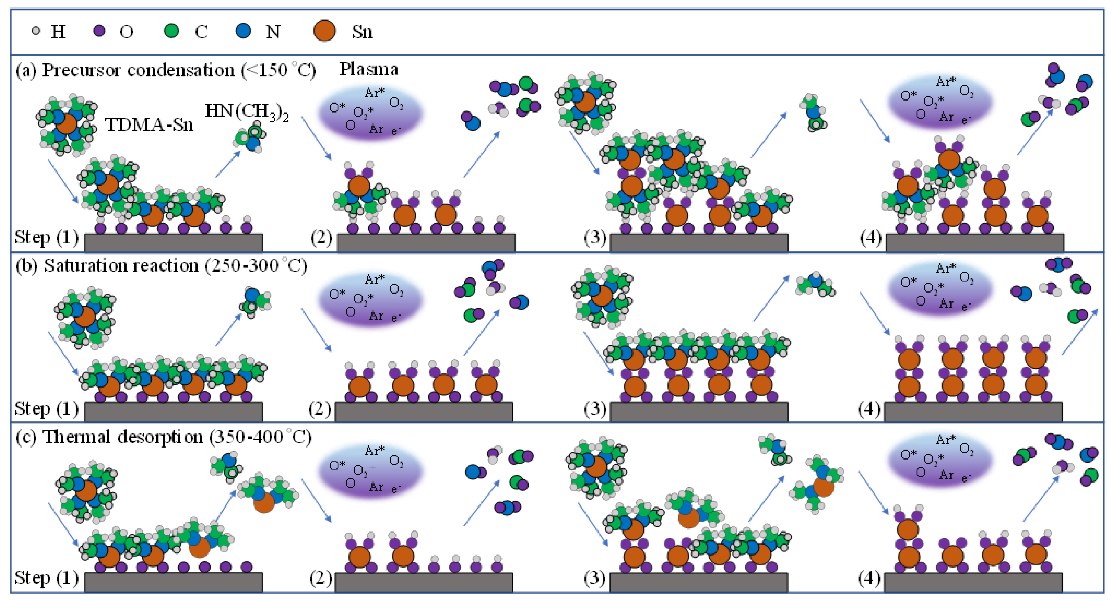
Figure 1. Deposition mechanism of PEALD SnOx films at different substrate temperatures dividing into three growth modes: (a) precursor condensation (<150 °C), (b) saturation reaction (250–300 °C), and (c) thermal desorption (350–400 °C).
Nanomaterials 12 02859 g001
Figure 2. (a) The substrate temperature-dependent growth per cycle (GPC) of PEALD SnOx films and (b) its trend line of corresponding thickness at each GPC.
Nanomaterials 12 02859 g002
Figure 3. (a) XPS spectrums for the PEALD SnOx films deposited at substrate temperatures from 100 and 400 °C. The spectra of (b) Sn 3d and (c) O 1s core level with (d) the peak area ratio of OL–Sn2+/[(OL–Sn2+) + (OL–Sn4+)] and OV/(OL + OV), and (e) the atomic ratio of O, Sn, and N elements.
Nanomaterials 12 02859 g003
Figure 4. (a) XRD patterns of PEALD SnOx films deposited at substrate temperatures where the red star mark with red dash line presents another (101) orientation of Sn3O4. (b) The variation for the FWHM of the preferential (110) orientation and the average crystallite size, showing (c) the dependence of the average dspacing of (110) planes, the dislocation density, and the micro-strain value. (d) The deconvolution results of the (110) orientation deposited at 300 °C in the 2 theta of 22–31°. (e) The variation of the area ration of (110)SnO2 to [(110)SnO2 + (101)Sn3O4 + (111)Sn3O4].
Nanomaterials 12 02859 g004
Figure 5. Topographic and stereoscopic surface morphologies of AFM with a scanning area of 5 × 5 µm2 and top-view images of FESEM for PEALD SnOx films deposited at various substrate temperatures from (ag) 100 °C to 400 °C on a Si wafer.
Nanomaterials 12 02859 g005
Figure 6. The TEM results of SnOx films deposited at different substrate temperatures of (a) 100 °C, (b) 250 °C, and (c) 400 °C, including cross-sectional and high-resolution images.
Nanomaterials 12 02859 g006
Figure 7. (a) The optical transmittance with reflectance and (b) absorption coefficient spectrums to extract (c) the band gap values for the PEALD SnOx films with increasing substrate temperatures from 100 to 400 °C.
Nanomaterials 12 02859 g007
Figure 8. The wavelength-dependent refractive index of PEALD SnOx films with increasing substrate temperatures from 100 to 400 °C.
Nanomaterials 12 02859 g008
Figure 9. Hall-effect measurement for the (a) carrier concentration (Ne) accompanied by the mobility (µ) and (b) resistivity (ρ) of SnOx films deposited at various substrate temperatures at 100–400 °C. Five samples were measured in each series, and standard deviations are included.
Nanomaterials 12 02859 g009
Table 1. Preparation parameters of PEALD SnO2 films.
ParameterValue
Bubbler temperature (°C)50
Substrate temperature (°C)100–400
TDMA-Sn pulse time (s)1.6
TDMA-Sn purge time (s)6
O2 pulse time (s)11
O2 purge time (s)5
Ar flow rate (sccm)80
O2 flow rate (sccm)150
O2 plasma power (W)2000
TDMA-Sn carry gas flow rate (sccm)120
TDMA-Sn dilute gas flow rate (sccm)400
Publisher’s Note: MDPI stays neutral with regard to jurisdictional claims in published maps and institutional affiliations.