Article

The Strong Protective Action of Ce3+/F Combined Treatment on Tooth Enamel and Epithelial Cells

by
Anton L. Popov
1,2,
Nadia M. Zholobak
3,
Alexander B. Shcherbakov
3,
Taisiya O. Kozlova
1,
Danil D. Kolmanovich
1,2,
Artem M. Ermakov
2,4,
Nelli R. Popova
2,
Nikita N. Chukavin
2,4,
Ernest A. Bazikyan
5 and
Vladimir K. Ivanov
1,*
1
Kurnakov Institute of General and Inorganic Chemistry of the Russian Academy of Sciences, Moscow 119991, Russia
2
Institute of Theoretical and Experimental Biophysics, Russian Academy of Sciences, Pushchino 142290, Moscow Region, Russia
3
Zabolotny Institute of Microbiology and Virology, National Academy of Sciences of Ukraine, Kyiv 03680, Ukraine
4
Moscow Region State University, Moscow 141014, Russia
5
Yevdokimov Moscow State University of Medicine and Dentistry, Moscow 127473, Russia
*
Author to whom correspondence should be addressed.
Nanomaterials 2022, 12(17), 3034; https://doi.org/10.3390/nano12173034
Submission received: 26 July 2022 / Revised: 14 August 2022 / Accepted: 28 August 2022 / Published: 1 September 2022
(This article belongs to the Special Issue Advancements of Nanomaterials in Oral Health and Clinical Dentistry)

Abstract

:
We studied the toxic effects of cerium and fluoride species on human dental pulp stem cells and epithelial cells of Cercopithecus aethiops as a surrogate for the human oral mucosa. The sequential use of CeCl3 and NH4F solutions in equimolar sub-toxic concentrations enabled the possible toxic effects of individual components to be avoided, ensuring the preservation of the metabolic activity of the cells due to the formation of CeF3 nanoparticles. Cerium fluoride nanoparticles and terbium-doped cerium fluoride nanoparticles exhibited neither cytotoxicity nor genotoxicity to dental pulp stem cells, even at high concentrations (10−4 M). In millimolar concentrations (from 10−5–10−6 M), these nanoparticles significantly increased the expression of genes responsible for the cell cycle, differentiation and proliferation. The formation of cerium fluoride on the surface of the mucous membrane and teeth provided protection against the development of carious lesions, periodontitis, ROS attacks and other inflammatory diseases of the oral cavity. Luminescent CeF3: Tb nanoparticles enabled the visualization of tooth enamel microcracks.

Graphical Abstract

1. Introduction

Caries is a dental disease that occurs with the destruction of hard tooth tissues, generally in acidic erosion conditions. The inorganic part of tooth hard tissue consists mainly of hydroxyapatite crystalline species (calcium phosphates). In the crystal lattice of hydroxyapatite, hydroxyl ions can be substituted by fluoride ions, thus, forming fluorapatite, which possesses enhanced microhardness and demonstrates higher resistance to the action of acids formed in the oral cavity.
The beneficial effects of fluoride on human oral health are well-known; fluorides have a protective effect against both dental hard tissue erosion and caries formation through demineralization inhibition and remineralization promotion. Fluoride ions can also protect against fermentation attack by Streptococcus sobrinus artificial plaque on tooth enamel [1]; however, an excess of fluoride ions has several adverse effects [2,3]. Exposure to high fluoride doses has been reported to induce apoptotic cell death in ameloblasts, odontoblasts and osteoblasts.
Tabuchi et al. studied the effects of fluoride ions on rat oral epithelial cells and observed increased cell death with an increasing concentration of NaF from 2 to 4 mM [4]. The induction of apoptosis by this compound was confirmed by the activation of caspase-3 with a concomitant increase in the expression of Atf3, Ddit3 and Fos genes. The differentially expressed genes are likely to be involved in cell death accompanying endoplasmic reticulum (ER) stress.
A recent study investigating the effects of fluoride ions on the proliferation of cells and gene expression showed that fluoride exposure increased human B lymphocytes’ viability at relatively low levels (10–160 µM); however, when the concentration reached 320 µM, cell proliferation was significantly inhibited. Fluoride ions induce apoptosis in mammalian cells, both in vitro and in vivo [5]. At a concentration of 50 μM, fluoride ions are able to increase the production of superoxide anion radicals in the mitochondria of osteoblasts, while reducing their oxygen consumption [6].
Increased production of reactive oxygen species (ROS) occurs due to oxidant system suppression: superoxide dismutase and catalase dramatically reduce their activity upon fluoride intoxication. At the same time, insoluble cerium fluoride is capable of protecting cells from oxidative stress [7] and accelerating mitogenesis via neoblastic activation [8].
Cerium species are promising for biomedical practice [9], including dental care, e.g., in ceria-containing orthodontic polymers [10]. Cerium oxide nanoparticles protect human dental stem cells from oxidative insult [11]. Recently, cerium oxide particles were successfully used as a filler to tune the radiopacity of dental adhesives [12]. Soluble and insoluble cerium compounds possess notable antibacterial activity [13] and appear to have a high potential for use against dental bacteria (including Streptococcus mutans [14]) that are responsible for the cariogenic process.
In their experiments with four common bacteria (i.e., E. coli, S. aureus, B. subtilis and B. cereus), Anastasiou et al. showed that doping with Ce3+ enhanced the regenerative potential of dental pulp stem cells, while retaining the good antibacterial profile of fluorapatite [15]. The incorporation of cerium ions into dental enamel and the formation of cerium-apatite has been reported [16]. Wegehaupt et al. established that cerium chloride is able to significantly reduce the acid susceptibility of dentine [17] and demonstrated the anti-erosive potential of the combination of fluoride ions, cerium chloride and laser irradiation on dentine [18].
Cerium chloride and its combination with fluoride species can significantly reduce carious mineral loss and the progression of lesion depth [19]. Zhang et al. showed that the combination of fluoride and cerium ions was more efficient in the prevention of root surface caries than lanthanide ions alone [20].
Later, Jaisingh et al. demonstrated that cerium chloride reduced demineralization, and improved remineralization, for artificial caries lesions in human enamel in vitro when only subjected to pH cycling [21]. Lanthanum, cerium and fluoride species (500 ppm) were shown to provide demineralization resistance in an acid resistance and remineralization study on human root surface [22]. Jin et al. demonstrated the beneficial effect of lanthanum, cerium and fluoride species on the prevention of root surface caries [23].
Soluble calcium species are well-known fluoride neutralizers; they reduce the fluoride bioavailability in lethal models of fluoride poisoning [2,24,25]. The mechanism of their protective action is thought to be the formation of insoluble calcium fluoride salts, which results in binding and inactivating the fluoride ions. The solubility constant of CeF3 (Ksp = 8 × 10–16) is substantially lower than that of fluorspar, CaF2 (Ksp = 4 × 10–11) [7]; this leads to the expectation of even more effective protection against fluoride ions when using cerium soluble salts.
The current study features the toxic effects of cerium and fluoride species on human dental pulp stem cells (DPSc) and epithelial cells of Cercopithecus aethiops as a surrogate for the human oral mucosa. The study also analyses the protective effect of CeF3:Tb nanoparticles in enamel defect zones and includes a comprehensive analysis of cell metabolic activity and gene expression. An analysis was made of the possible benefits of the combined application of cerium and fluoride species in dental healthcare.

2. Materials and Methods

2.1. Cell Cultures, Dental Samples, Chemicals

Cerium(III) chloride heptahydrate, terbium(III) chloride hexahydrate, calcium chloride, ammonium fluoride, hydrofluoric acid, lactic acid, sodium lactate and Arsenazo III were purchased from Sigma-Aldrich (St. Louis, MO, USA). Imidazole buffer (Glyoxaline solution) was purchased from Merck (Darmstadt, Germany). Cerium fluoride nanoparticles (both bare and terbium-doped) and cerium oxide nanoparticles were synthesized according to protocols reported elsewhere [7].
Epithelial cells MA-104 (ATCC® CRL-2378) isolated from an African green monkey fetal kidney were obtained from the biobank of the Zabolotny Institute of Microbiology and Virology. Dental pulp stem cells (DPSc) were isolated from the tooth pulp of a healthy donor according to orthodontic prescription and with the informed consent of the patient. All procedures were performed in accordance with the approved clinical rules for biomaterial sampling.
The cells were cultivated in a DMEM/F12 (1:1) medium containing 10% of fetal bovine serum (FBS), 50 μg/mL of penicillin, 50 μg/mL of streptomycin and 3 mg/mL of L-glutamine, at 37 °C in a humid atmosphere with 5% CO2. The cells were seeded at a density of 2 × 10⁴–2.5 × 10⁴ per cm2. Six hours after cell attachment, cerium salt solutions or ceria nanoparticles were added, in different concentrations (10−3–10−7 M). In the control experiments, the cells were cultured without cerium salt solutions or ceria nanoparticles.
Extracted for orthodontic reasons, human molars with sound enamel and approximately equal habit and weight (2.0 ± 0.4 g) were used; teeth were stored in a 0.5% formaldehyde solution adjusted to pH 7.5 with sodium hydroxide.

2.2. Visualization of Cerium and Fluoride Species Absorption and Confirmation of Cerium Fluoride Formation

For the verification of lanthanide and fluoride species adsorption on the tooth surface, human molars with sound enamel were used. To simulate a caries-like defect, part of the enamel was chipped out mechanically. We dissolved 0.425 mmol of cerium chloride heptahydrate (158.5 mg, MW = 372.58) and 0.075 mmol of terbium chloride hexahydrate (28 mg, MW = 373.38) in 5 mL of water (Ce:Tb = 0.85:0.15 mol) and named solution #1. Separately, 1.5 mmol of ammonium fluoride (55.5 mg, MW = 37) was dissolved in 5 mL of water and named solution #2.
Each molar was immersed in lanthanide chloride solution, exposed for 5 min and immersed in ammonium fluoride solution, then rinsed with distilled water and dried. Tooth luminescence was studied under irradiation, using a Philips TUV G15 T8 (Philips, Hamburg, Germany) germicidal quartz lamp with a maximum emission at 253.7 nm. For the independent confirmation of CeF3:Tb nanoparticles formation, the following experiment was performed. A few drops (~0.5 mL) of saliva were added to solution #2. Solution #1 was quickly added to solution #2 under agitation. The terbium-doped cerium fluoride nanoparticles formed were studied by luminescent spectrophotometry using a QE-65000 spectrometer, Ocean Optics (Orlando, FL, USA) and TEM microscopy (Leo912 AB Omega, Carl Zeiss, Oberkochen, Germany).

2.3. The Cytotoxicity of the Cerium Ions and Fluoride Ions In Vitro

The cytotoxicity of the substances was analysed using the colorimetric MTT assay; the test protocol for cytotoxicity evaluation was adopted from elsewhere [26]. Briefly, different concentrations of the substances (10 μM–2 mM) were added and cells were incubated for 24 h, at 37 °C in humid air (98%) containing 5% CO2. Four hours prior to the end of the exposure period, the supernatant was removed, and MTT (3-(4,5-Dimethyl-2-thiazolyl)-2,5-diphenyl-2H-tetrazolium bromide, Sigma-Aldrich, St. Louis, MO, USA, #M5655) solution in PBS (0.1 mg/mL, 100 μL/well) was added to the cells.
Upon the completion of the exposure period, the supernatant was removed, and a lysis solution containing 0.1% SDS (Sigma-Aldrich, #L3771) solution in DMSO (Sigma-Aldrich, #D8418) was added. Plates were shaken for 5 min, placed on a Multiskan MS Microplate Reader (Thermo Labsystems, Santa Rosa, CA, USA), and the absorbance was read colorimetrically at 492 nm. Each experiment was repeated three times, with four replications.

2.4. The Protective Effect of Cerium or Calcium Species against Fluoride Ions In Vitro

The protective effect of cerium or calcium species was studied according to therapeutic, and prophylactic, schemes. Briefly, in the prophylactic scheme, solutions of cerium chloride or calcium chloride, of different concentrations (10 μM–2 mM), were added to the cell monolayers; 24 h later, a solution of ammonium fluoride was added to the cells. The concentration of fluoride ions in each well was 0.5 mM, corresponding to 50% viability in the acute toxicity test. The cells were incubated in fluoride media for 4 h and subsequently washed and stained with crystal violet.
The excess dye was then removed and the stained monolayer was washed with distilled water and dried. The absorbance of stained cells was measured at 492 nm using a Thermo/LabSystems Multiskan MS Microplate Reader. The optical density of stained cells corresponded to their viability. The percentage of the cells absorbing crystal violet was determined according to the formula D e x D c o n t r · 100 , where Dex is the optical density of the experimental wells and Dcontr is the optical density of the intact (control) wells.
Statistical analysis of the data obtained was performed using BioStat 2009 Professional 5.8.1 (Analystsoft, Walnut, CA, USA) software, in accordance with standard recommendations. Experimental data were presented in terms of the median and interquartile range, Me (LQ-UQ), where Me = median (50% percentile), LQ = 25% percentile and UQ = 75% percentile. For the therapeutic scheme, the sequence of cerium/calcium and fluoride species addition was reversed. Each series of experiments was repeated five times.

2.5. The Protective Effect of Cerium-Containing Species against ROS In Vitro

The protective action of cerium ions and ceria nanoparticles against ROS was determined as described elsewhere [7]. Briefly, the solutions of tested species (0.005–0.3 mM) were introduced to the cell monolayers, and 18 h later hydrogen peroxide was added to the cells, (the H2O2 concentration in each well was 0.5 mM, corresponding to 5% viability in the toxicity test). The cells were incubated for 4 h under conditions of oxidative stress, then washed and stained with crystal violet. Subsequent protective effect analysis and data processing were essentially the same as in the previous series of experiments (see above, “The protective effect of cerium or calcium species against fluoride ions in vitro”). Each series of experiments was repeated five times.

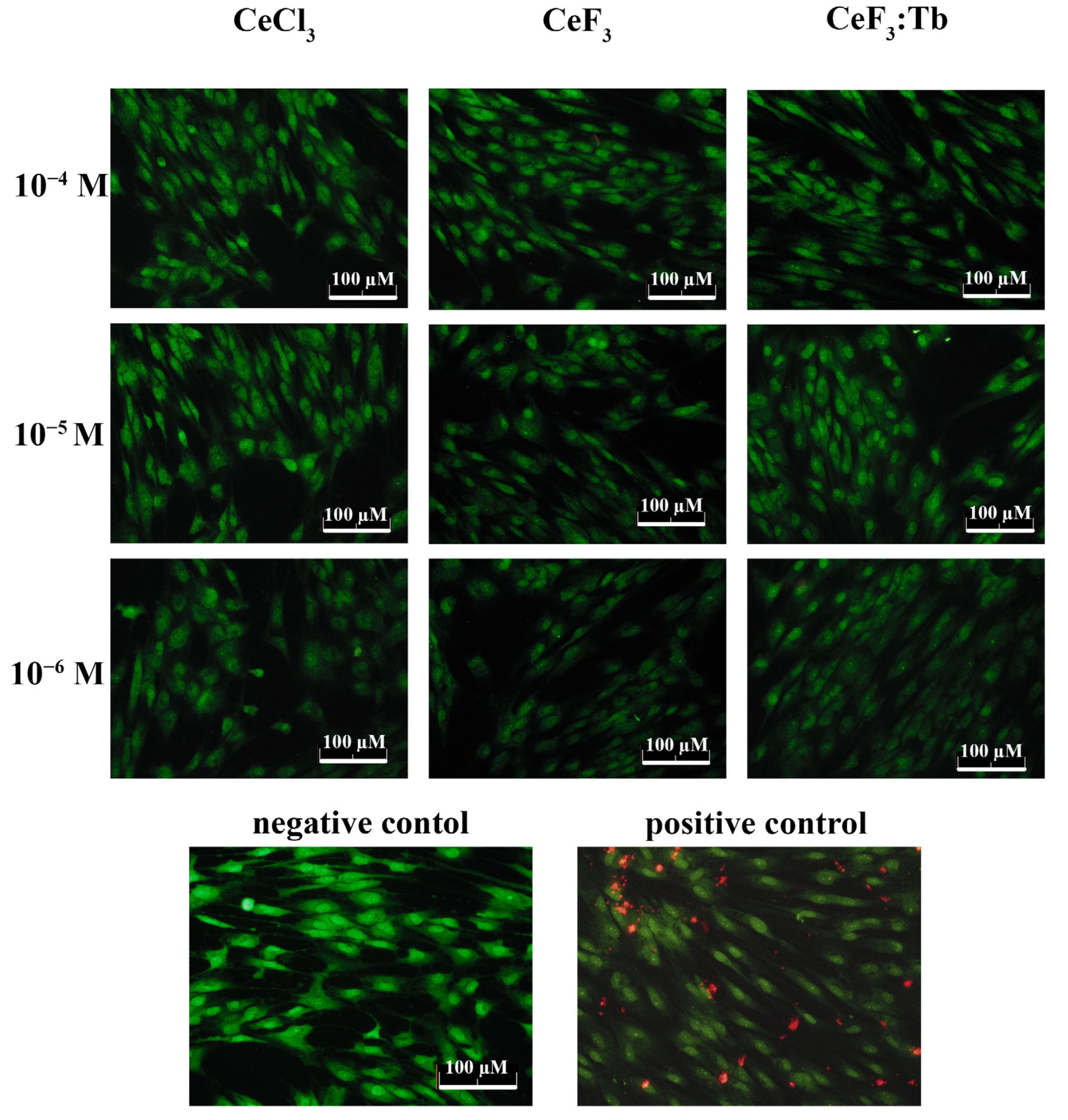
2.6. Live/Dead Assay

A Live/Dead Viability Kit (Invitrogen, Thermo Fischer, Carlsbad, CA, USA) was used to evaluate the cytotoxic effects of cerium-containing nanoparticles. The kit contained two fluorescent dyes for selective cell labelling. The green signal (SYTO 9 dye, λ = 485/498 nm) characterized the live cells, and the red signal (propidium iodide dye, λ = 535/617 nm) characterized the dead cells. Cells were seeded into 96-well plates and the kit was used according to the manufacturer’s protocol. Cells were visualized and photographed 25 min after adding the dyes using an Axiovert 200 fluorescent microscope (Carl Zeiss, Oberkochen, Germany) and a Canon A620 digital camera (Canon, Tokyo, Japan). For each cell group, four fields in each well were examined.

2.7. Analysis of Mitochondrial Membrane Potential (MMP)

Mitochondrial membrane potential (MMP) was determined by TMRE (tetramethylrhodamine, ethyl ester, ThermoFisher, Carlsbad, CA, USA) dye using fluorescence microscopy. TMRE accumulates in the mitochondrial membrane in a potential-dependent manner.
Cells were seeded into 96-well tissue culture plates (Greiner, Kremsmünster, Austria) at a density of 5 × 10⁴ cells/well and cultured in a CO2 incubator at 37 °C for 24, 48 and 72 h, with different concentrations of nanoparticles. The cells were preincubated with 1 μM TMRE in the HBSS in a CO2 incubator at 37 °C for 30 min. Next, the cells were washed twice by HBSS and analysed using an Axiovert 200M (Carl Zeiss, Oberkochen, Germany) inverted fluorescence microscope (Carl Zeiss, Germany) at 200× magnification.

2.8. Real-Time PCR

A kit for mRNA isolation with magnetic particles was used, according to the manufacturer’s protocol (Sileks, Moscow, Russia). Reverse transcription was performed using a kit supplied by Sileks (Russia), using oligo(dT) primer, following the manufacturer’s protocol. The cDNA derived was used as a template for real-time PCR. The reaction was conducted using a mixture with SybrGreen (Syntol, Moscow, Russia), using a CFX-96 thermal cycler (BioRad, Hercules, CA, USA) or ABI 7500 Fast Real-Time PCR System (Thermo Fisher Scientific, Waltham, MA, USA). The expression level of 96 genes, responsible for 25 key cellular processes, was estimated (Table 1). The analysed genes were selected using the database http://www.sabiosciences.com/ (accessed on 1 April 2022) for PCR profiling of different biological processes.
The transcription level was normalized using the expression of housekeeping genes encoding β-actin, RPLP0 (ribosomal protein, large, P0) and GAPDH (glyceraldehyde-3-phosphate dehydrogenase). Gene-specific primers were picked using Primer Express software (Applied Biosystems, USA). Each measurement was made with two replications (internal replication) and averaged for two independent samples. Samples without reverse transcription were used as a control. Analysis of the expression data was performed using online services at http://www.sabiosciences.com/ (accessed on 1 April 2022), mayday-2.14 software (Center for Bioinformatics, Tübingen, Germany) (accessed on 15 April 2022) and Genesis software.

2.9. Assessment of Tooth Enamel Protection

For the evaluation of tooth enamel protection against acidic dissolution by cerium- and fluoride-containing species, human molars were used. Twelve teeth with sound enamel, of approximately equal weight (2.0 ± 0.2 g) and habitus, were divided into four groups: nontreated (control); treated with 10 mM cerium chloride; treated with 10 mM ammonium fluoride; and treated with 10 mM cerium chloride and then by 10 mM ammonium fluoride.
An etching lactate buffer (5% w/v) was prepared using the solution containing 0.4510 M lactic acid and 0.1041 M sodium lactate, adjusted to pH 3.0 with sodium hydroxide. The release of calcium ions was measured using the colorimetric method, with Arsenazo III indicator [27]. The reagent used for the determination of calcium ions was composed of 100 mM imidazole buffer (pH 6.5) and 0.12 mM Arsenazo III. The absorption was determined at λ = 650 nm.
Each molar was immersed in the etching buffer solution (5 mL in glass tube) to the level of the enamel zone and exposed for 1 min; after 1 min of exposure, each molar was transferred to the next tube containing a fresh lactate buffer, and the remaining supernatant solution used was analysed. The total erosive attack time for each molar was 10 min. The square of enamel immersed was calculated for every molar, considering the tooth habit.

2.10. Statistical Data Processing

The results obtained were processed using statistical criteria. Variation statistics were used to estimate the significance of the results. Any significant difference from the control was assessed using the nonparametric Mann–Whitney U test.

3. Results

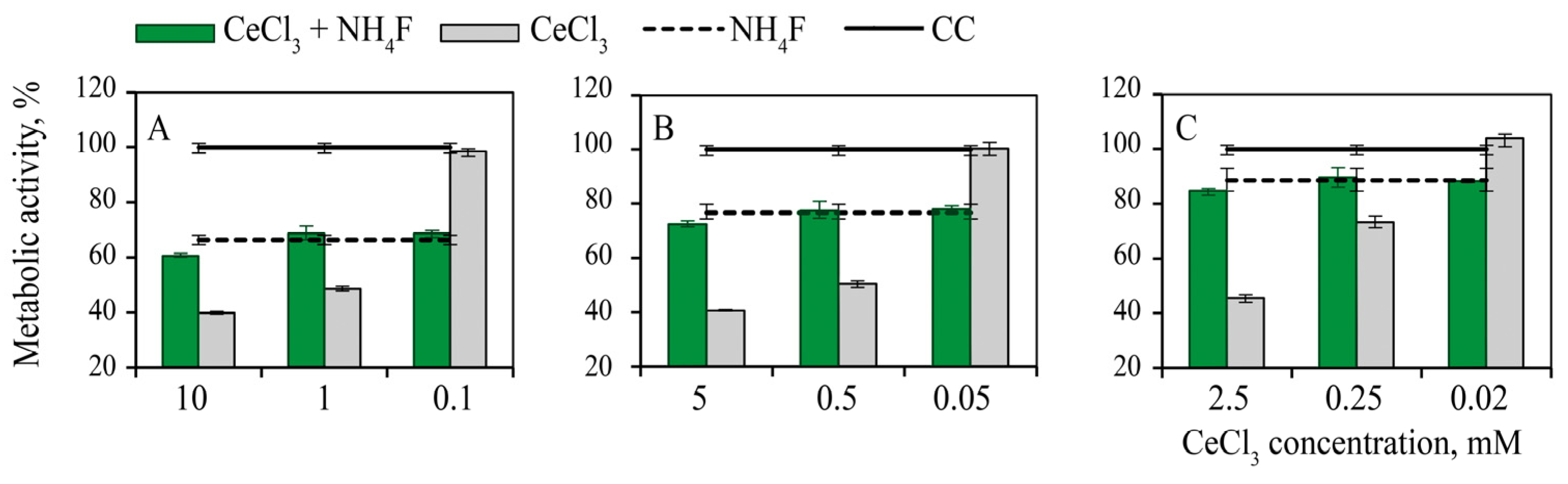
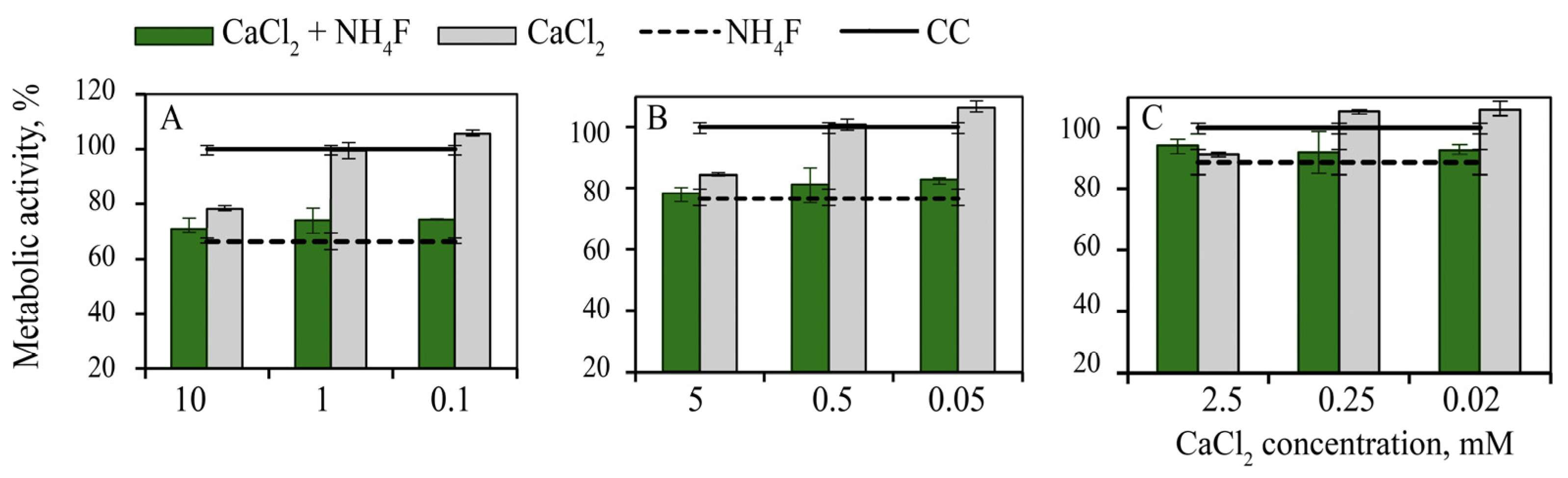
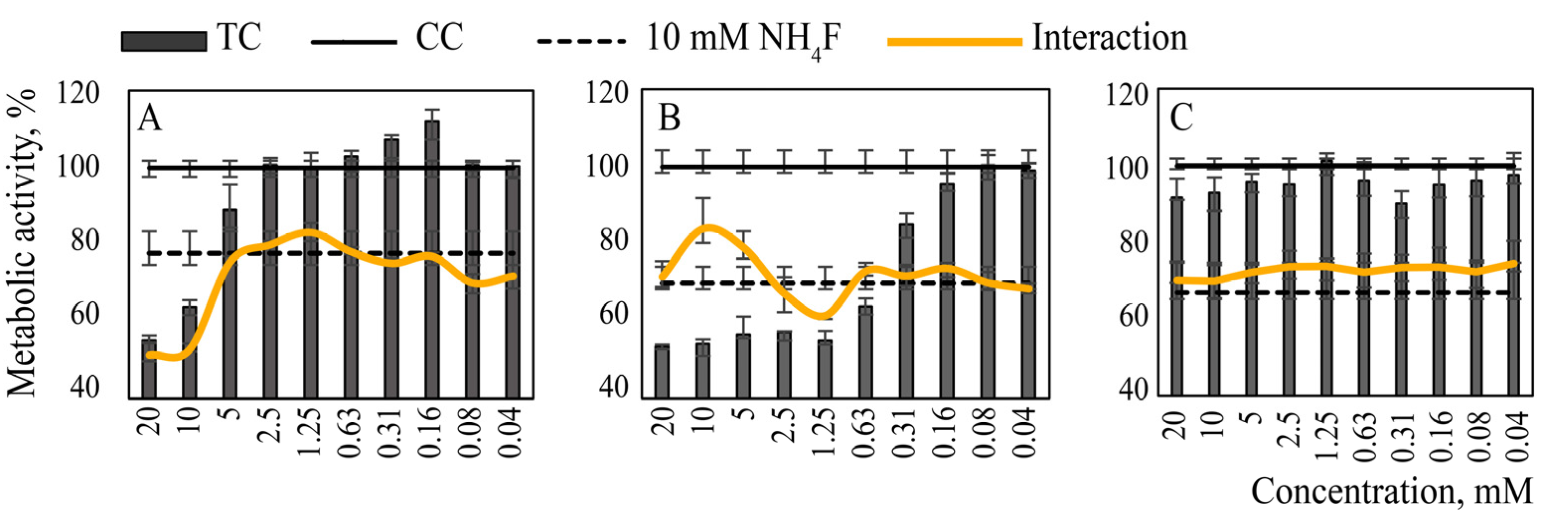
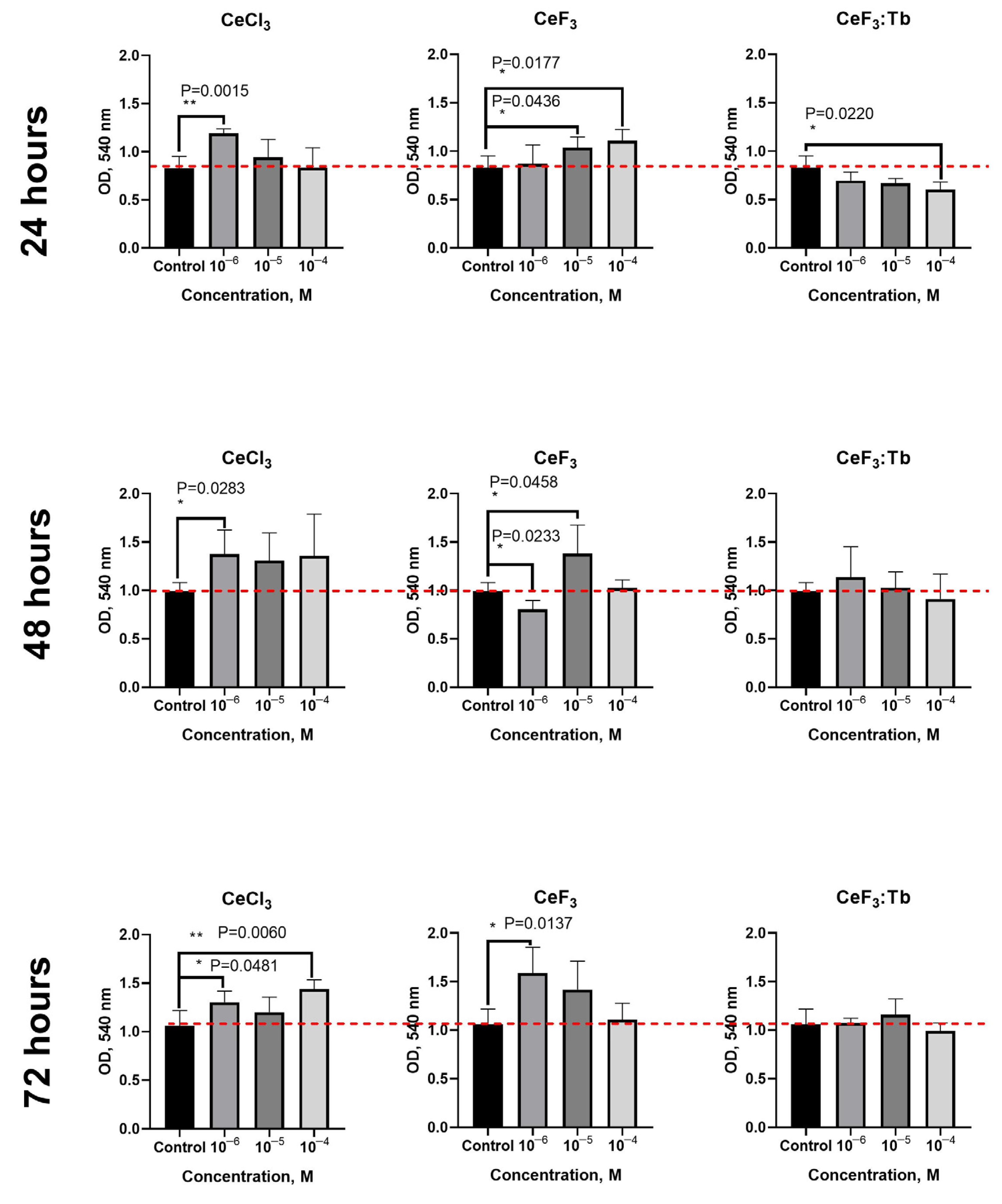
3.1. Toxicity and Cellular Metabolic Activity

The use of cerium and fluoride compounds in human dental healthcare is directly related to their effect on the oral mucosa; therefore, monkey epithelial cells MA-104 (having the maximum species proximity to humans) were selected for cytotoxicity and metabolic activity studies. Ce3+ and Ca2+ ions are close in terms of their biochemical properties [28]; thus, calcium salt solution was used as a reference. Equimolar toxic concentrations of cerium or calcium salts as well as 10-fold higher concentrations of salts were used.
The 24-h contact of MA-104 cells with NH4F solution was accompanied by 60–70% inhibition of their metabolism (MTT test), CC50 = 7.0 mM, CC0 = 1.3 mM (Figure 1A). For hydrofluoric acid, CC50 = 3.0 mM, CC0 was not determined. According to the results obtained on cytotoxicity, a 10 mM NH4F concentration was considered as toxic in further experiments in vitro.
Studies of the metabolic activity of MA-104 cells in the presence of cerium and calcium salts showed that calcium chloride had low toxicity (CC0 = 1.0 mM), while cerium chloride was much more toxic (CC0 = 0.1 mM) (Figure 1B). In the presence of calcium chloride, no changes in metabolic activity of the cells were observed; for cerium chloride, there was a short period of transition from non-toxic behaviour to metabolism-inhibiting action: a five-fold increase in salt concentration was accompanied by a 50% decrease in cell metabolism (CC50 = 0.5 mM). For calcium chloride, the calculated CC50 value was about 50 mM, i.e., 100-times higher.
The inhibition of cell metabolism caused by cerium chloride addition was found to decrease significantly after subsequent treatment with equimolar concentrations of NH4F (Figure 2). The same effect was observed when the NH4F concentration was an order of magnitude higher than the concentration of CeCl3. Thus, the addition of equimolar, or even significantly higher, concentrations of NH4F to the cells, treated with a CeCl3 solution in toxic concentrations, cancelled the development of the toxic effects induced by Ce3+ ions. The metabolic activity of the cells treated sequentially with cerium chloride and ammonium fluoride slightly differed from the metabolic activity of the cells treated solely with NH4F. Interestingly, non-toxic CeCl3 concentrations had no effect on the metabolic activity of cells subsequently treated with NH4F.
Ca ions in the concentration range of 0.025–0.25 mM caused a small, but statistically significant, increase in cell metabolic activity, while 2.5–10.0 mM Ca concentrations caused the inhibition of cell metabolism by 10–20%. Ammonium fluoride (2.5–10.0 mM) also caused a decrease in the metabolic activity of the cells, by 10–35%. Over the entire range of CaCl2 concentrations, the addition of NH4F to the cells caused an additional decrease in their metabolic activity compared with treatment with NH4F alone (Figure 3).
Based on the results obtained, a wider concentration range of CeCl3 and CaCl2 solutions was tested in combination with a known toxic concentration of NH4F (10 mM), causing 30% (22–32%) inhibition of metabolic activity. The interval between the treatment of the cells with CeCl3 or CaCl2 solutions and NH4F solutions was set at 5 min, which could be used in practice for a mouth rinse or for a similar medical tooth treatment. A colloidal solution of citrate-stabilized cerium dioxide nanoparticles of the same concentrations was used as an additional control. The results are shown in Figure 4.
The sequential treatment of the cells with CeCl3 and NH4F solutions at equimolar concentrations ensured the preservation of cell metabolic activity; for the cells treated with 10 mM CeCl3 only, the metabolic activity was 55% of that of the control; for the cells treated with NH4F: 70%; and for the cells treated sequentially with both CeCl3 and NH4F: 80–95%. The use of CeO2 colloidal solution did not increase the metabolic activity of cells treated with NH4F. The addition of CaCl2 solution to the cells before their treatment with NH4F did not lead to any statistically significant change in cell metabolism in comparison with the cells treated with NH4F only.
Thus, we found that contact of monkey kidney epithelial cells MA-104 with cerium chloride solution with concentrations of 0.1 mM or higher was accompanied by a sharp concentration-dependent decrease in their metabolic activity. The addition of NH4F solution in an equimolar, or even a ten-times higher concentration, to the cells after 40 min of CeCl3 treatment eliminated the adverse effects of cerium species. The ex tempore (5 min after treatment) addition of an equimolar NH4F solution also ensured good cell viability and even exceeded that of the cells treated with either CeCl3 only or NH4F only.

3.2. Protective Effect of Cerium and Fluoride Species against H2O2-Induced Oxidation Stress

The protective action of calcium chloride, cerium chloride, ammonium fluoride and cerium fluoride against oxidative stress caused by ROS (hydrogen peroxide) action on MA-104 cells was studied in the concentration range of 0.005–0.31 mM; these concentrations were selected as being non-toxic for MA-104 cells, according to the data presented above. Since cerium oxide nanoparticles are a well-known ROS protector [9], this material was also included in the research design as a reference; the protective action of calcium ions was evaluated for comparison, too.
MA-104 cells were treated with cerium or calcium species for 18 h and cell viability analysis was performed (Figure 5A). 0.15–0.31 mM concentrations of calcium chloride have been shown to provide cells with some resistance to peroxide-induced oxidative stress (about 50%), while lower concentrations do not protect cells. Unlike calcium species, cerium compounds provide protection for cells at lower concentrations; in addition, the efficiency of protection is significantly higher.
Thus, 0.31 mM cerium chloride provides 100% protection of the cells from oxidative stress induced by hydrogen peroxide; 50% and higher levels of protection were observed up to a CeCl3 concentration of 0.02 mM. It should be noted that CeF3 protects cells even more effectively than other species: a 100% level of protection was observed down to a concentration of 0.04 mM. A two-fold decrease in CeF3 concentration (to 0.02 mM) was accompanied by a two-fold decrease in the protective effect, although a further decrease in CeF3 concentration to 0.01 mM did not cause such a sharp decrease—the level of protection being 40 ± 15%.
Terbium-doped cerium fluoride protects cells against ROS in the same manner (not shown) as a pristine CeF3, with some minor differences (±5–10%). The use of nanoceria was accompanied by a concentration-dependent protective action: 100% protection of the cells was observed at concentrations of 0.16–0.31 mM; the concentration range of 0.04–0.08 mM provided more than 70% protection and 0.01–0.02 mM concentrations resulted in 40% cell protection. As expected, non-toxic concentrations of HF and NH4F provided no protective action and did not affect the cell viability (see Figure 5B).

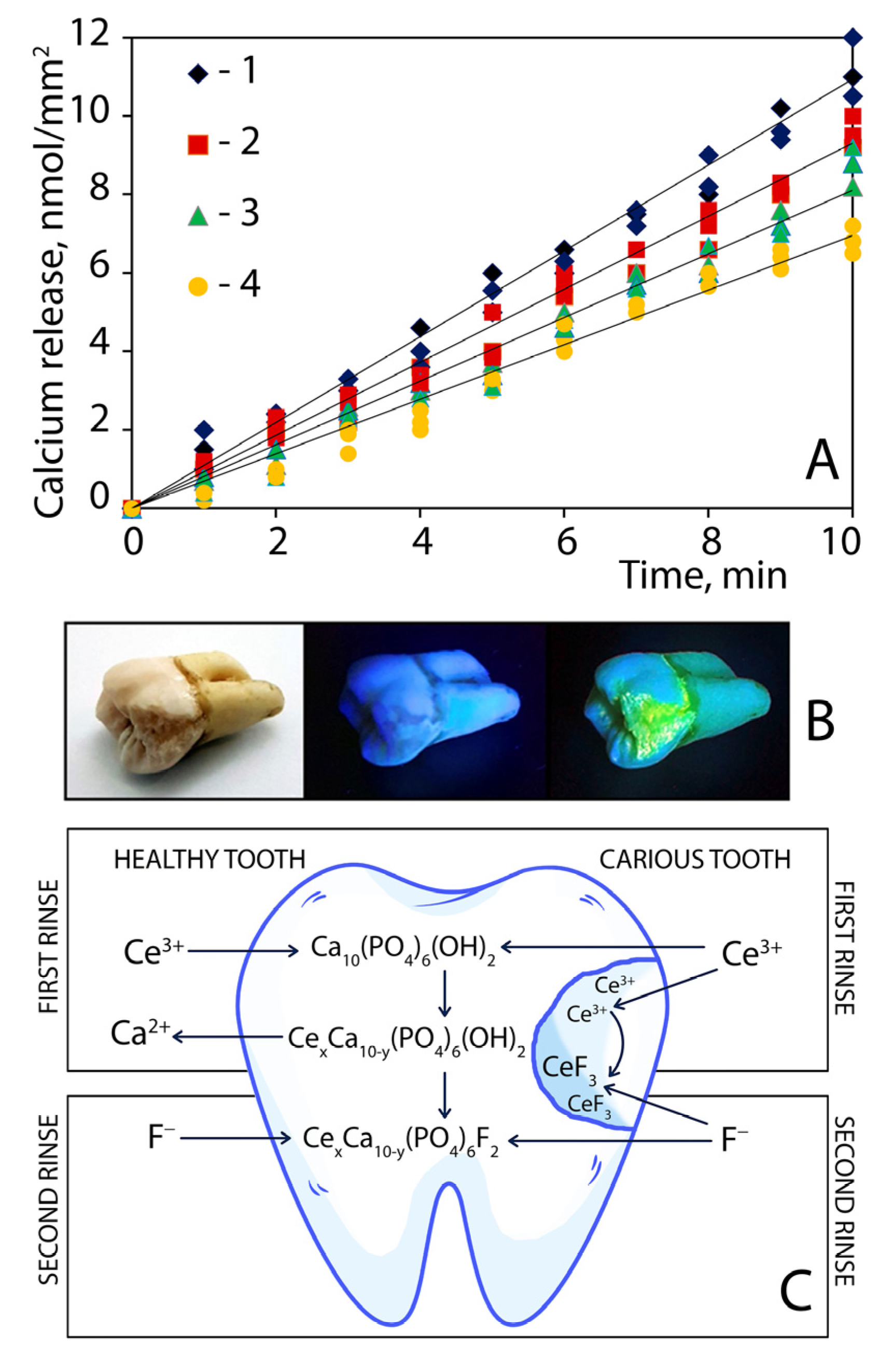
3.3. Tooth Enamel Erosion Protection and Carious Cavity Sealing by Sequential Treatment of Teeth with Cerium Chloride and Ammonium Fluoride

The end-product of bacterial glycolysis under anaerobic conditions is lactic acid; it creates acidity high enough to dissolve calcium phosphate in the tooth enamel, leading to the start of cavity formation. Clearly, acidic lactate buffer would be the best choice of surrogate oral solution for caries formation.
The amount of calcium released into the lactate buffer solution during each of the ten erosive attacks for the four treatment groups of the teeth is presented in Figure 6A. The amount of calcium decreased from 11 ± 1 nmol/mm2 (untreated teeth) to 6.0 ± 0.6 nmol/mm2 (teeth treated with cerium chloride following ammonium fluoride solutions), depending on treatment conditions. Figure 6A shows that sequential treatment of teeth with cerium chloride and ammonium fluoride most significantly reduced acid corrosion of tooth enamel. The effect was much higher than in the case of using each of the components separately.
If a tooth with damaged enamel is subjected to sequential rinsing with cerium and fluoride solutions, a layer of cerium fluoride nanoparticles forms at the carious zone. This fact can be confirmed by adding a small amount of terbium ions (~15 mol%) to the cerium salt solution. CeF3:Tb nanoparticles formed can easily be detected due to the bright luminescence (Figure 6B), which has well-known optical characteristics (excitation and emission wavelengths).
Moreover, the luminescence of terbium-doped cerium fluoride nanoparticles does not appear under either visible light or UVA irradiation; it can be registered under shortwave ultraviolet radiation (UVC) only and thus it does not impart the natural appearance of teeth (Figure 6B, left to right). Terbium-doped cerium fluoride nanoparticles formed in situ seem to be useful for the in vivo defectoscopy of enamel integrity and tooth surface behaviour, enabling the detection of problem areas and the prevention of caries formation.
According to the SAED pattern and TEM image analyses (Figure 7), crystalline and sufficiently monodispersed (15–20 nm) CeF3 and CeF3: Tb3+ nanoparticles were formed upon the simple mixing of lanthanide and fluoride ions in saliva-containing media. Terbium excitation at 254 nm (characteristic 489 nm 5D47F6, 543 nm 5D47F5, 583 nm 5D47F4 and 620 nm 5D47F3 transitions) suggests energy transfer from Ce3+ to Tb3+ in the crystalline lattice; such a transfer mechanism has been thoroughly investigated for CeF3: Tb3+ nanoparticles [7].
Possible mechanisms of tooth modification by rinsing with cerium and fluoride solutions are represented in Figure 6C. Healthy enamel consists mainly of hydroxyapatite Ca10(PO4)6(OH)2. The diagram in Figure 6C (left) demonstrates the mechanism of enamel strengthening due to the sequential modification of hydroxyapatite with cerium and fluoride ions by replacing calcium and hydroxide moieties. In the right part of the diagram in Figure 6C, a protective mechanism is suggested by sequential rinsing with cerium and fluoride solutions in the case of caries.
When the integrity of the enamel is violated, dentin (consisting mainly of collagens) channels are opened. Moreover, carious lesions contain numerous surface defects that are colonized by bacteria and covered with biofilms. Cerium ions are readily adsorbed by collagens, polysaccharides and other biopolymers. Then, during treatment with fluoride ions, a protective layer consisting of cerium fluoride nanoparticles is formed in situ on the surface of the caries zone in the saliva environment. If a certain amount of terbium is added to the solution of cerium ions (as a label), this layer of nanoparticles becomes visible, due to the activated luminescence, which is shown in Figure 6B. At the same time, the mechanism of enamel strengthening in the healthy part of the carious tooth also takes place as can be seen in diagram Figure 6C (left).

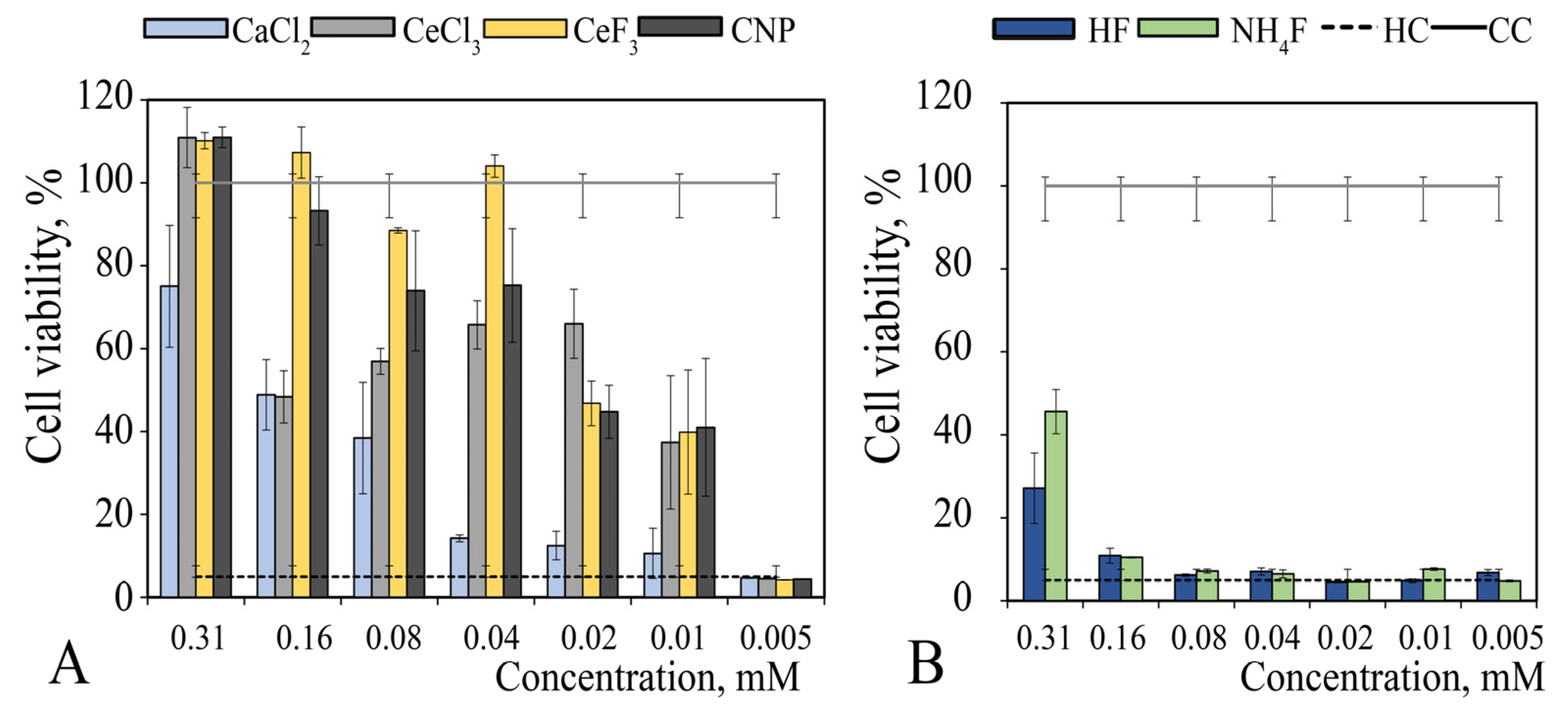
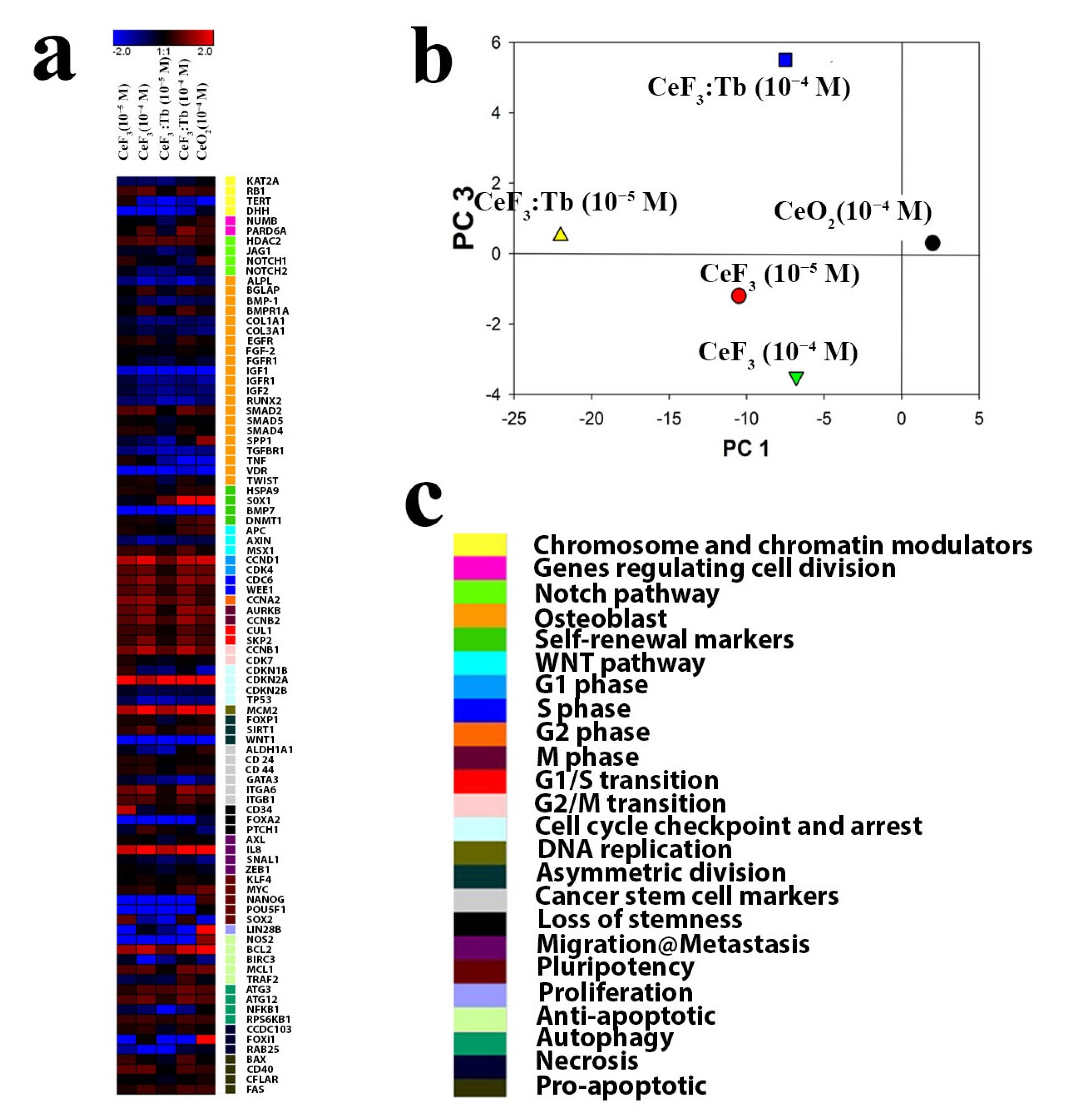
3.4. Toxicity Analysis of the Effect of Cerium Fluoride Nanoparticles on Dental Pulp Stem Cells (DPSc)

After treatment of the oral cavity with fluoride and cerium ions, cerium fluoride nanoparticles can deposit on the mucous membranes and dental surfaces and can remain there for a long time. Therefore, it was decided to study the biological behaviour of such nanoparticles on dental pulp stem cells (DPSc) in more detail. First, the cytotoxicity of CeF3 and CeF3:Tb nanoparticles was analysed on DPSc culture after 24, 48 and 72 h of cultivation (Figure 8). Additionally, cerium chloride was used as a reference for the comparative analysis of cerium-containing nanoparticles’ bioactivity. It was observed that cerium chloride at a concentration of 10–6 M caused a significant increase in the optical density of formazan, which confirmed the increased level of metabolic activity of the DPSc.
CeF3 nanoparticles also increased the level of metabolic activity of cell culture at 10−4 M (p ≤ 0.0177) and 10−5 M (p ≤ 0.0436) concentrations, compared with the untreated control group. In turn, CeF3:Tb nanoparticles throughout the concentration range studied did not significantly increase the level of metabolic activity of the cells after 24 h of cultivation. After 48 h, a similar effect was observed for CeF3 nanoparticles and cerium chloride. After 72 h, CeF3 nanoparticles at a concentration of 10−6 M significantly increased (by ~30%) the level of metabolic activity of DPSc, while CeF3:Tb did not show similar biological activity. An increase in human mesenchymal stem cells proliferation rate by cerium oxide nanoparticles has already been shown [29,30], whereas the positive effect of cerium fluoride nanoparticles has not been observed previously.
Next, an analysis was made of the viability of DPSc after incubation with CeF3 and CeF3:Tb nanoparticles (10−4–10−6 M) using a Live/Dead Viability Kit (Figure 9). Treatment with hydrogen peroxide (1 mM H2O2 for 1 h) was used as a positive control (100% death). None of the investigated concentrations of CeF3 and CeF3:Tb nanoparticles caused the appearance of dead (positively stained with propidium iodide) cells after 24 h of incubation. The morphological characteristics of the cells did not change after incubation with CeF3 and CeF3:Tb nanoparticles; DPSc retained their elongated shape, characterizing active proliferation and migration. Even the highest concentrations of CeF3 and CeF3:Tb nanoparticles (10−4 M) caused neither cell death nor changes in the cells’ morphology.
Considering that rare earth fluoride nanoparticles exhibit notable redox activity [31] and are able to modulate the redox status of a cell, we decided to measure the level of membrane mitochondrial potential (MMP), which is an indicator of the metabolic activity of cells (Figure 10) and is significantly reduced upon the development of intracellular oxidative stress. Using the potential-sensitive dye TRME, the mitochondrial potential level was analysed after 24 h of incubation with CeF3 and CeF3:Tb nanoparticles (10−4–10−6 M). Valinomycin (1 μM) was used as a positive control. This substance is a depolarizing agent that significantly reduces the MMP level. None of the tested concentrations of cerium fluoride nanoparticles and cerium chloride solutions (10−4–10−6 M) caused a decrease in the MMP level.
The genotoxicity analysis of cerium fluoride and terbium-doped cerium fluoride nanoparticles was performed by cell staining with Hoechst 33342 (Figure 11). As a positive control, UV-irradiation (365 nm, 10 min) was used, causing a significant increase in the number of dead cells, as observed in chromatin condensation or its defragmentation. None of the investigated concentrations of cerium fluoride and terbium-doped cerium fluoride nanoparticles caused any morphological changes in the nuclear apparatus of human mesenchymal stem cells.
Next, a comprehensive analysis was performed regarding the gene expression (more than 90 genes) responsible for proliferation, differentiation, pluripotency, migration, apoptosis and other important cellular functions after incubation with CeF3 and CeF3:Tb nanoparticles (Figure 12). It should be noted that both types of nanoparticle significantly increased the expression of genes responsible for the cell cycle, differentiation and proliferation. These data make it possible to confirm the stimulating effect of cerium fluoride nanoparticles on the proliferation of human DPSc.
The authors have previously shown that citrate-stabilized cerium oxide nanoparticles increase the proliferation rate of human MSCs isolated from dental pulp [32] and human vartan jelly [33]. This stimulation was associated with the regulation of the redox status of DPSc and activating genes responsible for proliferative and migratory activity. CeF3:Tb nanoparticles (10−4 M) increased mRNA transcripts of SOX2, a stem cell marker gene, which is required to maintain the self-renewal or pluripotency of mesenchymal stem cells [34]. Interestingly, all types of tested nanoparticles activated cell proliferation marker genes (CCDN1, CDK4, CDC6, WEE1, CCNA2, AURKB, CCNB2, CUL1, SKP2, CCNB1 and CDKN2A), with a concentration dependence observed.
CeF3 and CeF3:Tb nanoparticles at a 10−4 M concentration led to the activation of far more genes than the lower concentration (10−5 M) of these nanoparticles. We note the overexpression of the MCM2 gene, which encodes the DNA replication licensing factor MCM2 and which is a DNA replication marker [35]. This MCM2 overexpression indirectly confirms the high replicative activity of human DPSc in the presence of cerium fluoride nanoparticles. All investigated concentrations upregulate the IL-8 gene, which is responsible for cell migration, and the BCL2 gene, a marker of anti-apoptotic activity.
A comparison of different concentrations of CeF3 nanoparticles by PCA analysis revealed a similar trend in the gene activation for both concentrations (10−4 and 10−5 M), while CeF3:Tb showed a dose-dependent activation (Figure 12b). These results indicate that the doping of cerium fluoride nanoparticles changes their biological activity, which is reflected in the level of gene expression.
In general, based on data on transcriptional activity, it can be concluded that both CeF3 and CeF3:Tb nanoparticles did not activate the osteogenic differentiation of stem cells but promoted their proliferation and migration in a dose-dependent manner. At the same time, there was no activation of genes responsible for apoptosis, inflammation and markers of cell death; in fact, on the contrary, there was a pronounced expression of the gene encoding the anti-apoptotic protein BCL2.

4. Discussion

A comprehensive analysis was performed of cerium and calcium species’ action on epithelial cells and interaction with such toxic compounds as ammonium fluoride or hydrofluoric acid. The results obtained revealed that the pre-treatment of epithelial cells with cerium chloride solutions at a high concentration led to a significant inhibition of their metabolic activity; however, in the case of their subsequent treatment with an equimolar concentration of ammonium fluoride, this toxic effect levelled. At the same time, pre-treatment of epithelial cells with a solution of calcium chloride did not provide such a protective effect.
This protective effect was also absent in the case of the pre-treatment of cells with cerium oxide nanoparticles. These data confirm the in situ formation of cerium fluoride nanoparticles upon sequential treatment with water soluble cerium and fluoride compounds. Considering that the antioxidant properties of various cerium compounds have been well studied recently, an analysis was made of the possible protective effect of such a pre-treatment of epithelial cells in a model of oxidative stress induced by hydrogen peroxide.
Pre-treatment of the cells, even with micromolar concentrations of cerium chloride, resulted in a pronounced antioxidant effect, in contrast to calcium compounds, which did not exhibit any protective activity at similar concentrations. Cerium fluoride nanoparticles demonstrated an even more pronounced protective effect than cerium ions, which confirms the promise of using this nanomaterial as an effective protective agent with high antioxidant activity.
At present, the problem of the development of carious lesions of tooth enamel remains unresolved, due to the influence of bacterial glycolysis products and the subsequent dissolution of calcium phosphates of tooth enamel. The proposed sequential scheme of teeth treatment with solutions of cerium chloride and ammonium fluoride significantly inhibits acid corrosion of tooth enamel, preventing calcium leaching.
When a tooth with damaged enamel is subjected to successive treatment with aqueous solutions of cerium and fluoride species, a layer of cerium fluoride nanoparticles is formed in the carious zone, which acts as a protective layer that prevents the processes of enamel destruction. In turn, the formation of luminescent terbium-doped cerium fluoride nanoparticles upon sequential teeth treatment with a mixture of cerium and terbium species and fluoride species enables the visualization of tooth enamel microcracks.
A comprehensive analysis of the biological activity of cerium fluoride nanoparticles showed that they can act as a mitogen, accelerating the proliferation of human dental cells, as well as activating the key signalling pathways of cells responsible for the proliferation, differentiation and redox status of human MSCs. These data are in good agreement with the authors’ previous report that demonstrated that cerium fluoride nanoparticles can act as mitogens for whole organisms [8].
The downregulation of more than 30 genes responsible for cell differentiation and redox status was shown in the presence of CeF3 and CeF3:Tb nanoparticles, which was not observed in the case of treatment with cerium oxide nanoparticles at the same concentration. At the same time, an increase in the expression of markers of apoptosis and inflammation was not observed at all, which confirms the high level of biocompatibility of nanoscale cerium fluoride and cerium oxide.
In the context of practical application, the proposed teeth treatment method has some issues that must be taken into account when considering future in vivo studies. The use of oral rinses inevitably results in them being swallowed. Most contemporary dentrifices contain fluorides, and so the risk of their ingestion has been thoroughly assessed [36,37]. On the other hand, the use of rare earth compounds in oral rinses has received barely any attention. Cerium species, however, have a long history of biomedical tests [9]. They have relatively low toxicity per os: LD50 4200 mg kg−1 for cerium nitrate (female rats) and LD50 1178 mg kg−1 (female mice) [38]. These values are close to the toxicity parameters of table salt (LD50 3000 mg kg−1 for rats [39]).
The low risk associated with the oral administration of cerium species is supported by their low absorption, with more than 95% of the species being excreted in the faeces [9,40]. Moreover, orally administered lanthanide compounds at low doses have even been reported to have beneficial effects for laboratory and farm animals [9,41] resulting in a significant increase in their immune level [9]. According to the most recent report, orally administered cerium species have great potential for use in dental practise as they prevent inflammatory changes in periodontal tissues in obese humans with generalized catarrhal gingivitis [42].
Further implementation of the current research requires the translation of in vitro experiments to preclinical and clinical trials, as well as conversion of the cellular half maximal inhibitory concentration IC50 (mmol/L) into corresponding toxicity values for animals and humans (mg/kg), such as median lethal dose LD50, no observed adverse effect level NOAEL and no observed effect level NOEL. According to a prediction model recommended by the International Workshop on In Vitro Methods for Assessing Acute Systemic Toxicity [43], LD50 values (in mmol/kg) can be estimated from IC50x values (in mmol/L) using the following relation: log (LD50) = 0.435 × log (IC50x) + 0.625.
For cerium (III) chloride, IC50 = 5–10 mM (see above), thus LD50 is estimated to be 8.5 –11.5 mmol/kg or 2100–2800 mg/kg, which is in good agreement with experimentally measured values of 2800 mg/kg (male and female rats per os) [44]. NOEL and NOAEL are also assessed prior to the initiation of human trials, in order to establish a safe clinical starting dose. For cerium nitrate, NOEL was reported to be 110 mg/kg/day and NOAEL was reported to be 330 mg/kg/day (47-day, rats per os) [44].
For a more accurate transfer of drug doses from animal to human trials, the body surface area (BSA) normalization method has been suggested [45]. BSA normalization was advocated for translation from animals to humans in phase I and phase II clinical trials: HED = Animal   dose × Animal   Km Human   Km (here, HED—human equivalent dose (mg/kg), Km—surface area factor). For rats, Km = 6 and, for humans, Km = 37 (adults) or 25 (children) [45].
Taking into account the above considerations, the NOEL value for water-soluble cerium species for adult humans can be estimated as 18 mg/kg/day or about 1 g/day (for the body weight ~60 kg). Common oral rinses contain ~0.05% (230 ppm F) NaF, and thus the accidental daily swallowing of 10 mL rinse solution is equal to the ingestion of ~2.3 mg of fluoride [46], while the probable toxic dose (PTD) of fluoride was estimated to be 5 mg/kg [36].
According to the principle of molar quantitative compliance (see above), the cerium-containing rinse solution should contain approximately 0.1% CeCl3, so that accidental daily swallowing of 10 mL rinse solution will result in the ingestion of only ~10 mg of CeCl3, which is an order of magnitude less than the estimated NOEL value. This confirms the suitability of the proposed protocol of two-component oral treatment for future clinical trials.

5. Conclusions

The sequential use of CeCl3 and NH4F solutions in equimolar sub-toxic concentrations enables avoidance of the possible toxic effects of each substance, ensuring the preservation of the metabolic activity of the cells. Cerium fluoride nanoparticles and terbium-doped cerium fluoride nanoparticles do not exhibit cytotoxicity and genotoxicity to DPSc, even at high concentrations (10−4 M).
Cerium fluoride nanoparticles in millimolar concentrations (from 10−5–10−6 M) are able to accelerate cell proliferation rate after 72 h of incubation, maintaining a high level of metabolic activity and mitochondrial potential of the cell culture. CeF3 and CeF3:Tb nanoparticles significantly increase the expression of genes responsible for the cell cycle, differentiation and proliferation. These findings make it possible to confirm the stimulating effect of cerium fluoride nanoparticles on the proliferation of human DPSc.
The formation of cerium fluoride on the surface of the mucous membrane and teeth can protect them from the development of carious lesions, periodontitis, ROS attacks and other inflammatory diseases of the oral cavity.

Author Contributions

Conceptualization, V.K.I. and E.A.B.; methodology, N.M.Z. and A.B.S.; investigation, A.L.P., T.O.K., D.D.K., A.M.E., N.N.C. and N.R.P.; resources, T.O.K., E.A.B. and A.L.P.; data curation, A.L.P. and N.R.P.; writing—original draft preparation, A.L.P.; writing—review and editing, N.M.Z., A.B.S., E.A.B. and V.K.I.; supervision, A.B.S. and V.K.I.; project administration, V.K.I.; funding acquisition, V.K.I. All authors have read and agreed to the published version of the manuscript.

Funding

This research was partly funded by the Russian Science Foundation, grant number 19-13-00416.

Institutional Review Board Statement

Not applicable.

Informed Consent Statement

Informed consent was obtained from all subjects involved in the study.

Data Availability Statement

The data are included within the article.

Conflicts of Interest

The authors declare no conflict of interest.

References

  1. Luoma, H.; Nykänen, I.; Seppä, L.; Alakuijala, P.; Spets-Happonen, S.; Räisänen, J. Protection by F, I, Sr, and Combinations against Fermentation Attack by Streptococcus sobrinus Artificial Plaque on Bovine Enamel. Caries Res. 1989, 23, 5–13. [Google Scholar] [CrossRef] [PubMed]
  2. Ullah, R.; Zafar, M.S.; Shahani, N. Potential Fluoride Toxicity from Oral Medicaments: A Review. Iran. J. Basic Med. Sci. 2017, 20, 841–848. [Google Scholar] [CrossRef] [PubMed]
  3. Agalakova, N.I.; Gusev, G.P. Molecular Mechanisms of Cytotoxicity and Apoptosis Induced by Inorganic Fluoride. ISRN Cell Biol. 2012, 2012, 403835. [Google Scholar] [CrossRef]
  4. Tabuchi, Y.; Yunoki, T.; Hoshi, N.; Suzuki, N.; Kondo, T. Genes and Gene Networks Involved in Sodium Fluoride-Elicited Cell Death Accompanying Endoplasmic Reticulum Stress in Oral Epithelial Cells. Int. J. Mol. Sci. 2014, 15, 8959–8978. [Google Scholar] [CrossRef] [PubMed]
  5. Ribeiro, D.A.; Cardoso, C.M.; Yujra, V.Q.; Viana, M.D.B.; Aguiar, O.; Pisani, L.P.; Oshima, C.T.F. Fluoride Induces Apoptosis in Mammalian Cells: In Vitro and In Vivo Studies. Anticancer. Res. 2017, 37, 4767–4777. [Google Scholar] [CrossRef]
  6. Fina, B.L.; Lombarte, M.; Rigalli, J.P.; Rigalli, A. Fluoride Increases Superoxide Production and Impairs the Respiratory Chain in ROS 17/2.8 Osteoblastic Cells. PLoS ONE 2014, 9, e100768. [Google Scholar] [CrossRef]
  7. Shcherbakov, A.B.; Zholobak, N.M.; Baranchikov, A.E.; Ryabova, A.V.; Ivanov, V.K. Cerium Fluoride Nanoparticles Protect Cells against Oxidative Stress. Mater. Sci. Eng. C 2015, 50, 151–159. [Google Scholar] [CrossRef]
  8. Ermakov, A.; Popov, A.; Ermakova, O.; Ivanova, O.; Baranchikov, A.; Kamenskikh, K.; Shekunova, T.; Shcherbakov, A.; Popova, N.; Ivanov, V. The First Inorganic Mitogens: Cerium Oxide and Cerium Fluoride Nanoparticles Stimulate Planarian Regeneration via Neoblastic Activation. Mater. Sci. Eng. C 2019, 104, 109924. [Google Scholar] [CrossRef]
  9. Shcherbakov, A.B.; Zholobak, N.M.; Ivanov, V.K. Biological, Biomedical and Pharmaceutical Applications of Cerium Oxide. In Cerium Oxide (CeO2): Synthesis, Properties and Applications; Elsevier: Amsterdam, The Netherlands, 2020; pp. 279–358. [Google Scholar]
  10. Shcherbakov, A.B.; Reukov, V.V.; Yakimansky, A.V.; Krasnopeeva, E.L.; Ivanova, O.S.; Popov, A.L.; Ivanov, V.K. CeO2 Nanoparticle-Containing Polymers for Biomedical Applications: A Review. Polymers 2021, 13, 924. [Google Scholar] [CrossRef]
  11. Mahapatra, C.; Singh, R.K.; Lee, J.-H.; Jung, J.; Hyun, J.K.; Kim, H.-W. Nano-Shape Varied Cerium Oxide Nanomaterials Rescue Human Dental Stem Cells from Oxidative Insult through Intracellular or Extracellular Actions. Acta Biomater. 2017, 50, 142–153. [Google Scholar] [CrossRef]
  12. Garcia, I.M.; Leitune, V.C.B.; Takimi, A.S.; Bergmann, C.P.; Samuel, S.M.W.; Melo, M.A.; Collares, F.M. Cerium Dioxide Particles to Tune Radiopacity of Dental Adhesives: Microstructural and Physico-Chemical Evaluation. J. Funct. Biomater. 2020, 11, 7. [Google Scholar] [CrossRef] [PubMed]
  13. Zholobak, N.M.; Ivanov, V.K.; Shcherbakov, A.B. Interaction of Nanoceria with Microorganisms. In Nanobiomaterials in Antimicrobial Therapy; Elsevier: Amsterdam, The Netherlands, 2016; pp. 419–450. [Google Scholar] [CrossRef]
  14. Dos Santos, C.; Passos Farias, I.; Reis Albuquerque, A.; e Silva, P.; Costa One, G.; Sampaio, F. Antimicrobial Activity of Nano Cerium Oxide (IV) (CeO2) against Streptococcus mutans. BMC Proc. 2014, 8, P48. [Google Scholar] [CrossRef]
  15. Anastasiou, A.D.; Nerantzaki, M.; Gounari, E.; Duggal, M.S.; Giannoudis, P.V.; Jha, A.; Bikiaris, D. Antibacterial Properties and Regenerative Potential of Sr2+ and Ce3+ Doped Fluorapatites; a Potential Solution for Peri-Implantitis. Sci. Rep. 2019, 9, 14469. [Google Scholar] [CrossRef] [PubMed]
  16. Bänöczy, J.; Gintner, Z.; Kiss, J. Incorporation of Lanthanides in Human Dental Enamel. Acta Stomatol. Croat. 1996, 30, 37–48. [Google Scholar]
  17. Wegehaupt, F.J.; Sener, B.; Attin, T.; Schmidlin, P.R. Application of Cerium Chloride to Improve the Acid Resistance of Dentine. Arch. Oral Biol. 2010, 55, 441–446. [Google Scholar] [CrossRef]
  18. Wegehaupt, F.J.; Sener, B.; Attin, T.; Schmidlin, P.R. Anti-Erosive Potential of Amine Fluoride, Cerium Chloride and Laser Irradiation Application on Dentine. Arch. Oral Biol. 2011, 56, 1541–1547. [Google Scholar] [CrossRef]
  19. Wegehaupt, F.J.; Buchalla, W.; Sener, B.; Attin, T.; Schmidlin, P.R. Cerium Chloride Reduces Enamel Lesion Initiation and Progression in Vitro. Caries Res. 2014, 48, 45–50. [Google Scholar] [CrossRef]
  20. Zhang, W.; Jin, H.; Liu, A. A Comparison of the Preventive Effects of Lanthanides and Fluoride on Human Experimental Root Surface Carious-like Lesions. Chin. J. Dent. Res. 1999, 2, 38–44. [Google Scholar]
  21. Jaisingh, R.; Shanbhog, R.; Nandlal, B.; Thippeswamy, M. Effect of 10% Cerium Chloride on Artificial Caries Lesions of Human Enamel Evaluated Using Quantitative Light-Induced Fluorescence: An in Vitro Study. Eur. Arch. Paediatr. Dent. 2017, 18, 163–169. [Google Scholar] [CrossRef]
  22. Jin, H.; Zhang, W.G.; Gao, H.; Liu, A.R. Effects of Demineral-Resistant of Lanthanum, Cerium and Fluoride on Human Root Surface:A Comparative Study In Vitro. Shanghai Kou Qiang Yi Xue 1995, 4, 151–154. [Google Scholar]
  23. Jin, H.; Zhang, W.; Liu, A. A Comparative Study of Lanthanum, Cerium and Fluoride on the Prevention of Root Surface Caries by Various Procedure. Zhonghua Kou Qiang Yi Xue Za Zhi 1997, 32, 143–145. [Google Scholar] [PubMed]
  24. Heard, K.; Hill, R.E.; Cairns, C.B.; Dart, R.C. Calcium Neutralizes Fluoride Bioavailability in a Lethal Model of Fluoride Poisoning. J. Toxicol. Clin. Toxicol. 2001, 39, 349–353. [Google Scholar] [CrossRef] [PubMed]
  25. Kao, W.; Deng, J.; Chiang, S.; Heard, K.; Yen, D.H.T.; Lu, M.; Kuo, B.I.; Kuo, C.; Liu, T.; Lee, C. A Simple, Safe, and Efficient Way to Treat Severe Fluoride Poisoning—Oral Calcium or Magnesium. J. Toxicol. Clin. Toxicol. 2004, 42, 33–40. [Google Scholar] [CrossRef] [PubMed]
  26. Mosmann, T. Rapid Colorimetric Assay for Cellular Growth and Survival: Application to Proliferation and Cytotoxicity Assays. J. Immunol. Methods 1983, 65, 55–63. [Google Scholar] [CrossRef]
  27. Hannig, C.; Hamkens, A.; Becker, K.; Attin, R.; Attin, T. Erosive Effects of Different Acids on Bovine Enamel: Release of Calcium and Phosphate in Vitro. Arch. Oral Biol. 2005, 50, 541–552. [Google Scholar] [CrossRef] [PubMed]
  28. Wang, X.; Chen, Y.; Meng, Z.; Zhang, Q.; Zhai, J. Effect of Trivalent “Calcium-like” Cations on Ionic Transport Behaviors of Artificial Calcium-Responsive Nanochannels. J. Phys. Chem. C 2018, 122, 24863–24870. [Google Scholar] [CrossRef]
  29. Sadidi, H.; Hooshmand, S.; Ahmadabadi, A.; Javad Hoseini, S.; Baino, F.; Vatanpour, M.; Kargozar, S. Cerium Oxide Nanoparticles (Nanoceria): Hopes in Soft Tissue Engineering. Molecules 2020, 25, 4559. [Google Scholar] [CrossRef]
  30. Giri, S.; Karakoti, A.; Graham, R.P.; Maguire, J.L.; Reilly, C.M.; Seal, S.; Rattan, R.; Shridhar, V. Nanoceria: A Rare-Earth Nanoparticle as a Novel Anti-Angiogenic Therapeutic Agent in Ovarian Cancer. PLoS ONE 2013, 8, e54578. [Google Scholar] [CrossRef]
  31. Kudinov, K.; Bekah, D.; Cooper, D.; Shastry, S.; Hill, C.; Bradforth, S.; Nadeau, J. Lanthanum Fluoride Nanoparticles for Radiosensitization of Tumors. In Conference on Colloidal Nanoparticles for Biomedical Applications XI, San Francisco, CA, USA, 13–15 February 2016; Parak, W.J., Osinski, M., Liang, X.J., Eds.; SPIE: Bellingham, WA, USA, 2016; p. 97220V. [Google Scholar] [CrossRef]
  32. Popov, A.L.; Ermakov, A.M.; Savintseva, I.V.; Selezneva, I.I.; Poltavtseva, R.A.; Zaraiskii, E.I.; Poltavtsev, A.M.; Stepanova, I.E.; Ivanov, V.K.; Sukhikh, G.T. Biosafety and Effect of Nanoparticles of CeO2 on Metabolic and Proliferative Activity of Human Mesenchymal Stem Cells In Vitro. Nanomech. Sci. Technol. An Int. J. 2016, 7, 165–175. [Google Scholar] [CrossRef]
  33. Popov, A.L.; Ermakov, A.M.; Savintseva, I.V.; Selezneva, I.I.; Poltavtseva, R.A.; Zaraisky, E.I.; Poltavtsev, A.M.; Stepanov, A.A.; Ivanov, V.K.; Sukhikh, G.T. Citrate-Stabilized Nanoparticles of CeO2 Stimulate Proliferation of Human Mesenchymal Stem Cells In Vitro. Nanomech. Sci. Technol. Int. J. 2016, 7, 235–246. [Google Scholar] [CrossRef]
  34. Park, S.B.; Seo, K.W.; So, A.Y.; Seo, M.S.; Yu, K.R.; Kang, S.K.; Kang, K.S. SOX2 Has a Crucial Role in the Lineage Determination and Proliferation of Mesenchymal Stem Cells through Dickkopf-1 and c-MYC. Cell Death Differ. 2012, 19, 534–545. [Google Scholar] [CrossRef] [PubMed] [Green Version]
  35. Meagher, M.; Epling, L.B.; Enemark, E.J. DNA Translocation Mechanism of the MCM Complex and Implications for Replication Initiation. Nat. Commun. 2019, 10, 3117. [Google Scholar] [CrossRef] [PubMed]
  36. Whitford, G.M. Acute Toxicity of Ingested Fluoride. Monogr. Oral Sci. 2011, 22, 66–80. [Google Scholar] [CrossRef] [PubMed]
  37. Zuanon, A.C.C.; Aranha, A.M.F. Mouthwash Ingestion by Preschool Children. J. Clin. Pediatr. Dent. 2006, 30, 15–18. [Google Scholar] [CrossRef]
  38. Bruce, D.W.; Hietbrink, B.E.; DuBois, K.P. The Acute Mammalian Toxicity of Rare Earth Nitrates and Oxides. Toxicol. Appl. Pharmacol. 1963, 5, 750–759. [Google Scholar] [CrossRef]
  39. Feldman, S.R. Sodium Chloride. In Kirk-Othmer Encyclopedia of Chemical Technology; John Wiley & Sons, Inc.: Hoboken, NJ, USA, 2011. [Google Scholar]
  40. Morganti, J.B.; Lown, B.A.; Stineman, C.H.; Massaro, E.J. Cerium Tissue/Organ Distribution and Alterations in Open Field and Exploratory Behavior Following Repeated Exposure of the Mouse to Citrate Complexed Cerium. Gen. Pharmacol. Vasc. Syst. 1978, 9, 257–261. [Google Scholar] [CrossRef]
  41. Aquilina, G.; Bach, A.; Bampidis, V.; De Lourdes Bastos, M.; Costa, L.G.; Flachowsky, G.; Gralak, M.A.; Hogstrand, C.; Leng, L.; López-Puente, S.; et al. Scientific Opinion on the Safety and Efficacy of Lancer (Lanthanide Citrate) as Feed Additive for Weaned Piglets. EFSA J. 2013, 11, 3206. [Google Scholar] [CrossRef]
  42. Skrypnyk, M.; Petrushanko, T.; Neporada, K.; Bubnov, R.; Shcherbakov, O.; Spivak, M. Effectiveness of Nanocrystalline Cerium Dioxide for Secondary Prevention of Inflammatory Periodontal Diseases in Young Individuals with Obesity. Lett. Appl. NanoBiosci. 2019, 8, 754–761. [Google Scholar] [CrossRef]
  43. NIH Publication No. 01-4499; Report of the International Workshop on In Vitro Methods for Assessing Acute Systemic Toxicity. National Institute of Environmental Health Services, Research Triangle Park: Durham, NC, USA, 2001.
  44. Shin, S.-H.; Kim, H.-O.; Rim, K.-T. Worker Safety in the Rare Earth Elements Recycling Process from the Review of Toxicity and Issues. Saf. Health Work 2019, 10, 409–419. [Google Scholar] [CrossRef]
  45. Reagan-Shaw, S.; Nihal, M.; Ahmad, N. Dose Translation from Animal to Human Studies Revisited. FASEB J. 2008, 22, 659–661. [Google Scholar] [CrossRef]
  46. Marinho, V.C.; Chong, L.-Y.; Worthington, H.V.; Walsh, T. Fluoride Mouthrinses for Preventing Dental Caries in Children and Adolescents. Cochrane Database Syst. Rev. 2016, 7, CD002284. [Google Scholar] [CrossRef] [PubMed]
Figure 1. Metabolic activity (MTT test) of MA-104 cells 2 h after the addition of various concentrations of: (A) HF and NH4F and (B) calcium chloride and cerium chloride. CC—cell control, 100%.
Nanomaterials 12 03034 g001
Figure 2. Metabolic activity of MA-104 cells pre-treated with cerium chloride 5 min before the addition of ammonium fluoride: (A) 10 mM, (B) 5 mM and (C) 2.5 mM. CC—cell control.
Nanomaterials 12 03034 g002
Figure 3. Metabolic activity of MA-104 cells pre-treated with calcium chloride 5 min before the addition of ammonium fluoride: (A) 10 mM, (B) 5 mM and (C) 2.5 mM. CC—cell control.
Nanomaterials 12 03034 g003
Figure 4. Metabolic activity of MA-104 cells pre-treated with toxic concentrations (TC) of: (A) cerium oxide nanoparticles, (B) cerium chloride and (C) calcium chloride. CC—cell control. “Interaction” lines demonstrate the effect of calcium or cerium species added 5 min before the addition of 10 mM NH4F.
Nanomaterials 12 03034 g004
Figure 5. Viability of MA-104 cells under H2O2-induced oxidative stress. Cells were pre-treated with: (A) cerium or calcium species and (B) fluoride species. CNP—ceria nanoparticles, HC—hydrogen peroxide toxicity control and CC—intact cell control.
Nanomaterials 12 03034 g005
Figure 6. (A) Calcium release: 1—untreated molars (control); 2—molars treated with cerium chloride solution; 3—molars treated with ammonium fluoride solution; 4—molars treated with cerium chloride following ammonium fluoride solutions. (B) Molar with protective layer of CeF3:Tb nanoparticles in the enamel defect zones. (C) The proposed mechanisms of tooth protection by sequential treatment with cerium and fluoride ions.
Nanomaterials 12 03034 g006
Figure 7. TEM images of CeF3:Tb3+ (A) and CeF3 (B) nanoparticles. Room temperature emission spectra of CeF3:Tb3+ (15 mol%) nanoparticles in aqueous solutions upon 254 nm excitation (C).
Nanomaterials 12 03034 g007
Figure 8. Metabolic activity (via MTT assay) of DPSc in the presence of CeCl3, CeF3 and CeF3:Tb nanoparticles (10−4–10−6 M) after 24, 48 and 72 h of incubation. The data are presented as the mean ± SD. * Significant differences were assessed using the Mann–Whitney U criterion.
Nanomaterials 12 03034 g008
Figure 9. Live/Dead assay for DPSc after 24 h incubation with different concentrations (10−4–10−6 M) of CeCl3 solution, CeF3 and CeF3:Tb nanoparticles. Negative control—cells with no nanoparticles. Positive control—1 mM H2O2 for 1 h.
Nanomaterials 12 03034 g009
Figure 10. Analysis of the membrane mitochondrial potential of DPSc after 24 h of incubation with different concentrations (10−4–10−6 M) of CeCl3 solution, CeF3 and CeF3:Tb nanoparticles. Negative control—cells without nanoparticles. Positive control—valinomycin 1 µM.
Nanomaterials 12 03034 g010
Figure 11. Fluorescent microscopy analysis of nuclei fragmentation in DPSc after 24 h incubation with different concentrations (10–4–10–6 M) of CeCl3 solution, CeF3 and CeF3:Tb nanoparticles, using Hoechst 33342. Negative control—cells without nanoparticles. Positive control—10 min UVB irradiation.
Nanomaterials 12 03034 g011
Figure 12. Heat map of gene expression in DPSc after 24 h incubation with CeF3 (10−4 and 10−5 M) and CeF3:Tb (10−4 and 10−5 M) nanoparticles (a). The intensity scale of the standardized expression values ranges from −3 (green: low expression) to +3 (red: high expression), with a 1:1 intensity value (black) representing the control (nontreated). Principal Component Analysis (PCA) of qRT-PCR data for different concentrations of CeF3 and CeF3:Tb nanoparticles (b). Cluster groups of genes and their functionality (c).
Nanomaterials 12 03034 g012
Table 1. Selected genes and their functions.
GeneFunctionGeneFunctionGeneFunction
KAT2AChromatin and chromosome modulatorsHSPA9Self-renewal markersFOXA2Stemness reduction markers
RB1SOX1PTCH1
TERTBMP7CD34
DNMT1
DHHRegulators of cell division symmetryAXLCell migration markers
NUMBAPCStemness maintenance (Wnt signalling)IL8
PARD6AAXINSNAI1
HDAC2Stemness maintenance (Notch signalling)MSX1TWIST1
JAG1CCND1Proliferation markersZEB1
NOTCH1CDK4KLF4Pluripotency markers
NOTCH2CDC6MYC
ALPLWEE1NANOG
BGLAPMarkers of MSCs and their differentiationCCNA2POU5F1
BMP1AURKBSOX2
BMPR1ACCNB2NOS2Anti-apoptotic markers
COL1A1CUL1BCL2
COL3A1SKP2BIRC3
EGFRCCNB1MCL1
FGF-2CDK7TRAF2
FGFR1CDKN1BATG3Autophagy markers
IGF1CDKN2AATG12
IGFR1CDKN2BNFKB1
RUNX2TP53RPS6KB1
SMAD2MCM2CCDC103Necrosis markers
SMAD4LIN28BFOXI1
SMAD5FOXP1Markers of asymmetric cell divisionJPH3
SPP1SIRT1RAB25
TGFBR1WNT1BAXPro-apoptotic markers
TNFALDH1AStemness markersCD40
VDRCD44CFLAR
GATA3FAS
ITGA6TNFRSF10A
ITGB1
Publisher’s Note: MDPI stays neutral with regard to jurisdictional claims in published maps and institutional affiliations.

