Article

An Efficient Voltammetric Sensor Based on Graphene Oxide-Decorated Binary Transition Metal Oxides Bi2O3/MnO2 for Trace Determination of Lead Ions

1
College of Life Sciences and Chemistry, Hunan University of Technology, Zhuzhou 412007, China
2
Department of Neurology, Zhuzhou People’s Hospital, Zhuzhou 412008, China
3
Zhuzhou Institute for Food and Drug Control, Zhuzhou 412011, China
*
Author to whom correspondence should be addressed.
Nanomaterials 2022, 12(19), 3317; https://doi.org/10.3390/nano12193317
Submission received: 22 August 2022 / Revised: 18 September 2022 / Accepted: 20 September 2022 / Published: 23 September 2022

Abstract

:
Herein we present a facile synthesis of the graphene oxide-decorated binary transition metal oxides of Bi2O3 and MnO2 nanocomposites (Bi2O3/MnO2/GO) and their applications in the voltammetric detection of lead ions (Pb2+) in water samples. The surface morphologies, crystal structures, electroactive surface area, and charge transferred resistance of the Bi2O3/MnO2/GO nanocomposites were investigated through the scanning electron microscopy (SEM), power X-ray diffraction (XRD), cyclic voltammetry (CV), and electrochemical impedance spectroscopy (EIS) techniques, respectively. The Bi2O3/MnO2/GO nanocomposites were further decorated onto the surface of a glassy carbon electrode (GCE), and Pb2+ was quantitatively analyzed by using square-wave anodic stripping voltammetry (SWASV). We explored the effect of the analytical parameters, including deposition potential, deposition time, and solution pH, on the stripping peak current of Pb2+. The Bi2O3/MnO2/GO nanocomposites enlarged the electroactive surface area and reduced the charge transferred resistance by significant amounts. Moreover, the synergistic enhancement effect of MnO2, Bi2O3 and GO endowed Bi2O3/MnO2/GO/GCE with extraordinary electrocatalytic activity toward Pb2+ stripping. Under optimal conditions, the Bi2O3/MnO2/GO/GCE showed a broad linear detection range (0.01–10 μM) toward Pb2+ detection, with a low limit of detection (LOD, 2.0 nM). The proposed Bi2O3/MnO2/GO/GCE electrode achieved an accurate detection of Pb2+ in water with good recoveries (95.5–105%).

1. Introduction

As a common heavy metal ion (HMI), Pb2+ has adverse effects on our health and the environment because of its high toxicity, even at low concentrations. Pb2+ in the aquatic environment barely degrades and is easily enriched in aquatic food [1,2]. Therefore, Pb2+ chronically endangers human health via the food chain and gradually induces life-threatening circumstances. Excessive levels of Pb2+ in human body can severely destroy our organs and nervous system, which is highly associated with various cancers such as lung, kidney, and brain cancers [3]. Hence, a highly efficient determination of Pb2+ in water is quite essential to guarantee our health.
Over the last few decades, conventional analytical techniques have been developed to reliably detect Pb2+, including X-ray fluorescence spectrometry [4], UV-Vis spectroscopy [5], atomic absorption spectrophotometry [6], inductively coupled plasma mass spectrometry [7], and inductively coupled plasma-atomic emission spectrometry [8]. These analytical techniques are very robust and accurate, even in complex sample matrixes; however, they often require expensive and bulky equipment, cumbersome and time-consuming operation procedures, and highly skilled personnel. Without a doubt, they are not suitable for an on-field analysis. In recent years, stripping voltammetry, especially SWASV, has emerged as a powerful alternative for the trace determination of HMIs due to its advantages of portability, low cost, rapid response, excellent sensitivity, and feasibility for on-site analyses [9,10]. A voltammetric determination of Pb2+ often involves hanging mercury drop electrodes or mercury film electrodes. Owing to their superior stripping characteristics, these mercury-based electrodes are excellent in their sensitivity and reproducibility [11,12]. However, the toxic mercury contaminates samples and poses health risks to analysts. Alternatively, eco-friendly bismuth film electrodes can provide comparable sensing properties for HMI determination [13,14]. Unlike mercury electrodes, bismuth film electrodes usually suffer from surface passivation, which degrades their stripping signals. Therefore, designing novel materials with extraordinary sensing performance toward Pb2+ is highly desirable and challenging.
Transition metal oxide nanostructures have been extensively used for the voltammetric detection of HMIs because of their natural abundance, high adsorption capacity, and favorable catalytic activity [15,16]. Among transition metal oxides, MnO2 has attracted increasing attention due to its earth abundance, low cost, eco-friendliness, favorable electrocatalytic activity, and excellent adsorption capability [17,18]. Nanostructured α-MnO2 has demonstrated a high affinity for adsorption of Cu2+, Pb2+, Zn2+, Cd2+, Hg2+, etc. [19,20]. Therefore, MnO2 nanostructures have recently been used for the voltammetric detection of HMIs [21,22,23]. Owing to its nontoxicity, cost-effectiveness, relatively narrow band gap, high adsorption capacity, and admirable catalytic properties, nanoscale Bi2O3 has also found growing interest in various fields such as photocatalysis [24], electroreduction [25], supercapacitors [26,27,28], and voltammetric sensors [29,30]. It has been reported that nanoscale Bi2O3 displayed a high affinity to HMIs such as Cd2+ [31,32]. The electrochemical reduction of Bi2O3 can produce a porous Bi layer and further form a “fused alloy” with the heavy metal, which accumulates more HMIs on its surface and eventually enhances the sensitivity [33]. For these reasons, Bi2O3-based electrodes have emerged as promising alternatives to mercury-based electrodes for HMI determination.
In contrast to single transition metal oxides, binary transition metal oxide electrocatalysts generally show a higher electrocatalytic activity [34,35]. However, binary transition metal oxides have rarely been used to detect HMIs [36,37,38]. Fe2O3/NiO heterojunctions possess a lower diffusion energy barrier for lead atoms, thus significantly improving the anti-interference ability for detecting Pb2+ [37]. Bi2O3/Fe2O3-decorated graphene oxide (GO) has demonstrated a remarkable electrocatalytic activity toward Cd2+ determination, having a low LOD of 1.85 ng L−1 [38]. In our recent work, the synergistic interaction between β-Bi2O3 microspheres and shuttle-like α-Fe2O3 nanoparticles enabled the concurrent determination of Cd2+ and Pb2+ in environmental and food samples at the nanomolar levels [30].
Binary transition metal oxides such as Bi2O3/MnO2 have been successfully used in supercapacitors [39,40] and the voltammetric detection of H2O2 [41]. In addition, the individual Bi2O3 or MnO2 nanostructures have also been used to detect Pb2+. However, to the best of our knowledge, GO-decorated Bi2O3/MnOx composites have not yet been reported. Herein, we fabricated GO-decorated binary transition metal oxides of Bi2O3 and MnO2 nanocomposites (Bi2O3/MnO2/GO) and used them as a delicate electrocatalyst for Pb2+ determination. GO nanoflakes are an electron-rich species that can reduce Pb2+ into metallic Pb by applying a suitable potential. In addition, abundant oxygen-containing functional groups (OxFGs) such as carboxyl, hydroxy, carbonyl, and epoxide groups in the edge of GO flakes can firmly bind Pb2+ onto their surface through electrostatic and coordination interactions, which facilitates the adsorption of Pb2+ [42,43]. Generally, the sensing performance for HMIs mainly relies on the adsorption capacity and electrocatalytic activity of the sensing material that is decorated on the electrode [44], which can be readily tailored using morphology engineering [45,46]. In this regard, we synthesized dandelion-like α-MnO2 and flower-like β-Bi2O3 nanocomposites to enhance the Pb2+ adsorption and electrocatalytic activity. With the synergistic interaction of both MnO2 and Bi2O3, Bi2O3/MnO2/GO nanocomposites were expected to boost the stripping voltammetric responses of Pb2+. The Bi2O3/MnO2/GO-modified glassy carbon electrode (Bi2O3/MnO2/GO/GCE) showed an extraordinary electrocatalytic activity toward the stripping voltammetric behavior of Pb2+, with a wide linear detection range (LDR, 0.01–10 μM), low LOD (2.6 nM), and high sensitivity (53.43 μA μM−1). Furthermore, the Bi2O3/MnO2/GO/GCE could reliably determine Pb2+ in water with good recoveries.

2. Materials and Methods

2.1. Chemicals and Solutions

GO nanoflakes were purchased from Xianfeng Nanotechnology, Inc. (Nanjing, China). Manganese sulfate monohydrate (MnSO4·H2O), bismuth nitrate pentahydrate (Bi(NO3)3·5H2O), lead nitrate (Pb(NO3)2), potassium peroxydisulfate (K2S2O8), potassium sulfate (K2SO4), N, N-dimethylformamide (DMF), potassium ferri/ferro-cyanide (K3/4[Fe(CN)6]), sodium acetate (NaAc), acetic acid (HAc), concentrated H2SO4, and anhydrous alcohol were purchased from Sinopharm Chemical Reagent Co., Ltd. (Shanghai, China). All reagents were of analytical grade and directly used as received. Water samples were collected from a local lake and from our laboratory. 0.3312 g of Pb(NO3)2 was completely dissolved in 100 mL of 0.1 M HAc-NaAc buffer (pH = 5.5) to prepare a 0.01 M Pb2+ stock solution. A series of standard solutions of Pb2+ with different concentrations were prepared by appropriately diluting the stock solution with the 0.1 M HAc-NaAc buffer (pH = 5.5). Deionized water (DI water, resistivity of 18.2 MΩ·m) was used for all experiments.

2.2. Preparation of Bi2O3/MnO2/GO Nanocomposites

2.2.1. Preparation of Dandelion-like α-MnO2 Microspheres

Dandelion-like α-MnO2 microspheres were prepared via a facile hydrothermal treatment route [47]. Typically, 1.3522 g of MnSO4·H2O, 2.1626 g of K2S2O8 and 1.3941 g of K2SO4 were sequentially added to 60 mL of 0.6 M H2SO4 and magnetically stirred for 30 min to completely dissolve. Then, the mixture solution was decanted into a 100 mL Teflon-lined stainless steel container and heated at a temperature of 140 °C for 12 h. The resultant product was repeatedly rinsed with DI water and dried at 60 °C overnight for further use.

2.2.2. Synthesis of Flower-like β-Bi2O3 Microspheres

Flower-like β-Bi2O3 microspheres were synthesized by a simple hydrothermal treatment followed by a thermal decomposition at high temperature [30]. In brief, 0.03 mol of Bi(NO3)3·5H2O was dissolved into 11 mL of HAc, and 14 mL of anhydrous ethanol was then added to form a white suspension. The resulting suspension was ceaselessly stirred for 45 min, and 28 mL of DMF was then added to yield a clear solution. Subsequently, the mixture solution was poured into a 100 mL Teflon-lined stainless steel autoclave and reacted at a temperature of 120 °C for 40 min. The precursor was centrifuged at 10,000 rpm for 5 min, where it was alternately rinsed with anhydrous alcohol and DI water and allowed to dry at 80 °C overnight. Finally, the resultant β-Bi2O3 precursor was further transferred to a porcelain boat and calcinated at 350 °C for 4 h at a heating rate of 2 °C min−1 in an air atmosphere to yield orange-yellow β-Bi2O3 microspheres.

2.2.3. Preparation of Bi2O3/MnO2/GO Nanocomposites

At first, 5 mg of dandelion-like α-MnO2 microspheres, flower-like β-Bi2O3 microspheres, and GO nanoflakes were separately dispersed into 10 mL of DI water under ultrasonication to form their respective uniform dispersions at a concentration of 0.5 mg mL−1. Then, 1 mL of the α-MnO2, β-Bi2O3, and GO dispersions were further mixed and subjected to a 30 min ultrasonication to obtain a uniform Bi2O3/MnO2/GO dispersion. The amount of the three materials were optimized during our preliminary experiments. Bi2O3/MnO2/GO composites containing 5 mg of each of the three materials showed the largest stripping peak current of Pb2+. Therefore, we selected the composite with this component content as the sensing material.

2.3. Characterizations of Sensing Materials

The microscopic morphologies of the GO nanoflakes, dandelion-like α-MnO2 microspheres, flower-like β-Bi2O3 microspheres, and Bi2O3/MnO2/GO nanocomposites were observed using field-emission SEM (Sigma HD, Zeiss, Oberkochen, Germany). Before taking the SEM measurements, a few thin layers of Au were coated onto the surface of the samples. The crystalline structures of these materials were studied using a powder XRD (Rigaku Ultima IV, Tokyo, Japan) with monochromatized Cu Kα radiation (λ = 1.54 A).

2.4. Fabrication of Modified Electrodes

Before electrode modification, the GCE was thoroughly polished to a shining mirror-like surface with 0.05 μm of alumina slurry, and it was ultrasonically cleaned with anhydrous alcohol and DI water for three cycles to remove residual contaminants. Then, the polished GCE was exposed to infrared light to allow the material to adequately dry. The Bi2O3/MnO2/GO/GCE electrode was prepared by using a conventional drop-casting method. Specifically, 5 μL of the Bi2O3/MnO2/GO dispersion was cast on the surface of the freshly polished GCE and dried under the exposure of infrared light to form a firm sensing film. For comparison, the MnO2/GO/GCE, Bi2O3/GO/GCE, and GO/GCE were also fabricated using the same procedure, aside from the dispersion used.

2.5. Electrochemical Measurements

All electrochemical measurements were performed on a CHI 660E electrochemical workstation (Chenhua Inc., Shanghai, China) equipped with a classic three-electrode system, which consists of the Bi2O3/MnO2/GO/GCE electrode, a Pt wire, and a saturated calomel electrode (SCE) as the working, counter, and reference electrodes, respectively. A 10 mL electrochemical cell made of glass was used for the electrochemical measurements. Unless otherwise specified, the 0.1 M HAc-NaAc buffer (pH = 5.5) functioned as the supporting electrolyte. To assess the electrochemical performance, the CV curves and Nyquist plots of different modified electrodes were recorded in a solution of 2 mM [Fe(CN)6]3−/4− and 0.1 M KCl. To improve the stripping responses, a suitable deposition was employed in the Pb2+ standard solutions. After 30 s of rest, the stripping peak currents of Pb2+ were recorded between −1.0 V and −0.5 V using the SWASV technique. The frequency, step potential, and pulse amplitude of the SWASV were set at 15 Hz, 4 mV, and 25 mV, respectively. When not in use, the Bi2O3/MnO2/GO/GCE electrode was stored in the air. After each determination, the electrode surface was refreshed by immersing it into a blank solution and applying +0.3 V for 150 s to ensure the complete removal of the residual metals.

3. Results and Discussion

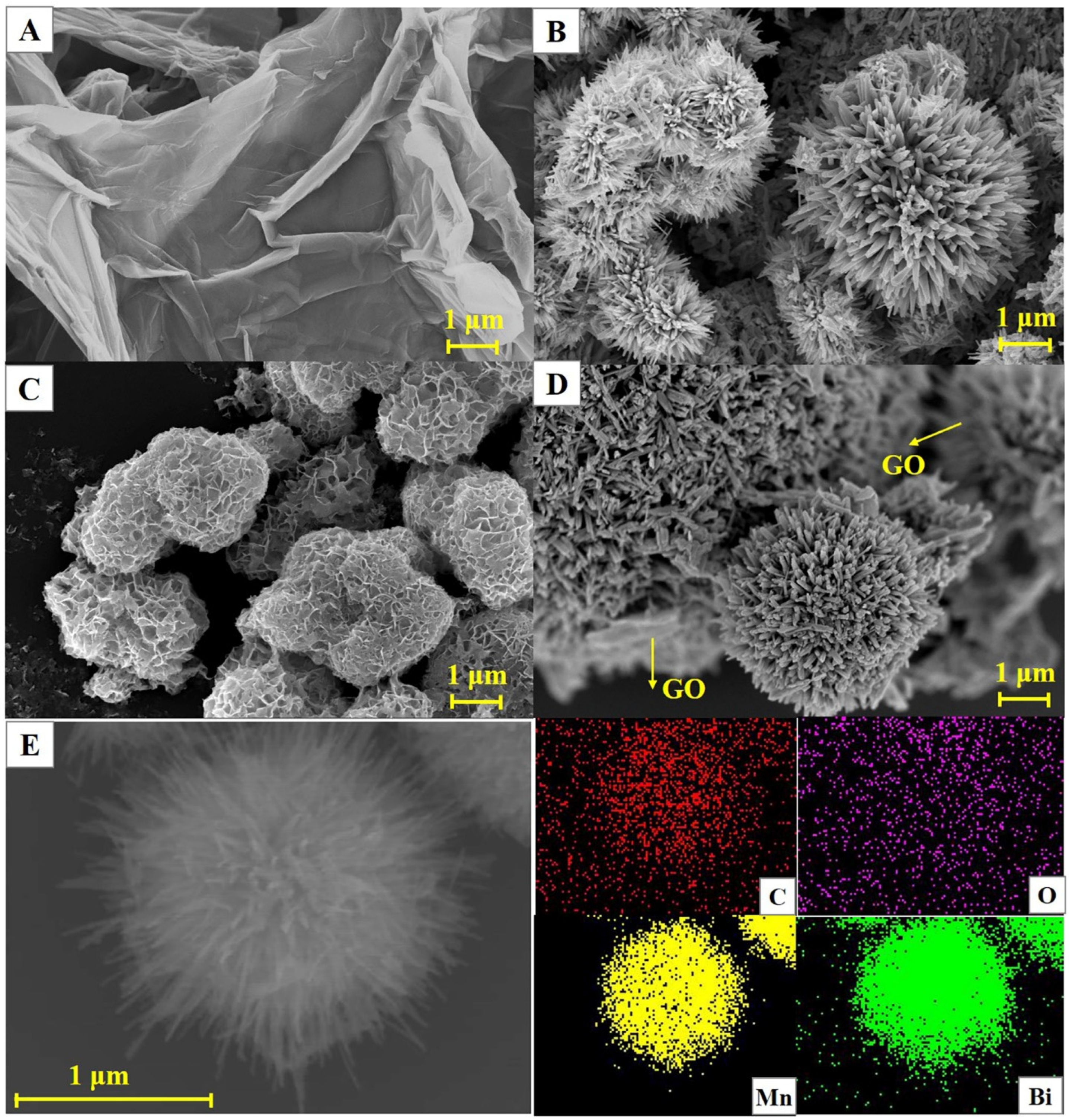
3.1. Physical Characterization

The microscopic morphologies of the GO nanoflakes, dandelion-like α-MnO2 microspheres, flower-like β-Bi2O3 microspheres, and Bi2O3/MnO2/GO nanocomposites were observed using the SEM technique, and their SEM images are shown in Figure 1. The GO nanosheets exhibited a typical lamellar structure with obvious wrinkles (Figure 1A). Dandelion-like nanostructures are found in the image of the α-MnO2, consisting of many radially distributed nanorods (Figure 1B). Typical flower-like β-Bi2O3 microspheres are observed in Figure 1C, which consist of many interconnected thin nanosheets. The unique dandelion-like α-MnO2 and flower-like Bi2O3 structures enlarge the electroactive surface area, thereby improving the sensing performance. In addition, the interconnected porous microstructures are found in the dandelion-like α-MnO2 and flower-like Bi2O3 microspheres, which facilitate the electrolyte infiltration and adsorption of HMIs. As illustrated in Figure 1D, typical microspheres are observed in the SEM image of the Bi2O3/MnO2/GO nanocomposites. In addition, the microspheres are partially wrapped by GO nanosheets. To conform the composition of the microspheres, the energy-dispersive X-ray spectroscopy (EDS) mappings of the Bi2O3/MnO2/GO nanocomposites were also measured (Figure 1E). The uniformly dispersed C, O, Mn, and Bi distribution suggests the presence of C, O, Mn, and Bi elements. In addition, the distributions of Mn and Bi exhibit obvious microsphere structures, indicating that the microsphere consists of both Bi2O3 and MnO2. All of these results indicate the successful synthesis of Bi2O3/MnO2/GO nanocomposites.
Figure 2 displays the XRD patterns of the GO nanoflakes, dandelion-like α-MnO2 microspheres, flower-like β-Bi2O3 microspheres, and Bi2O3/MnO2/GO nanocomposites. A sharp diffraction peak was observed at 2θ of 9.68° in the XRD pattern of the GO, which is attributed to the (001) crystal plane of the GO [48]. α-MnO2 nanostructures displayed distinct diffraction peaks at 2θ of 12.78°, 18.08°, 28.64°, 37.62°, 50°, 56.14°, 60.18°, and 69.54°, corresponding to the (110), (200), (310), (211), (411), (600), (521), and (541) planes of α-MnO2 (JCPDS 440141), respectively [49]. Additionally, we detected sharp diffraction peaks without any apparent impurity peaks, demonstrating that the as-prepared α-MnO2 microspheres were of high purity. Broad diffraction peaks were observed for the flower-like β-Bi2O3 at 28.00°, 32.52°, 46.32°, and 55.64°, which can be indexed to the (201), (220), (222), and (213) crystal facets (JCPDS 651209) [24]. The characteristic diffraction peaks of both the GO α-MnO2 and β-Bi2O3 can be observed in the XRD pattern of the Bi2O3/MnO2/GO nanocomposites. However, the intensity of the diffraction peaks of the α-MnO2 and β-Bi2O3 microspheres were significantly weakened, which was mainly due to the presence of a large amount of GO partially masking the diffraction peaks of the α-MnO2 and β-Bi2O3. This further confirmed that the Bi2O3/MnO2/GO nanocomposites were successfully synthesized.

3.2. Electrochemical Properties of Different Electrodes

The CV curves for the different electrodes were scanned in a solution of 2.0 mM [Fe(CN)6]3−/4− and 0.1 M KCl to assess their electrochemical properties. As shown in Figure 3A, a pair of sharp and symmetric redox peaks occur at all electrodes, with an almost identical anodic and cathodic peak current (Ipa and Ipc), indicating that the redox of Fe(III)/Fe(II) is a quasi-reversible process. After the modification of the GO, Bi2O3/GO, MnO2/GO and MnO2/Bi2O3/GO, the redox peak currents were sequentially enhanced. The corresponding effective electroactive areas were also estimated based on the Randles–Sevcik equation:
I pc = 2.69 × 10 5 × n 3 2 v 1 2 D 1 2 A C 0
R f = A A geom
where A is the effective electroactive area, Ageom is the geometric surface area (diameter of 3.0 mm, 7.07 mm2), and the other symbols retain their usual meanings. The effective electroactive area and roughness factor of these electrodes were estimated according to Equations (1) and (2) (Table 1). The effective electroactive area of the bare GCE was very close to its actual geometric area. The effective electroactive area of the Bi2O3/MnO2/GO/GCE was 1.8 and 1.6 times greater than that of the bare GCE and GO/GCE, respectively. This indicates that the Bi2O3/MnO2/GO nanocomposites significantly boosted the electroactive surface area, which is closely related to the high specific area of the GO, dandelion-like MnO2, and flower-like Bi2O3 nanostructures. The large electroactive surface area of the Bi2O3/MnO2/GO not only increases the accessible catalytic active sites, but also facilitates the adsorption of more Pb2+, which ultimately results in the improvement of the Pb2+ sensing performance.
EIS is a useful technique to assess the interfacial properties, mass-transport, and kinetic parameters, in addition to the charge transferred resistance (Rct) of electrodes by observing the change in a semicircle diameter [50,51,52]. Figure 3B displays the Nyquist plots of the different electrodes. Typically, a Nyquist diagram includes a semicircle at the higher frequency domain and a straight line at the lower frequency region, which is closely related to the electron-transfer-limited and diffusion-controlled processes, respectively [53,54]. Clearly, the bare GCE showed the largest semicircle (Rct = 4126 Ω), suggesting that the electron transfer was severely retarded in the unmodified bare. When GO was decorated on the GCE, the Rct reduced to 2815 Ω due to the good electrical conductivity of GO. When the β-Bi2O3 and α-MnO2 microspheres were further introduced into the GO/GCE, the respective Rct reduced to 2632 Ω and 1898 Ω, respectively. As anticipated, the smallest semicircle diameter was achieved in the Bi2O3/MnO2/GO/GCE (Rct = 1761 Ω). This indicates that the Bi2O3/MnO2/GO effectively promotes the electron transfer, which ultimately improves the electrochemical sensing performance.

3.3. Stripping Voltammetric Responses of Pb2+ on Different Electrodes

The voltammetric behavior of 1.0 μM of Pb2+ on the different modified electrodes were studied using the SWASV technique (Figure 4). As a control, we also recorded the SWASV curves of the different electrodes in the absence of Pb2+. In the absence of Pb2+, no noticeable response peaks were found in any of the electrodes (Figure S1). In unmodified GCE, a weak stripping peak was observed at −0.656 V with an anodic stripping peak current (Ipa) of 4.118 μA, indicating that a sluggish oxidation process occurred in unmodified GCE. When GO nanoflakes were decorated on the GCE surface, the Ipa (Pb2+) increased to 6.277 μA because GO, with its large surface area and abundant OXFGs, facilitates the adsorption of Pb2+. When the flower-like β-Bi2O3 and dandelion-like α-MnO2 microspheres were introduced into the GO/GCE, their Ipa (Pb2+) significantly increased to 9.541 μA and 10.95 μA, respectively, while their respective anodic stripping peak potentials (Epa) also decreased. This suggests that the decoration of flower-like β-Bi2O3 and dandelion-like α-MnO2 microspheres promotes an efficient electron transfer, which is closely related to high affinity capacity and extraordinary electrocatalytic activity toward Pb2+. As expected, the GO-coated binary transition metal oxides of Bi2O3/MnO2 remarkably improved the stripping voltammetric response of Pb2+, with the highest Ipa of 58.07 μA and the lowest Epa (−0.667 V). Notably, the stripping peak current for the GO-coated binary transition metal oxide was about five times higher than that of the GO-coated single metal oxides, suggesting that the synergistic effect between the flower-like β-Bi2O3 and dandelion-like α-MnO2 microspheres is attributed to the enhanced Ipa and reduction in overpotential.

3.4. Optimization of Determination Conditions

3.4.1. Effect of Deposition Parameters

Deposition parameters have a prominent effect on the voltammetric behavior of Pb2+. As illustrated in Figure 5A, the Ipa (Pb2+) gradually increased when the deposition potential shifted from −1.3 V to −1.0 V, then sharply declined as the deposition potentials shifted further. At an excessively negative deposition potential, hydrogen bubbles would be generated on the surface of the Bi2O3/MnO2/GO/GCE, resulting in the exfoliation of the deposited Pb2+. When the deposition potential was higher than –1.0 V, the electrochemical energy was not sufficient to reduce the deposited Pb2+. Therefore, the optimal deposition potential was set at −1.0 V. Generally, prolonging the deposition time can enhance the adsorption amount of HMIs on the electrode surface, thereby increasing the stripping peak current. As presented in Figure 5B, the Ipa (Pb2+) steadily increased with deposition time until reaching a plateau at 300 s. This was mainly because the surface adsorption sites of the Bi2O3/MnO2/GO/GCE were saturated at 300 s. Thus, the optimum deposition time was set at 300 s.

3.4.2. Effect of Solution pH

It is well-known that a solution’s pH has a significant impact on the Ipa (Pb2+). Therefore, the influence of the solution’s pH was also explored. As illustrated in Figure 6, the Ipa (Pb2+) slowly increased as the pH increased from 3.0 to 4.5, and then sharply increased until the pH of 5.5. Afterwards, the Ipa (Pb2+) dramatically decreased when the pH exceeded 5.5. Therefore, pH = 5.5 was chosen as the optimal solution pH. This phenomenon can be interpreted as follows. At lower pH values, the H+ adsorption on the electrode surface neutralizes the negative charge on the electrode surface, which reduces the adsorption of Pb2+, resulting in a decrease in the Ipa (Pb2+). Pb2+ tends to be hydrolyzed in a solution with a higher pH so that the concentration of free Pb2+ in the solution decreases and the Ipa (Pb2+) decreases.

3.5. Stripping Kinetics of Pb2+ on the Bi2O3/MnO2/GO/GCE

In order to study the stripping kinetics of Pb2+, the cyclic voltammograms of 1.0 μM of Pb2+ were measured by the Bi2O3/MnO2/GO/GCE at different scanning rates (0.05–0.40 V s−1). Figure 7A shows the CV curves of 1.0 μM of Pb2+ at various scanning rates. A pair of well-shaped redox peaks occurred at all scanning rates with almost identical Ipa and Ipc (Ipa/Ipc ≈ 1), suggesting that Pb2+ stripping is a quasi-reversible process. As the scanning rate increased, the Ipa and Ipc gradually increased. In addition, the anodic peaks shift to more positive potential while the cathodic peaks shift to more negative potential. As illustrated in Figure 7B, both the Ipa and Ipc are linearly correlated to the square root of scanning rates (v1/2), demonstrating that Pb2+ stripping was primarily controlled by the diffusion.

3.6. Calibration Plot, LDR, and LOD

Under optimal determination conditions, the Ipa (Pb2+) at various concentrations were measured on the Bi2O3/MnO2/GO/GCE via the SWASV technique. As illustrated in Figure 8A, well-defined stripping peaks of Pb2+ occurred at about −0.65 V with a slight positive shift at higher concentrations. As shown in the inset of Figure 8A, the stripping peaks of low concentrations of Pb2+ slightly shifted to more negative biases, probably due to the electrode surfaces not being exactly the same. However, the obvious positive shift in the peak potential at higher concentrations is probably due to the occurrence of concentration polarization. Moreover, the Ipa (Pb2+) gradually increased with Pb2+ concentration. The Ipa (Pb2+) are in good proportion to Pb2+ concentration from 0.01 to 10 μM (Figure 8B). The corresponding linear regression equation was expressed as Ipa(μA) = 53.45C (μM) + 0.578, with a good correlation coefficient (R2) of 0.998. The LOD was calculated as 2.0 nM (0.41 μg L−1) based on 3σ/s (where σ is the standard deviation in blank solution and s is the slope of the calibration plot). A comparison of the analytical properties for Pb2+ was also made between the Bi2O3/MnO2/GO/GCE composite and previously reported ones. As shown in Table 2, the analytical properties of the Bi2O3/MnO2/GO/GCE composite, including the LDR, LOD, and sensitivity, well matches or even exceeds the previously reported electrodes.

3.7. Anti-Interference Ability

Excellent selectivity is essential for trace determination of HMIs in complex sample matrix. Therefore, the anti-interfering ability of the Bi2O3/MnO2/GO/GCE was also studied. To explore the anti-interfering ability, the SWASV responses of 1.0 μM Pb2+ were recorded on the Bi2O3/MnO2/GO/GCE in presence of 100-fold concentration interfering species, such as common cations (i.e., Na+, K+, Ca2+, Mg2+, Zn2+, Fe2+, Co2+, Cu2+, Cd2+, Al3+) and anions (i.e., Cl, NO3, SO42−, PO43−). The relative errors are less than 5% in presence of these potential interfering species (Table S1), indicating that the Bi2O3/MnO2/GO/GCE possesses excellent selectivity. The extraordinary selectivity of the Bi2O3/MnO2/GO/GCE may due to the higher affinity of Bi2O3/MnO2/GO for Pb2+. Interestingly, a sharp stripping peak of Cd2+ was also observed at −0.865 V on the Bi2O3/MnO2/GO/GCE. In addition, the stripping peaks of Pb2+ and Cd2+ did not overlap each other with a broad potential separation of 0.215 V, suggesting the feasibility of simultaneous detection of Pb2+ and Cd2+.

3.8. Reproducibility, Repeatability and Stability

To assess the practicability of the Bi2O3/MnO2/GO/GCE composite, we also studied their reproducibility, repeatability, and stability. The relative standard deviation (RSD) for parallel detections of 10 μM of Pb2+ using five independent Bi2O3/MnO2/GO/GCEs was 4.59% (Figure S2), indicating satisfactory reproducibility. The RSD for five consecutive detections of 10 μM of Pb2+ was 5.38% (Figure S3), suggesting admirable repeatability. Moreover, the Ipa of 10 μM of Pb2+ retained 92.05% of its initial values after one week (Figure S4), indicating excellent storage stability.

3.9. Determination of Trace Pb2+ in Water Samples

Under the optimal determination conditions, the Pb2+ concentrations in the water samples were quantitatively determined by the SWASV technique using the Bi2O3/MnO2/GO/GCE composite. As summarized in Table 3, the Pb2+ concentration from a local lake was determined to be 0.121 μM, while no Pb2+ was determined in the tap water. To further confirm the accuracy and precision, a series of Pb2+ standard solutions of known concentrations were separately spiked into the water samples, and recovery assays were then performed. The Bi2O3/MnO2/GO/GCE exhibited acceptable RSD values (3.83–5.89%) and satisfactory recoveries (95.5–105%). The Bi2O3/MnO2/GO/GCE has demonstrated tremendous prospects in the sensitive determination of Pb2+ from complex matrixes.

4. Conclusions

In this study, GO-coated binary transition metal oxides of Bi2O3/MnO2 nanocomposites were used to fabricate a sensitive voltammetric sensor for the trace detection of lead ions in water samples. The Bi2O3/MnO2/GO nanocomposites boosted the electroactive surface area and significantly reduced the charge transferred resistance. Moreover, the synergistic enhancement effect from the GO nanoflakes, dandelion-like α-MnO2 microspheres, and flower-like β-Bi2O3 microspheres endowed Bi2O3/MnO2/GO/GCE with extraordinary electrocatalytic activity toward the stripping voltammetric behavior of Pb2+. Under optimal detection conditions, the Bi2O3/MnO2/GO/GCE exhibited a relatively wide LDR (0.01–10 μM), low LOD (2.0 nM) and high sensitivity (53.43 μA μM−1). Moreover, the Bi2O3/MnO2/GO/GCE exhibited an anti-interference ability even in presence of a 100-fold concentration of common cations and anions, as well as outstanding reproducibility, repeatability, and stability. The MnO2/Bi2O3/GO/GCE composite realized the sensitive detection of trace Pb2+ in water samples with satisfactory recovery. Together with portable and smart electrochemical devices, the proposed Bi2O3/MnO2/GO nanocomposites demonstrate promising prospects in the in situ detection of HMIs.

Supplementary Materials

The following supporting information can be downloaded at https://www.mdpi.com/article/10.3390/nano12193317/s1: Figure S1: The SWASV curves for the Bi2O3/MnO2/GO/GCE recorded in the absence of Pb2+ (0.1 M HAc-NaAc buffer, pH = 5.5); Figure S2: The stripping peak current for parallel detections of 10 μM of Pb2+ using five independent Bi2O3/MnO2/GO/GCEs; Figure S3: The stripping peak current for five consecutive detections of 10 μM of Pb2+ using the same Bi2O3/MnO2/GO/GCE; Figure S4: The change in the stripping peak current of 10 μM of Pb2+ within a week; and Table S1: The interference of selected ions on the determination of Pb2+ with Bi2O3/MnO2/GO/GCE.

Author Contributions

Conceptualization, G.L. and C.Y.; methodology, G.L., X.Q. and Y.X. (Yonghui Xia).; validation, Y.X. (Yang Xiao) and Y.Z.; formal analysis, X.W. and J.W.; investigation, G.L. and X.Q.; writing—original draft preparation, G.L. and C.Y.; writing—review and editing, C.Y.; visualization, G.L. and Y.X. (Yonghui Xia); project administration, K.L.; funding acquisition, G.L., K.L., Y.X. (Yang Xiao), Y.X. (Yonghui Xia) and C.Y.; All authors have read and agreed to the published version of the manuscript.

Funding

This research was funded by the Hunan Provincial Natural Science Foundation (Grant No. 2022JJ80123 and 2020JJ5126), Scientific Research Fund of Hunan Provincial Education Department (Grant No. 21B0532 and 20C0644), Zhuzhou Municipal Science and Technology Bureau (Grant No. 2020015) and Innovation and Entrepreneurship Training Program for College Students in Hunan Province (Grant No. S202111535026).

Institutional Review Board Statement

Not applicable.

Informed Consent Statement

Not applicable.

Data Availability Statement

The data are available upon reasonable request from the corresponding author.

Conflicts of Interest

The authors declare no conflict of interest.

References

  1. Guo, H.; Wang, D.; Chen, J.; Weng, W.; Huang, M.; Zheng, Z. Simple fabrication of flake-like NH2-MIL-53(Cr) and its application as an electrochemical sensor for the detection of Pb2+. Chem. Eng. J. 2016, 289, 479–485. [Google Scholar] [CrossRef]
  2. Hwang, J.-H.; Wang, X.; Zhao, D.; Rex, M.M.; Cho, H.J.; Lee, W.H. A novel nanoporous bismuth electrode sensor for in situ heavy metal detection. Electrochim. Acta 2019, 298, 440–448. [Google Scholar] [CrossRef]
  3. Steenland, K.; Boffetta, P. Lead and cancer in humans: Where are we now? Am. J. Ind. Med. 2000, 38, 295–299. [Google Scholar] [CrossRef]
  4. Beltrán, B.; Leal, L.O.; Ferrer, L.; Cerdà, V. Determination of lead by atomic fluorescence spectrometry using an automated extraction/pre-concentration flow system. J. Anal. At. Spectrom. 2015, 30, 1072–1079. [Google Scholar] [CrossRef]
  5. Sengan, M.; Kamlekar, R.K.; Veerappan, A. Highly selective rapid colorimetric sensing of Pb2+ ion in water samples and paint based on metal induced aggregation of N-decanoyltromethamine capped gold nanoparticles. Spectrochim. Acta A 2020, 239, 118485. [Google Scholar] [CrossRef] [PubMed]
  6. Ghaedi, M.; Ahmadi, F.; Shokrollahi, A. Simultaneous preconcentration and determination of copper, nickel, cobalt and lead ions content by flame atomic absorption spectrometry. J. Hazard. Mater. 2007, 142, 272–278. [Google Scholar] [CrossRef]
  7. Townsend, A.T.; Miller, K.A.; McLean, S.; Aldous, S. The determination of copper, zinc, cadmium and lead in urine by high resolution ICP-MS. J. Anal. At. Spectrom. 1998, 13, 1213–1219. [Google Scholar] [CrossRef]
  8. Kagaya, S.; Mizuno, T.; Tohda, K. Inductively coupled plasma atomic emission spectrometric determination of 27 trace elements in table salts after coprecipitation with indium phosphate. Talanta 2009, 79, 512–516. [Google Scholar] [CrossRef] [PubMed]
  9. Li, Q.; Wu, J.-T.; Liu, Y.; Qi, X.-M.; Jin, H.-G.; Yang, C.; Liu, J.; Li, G.-L.; He, Q.-G. Recent advances in black phosphorus-based electrochemical sensors: A review. Anal. Chim. Acta 2021, 1170, 338480. [Google Scholar] [CrossRef] [PubMed]
  10. Sawan, S.; Maalouf, R.; Errachid, A.; Jaffrezic-Renault, N. Metal and metal oxide nanoparticles in the voltammetric detection of heavy metals: A review. TrAC Trends Anal. Chem. 2020, 131, 116014. [Google Scholar] [CrossRef]
  11. Economou, A.; Fielden, P.R. Mercury film electrodes: Developments, trends and potentialities for electroanalysis. Analyst 2003, 128, 205–213. [Google Scholar] [CrossRef] [PubMed]
  12. Shen, Y.; Li, X.; Chen, W.; Cheng, F.; Song, F. Electrochemical determination of indole butyric acid by differential pulse voltammetry on hanging mercury drops electrode. J. Plant Biochem. Biotechnol. 2013, 22, 319–323. [Google Scholar] [CrossRef]
  13. Lezi, N.; Economou, A.; Dimovasilis, P.A.; Trikalitis, P.N.; Prodromidis, M.I. Disposable screen-printed sensors modified with bismuth precursor compounds for the rapid voltammetric screening of trace Pb(II) and Cd(II). Anal. Chim. Acta 2012, 728, 1–8. [Google Scholar] [CrossRef] [PubMed]
  14. Švancara, I.; Prior, C.; Hočevar, S.B.; Wang, J. A Decade with Bismuth-Based Electrodes in Electroanalysis. Electroanalysis 2010, 22, 1405–1420. [Google Scholar] [CrossRef]
  15. Hu, J.-S.; Zhong, L.-S.; Song, W.-G.; Wan, L.-J. Synthesis of Hierarchically Structured Metal Oxides and their Application in Heavy Metal Ion Removal. Adv. Mater. 2008, 20, 2977–2982. [Google Scholar] [CrossRef]
  16. Jin, W.; Fu, Y.; Hu, M.; Wang, S.; Liu, Z. Highly efficient SnS-decorated Bi2O3 nanosheets for simultaneous electrochemical detection and removal of Cd(II) and Pb(II). J. Electroanal. Chem. 2020, 856, 113744. [Google Scholar] [CrossRef]
  17. Ma, S.-B.; Nam, K.-W.; Yoon, W.-S.; Yang, X.-Q.; Ahn, K.-Y.; Oh, K.-H.; Kim, K.-B. Electrochemical properties of manganese oxide coated onto carbon nanotubes for energy-storage applications. J. Power Sources 2008, 178, 483–489. [Google Scholar] [CrossRef]
  18. Li, Q.; Xia, Y.; Wan, X.; Yang, S.; Cai, Z.; Ye, Y.; Li, G. Morphology-dependent MnO2/nitrogen-doped graphene nanocomposites for simultaneous detection of trace dopamine and uric acid. Mater. Sci. Eng. C 2020, 109, 110615. [Google Scholar] [CrossRef]
  19. Su, Q.; Pan, B.; Wan, S.; Zhang, W.; Lv, L. Use of hydrous manganese dioxide as a potential sorbent for selective removal of lead, cadmium, and zinc ions from water. J. Colloid Interface Sci. 2010, 349, 607–612. [Google Scholar] [CrossRef]
  20. Tripathy, S.S.; Bersillon, J.-L.; Gopal, K. Adsorption of Cd2+ on hydrous manganese dioxide from aqueous solutions. Desalination 2006, 194, 11–21. [Google Scholar] [CrossRef]
  21. Wang, L.; Lei, T.; Ren, Z.; Jiang, X.; Yang, X.; Bai, H.; Wang, S. Fe3O4@PDA@MnO2 core-shell nanocomposites for sensitive electrochemical detection of trace Pb(II) in water. J. Electroanal. Chem. 2020, 864, 114065. [Google Scholar] [CrossRef]
  22. Zhang, Q.-X.; Wen, H.; Peng, D.; Fu, Q.; Huang, X.-J. Interesting interference evidences of electrochemical detection of Zn(II), Cd(II) and Pb(II) on three different morphologies of MnO2 nanocrystals. J. Electroanal. Chem. 2015, 739, 89–96. [Google Scholar] [CrossRef]
  23. Mališić, M.; Janošević, A.; Šljukić Paunković, B.; Stojković, I.; Ćirić-Marjanović, G. Exploration of MnO2/carbon composites and their application to simultaneous electroanalytical determination of Pb(II) and Cd(II). Electrochim. Acta 2012, 74, 158–164. [Google Scholar] [CrossRef]
  24. Wang, J.; Yang, X.; Zhao, K.; Xu, P.; Zong, L.; Yu, R.; Wang, D.; Deng, J.; Chen, J.; Xing, X. Precursor-induced fabrication of β-Bi2O3 microspheres and their performance as visible-light-driven photocatalysts. J. Mater. Chem. A 2013, 1, 9069–9074. [Google Scholar] [CrossRef]
  25. Li, L.; Ma, D.-K.; Qi, F.; Chen, W.; Huang, S. Bi nanoparticles/Bi2O3 nanosheets with abundant grain boundaries for efficient electrocatalytic CO2 reduction. Electrochim. Acta 2019, 298, 580–586. [Google Scholar] [CrossRef]
  26. Mane, S.A.; Kashale, A.A.; Kamble, G.P.; Kolekar, S.S.; Dhas, S.D.; Patil, M.D.; Moholkar, A.V.; Sathe, B.R.; Ghule, A.V. Facile synthesis of flower-like Bi2O3 as an efficient electrode for high performance asymmetric supercapacitor. J. Alloys Compd. 2022, 926, 166722. [Google Scholar] [CrossRef]
  27. Yang, S.; Qian, L.; Ping, Y.; Zhang, H.; Li, J.; Xiong, B.; Fang, P.; He, C. Electrochemical performance of Bi2O3 supercapacitors improved by surface vacancy defects. Ceram. Int. 2021, 47, 8290–8299. [Google Scholar] [CrossRef]
  28. Li, J.; Chen, D.; Zhang, Q.; Zhang, Y.; Wang, X.; Yang, C.; Wu, Q. Synthesis of Sponge-Like Bi2O3 by Using a Soft/Hard-Combined Biomembrane Support System for Application as Supercapacitor. Eur. J. Inorg. Chem. 2018, 2018, 1688–1692. [Google Scholar] [CrossRef]
  29. Manjula, N.; Chen, T.-W.; Chen, S.-M.; Lou, B.-S. Sonochemical Synthesis and Characterization of Rod-Shaped Bi2O3/ZnO Anchored with f-MWCNT Nanocomposite for the Electrochemical Determination of Ofloxacin. J. Electrochem. Soc. 2021, 168, 087506. [Google Scholar] [CrossRef]
  30. Li, G.; Qi, X.; Zhang, G.; Wang, S.; Li, K.; Wu, J.; Wan, X.; Liu, Y.; Li, Q. Low-cost voltammetric sensors for robust determination of toxic Cd(II) and Pb(II) in environment and food based on shuttle-like α-Fe2O3 nanoparticles decorated β-Bi2O3 microspheres. Microchem. J. 2022, 179, 107515. [Google Scholar] [CrossRef]
  31. Zeinu, K.M.; Hou, H.; Liu, B.; Yuan, X.; Huang, L.; Zhu, X.; Hu, J.; Yang, J.; Liang, S.; Wu, X. A novel hollow sphere bismuth oxide doped mesoporous carbon nanocomposite material derived from sustainable biomass for picomolar electrochemical detection of lead and cadmium. J. Mater. Chem. A 2016, 4, 13967–13979. [Google Scholar] [CrossRef]
  32. Pauliukaite, R.; Metelka, R.; Švancara, I.; Królicka, A.; Bobrowski, A.; Vytřas, K.; Norkus, E.; Kalcher, K. Carbon paste electrodes modified with Bi2O3 as sensors for the determination of Cd and Pb. Anal. Bioanal.Chem. 2002, 374, 1155–1158. [Google Scholar] [CrossRef] [PubMed]
  33. Pérez, M.A.; López Teijelo, M. Cathodic behavior of bismuth. I. Ellipsometric study of the electroreduction of thin Bi2O3 films. J. Electroanal. Chem. 2005, 583, 212–220. [Google Scholar] [CrossRef]
  34. Wei, L.; Karahan, H.E.; Zhai, S.; Liu, H.; Chen, X.; Zhou, Z.; Lei, Y.; Liu, Z.; Chen, Y. Amorphous Bimetallic Oxide–Graphene Hybrids as Bifunctional Oxygen Electrocatalysts for Rechargeable Zn–Air Batteries. Adv. Mater. 2017, 29, 1701410. [Google Scholar] [CrossRef] [PubMed]
  35. Chen, D.; Chen, C.; Baiyee, Z.M.; Shao, Z.; Ciucci, F. Nonstoichiometric Oxides as Low-Cost and Highly-Efficient Oxygen Reduction/Evolution Catalysts for Low-Temperature Electrochemical Devices. Chem. Rev. 2015, 115, 9869–9921. [Google Scholar] [CrossRef]
  36. Pu, Y.; Wu, Y.; Yu, Z.; Lu, L.; Wang, X. Simultaneous determination of Cd2+ and Pb2+ by an electrochemical sensor based on Fe3O4/Bi2O3/C3N4 nanocomposites. Talanta Open 2021, 3, 100024. [Google Scholar] [CrossRef]
  37. Wei, J.; Zhao, J.; Li, C.-Y.; Xie, X.-Y.; Wei, Y.-Y.; Shen, W.; Wang, J.-P.; Yang, M. Highly sensitive and selective electrochemical detection of Pb(II) in serum via an α-Fe2O3/NiO heterostructure: Evidence from theoretical calculations and adsorption investigation. Sens. Actuators B Chem. 2021, 344, 130295. [Google Scholar] [CrossRef]
  38. Das, T.R.; Sharma, P.K. Sensitive and selective electrochemical detection of Cd2+ by using bimetal oxide decorated Graphene oxide (Bi2O3/Fe2O3@GO) electrode. Microchem. J. 2019, 147, 1203–1214. [Google Scholar] [CrossRef]
  39. Shaikh, Z.A.; Shinde, P.V.; Shaikh, S.F.; Al-Enizi, A.M.; Mane, R.S. Facile synthesis of Bi2O3@MnO2 nanocomposite material: A promising electrode for high performance supercapacitors. Solid State Sci. 2020, 102, 106158. [Google Scholar] [CrossRef]
  40. Ng, C.H.; Lim, H.N.; Hayase, S.; Zainal, Z.; Shafie, S.; Huang, N.M. Effects of Temperature on Electrochemical Properties of Bismuth Oxide/Manganese Oxide Pseudocapacitor. Ind. Eng. Chem. Res. 2018, 57, 2146–2154. [Google Scholar] [CrossRef]
  41. Ray, C.; Dutta, S.; Roy, A.; Sahoo, R.; Pal, T. Redox mediated synthesis of hierarchical Bi2O3/MnO2 nanoflowers: A non-enzymatic hydrogen peroxide electrochemical sensor. Dalton Trans. 2016, 45, 4780–4790. [Google Scholar] [CrossRef] [PubMed]
  42. Zheng, D.; Hu, H.; Liu, X.; Hu, S. Application of graphene in elctrochemical sensing. Curr. Opin. Colloid Interface Sci. 2015, 20, 383–405. [Google Scholar] [CrossRef]
  43. Liu, J.; Liu, W.; Wang, Y.; Xu, M.; Wang, B. A novel reusable nanocomposite adsorbent, xanthated Fe3O4-chitosan grafted onto graphene oxide, for removing Cu(II) from aqueous solutions. Appl. Surf. Sci. 2016, 367, 327–334. [Google Scholar] [CrossRef]
  44. Heitzmann, M.; Bucher, C.; Moutet, J.-C.; Pereira, E.; Rivas, B.L.; Royal, G.; Saint-Aman, E. Complexation of poly(pyrrole-EDTA like) film modified electrodes: Application to metal cations electroanalysis. Electrochim. Acta 2007, 52, 3082–3087. [Google Scholar] [CrossRef]
  45. Huang, W.; Zhang, Y.; Li, Y.; Zeng, T.; Wan, Q.; Yang, N. Morphology-controlled electrochemical sensing of environmental Cd2+ and Pb2+ ions on expanded graphite supported CeO2 nanomaterials. Anal. Chim. Acta 2020, 1126, 63–71. [Google Scholar] [CrossRef]
  46. Zhang, Q.-X.; Peng, D.; Huang, X.-J. Effect of morphology of α-MnO2 nanocrystals on electrochemical detection of toxic metal ions. Electrochem. Commun. 2013, 34, 270–273. [Google Scholar] [CrossRef]
  47. Zheng, X.; Yu, L.; Lan, B.; Cheng, G.; Lin, T.; He, B.; Ye, W.; Sun, M.; Ye, F. Three-dimensional radial α-MnO2 synthesized from different redox potential for bifunctional oxygen electrocatalytic activities. J. Power Sources 2017, 362, 332–341. [Google Scholar] [CrossRef]
  48. Li, G.; Wu, J.; Jin, H.; Xia, Y.; Liu, J.; He, Q.; Chen, D. Titania/Electro-Reduced Graphene Oxide Nanohybrid as an Efficient Electrochemical Sensor for the Determination of Allura Red. Nanomaterials 2020, 10, 307. [Google Scholar] [CrossRef]
  49. He, Q.; Liu, J.; Liu, X.; Li, G.; Deng, P.; Liang, J. Manganese dioxide Nanorods/electrochemically reduced graphene oxide nanocomposites modified electrodes for cost-effective and ultrasensitive detection of Amaranth. Colloids Surf. B 2018, 172, 565–572. [Google Scholar] [CrossRef]
  50. Li, G.; Wang, S.; Li, M.; Duan, Y.Y. Towards real-life EEG applications: Novel superporous hydrogel-based semi-dry EEG electrodes enabling automatically ‘charge–discharge’ electrolyte. J. Neural Eng. 2021, 18, 046016. [Google Scholar] [CrossRef]
  51. Li, G.; Ren, M.; Zhou, H. Observably boosted electrochemical performances of roughened graphite sheet/polyaniline electrodes for use in flexible supercapacitors. Surf. Interfaces 2022, 30, 101874. [Google Scholar] [CrossRef]
  52. Li, G.; Wu, J.; Xia, Y.; He, Q.; Jin, H. Review of semi-dry electrodes for EEG recording. J. Neural Eng. 2020, 17, 051004. [Google Scholar] [CrossRef] [PubMed]
  53. Li, G.; Qi, X.; Wu, J.; Xu, L.; Wan, X.; Liu, Y.; Chen, Y.; Li, Q. Ultrasensitive, label-free voltammetric determination of norfloxacin based on molecularly imprinted polymers and Au nanoparticle-functionalized black phosphorus nanosheet nanocomposite. J. Hazard. Mater. 2022, 436, 129107. [Google Scholar] [CrossRef]
  54. Li, G.; Wu, J.; Qi, X.; Wan, X.; Liu, Y.; Chen, Y.; Xu, L. Molecularly imprinted polypyrrole film-coated poly(3,4-ethylenedioxythiophene):polystyrene sulfonate-functionalized black phosphorene for the selective and robust detection of norfloxacin. Mater. Today Chem. 2022, 26, 101043. [Google Scholar] [CrossRef]
  55. Ping, J.; Wang, Y.; Wu, J.; Ying, Y. Development of an electrochemically reduced graphene oxide modified disposable bismuth film electrode and its application for stripping analysis of heavy metals in milk. Food Chem. 2014, 151, 65–71. [Google Scholar] [CrossRef] [PubMed]
  56. Zhou, W.; Li, C.; Sun, C.; Yang, X. Simultaneously determination of trace Cd2+ and Pb2+ based on L-cysteine/graphene modified glassy carbon electrode. Food Chem. 2016, 192, 351–357. [Google Scholar] [CrossRef]
  57. Yao, Y.; Wu, H.; Ping, J. Simultaneous determination of Cd(II) and Pb(II) ions in honey and milk samples using a single-walled carbon nanohorns modified screen-printed electrochemical sensor. Food Chem. 2019, 274, 8–15. [Google Scholar] [CrossRef] [PubMed]
  58. Maleki, B.; Baghayeri, M.; Ghanei-Motlagh, M.; Mohammadi Zonoz, F.; Amiri, A.; Hajizadeh, F.; Hosseinifar, A.; Esmaeilnezhad, E. Polyamidoamine dendrimer functionalized iron oxide nanoparticles for simultaneous electrochemical detection of Pb2+ and Cd2+ ions in environmental waters. Measurement 2019, 140, 81–88. [Google Scholar] [CrossRef]
  59. Yang, Z.-H.; Wu, X.-Y.; Liu, X.-C.; Xu, M.-M. One-step Bridging of g-C3N4 and Graphene Oxide by Successive Electrolysis for Constructing Electrochemical Sensor of Pb2+. Chin. J. Anal. Chem. 2021, 49, e21179–e21186. [Google Scholar] [CrossRef]
  60. Nie, J.; He, B.; Cheng, Y.; Yin, W.; Hou, C.; Huo, D.; Qian, L.; Qin, Y.; Fa, H. Design of L-cysteine functionalized Au@SiO2@ Fe3O4/nitrogen-doped graphene nanocomposite and its application in electrochemical detection of Pb2+. Chem. Res. Chin. Univ. 2017, 33, 951–957. [Google Scholar] [CrossRef]
  61. Xu, X.; Duan, G.; Li, Y.; Liu, G.; Wang, J.; Zhang, H.; Dai, Z.; Cai, W. Fabrication of Gold Nanoparticles by Laser Ablation in Liquid and Their Application for Simultaneous Electrochemical Detection of Cd2+, Pb2+, Cu2+, Hg2+. ACS Appl. Mater. Interfaces 2014, 6, 65–71. [Google Scholar] [CrossRef]
  62. Wang, D.; Ke, Y.; Guo, D.; Guo, H.; Chen, J.; Weng, W. Facile fabrication of cauliflower-like MIL-100(Cr) and its simultaneous determination of Cd2+, Pb2+, Cu2+ and Hg2+ from aqueous solution. Sens. Actuators B 2015, 216, 504–510. [Google Scholar] [CrossRef]
  63. Jarczewska, M.; Kierzkowska, E.; Ziółkowski, R.; Górski, Ł.; Malinowska, E. Electrochemical oligonucleotide-based biosensor for the determination of lead ion. Bioelectrochemistry 2015, 101, 35–41. [Google Scholar] [CrossRef] [PubMed]
  64. Yang, D.; Wang, L.; Chen, Z.; Megharaj, M.; Naidu, R. Anodic stripping voltammetric determination of traces of Pb (II) and Cd (II) using a glassy carbon electrode modified with bismuth nanoparticles. Microchim. Acta 2014, 181, 1199–1206. [Google Scholar] [CrossRef]
Figure 1. SEM images of the GO nanoflakes (A), dandelion-like α-MnO2 microspheres (B), flower-like β-Bi2O3 microspheres (C), and Bi2O3/MnO2/GO nanocomposites (D). (E) EDS mapping of the Bi2O3/MnO2/GO nanocomposites.
Nanomaterials 12 03317 g001
Figure 2. XRD pattern of the GO nanoflakes, dandelion-like α-MnO2 microspheres, flower-like β-Bi2O3 microspheres, and Bi2O3/MnO2/GO nanocomposites.
Nanomaterials 12 03317 g002
Figure 3. The CV curves (A) and Nyquist plots (B) of the bare GCE, GO/GCE, Bi2O3/GO/GCE, MnO2/GO/GCE, and Bi2O3/MnO2/GO/GCE recorded in a 10 mL solution of 2.0 mM [Fe(CN)6]3−/4 and 0.1 M KCl.
Nanomaterials 12 03317 g003
Figure 4. The SWASV curves (A) and their respective stripping peak currents (B) of Pb2+, measured on different electrodes in 10 mL of 0.1 M HAc-NaAc buffer (pH = 5.5) containing 1.0 μM of Pb2+. Deposition was applied at −1.0 V for 300 s.
Nanomaterials 12 03317 g004
Figure 5. (A) The effect of the deposition potential on the Ipa of 1.0 μM of Pb2+ with the deposition time fixed at 120 s. (B) The effect of the deposition time on the Ipa of 1.0 of μM Pb2+ with the deposition potential fixed at −1.0 V.
Nanomaterials 12 03317 g005
Figure 6. The effect of the solution pH on the Ipa of 1.0 μM of Pb2+. Deposition was applied at −1.0 V for 300 s.
Nanomaterials 12 03317 g006
Figure 7. (A) The CV curves of Pb2+ recorded at various scanning rates in 10 mL of 0.1 M HAc-NaAc buffer (pH = 5.5) containing 1.0 μM of Pb2+. (B) A linear plot of Ipa (Pb2+) versus square root of scanning rate (v1/2).
Nanomaterials 12 03317 g007
Figure 8. (A) The SWASV curves of Pb2+ recorded on the Bi2O3/MnO2/GO/GCE in 10 mL of 0.1 M HAc-NaAc buffer (pH = 5.5) containing various concentrations of Pb2+; the inner inset represents the magnification of the SWASV curves of Pb2+ at low concentration (0.01–1.0 μM). (B) A linear plot of the SWASV responses of Pb2+ versus Pb2+ concentration.
Nanomaterials 12 03317 g008
Table 1. A comparison of the electrochemical properties of different electrodes.
ElectrodeIpcElectroactive AreaRoughness FactorRct
GCE32.83 μA7.00 mm20.9904126 Ω
GO/GCE35.86 μA7.65 mm21.0822815 Ω
Bi2O3/GO/GCE44.58 μA9.50 mm2 1.3442632 Ω
MnO2/GO/GCE48.94 μA10.43 mm21.4751898 Ω
Bi2O3/MnO2/GO/GCE58.94 μA12.57 mm21.7781761 Ω
Table 2. A comparison on the analytical properties for Pb2+ determination.
ElectrodesMethodLDR
(μg L−1)
LOD
(μg L−1)
Refs.
α-Fe2O3/NiO/GCESWASV10.4–1864.14[37]
BiF/ERGO/SPESWASV1.00–60.00.80[55]
Fe3O4/Bi2O3/C3N4/GCESWASV2.07–6220.21[36]
SnS-Bi2O3/GCESWASV20.7–2070.29[16]
L-Cys/GR–CS/GCEDPASV1.04–64.10.12[56]
SWCNHs/SPESWASV1.0–60.00.40[57]
Bi2O3/CPEDPASV10.0–1005.00[32]
Fe3O4@G2-PAD/CPESWASV0.50–80.00.17[58]
g-C3N4/r-GO/GCESWASV1.00–3000.15[59]
L-cysine/Au@SiO2 @Fe3O4/NG/GCESWASV5.00–800.60[60]
AuNPs/GCEDPASV62.1–29062.0[61]
MIL-100(Cr)/GCESWASV207–20709.94[62]
TBA/MCH-AuSWASV10.4–2077.18[63]
BiNPs/GCESWASV5.00–600.80[64]
Bi2O3/MnO2/GO/GCESWASV2.07–20720.41This work
Table 3. Determination of Pb2+ in the water samples using the Bi2O3/MnO2/GO/GCE composite (n = 3).
SamplesDetected
(μM)
Added
(μM)
Found
(μM)
RSD
(%)
Recovery
(%)
Lake water0.1210.1000.2265.89105%
Lake water0.1210.2000.3124.7295.5%
Tap waterND0.1000.1044.26104%
Tap waterND0.5000.4923.8398.4%
ND: not detected.
Publisher’s Note: MDPI stays neutral with regard to jurisdictional claims in published maps and institutional affiliations.

