Article

Enhanced Electrocatalytic Water Oxidation of Ultrathin Porous Co3O4 Nanosheets by Physically Mixing with Au Nanoparticles

1
School of Materials Science & Engineering, Shanghai Institute of Technology, Shanghai 201418, China
2
Department of Materials Science and Engineering, University of Science and Technology of China, Hefei 230026, China
3
Hefei National Laboratory for Physical Sciences at the Microscale, Hefei 230026, China
*
Authors to whom correspondence should be addressed.
These authors contributed equally to this work.
Nanomaterials 2022, 12(24), 4419; https://doi.org/10.3390/nano12244419
Submission received: 2 November 2022 / Revised: 21 November 2022 / Accepted: 30 November 2022 / Published: 11 December 2022

Abstract

:
Ultrathin porous Co3O4 nanosheets are synthesized successfully, the thickness of which is about three unit-cell dimensions. The enhanced oxygen evolution reaction (OER) performance and electronic interaction between Co3O4 and Au is firstly reported in Co3O4 ultrathin porous nanosheets by physically mixing with Au nanoparticles. With the loading of the Au nanoparticles, the current density of ultrathin porous Co3O4 nanosheets is enhanced from 9.97 to 14.76 mA cm−2 at an overpotential of 0.5 V, and the overpotential required for 10 mA cm−2 decreases from 0.51 to 0.46 V, smaller than that of commercial IrO2 (0.54 V). Furthermore, a smaller Tafel slope and excellent durability are also obtained. Raman spectra, XPS measurement, and X-ray absorption near edge structure spectra (XANES) show that the enhanced OER ascribed to a higher Co2+/Co3+ ratio and quicker charge transfer due to the electronic interaction between Au and ultrathin Co3O4 nanosheets with low-coordinated surface, and Co2+ ions are beneficial for the formation of CoOOH active sites.

1. Introduction

Water electrolysis is a promising approach to store and supply clean and sustainable sources of energy [1,2], the development of which is driven by the dwindling energy supplies and environmental pollution [3,4]. Unfortunately, its efficiency and commercial implementation are severely restricted due to sluggish oxygen evolution reaction (OER) kinetics, which are of the thermodynamic uphill reaction that involves complex four-electron process [5,6], and the formation of O-O bond needs to overcome the high energy barrier, which means that a high overpotential is required to drive the reaction. Owing to the scarcity and high cost of current catalysts, e.g., IrO2 and RuO2 [7,8], Co3O4 is developed as a replacement for noble-metal-based catalysts, which is low-cost, abundant, and has high chemical stability. However, it still suffers from low electron conductivity, poor cycling stability, and relatively high overpotential [9,10,11,12].
The current research demonstrates that ultrathin nanosheets with limited atomic layer stacking possess better electrocatalytic water oxidation than bulk materials due to their large surface-to-volume ratio and abundant active sites. Xie’s group designs NiCo2O4 nanosheets with ultrathin thickness, which show a higher current density and lower overpotential and larger turnover frequency (TOF) than the bulk sample, and the authors attribute them to the synergistic effects between the oxygen vacancies and ultrathin thickness, which could lead to a high interfacial contact area with the electrolyte, short ion diffusion paths, and fast adsorption of the H2O molecules. Subsequently, the OER process is accelerated [13]. Recently, as a highly efficient catalyst for the OER, graphene-like holey Co3O4 nanosheets were reported, which exhibit low onset potential of 0.617 V vs. Hg/HgO, a high current density of 12.26 mA cm−2 at 0.8 V vs. Hg/HgO, and long-term stability with negligible fading in current density after 2000 cycles [14]. On the other hand, catalyst supports are also important to OER activity. Yeo and Bell’s research shows that Au-supported Co3O4 film exhibits better OER performance than other noble metal-supported catalysts [15]. However, such enhancement decreased rapidly with the increase in the catalyst’s thickness, leading to low catalyst loadings and limited enhancement [15,16,17]. Herein, developing ultrathin nanosheets catalysts with the beneficial effect of Au could be an effective strategy to increase catalyst loading and improve OER performance. To the best of our knowledge, there are few reports on the Au-enhanced OER performance of Co3O4 ultrathin nanosheets; furthermore, such a promoting effect does not exist in the physically mixed Au/catalysts.
Here, ultrathin porous Co3O4 nanosheets are synthesized successfully by a facile method, and the corresponding OER performance is firstly enhanced by physically mixing Co3O4 ultrathin porous nanosheets with Au nanoparticles. With the loading of Au nanoparticles, the overpotential at a current density of 10 mA cm−2 decreases from 0.51 to 0.46 V, both of which are smaller than that of commercial IrO2 (0.54 V), and the current density of ultrathin porous Co3O4 nanosheets is enhanced from 9.97 to 14.76 mA cm−2 at an overpotential of 0.5 V. Furthermore, Co3O4/Au nanocomposites possess a smaller Tafel slope (88 mV dec−1) than Co3O4 ultrathin nanosheets (100 mV dec−1). X-ray photoelectron spectroscopy (XPS), Raman spectra, and XANES spectra indicate that the enhanced OER performance is ascribed to the higher Co2+/Co3+ ratio and quicker charge transfer in Co3O4/Au nanocomposites than that in the ultrathin porous Co3O4 nanosheets, and Co2+ ions benefit from the formation of CoOOH, which serves as an active site for the OER.

2. Experimental Section

2.1. Synthesis of Co3O4 Bulk

Co3O4 bulk was synthesized by the thermal decomposition method. The detailed procedures were as follows: 1 g Co(NO3)2•6H2O was firstly calcined at 900 °C for 24 h in an air-filled furnace, and after cooling to room temperature, the final Co3O4 bulk was obtained.

2.2. Synthesis of Co3O4/Au Bulk Composites

The Co3O4/Au bulk catalysts were prepared by an impregnation method. The NaOH solution (0.1 M) was firstly added dropwise into the HAuCl4 solution (4 g/L) until pH = 6. Then, Co3O4 bulk was dispersed in the above solution. The composites were formed after being dried and calcined at 400 °C for 30 min.

2.3. Synthesis of Au Nanoparticles

Au nanoparticles were synthesized through the Frens’s approach [18]. In a typical procedure, a trisodium citrate solution (4 mL, 1 wt%) was firstly injected rapidly into a boiling HAuCl4 aqueous solution (100 mL, 0.01 wt%) under vigorous stirring. After refluxing for 15 min., the product was cooled to room temperature, and the purple transparent solution was obtained.

2.4. Synthesis of Co3O4 Ultrathin Porous Nanosheets

The ultrathin porous Co3O4 nanosheets were synthesized by a previously reported procedure [19]. Firstly, polyethylene oxide–polypropylene oxide–polyethylene oxide (PEO-PPO-PEO, Pluronic (P123, 0.2 g) was dissolved in the mixture of 0.8 g H2O and 13 g ethanol. Secondly, 0.125 g Co(Ac)2•4H2O and 0.07 g hexamethylenetetramine (HMTA) were added, and a purple solution formed after stirring for 15 min. Subsequently, 13 mL ethylene glycol (EG) was added, and a transparent solution was obtained after continuous stirring for 30 min. After aging for 1 day, the obtained solution was transferred into a 45 mL autoclave for solvothermal treatment at 170 °C for 2 h. The resultant product was washed for several times by ethanol and water, and Co3O4 nanosheets were collected after freeze drying and heated at 300 °C for 0.5 h.

2.5. Synthesis of Co3O4/Au Nanocomposites

After being washed with deionized water, the as-obtained Au solution (25 mL) was mixed with ultrathin porous Co3O4 nanosheets (15 mg) in water. Then, the obtained suspension was stirred for 2 h. Finally, the brown powders were collected after freeze drying.

2.6. Characterization

The composition and crystallinity of the samples were characterized by X-ray diffraction (XRD, Rigaku-TTR III X-ray diffractometer, Tokyo, Japan) employing Cu-Kα radiation (λ = 1.5405 Å). Transmission electron microscopy (TEM) measurements were conducted on a JEOL-JEM 2010 equipment. Scanning transmission electron microscopy high-angle annular dark field (STEM-HAADF) was carried out on a JEM-ARM200F high-resolution transmission electron microscopy (HRTEM, JEOL, Tokyo, Japan). Scanning electron microscopy (SEM, JEOL, JSM-6700F, Japan) was employed to characterize the morphologies of the samples. The thickness of nanosheets was observed by atomic force microscope (AFM, MultiMode V, Veeco, Plainview, NY, USA). The X-ray photoelectron spectroscopy (XPS) of samples was collected on a Thermo ESCALAB 250 with Al-Kα radiation (ThermoFisher Scientific, Waltham, MA, USA). The soft X-ray absorption near edge-structure (XANES) spectra for Co L-edge and O K-edge was conducted at the BL12B-a Line of National Synchrotron Radiation Laboratory, China, and the total electron yield (TEY) mode was adopted. SPEX-1403 laser Raman spectrometer (SPEX, Costa Mesa, CA, USA) was employed to measure the Raman spectra of the samples.
All the electrochemical measurements were conducted in a three-electrode system on an electrochemical workstation (CHI instruments 660E, Shanghai City, China). Firstly, 5 mg catalysts were dispersed in a mixed solvent of water/isopropanol with a volume ration of 3:1 (1 mL). Subsequently, an 80 μL Nafion solution was added, and homogeneous ink formed after sonication for 1 h. Then, 3 μL dispersion was loaded on the glass carbon working electrode, and Pt foil and Ag/AgCl (3.5 M KCl) were adopted as the counter electrode and the reference, respectively. Linear sweep voltammetry (LSV) was carried out in 0.1 M KOH solution to obtain the polarization curves. The cycling stability was characterized by cyclic voltammetry (CV) from 0 to 1.0 V (vs. Ag/AgCl). The electrochemical impendence spectra (EIS) were measured at 0.5 V (vs. Ag/AgCl) with a frequency ranging from 1 Hz to 1000 kHz. The measured potential values vs. Ag/AgCl were converted to RHE according to the formula E (RHE) = 0.205 + E (vs. Ag/AgCl) + 0.059 pH.

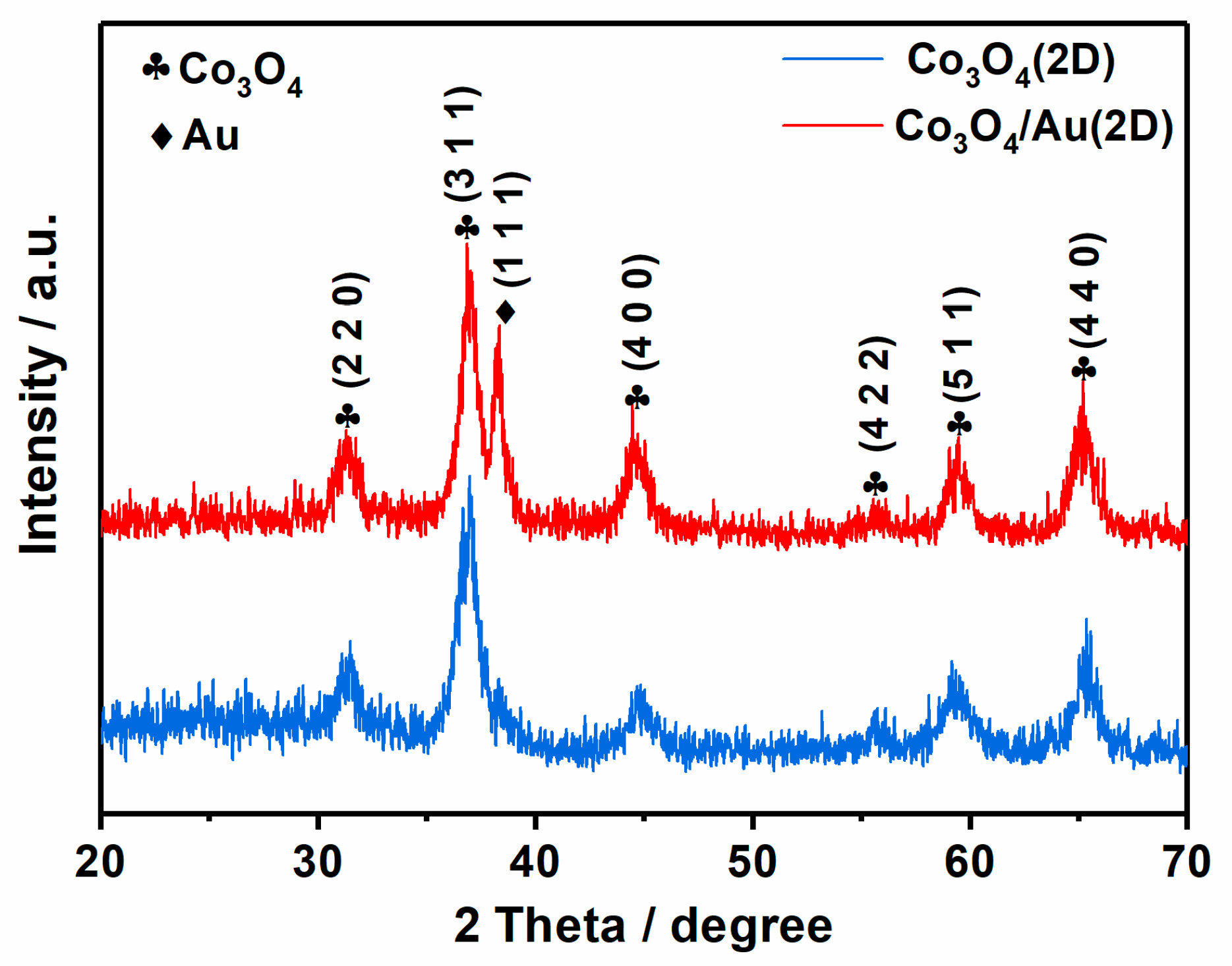
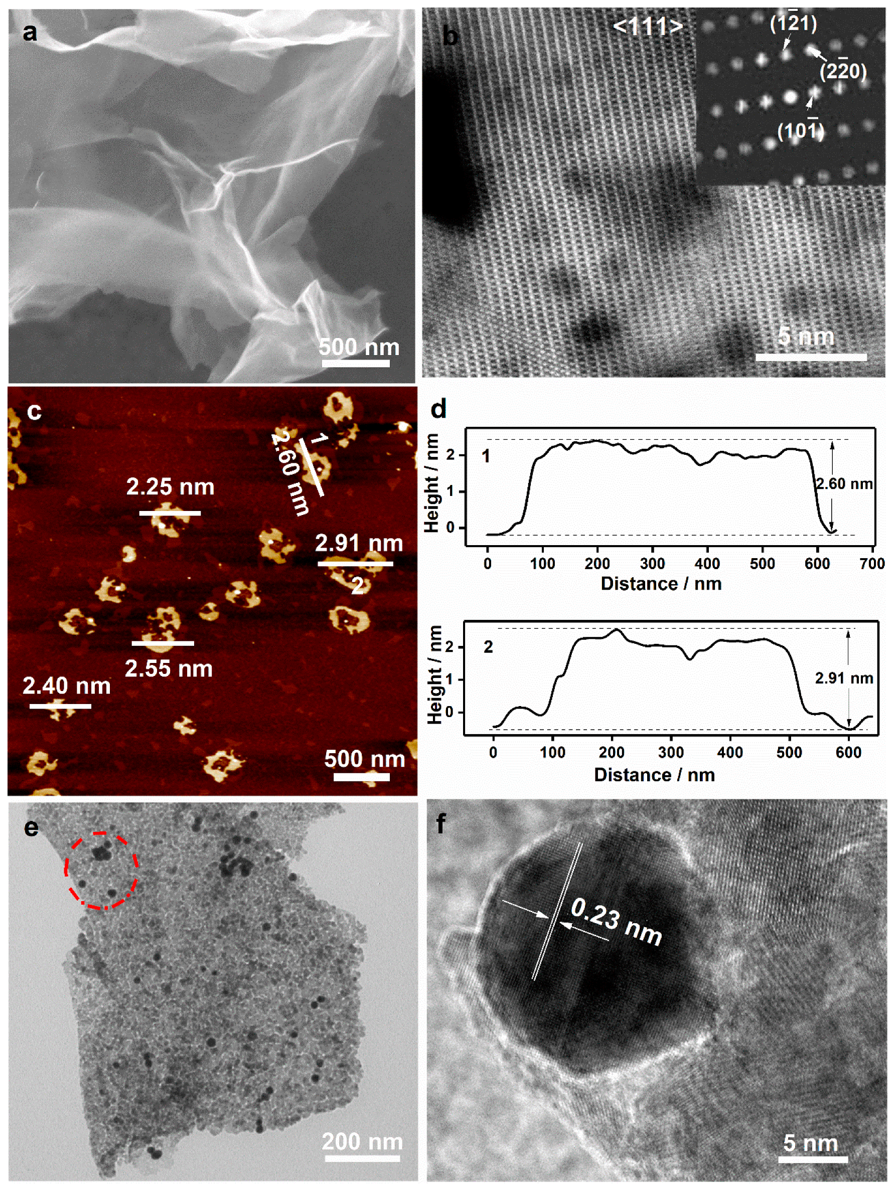
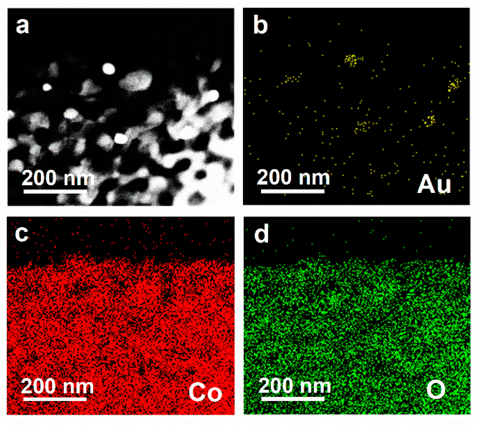
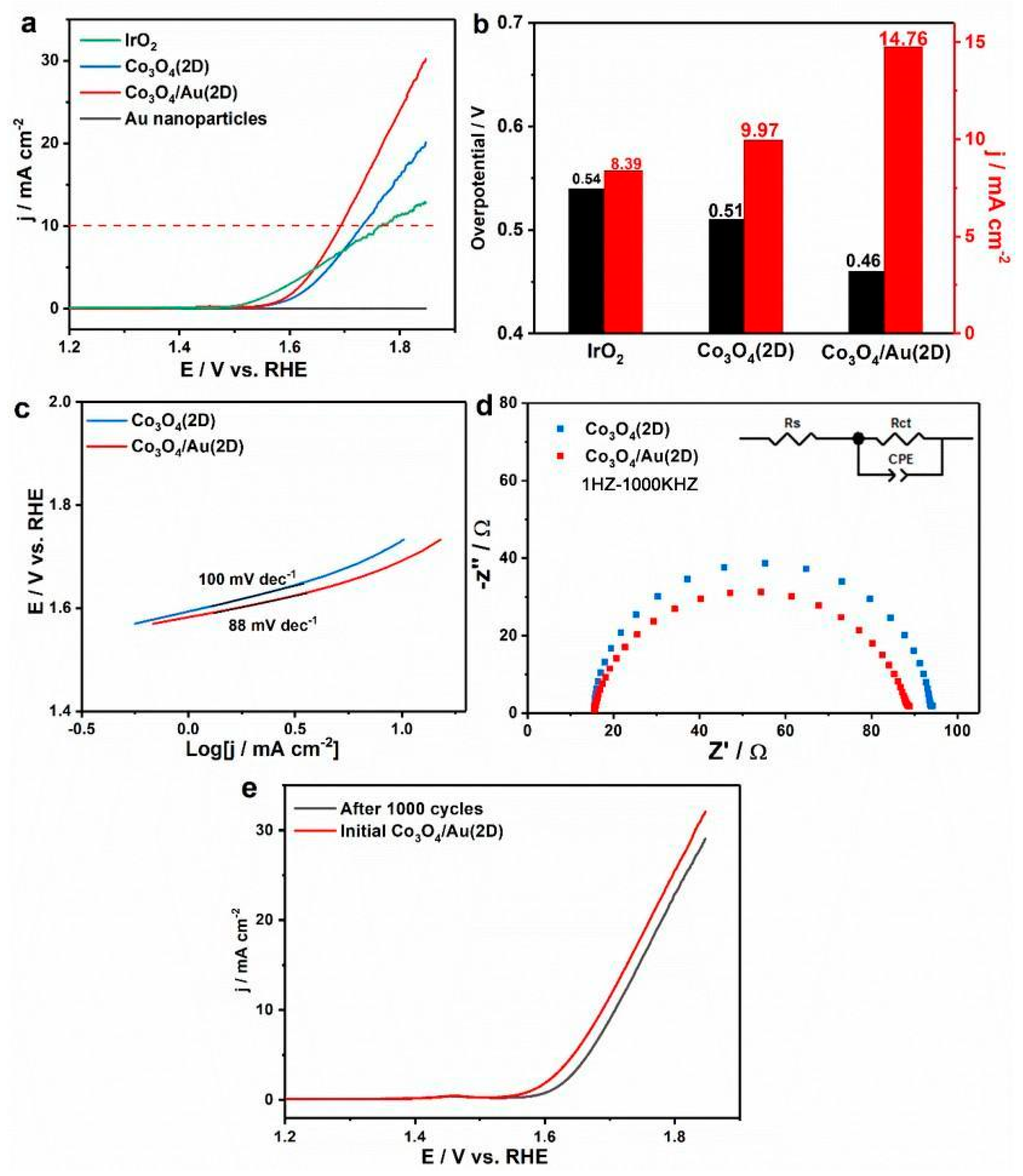
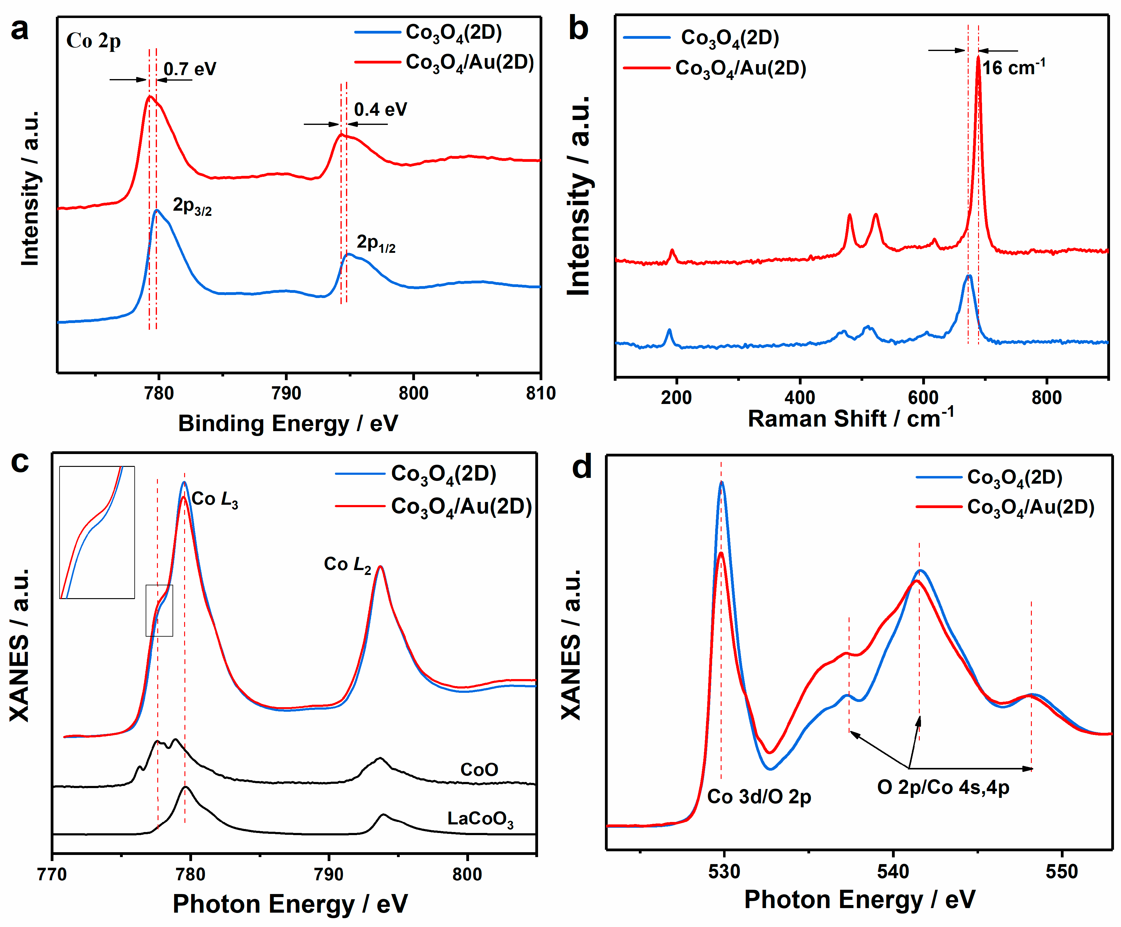
3. Results and Discussion

Figure 1 exhibits XRD patterns of Co3O4 samples with or without Au. The diffraction peaks of the sample without Au loading are indexed to the standard spinel-structured Co3O4 (JCPDS File No. 65-3103) [20], indicating that cobalt precursors have been completely transformed to the Co3O4 phase. After decorating with Au NPs, the (111) peak of metallic Au (JCPDS No. 65-8601) is observed [21], suggesting the successful syntheses of Co3O4/Au nanocomposites by simply mixing Co3O4 with Au nanoparticles. Moreover, Co3O4 bulk and the corresponding Co3O4/Au bulk composites are also prepared successfully for comparison (Figures S1 and S2).
The as-prepared samples are further examined by SEM, TEM, and AFM. The SEM image in Figure 2a shows that the wrinkles and transparent Co3O4 nanosheets are obtained, which have a size of 2–5 μm. Moreover, the porous structure with a size of ~5 nm is also formed in Co3O4 nanosheets after calcining at 300 °C for 0.5 h (see HRTEM images in S3-5), suggesting the successful synthesis of ultrathin porous Co3O4 nanosheets. HAADF-STEM is also conducted to further affirm the structure of Co3O4 nanosheets (Figure 2b), where the bright points represent Co atoms. The selected area electron diffraction (SAED) analysis shows that the nanosheets could be indexed to the (111) plane of cubic spinel Co3O4. The nonlinear arrangement of Co atoms in the HAADF image (Figure 2b) suggests the slight lattice distortion in Co3O4 nanosheets due to the ultrathin thickness and porous structure. As shown in Figure 2c,d, the height of nanosheets is measured by AFM, which is approximate 2.6 nm, about three times higher than 0.8 nm, the unit-cell dimension of cubic Co3O4 (JCPDS No. 65-3103). Moreover, the morphology and detailed microstructure of Co3O4/Au nanocomposites are also investigated by TEM and HRTEM. From Figure 2e, it can be clearly observed that Au nanoparticles (black points) are loaded successfully onto the surface of Co3O4 nanosheets, and the closest lattice fringes of Au (the red circle in Figure 2e) are ~0.23 nm, corresponding to the (111) lattice plane of Au. Further, STEM-EDS elemental mapping is used to characterize the elemental distribution of Co3O4/Au nanocomposites (Figure 3), and the elemental mapping images of the Co, O, and Au elements (Figure 3b,d) suggest that Au nanoparticles are loaded onto Co3O4 nanosheets uniformly.
Having studied the structure features of ultrathin porous Co3O4 nanosheets and Co3O4/Au nanocomposites, we investigate the OER activities of nanocatalysts in 0.1 M KOH (experimental section). Among Co3O4/Au nanocomposites with different Au loadings (10 mL, 15 mL, 20 mL, 25 mL, and 30 mL), the Co3O4/Au (15 mg/25 mL) sample shows the best OER performance (Figure S7). Figure 4a is the LSV curves of Au nanoparticles, IrO2, ultrathin Co3O4 nanosheets, and Co3O4/Au nanocomposites (15 mg/25 mL) in O2-saturated 0.1 M KOH with a scan rate of 5 mVs−1. Au nanoparticles show little OER performance, indicating that Au nanoparticles are OER inactive. After decorating Au nanoparticles onto Co3O4 ultrathin porous nanosheets, Co3O4/Au nanocomposites exhibit significant improvement of OER performance compared with Co3O4 nanosheets. Additionally, it is also worth noting that both Co3O4/Au nanocomposites and Co3O4 nanosheets display superior OER activities to commercial IrO2. Moreover, it is important to compare the overpotential required for getting a current density of 10 mA cm−2, which corresponds to the significant level of high efficiency of the solar water splitting system [22,23]. At the current density of 10 mA cm−2 (Figure 4b), Co3O4/Au nanocomposites display a smaller overpotential (0.46 V) than Co3O4 nanosheets (0.51 V) and commercial IrO2 (0.54 V). Further, the current density of ultrathin porous Co3O4 nanosheets and Co3O4/Au nanocomposites at an overpotential of 0.5 V (1.73 V vs. RHE) is also compared in Figure 4b. With the loading of Au nanoparticles, the current density is enhanced from 9.97 mA cm−2 for Co3O4 nanosheets to 14.76 mA cm−2 for Co3O4/Au nanocomposites, much higher than 0.73 mA cm−2 and 1.33 mA cm−2 of bulk Co3O4 and bulk Co3O4/Au (Figure S8), respectively. Furthermore, the corresponding Tafel plots in Figure 4c suggest that Co3O4/Au nanocomposites possess smaller Tafel slopes (88 mV dec−1) than Co3O4 ultrathin nanosheets (100 mV dec−1), meaning that Au loading onto Co3O4 ultrathin porous nanosheets accelerates the catalytic process of the OER. It can be concluded that Au nanoparticles could enhance the OER performance of ultrathin porous Co3O4 nanosheets, which is firstly observed in the composites obtained by physically mixed Co3O4 nanosheets with Au nanoparticles. The possible reason is that ultrathin nanosheets possess more low-coordinated surface atoms than bulk materials [13], meaning that the interaction between the physically mixed Co3O4 ultrathin nanosheets and Au nanoparticles could happen more easily. To further comprehend the kinetics process for the OER, the electrochemical impendence spectra (EIS) data are fitted using an equivalent circuit diagram consisting of the charger transfer resistance (Rct), the solution resistance (RS), and the constant phase element (CPE) related to the electrochemical double capacitance. The Rct is related to the kinetics process of the OER. Figure 4d shows that the Rct of Au-decorated nanosheets (73 Ω) is smaller than pure Co3O4 nanosheets (78 Ω), demonstrating the faster charge transfer kinetics during electrochemical reaction. Furthermore, the polarization curve slightly decreases after 1000 cycles, revealing excellent durability of Co3O4/Au nanocomposites, as shown in Figure 4e.
To reveal the role of Au nanoparticles in enhancing the OER performance of ultrathin porous Co3O4 nanosheets, the corresponding XPS and Raman spectra were conducted. As shown in Figure 5a, compared with ultrathin porous Co3O4 nanosheets, Co3O4/Au nanocomposites show a ~0.7 eV decreased electron binding energy for Co 2P3/2 and ~0.4 eV decreased electron binding energy for 2P1/2. The binding energy shift is due to the increased electron density around Co atoms in Co3O4/Au nanocomposites [17], which indicates the interaction between the nanosheets and Au loading. To further verify the above speculation that the interaction happens between the physically mixed Co3O4 ultrathin nanosheets and Au nanoparticles, the Raman measurement is conducted (Figure 5b). Five peaks at 671, 606, 510, 471, and ~188cm−1 are observed in both the Raman spectra of the Co3O4/Au nanocomposites and Co3O4 nanosheets, which correspond to A1g, F2g, F2g, Eg, and F2g modes of the Co3O4 spinel structure, respectively [24]. Compared with the nanosheets, the Raman peaks of the Co3O4/Au nanocomposites exhibit a blue shift, and the peak at 671 even shifts to 687cm−1 by an increase of 18 cm−1, which further certifies the electronic interaction between ultrathin Co3O4 porous nanosheets and Au nanoparticles [17].
To get an in-depth understanding of the electronic interaction between the Co3O4 ultrathin nanosheets and Au nanoparticles and the corresponding effect on the OER performance of the Co3O4/Au nanocomposites, XANES is also performed, which is sensitive to the electronic states of the special elements. Figure 5c shows the normalized Co L-edge XANES for the Co3O4 nanosheets and the Co3O4/Au nanocomposites, along with the reference samples CoO and LaCoO3 [25]. The Co L-edge XANES spectra correspond to the excitation of 2p electron to the unoccupied 3d state. Additionally, for the Co L3-edge spectrum in Co3O4, there are two peaks at 779.5 eV and 777.6 eV, originating from the octahedrally coordinated Co3+ and tetrahedrally coordinated Co2+, respectively. With the loading of Au nanoparticles onto ultrathin Co3O4 nanosheets, the peak at 777.6 eV becomes stronger, while the peak at 779.5 eV weakens, indicating that the ratio of Co2+/Co3+ increases in Co3O4 nanocomposites with the loading of Au nanoparticles. Furthermore, the O K-edge XANES spectra of Co3O4/Au nanocomposites (Figure 5d) show an obvious decrease in the unoccupied O 2p-Co 3d hybridized state (~529.8 eV) compared with that of the Co3O4 nanosheets, suggesting a higher electronic density at the O sites, which strengthens the covalent Co-O bonding in the Co3O4/Au composite, thus improving the conductivity of the nanocomposites [26], which is consistent with the EIS results. It has been well-documented that Co2+ can release electrons under an applied bias, thus promoting an affinity to oxygen ions on the surface of the catalyst to form CoOOH, i.e., the main active site for OER [27,28,29,30,31]. In conclusion, due to the addition of Au nanoparticles, the increased Co2+/Co3+ ratio and conductivity result in the enhanced OER performance of the Co3O4/Au nanocomposites. For comparison, the bulk Co3O4 and bulk Co3O4/Au composites are also prepared, and no obvious OER enhancement is observed with the loading of the Au nanoparticles (Figure S8). Compared with the formed reports [32,33,34,35], the ultrathin porous Co3O4 nanosheets do not exhibit superior electrocatalytic properties, which could be due to the low electron conductivity and imperfect synthesis process of the Co3O4 nanosheets.

4. Conclusions

In summary, ultrathin porous Co3O4 nanosheets are synthesized successfully by a facile method. The thickness of the nanosheet is about three times that of the unit-cell dimension (0.8 nm) given for Co3O4. Additionally, the corresponding OER performance is firstly enhanced by physically mixing Co3O4 ultrathin porous nanosheets with Au nanoparticles, which originated from the electronic interaction between Co3O4 and Au nanoparticles due to the unsaturated bonds on the surface of Co3O4 ultrathin porous nanosheets. With the loading of Au nanoparticles, the current density of ultrathin porous Co3O4 nanosheets is enhanced from 9.97 to 14.76 mA cm−2 at an overpotential of 0.5 V. Meanwhile, Co3O4/Au nanocomposites also exhibit a lower overpotential and a Tafal slope, as well as good stability. The enhanced OER performance is ascribed to a higher Co2+/Co3+ ratio and quicker charge transfer in the Co3O4/Au nanocomposites than in the ultrathin porous Co3O4 nanosheets, and Co2+ ions are beneficial to the formation of CoOOH, which acts as the main active site for OER.

Supplementary Materials

The following supporting information can be downloaded at: https://www.mdpi.com/article/10.3390/nano12244419/s1, Figure S1: (a) SEM image and (b) XRD patterns of Co3O4 bulk; Figure S2: XRD patterns of Co3O4 bulk and Co3O4/Au bulk; Figure S3: XRD pattern of Co(OH)2 precursors; Figure S4: (a) SEM and (b) TEM images of Co(OH)2 precursors; Figure S5: HRTEM image of the ultrathin porous Co3O4 nanosheets; Figure S6: HRTEM image of Au nanoparticles; Figure S7: LSV curves of ultrathin porous Co3O4 nanosheets loading with different amount of Au nanoparticles; Figure S8: LSV curves of Co3O4/Au bulk loading with different amount of Au nanoparticles.

Author Contributions

Conceptualization, M.L.; methodology, C.H.; formal analysis, C.H. and D.S.; investigation, J.L.; resources, Q.Z.; data curation, X.L. and H.F.; writing—original draft preparation, C.H.; writing—review and editing, M.L.; visualization, J.X. and G.J.; supervision, M.L.; project administration, M.L.; funding acquisition, M.L. and Y.L. All authors have read and agreed to the published version of the manuscript.

Funding

This research was funded by the National Key Research and Development Project (2016YFA0400904), the Scientific Research Foundation for the Talented Young Scholars of Shanghai Institute of Technology (Grant No. ZQ2020-7 and YJ2021-12), and the scientific Startup Foundation of Shanghai Institute of Technology (Grant No YJ2020-26).

Conflicts of Interest

The authors declare no conflict of interest.

References

  1. Cook, T.R.; Dogutan, D.K.; Reece, S.Y.; Surendranath, Y.; Teets, T.S.; Nocera, D.G. Solar Energy Supply and Storage for the Legacy and Nonlegacy Worlds. Chem. Rev. 2013, 110, 6474–6502. [Google Scholar] [CrossRef] [PubMed]
  2. Armand, M.; Tarascon, J.-M. Building better batteries. Nature 2008, 451, 652–657. [Google Scholar] [CrossRef] [PubMed]
  3. Lim, B.; Jiang, M.; Camargo, P.H.; Cho, E.C.; Tao, J.; Lu, X.; Zhu, Y.; Xia, Y. Pd-Pt bimetallic nanodendrites with high activity for oxygen reduction. Science 2009, 324, 1302–1305. [Google Scholar] [CrossRef] [Green Version]
  4. Jensen, B.W.; Jensen, O.W.; Forsyth, M.; Macfarlane, D.R. High rates of oxygen reduction over a vapor phase-polymerized PEDOT electrode. Science 2008, 321, 671–674. [Google Scholar] [CrossRef] [PubMed]
  5. Lee, S.W.; Carlton, C.; Risch, M.; Surendranath, Y.; Chen, S.; Furutsuki, S.; Yamada, A.; Nocera, D.G.; Shao-Horn, Y. The nature of lithium battery materials under oxygen evolution reaction conditions. J. Am. Chem. Soc. 2012, 134, 16959–16962. [Google Scholar] [CrossRef] [PubMed]
  6. Suntivich, J.; May, K.J.; Gasteiger, H.A.; Goodenough, J.B.; Shao-Horn, Y. A perovskite oxide optimized for oxygen evolution catalysis from molecular orbital principles. Science 2011, 334, 1383–1385. [Google Scholar] [CrossRef] [PubMed]
  7. Gerken, J.B.; McAlpin, J.G.; Chen, J.Y.; Rigsby, M.L.; Casey, W.H.; Britt, R.D.; Stahl, S.S. Electrochemical water oxidation with cobalt-based electrocatalysts from pH 0-14: The thermodynamic basis for catalyst structure, stability, and activity. J. Am. Chem. Soc. 2011, 133, 14431–14442. [Google Scholar] [CrossRef] [PubMed]
  8. Sun, Y.; Gao, S.; Lei, F.; Liu, J.; Liang, L.; Xie, Y. Atomically-thin non-layered cobalt oxide porous sheets for highly efficient oxygen-evolving electrocatalysts. Chem. Sci. 2014, 5, 3976–3982. [Google Scholar] [CrossRef]
  9. Liang, Y.; Li, Y.; Wang, H.; Zhou, J.; Wang, J.; Regier, T.; Dai, H. Co3O4 nanocrystals on graphene as a synergistic catalyst for oxygen reduction reaction. Nat. Mater. 2011, 10, 780–786. [Google Scholar] [CrossRef] [Green Version]
  10. Qu, Q.; Zhang, J.H.; Wang, J.; Li, Q.Y.; Xu, C.W.; Lu, X. Three-dimensional ordered mesoporous Co3O4 enhanced by Pd for oxygen evolution reaction. Sci. Rep. 2017, 7, 41542. [Google Scholar] [CrossRef]
  11. Wu, J.; Xue, Y.; Yan, X.; Yan, W.; Cheng, Q.; Xie, Y. Co3O4 nanocrystals on single-walled carbon nanotubes as a highly efficient oxygen-evolving catalyst. Nano Res. 2012, 5, 521–530. [Google Scholar]
  12. Zhang, J.J.; Wang, H.H.; Zhao, T.J.; Zhang, K.X.; Wei, X.; Jiang, Z.D.; Hirano, S.I.; Li, X.H.; Chen, J.S. Oxygen Vacancy Engineering of Co3O4 Nanocrystals through Coupling with Metal Support for Water Oxidation. ChemSusChem 2017, 10, 2875–2879. [Google Scholar] [CrossRef] [PubMed]
  13. Bao, J.; Zhang, X.; Fan, B.; Zhang, J.; Zhou, M.; Yang, W.; Hu, X.; Wang, H.; Pan, B.; Xie, Y. Ultrathin Spinel-Structured Nanosheets Rich in Oxygen Deficiencies for Enhanced Electrocatalytic Water Oxidation. Angew. Chem. Int. Ed. Engl. 2015, 54, 7399–7404. [Google Scholar] [CrossRef] [PubMed]
  14. Dou, Y.; Liao, T.; Ma, Z.; Tian, D.; Liu, Q.; Xiao, F.; Sun, Z.; Ho Kim, J.; Xue Dou, S. Graphene-like holey Co3O4 nanosheets as a highly efficient catalyst for oxygen evolution reaction. Nano Energy 2016, 30, 267–275. [Google Scholar] [CrossRef]
  15. Yeo, B.S.; Bell, A.T. Enhanced activity of gold-supported cobalt oxide for the electrochemical evolution of oxygen. J. Am. Chem. Soc. 2011, 133, 5587–5593. [Google Scholar] [CrossRef] [PubMed]
  16. Ng, J.W.D.; García-Melchor, M.; Bajdich, M.; Chakthranont, P.; Kirk, C.; Vojvodic, A.; Jaramillo, T.F. Gold-supported cerium-doped NiOx catalysts for water oxidation. Nature Energy 2016, 1, 16053–16060. [Google Scholar] [CrossRef] [Green Version]
  17. Zhao, S.; Jin, R.; Abroshan, H.; Zeng, C.; Zhang, H.; House, S.D.; Gottlieb, E.; Kim, H.J.; Yang, J.C.; Jin, R. Gold Nanoclusters Promote Electrocatalytic Water Oxidation at the Nanocluster/CoSe2 Interface. J. Am. Chem. Soc. 2017, 139, 1077–1080. [Google Scholar] [CrossRef]
  18. Frens, G. Controlled Nucleation for the Regulation of the Particle Size in Monodisperse Gold Suspensions. Nat. Phys. Sci. 1973, 241, 20–22. [Google Scholar] [CrossRef]
  19. Sun, Z.; Liao, T.; Dou, Y.; Hwang, S.M.; Park, M.S.; Jiang, L.; Kim, J.H.; Dou, S.X. Generalized self-assembly of scalable two-dimensional transition metal oxide nanosheets. Nat. Commun. 2014, 5, 3813–3821. [Google Scholar] [CrossRef] [Green Version]
  20. Meher, S.K.; Rao, G.R. Effect of Microwave on the Nanowire Morphology, Optical, Magnetic, and Pseudocapacitance Behavior of Co3O4. J. Phys. Chem. C 2011, 115, 25543–25556. [Google Scholar] [CrossRef]
  21. Wang, Z.L.; Wang, H.L.; Yan, J.M.; Ping, Y.; Song, O., II; Li, S.J.; Jiang, Q. DNA-directed growth of ultrafine CoAuPd nanoparticles on graphene as efficient catalysts for formic acid dehydrogenation. Chem. Commun. 2014, 50, 2732–2734. [Google Scholar] [CrossRef] [PubMed]
  22. Zhao, S.; Jin, R.; Song, Y.; Zhang, H.; House, S.D.; Yang, J.C.; Jin, R. Atomically Precise Gold Nanoclusters Accelerate Hydrogen Evolution over MoS2 Nanosheets: The Dual Interfacial Effect. Small 2017, 13, 1701519–1701525. [Google Scholar] [CrossRef]
  23. Zheng, Y.R.; Gao, M.R.; Gao, Q.; Li, H.H.; Xu, J.; Wu, Z.Y.; Yu, S.H. An efficient CeO2/CoSe2 Nanobelt composite for electrochemical water oxidation. Small 2015, 11, 182–188. [Google Scholar] [CrossRef] [PubMed]
  24. Hadjiev, V.G.; Ilievl, M.N.; Vergilov, I.V. The Raman spectra of Co3O4. J. Phys. C Solid State Phys. 1988, 21, 199–201. [Google Scholar] [CrossRef]
  25. Cui, Z.; Xu, H.; Yun, Y.; Guo, J.; Chuang, Y.-D.; Huang, H.; Meng, D.; Wang, J.; Fu, Z.; Peng, R.; et al. Soft X-ray absorption spectroscopy investigations of Bi6FeCoTi3O18 and LaBi5FeCoTi3O18 epitaxial thin films. J. Appl. Phys. 2016, 120, 084101–084105. [Google Scholar] [CrossRef]
  26. Zhu, J.; Bai, L.; Sun, Y.; Zhang, X.; Li, Q.; Cao, B.; Yan, W.; Xie, Y. Topochemical transformation route to atomically thick Co3O4 nanosheets realizing enhanced lithium storage performance. Nanoscale 2013, 5, 5241–5246. [Google Scholar] [CrossRef] [PubMed]
  27. Chen, Y.; Hu, J.; Diao, H.; Luo, W.; Song, Y.F. Facile Preparation of Ultrathin Co3O4/Nanocarbon Composites with Greatly Improved Surface Activity as a Highly Efficient Oxygen Evolution Reaction Catalyst. Chemistry 2017, 23, 4010–4016. [Google Scholar] [CrossRef]
  28. Liang, Y.; Li, Y.; Wang, H.; Dai, H. Strongly coupled inorganic/nanocarbon hybrid materials for advanced electrocatalysis. J. Am. Chem. Soc. 2013, 135, 2013–2036. [Google Scholar] [CrossRef]
  29. Wang, H.Y.; Hung, S.F.; Chen, H.Y.; Chan, T.S.; Chen, H.M.; Liu, B. In Operando Identification of Geometrical-Site-Dependent Water Oxidation Activity of Spinel Co3O4. J. Am. Chem. Soc. 2016, 138, 36–39. [Google Scholar] [CrossRef]
  30. Wang, W.; Kuai, L.; Cao, W.; Huttula, M.; Ollikkala, S.; Ahopelto, T.; Honkanen, A.P.; Huotari, S.; Yu, M.; Geng, B. Mass-Production of Mesoporous MnCo2O4 Spinels with Manganese(IV)- and Cobalt(II)-Rich Surfaces for Superior Bifunctional Oxygen Electrocatalysis. Angew. Chem. Int. Ed. 2017, 56, 14977–14981. [Google Scholar] [CrossRef] [Green Version]
  31. Xu, L.; Jiang, Q.; Xiao, Z.; Li, X.; Huo, J.; Wang, S.; Dai, L. Plasma-Engraved Co3O4 Nanosheets with Oxygen Vacancies and High Surface Area for the Oxygen Evolution Reaction. Angew. Chem. Int. Ed. 2016, 55, 5277–5281. [Google Scholar] [CrossRef]
  32. Li, S.; Li, Z.; Ma, R.; Gao, C.; Liu, L.; Hu, L.; Zhu, J.; Sun, T.; Tang, Y.; Liu, D.; et al. A glass-ceramic with accelerated surface reconstruction toward the efficient oxygen evolution reaction. Angew. Chem. Int. Ed. 2021, 60, 3773–3780. [Google Scholar] [CrossRef] [PubMed]
  33. Olowoyo, J.O.; Kriek, R.J. Recent progress on bimetallic-based spinels as electrocatalysts for the oxygen evolution reaction. Small. 2022, 18, 2203125–2203174. [Google Scholar] [CrossRef]
  34. Li, J.; Stephanopoulos, M.F.; Xia, Y. Introduction: Heterogeneous single-atom catalysis. Chem. Rev. 2020, 120, 11699–11702. [Google Scholar] [CrossRef] [PubMed]
  35. Xiao, M.; Zhang, C.; Wang, P.; Zeng, W.; Zhu, J.; Li, Y.; Peng, W.; Liu, Q.; Xu, H.; Zhao, Y.; et al. Polymetallic phosphides evolved from MOF and LDH dual-precursors for robust oxygen evolution reaction in alkaline and seawater media. Mater. Today Phys. 2022, 60, 100684–100694. [Google Scholar] [CrossRef]
Figure 1. XRD patterns of Co3O4 nanosheets and Co3O4/Au nanocomposites.
Nanomaterials 12 04419 g001
Figure 2. (a) SEM and (b) STEM-HAADF images of Co3O4 ultrathin nanosheets; (c) AFM image and (d) the corresponding height profiles of Co3O4 ultrathin nanosheets; and (e) TEM and (f) HRTEM images of Co3O4/Au nanocomposites.
Nanomaterials 12 04419 g002
Figure 3. (a) Dark-field STEM micrograph of Co3O4/Au nanocomposites; and STEM-EDS elemental mapping of (b) Au, (c) Co, and (d) O elements.
Nanomaterials 12 04419 g003
Figure 4. OER activities of the nanocatalysts: (a) LSV curves of Au nanoparticles, IrO2, Co3O4 ultrathin nanosheets, and Co3O4/Au nanocomposites; (b) comparison of the current density at an overpotential of 0.5 V and the overpotential required for achieving a current density of 10 mA cm−2, for IrO2, Co3O4 ultrathin porous nanosheets, and Co3O4/Au nanocomposites; (c) Tafel plots for Co3O4 nanosheets and Co3O4/Au nanocomposites; (d) EIS of Co3O4 nanosheets and Co3O4/Au nanocomposites; and (e) LSV curves of the Co3O4/Au nanocomposites before and after 1000 cycles.
Nanomaterials 12 04419 g004
Figure 5. The electronical structure characterizations of Co3O4 ultrathin nanosheets and Co3O4/Au nanocomposites: (a) XPS spectra of Co 2p level; (b) Raman spectra between 100 and 900 cm−1; (c) XANES spectra of Co L-edge in Co3O4 ultrathin nanosheets and Co3O4/Au nanocomposites, and XANES spectra of LaCoO3 and CoO are cited from Ref [25]; and (d) XANES spectra of O K-edge in Co3O4 ultrathin nanosheets and Co3O4/Au nanocomposites.
Nanomaterials 12 04419 g005
Publisher’s Note: MDPI stays neutral with regard to jurisdictional claims in published maps and institutional affiliations.

