Review

Gene Therapy Using Efficient Direct Lineage Reprogramming Technology for Neurological Diseases

1
Laboratory of Regenerative Medicine for Neurodegenerative Disease, Stand Up Therapeutics, Hannamdaero 98, Seoul 04418, Republic of Korea
2
Department of Molecular Biology, Nuturn Science, Sinsadong 559-8, Seoul 06037, Republic of Korea
3
Department of Chemistry, Sungkyunkwan University, 2066, Seobu-ro, Jangan-gu, Suwon-si 16419, Republic of Korea
4
Department of Bio-Health Technology, College of Biomedical Science, Kangwon National University, 1 Kangwondeahak-gil, Chuncheon 24341, Republic of Korea
5
Research Animal Resource Center, Korea Institute of Science and Technology, Hwarang-ro 14-gil, Seongbuk-gu, Seoul 02792, Republic of Korea
6
Database Laboratory, Department of Computer Science and Engineering, Dongguk University-Seoul, Pildong-ro 1-gil 30, Jung-gu, Seoul 04620, Republic of Korea
*
Authors to whom correspondence should be addressed.
These authors contributed equally to this work.
Nanomaterials 2023, 13(10), 1680; https://doi.org/10.3390/nano13101680
Submission received: 24 April 2023 / Revised: 12 May 2023 / Accepted: 16 May 2023 / Published: 19 May 2023
(This article belongs to the Special Issue Functionalization Chemistry in Porous Nanomaterials)

Abstract

:
Gene therapy is an innovative approach in the field of regenerative medicine. This therapy entails the transfer of genetic material into a patient’s cells to treat diseases. In particular, gene therapy for neurological diseases has recently achieved significant progress, with numerous studies investigating the use of adeno-associated viruses for the targeted delivery of therapeutic genetic fragments. This approach has potential applications for treating incurable diseases, including paralysis and motor impairment caused by spinal cord injury and Parkinson’s disease, and it is characterized by dopaminergic neuron degeneration. Recently, several studies have explored the potential of direct lineage reprogramming (DLR) for treating incurable diseases, and highlighted the advantages of DLR over conventional stem cell therapy. However, application of DLR technology in clinical practice is hindered by its low efficiency compared with cell therapy using stem cell differentiation. To overcome this limitation, researchers have explored various strategies such as the efficiency of DLR. In this study, we focused on innovative strategies, including the use of a nanoporous particle-based gene delivery system to improve the reprogramming efficiency of DLR-induced neurons. We believe that discussing these approaches can facilitate the development of more effective gene therapies for neurological disorders.

1. Introduction

Cell fate conversion technology originated with the generation of induced pluripotent stem cells (iPSCs) via viral transduction of specific transcription factors, namely, octamer-binding transcription factor 4, SRY-Box transcription factor 2, KLF transcription factor 4, and MYC proto-oncogene, bHLH transcription factor (OSKM) [1]. This technology demonstrated that a fully differentiated somatic cell could be reprogrammed into a completely different cell type by introducing key transcriptional factors of starting cells. Based on this principle, numerous studies have shown the feasibility of direct lineage reprogramming (DLR) from fibroblasts to various cell types, such as neurons, cardiomyocytes, chondrocytes, and hepatocytes.
Specifically, for neuronal reprogramming, DLR technology can be used to reprogram fibroblasts into various neuronal types, including cholinergic motor, glutamatergic, GABAergic, and dopaminergic neurons [2,3,4,5,6]. In 2010, it was revealed that the ectopic expressions of achaete-scute homolog 1 (ASCL1), POU domain transcription factor (BRN2), and myelin transcription factor 1-like (MYT1L) (ABM) could induce DLR of fibroblasts into functional glutamatergic neurons, characterized by synaptic activity and the regulation of Na+/K+ currents. In 2014, Wernig et al. showed that ASCL1 alone could induce DLR of functional glutamatergic neurons, which demonstrated that ASCL1 is the key master regulator that drives neuronal lineage development, with the assistance of two other factors.
Another example is the generation of induced dopaminergic neurons (iDNs). To generate iDNs, it is crucial to cotransduce dopaminergic lineage transcriptional factors, nuclear receptor 4A2 (NURR1), paired-like homeodomain 3 (PITX3), and LIM homeobox transcription factor 1 alpha (LMX1A) with ASCL1 (ANPL) [7]. By generating iDNs using DLR technology, researchers have provided evidence that Parkinson’s disease (PD) could be treated. PD is a progressive neurodegenerative disease that mainly affects motor function. It is characterized by the degeneration of dopaminergic neurons in the substantia nigra-to-striatum circuit, which leads to neurotransmitter imbalances and Parkinsonian symptoms. The cardinal symptoms of this disease include bradykinesia (slowness of movement), rigidity (stiffness and increased resistance to passive limb movements), tremor (involuntary and rhythmic shaking), and postural instability (impaired balance and coordination). In research settings, 1-methyl-4-phenyl-1,2,3,6-tetrahydropyridine (MPTP) and 6-hydroxydopamine hydrobromide (6-OHDA)-induced PD mouse models exhibited a lack of dopaminergic neurons and generated small amounts of dopamine, thus, displaying a Parkinsonian phenotype. The conversion of astrocytes into dopaminergic neurons in the striatum region led to the production of tyrosine hydroxylase (TH)-positive cells from originally glial fibrillary acidic protein (GFAP)-positive cells, which reduced Parkinsonian symptoms.
Spinal cord injury (SCI) is another major neurological disorder that can cause movement disabilities. SCI is a devastating condition that can lead to partial or complete loss of motor and sensory functions below the level of an injury. The pathophysiology of the disease involves complex interplay among secondary injury mechanisms, including oxidative stress, inflammation, cellular toxicity, and demyelination. Despite extensive research efforts, there is currently no effective therapy for SCI. Gene therapy is a promising approach as it allows targeted delivery of therapeutic genes to an injured spinal cord.
Motor neurons are specialized neurons that control the contraction of skeletal muscles, which permits movement and locomotion. In SCI, the motor neurons that innervate the muscles below the level of injury are often damaged or destroyed, which leads to paralysis and motor malfunction. Recently, the cotransduction of transcriptional factors, ASCL1, BRN2, MYT1L, and LIM homeobox 3 (Lhx3) has been reported to induce DLR of motor neurons from fibroblasts [8].
DLR exhibits several advantages over iPSC redifferentiation techniques because it does not pass through a pluripotent stage, which enables the production of stem cell-free cell sources (e.g., astrocytes and fibroblasts) [5]. Furthermore, DLR can address safety issues, such as teratoma formation, and it is more cost-effective for clinical applications. The OSKM-induced iPSCs possess a risk of differentiating into other types of pluripotent stem cells because the early phase of intermediate iPSCs is unstable. Moreover, the regeneration of iPSCs is less cost effective than DLR because multiple steps are involved in creating iPSCs; thus, dealing with iPSC reprogramming, differentiation, and transplantation to the target organs is difficult. However, in vivo DLR could be more successful for direct differentiation at precise locations in several organs. In vivo DLR is accomplished by manipulating the expression of certain genes in the targeted cells, leading to their conversion into a different cell type without the need for cell transplantation or external manipulation (Figure 1a). In vivo DLR could be used to treat a wide range of diseases by converting the affected cells into healthy cells [9,10]. Guo et al. showed that targeted expression of NeuroD1 to astrocyte induced glutamatergic and GABAergic neurons after brain injury or in Alzheimer’s disease mouse brain [11]. Similarly, NeuroD1-induced reprogramming of astrocytes has been applied for the treatment of a stroke model [12]. In the adult mouse striatum, Cervo et al. showed that combinatorial expression of NeuroD1, Ascl1, Lmx1a, and miR-218 induced dopamine neurons in PD mouse model [13]. These studies provide evidence that in vivo DLR may hold potential for treating neurodegenerative disease by replacing lost or damaged cells with newly converted cells (Figure 1b).
Consequently, more than 100 studies since 2010 have investigated the feasibility of DLR technology for gene therapy. Nonetheless, the low efficiency and quality of reprogrammed cells remain to be significant obstacles to DLR-based gene therapy [14,15,16,17]. Therefore, various studies have attempted to enhance DLR efficiency by treating additional genetic factors, miRNAs, small molecules, or biocompatible materials [5,18,19,20,21,22,23].
The use of nanoparticles as a delivery system in the biomedical field is another promising subject in drug delivery. Improving the stability, bioavailability, and pharmacokinetics of therapeutic agents is possible by encapsulating them within nanoparticles. Moreover, side effects, such as cytotoxicity, inflammation, and reactive oxygen species (ROS) generation, can be reduced. Various types of nanoparticles, including liposomes, polymeric organic nanoparticles, and inorganic nanoparticles, have been employed for targeted drug delivery in cancer therapy. This method enables selective delivery of anticancer drugs to tumor cells while minimizing damage to other tissues [24]. Furthermore, nanoparticles can also be used for the delivery of cDNA, siRNA, and proteins. The use of nanoparticles in a delivery system instead of a viral transduction system or combination of small molecules offers several advantages, including enhanced safety, reduced immunogenicity, tunable physicochemical properties, and the ability to encapsulate and protect genetic material, such as DNA, RNA, and oligonucleotides. Additionally, nanoparticles facilitate targeted delivery and controlled release of therapeutic genes, ultimately improving the therapeutic efficacy and reducing off-target effects [25,26,27].
Nanoparticles can be employed in tissue engineering to construct biomimetic matrices. They are called nanotopographical structure and it can mimic the native extracellular matrix, thereby, providing a conducive environment for cell adhesion, proliferation, and reprogramming [5,28]. For example, a recent study by Yoo et al. demonstrated that an artificially engineered nanotopographical matrix could promote the direct conversion of dopaminergic neurons by modulating the focal adhesion kinase (FAK) molecule in the cell reprogramming process. This modulation increased H3K4me3 levels in the transcription start site of dopaminergic neuronal marker genes, such as tyrosine hydroxylase (Th), dopamine transporter (Dat), and microtubule-associated protein 2 (Map2) [23]. Furthermore, nanoparticles can also serve as catalytic activators for cellular processes, which facilitates cell differentiation or reprogramming. For instance, hydroxyapatite nanoparticles have been employed for the development of bone graft substitutes that promote osteointegration and bone regeneration. Additionally, solid AuNPs with electromagnetic properties have been shown to stimulate the cell reprogramming process by regulating histone acetylation H4K12ac [5].
Reducing oxidative stress is critical for the DLR process because the conversion of cell fate requires significant energy undertaking. A recent study by Lee et al. showed that the use of gold nanoporous rods (AuNpRs) to alleviate cell stress had a considerable impact on the efficacy of induced neuron direct conversion. The researchers observed that AuNpRs functioned as ROS scavengers in DLR, thus, lowering internal cellular conversion stress. Their discovery demonstrated that the use of AuNpRs enhanced the efficiency of DLR-induced dopaminergic (iDA) neurons originating from brain astrocytes, thereby, ameliorating the Parkinsonian phenotype in animal models [29].
Neurological disorders are one of the major interests in the field of gene therapy, with many researchers suggesting the possibility of treating neurological diseases, such as PD and SCI, using DLR technology. The use of master regulators that positively control cellular stress or activate the neuronal actuator (related genes) by opening the related chromatin can serve as a novel therapeutic approach for treating neurological diseases using DLR technology [29,30]. Another way to accelerate DLR is to apply modified nanoparticles. The use of nanotechnology in the biomedical engineering field is emerging as a novel therapeutic approach for treating neurological diseases, with over 10 studies having described how some nanosized gold or graphene nanostructures can facilitate the DLR process [5,14,23,31,32,33,34,35,36].
Nanotechnology has brought significant advancements in the biomedical engineering field and has revolutionized the treatment of various diseases. Various nanotechnology approaches have been implemented in this field including nanocarriers, which have provided numerous experimental and clinical benefits. One of the experimental advantages is their ability to facilitate targeted drug delivery. Nanocarriers such as gold nanoparticles and nanorods can transport growth factors or drugs to specific regions in the brain [37,38,39]. Delivery of targeted growth factors or drugs allows for higher doses of drugs to reach a target area while minimizing toxicity to healthy neurons, improving cargo efficacy, and reducing side effects. In addition, nanotechnology methods have also facilitated the development of regenerative medicine, which aims to repair or replace damaged tissues and organs. Scaffold nanomaterials can mimic the extracellular matrix and provide neuronal structural support for neuronal growth, allowing for the regeneration of neuronal networks in the brain [40,41]. Taken together, nanotechnology methods have significant experimental and clinical importance in the biomedical engineering field.

2. Generation of Induced Neurons via Cell Fate Conversion

2.1. Acceleration of the Direct Neuronal Reprogramming Process

Cell fate conversion techniques hold substantial promise within the realm of gene therapy for neurological diseases, and therefore, have garnered immense interest among researchers in the field. Consequently, numerous approaches aimed at expediting the process of cell fate conversion have been investigated [5,31,42,43]. Of particular note, cell DLR represents a cutting-edge technology within the field of regenerative medicine. A pioneering study discovered that the direct conversion of induced neurons could be achieved by transducing key transcriptional factors. The ectopic expression of ASCL1, BRN2, and MYT1L (ABM) induced cortical neuronal reprogramming; ASCL1, NURR1, PITX3, and LMX1A (ANPL) induced dopaminergic neuronal reprogramming; and ASCL1, BRN2, MYT1L, and Lhx3 produced motor neurons’ action potential, forming the neuromuscular junction [2,3,44]. Several studies have described various methodologies for promoting neuronal DLR. Table 1 presents an overview of the different types of DLR enhancers, including small molecules, such as CHIR99021, a GSK3 inhibitor; LDN193189, a BMP type 1 receptor ALK2/3 inhibitor; A83-01, a TGF-β type 1 receptor ALK4/5/7 inhibitor; and ascorbic acid. RNA interference (RNAi) using miR-124 and miR-9/9 and biocompatible materials such as AuNpRs, nanotopographical substrates, and graphene nanosheets, have also been shown to improve the efficiency of DLR [5,19,23,31,45,46]. Recent evidence suggests that inorganic materials can regulate cellular mechanisms by modulating epigenetic modifications. Graphene, a single-atom-thick sheet of carbon atoms with a two-dimensional honeycomb structure, possesses unique physical, chemical, and mechanical properties. This materials has been shown to significantly activate H3K4me3 by inducing mesenchymal-to-epithelial transition [28]. Additionally, nanogrooved substrates have been reported to upregulate H3K4me3 and decrease H3K9me3 and H3K27me3 levels, thereby, activating FAK and promoting dopaminergic neuronal fate [23]. Furthermore, the in vivo injection of electromagnetized AuNPs has been documented to significantly activate H4K12 acetylation in histones, which resulted in the generation of iDNs in a mouse model of PD [5].
The use of an appropriate DNA carrier for reprogramming transcriptional factors is necessary for applying DLR technology in the field of gene therapy. Currently, there is significant interest among researchers in using viral vectors for gene therapy, owing to their ability to effectively deliver the necessary genetic material to the target cells. Globally, lentivirus, retrovirus, adenovirus, and adeno-associated virus (AAV) are commonly used viral vectors for ex vivo cell therapy and in vivo gene therapy [47,48,49,50]. There are still some concerns surrounding the use of AAV vectors. One of the concerns is an AAV-induced autoimmune response, which can decrease the effectiveness of the treatment or cause adverse reactions in AAV-treated patients [51]. Additionally, AAV vectors have a limitation since they carry cargoes that are dependent on the size or complexity of the gene [52].
Nonetheless, AAV is a promising candidate for in vivo gene therapy because of its unique features and key advantages over other viral vectors. AAV is known for its nongenomic integration and low toxicity, which significantly alleviate the risk of immunogenicity, mutagenesis, and genotoxicity associated with other viral vectors. For example, systemically administered recombinant AAV used for cancer therapy has shown that rAAVV2/5 has no genotoxicity in nonhuman primate and human livers [53]. In research on the AAV5-cohPBGD vector used in cancer therapy, its intravenous administration had no side effects or integration of immunogenicity and genotoxicity in primate tissues, such as the liver, spleen, and adrenal gland [54]. As a result, AAV has gained widespread attention and is being increasingly used in clinical studies [55,56]. A review of gene therapy trials indicated that 149 clinical trials employed pAAV plasmid vector, of which 94 have been concluded, with 51 reaching the endpoint of efficacy [57]. Most ongoing gene therapy trials are phase 1/2 safety tests. Furthermore, clinical trials that employed AAV serotype 2 were primarily conducted in the field of neurological disorders [58]. AAV2 has the greatest safety and efficacy record in neurological research, as evidenced by over 40 completed clinical trials. The ongoing and completed clinical trials based on AAV2 are listed in Table 2 [58,59,60,61,62,63,64,65,66,67,68,69].

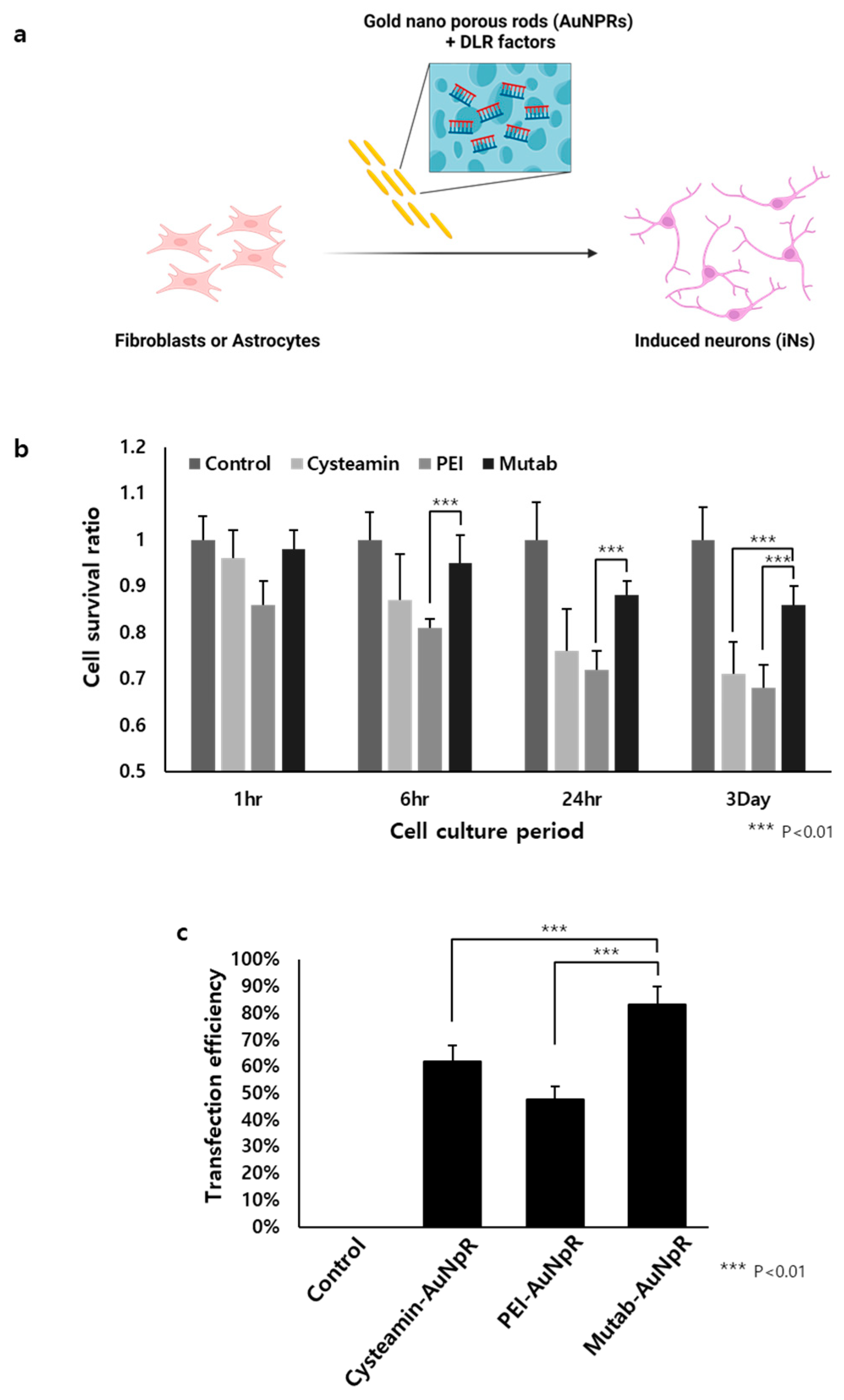
2.2. The Role of AuNpRs in the Direct Neuronal Reprogramming Process

The other potential clinical method for gene therapy in the future is to apply biocompatible nanomaterials, particularly AuNpRs. Several studies have shown that specifically modified or functionalized nanoparticles exert crucial effects on biological processes, such as cell survival and differentiation. AuNPs exhibit various therapeutic characteristics, including biocompatibility, ROS scavenging effect, high surface reactivity, and plasmon resonance [29,70,71,72,73]. Not all AuNPs have these effects; however, specifically modified AuNPs or AuNPs integrated with other materials have a drastic impact on the DLR process. Recently, Yoo and Park et al. reported that AuNpRs played a key role as ROS scavengers, ameliorating Parkinsonian phenotypes (i.e., slowness, muscle rigidity, and loss of movement) [29]. They injected AuNpRs into the substantia nigra region in 6-OHDA-induced Parkinsonian mouse model with ASCL1, NURR1, PITX3, and LMX1A, which are dopaminergic neuron conversion transcriptional factors [29]. In the group treated with AuNpRs, a higher number of TH-, DAT-, Forkhead Box A2 (FOXA2)-, and G protein-activated inward rectifier potassium channel 2 (GIRK2)-positive neurons were observed using immunostaining (Figure 2a). To investigate the underlying mechanism, the researchers performed RNA transcriptomic studies during the AuNpR treatment in the reprogramming process. The results revealed an increase in antioxidation-related molecules, particularly ROS scavenging molecules (i.e., Gsta4, Mt3, Sod1, Sod2, and Sirt3), in the AuNpR-treated groups (Figure 2b). Most gene ontology analyses were classified under the oxidation–reduction processes and detoxification.
DA neurons are particularly susceptible to oxidative stress owing to several factors, which contribute to their vulnerability in PD. First, excessive ROS can negatively affect the enzymes involved in dopamine synthesis. TH is the rate-limiting enzyme in dopamine biosynthesis, which converts tyrosine to levodopa. ROS can inhibit TH activity by causing oxidative modification of the enzyme, such as the formation of disulfide bonds and carbonyl groups. This modification results in reduced dopamine production. Second, dopaminergic neurons store dopamine in vesicles via the action of vesicular monoamine transporter (VMAT2). Excessive ROS can impair VMAT2 function, which decreases the vesicular storage of dopamine. This process increases cytosolic dopamine levels, which, in turn, makes more dopamine available for auto-oxidation and subsequent ROS generation, thus, leading to a vicious cycle of oxidative stress and damage. Lastly, dopaminergic neurons themselves are particularly susceptible to oxidative stress because of their high metabolic rate, low antioxidant defenses, and inherent vulnerability associated with dopamine metabolism.
Owing to these reasons, regulating ROS levels during iDA reprogramming is crucial. Yoo and Park et al. successfully demonstrated that antioxidants and ROS scavenging molecules, mediated by AuNpRs, could enhance iDA conversion and alleviate PD phenotypes in animal models [29].
In a study, AuNpRs were synthesized by codepositing Au and Au plating solution at a voltage of −0.95 V to produce Au/Ag alloy nanomaterials. Subsequently, 30% nitric acid was used to dealloy them. To create nanopores in Au nanorods, precise control of the atomic composition of the Au/Ag alloy was achieved by varying the volume ratio of the Au plating solution mixture. To confirm whether AuNpRs were well located in the cell membrane, Yoo and Park attempted to modify the surface of AuNpRs. The surface was modified by incorporating the fluorescence dye rhodamine B (Rho B). They observed that the induced neuron-converted cells exhibited red fluorescence, which could be attributed to the presence of the Rho B dye on AuNpRs. This result showed that the cellular component and biological process could be manipulated by administrating specific types of nanoparticles, such as AuNpRs. Hence, further investigations are being conducted to explore the potential of various shapes and types of nanoparticles in the field of gene therapy and regenerative medicine. Previously, Lee et al. revealed that nanoparticles could be modified by galvanic replacement or the Kirkendall effect [74,75,76]. For example, gold porous lens, gold porous ring, gold shell ring, or gold nanolens have been generated via these reactions (Figure 3) [74,75,76]. In Supplementary Figure S1, a detailed description of the method for producing AuNpRs (Supplementary Figure S1a,b) and nanotubes using a three-electrode system is provided. By employing AuNpRs, they successfully generated induced neurons.

2.3. The Role of Biocompatible Materials as Delivery Cargo for DLR

Recently, several types of biocompatible materials have emerged as promising DNA carriers for gene therapy [77]. Nanoparticles, such as liposomes, polymeric nanoparticles, and inorganic nanoparticles, have been engineered to improve the efficiency, specificity, and safety of gene delivery to target cells [78,79,80]. Versatile carriers can protect the encapsulated DNA from degradation, enhance cellular uptake, and facilitate controlled release, and thereby, increase the possibility of successful gene delivery [79]. In the context of gene therapy for neurological diseases, the use of nanoparticles as DNA carriers can overcome the challenges associated with crossing the blood–brain barrier, thus, enabling more effective and targeted delivery of therapeutic genes [81]. By leveraging the advances in nanoparticle technology, DLR strategies can be further optimized to provide innovative and safer therapeutic options for patients with neurological disorders.
Lee et al. found that AuNpRs could function as not only DLR accelerators but also DLR transcriptional factor carriers (Figure 4a) [29]. They hypothesized that DLR transcriptional factors could be loaded inside the pores of AuNpRs, thereby, accelerating the DLR process without administrating viruses. They used several positively charged materials (i.e, cysteamine, PEI, and Mutab) and observed that Mutab was the most stable and nontoxic material with the highest transfection efficiency (Figure 4b,c) [82,83,84]. From these results, they hypothesized that an efficient DLR process could be performed using a virus-free system. This method can avoid host genome integration and virus immunogenicity issues faced in viral transduction systems.

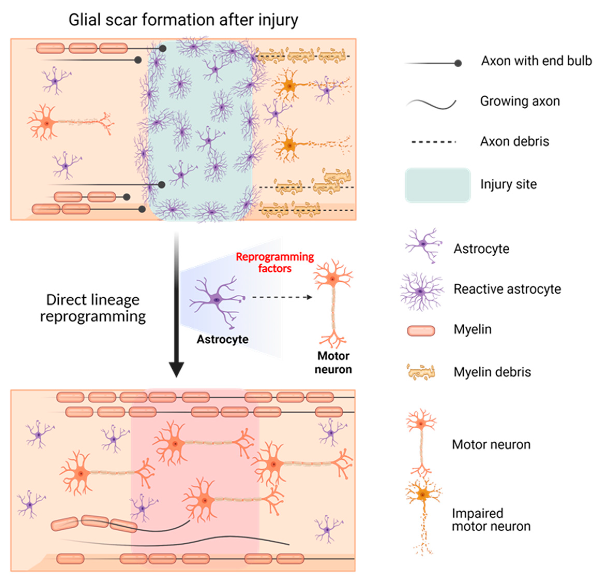
2.4. Gene Therapy for SCI Using DLR Technology

2.4.1. SCI: The Incurable Neurological Disorder

Over a decade ago, SCI was considered to lead to a lifetime of medical complications and reliance on a wheelchair, with extremely limited therapeutic options being available [85]. Patients with SCI often received frustrating and hopeless care. Since then, many researchers have been striving to identify the most effective treatment for patients with SCI. However, so far, no therapeutic methodology has been developed. Fortunately, recent advancements in neurosciences have provided new hope [86]. One promising avenue of research is gene therapy using AAV, which offers the potential for regeneration and functional restoration. Although therapeutic methodologies for SCI remain elusive, the prospects for successful treatment continue to improve with each new discovery [57].
In a study, the SCI animal group showed the formation of a physical barrier, known as “glial scar”, at the injury site owing to the recruitment of reactive astrocytes. This scar impeded axonal regeneration and was characterized by the expression of molecules, such as monoamine oxidase B (Figure 5) [87,88,89,90]. Furthermore, certain studies have indicated that premature removal of glial scars can enlarge an injured area and decrease functional recovery in animal models [90,91].

2.4.2. The Potential Therapeutic Method for SCI: AAV Gene Therapy Using DLR Technology

The possibility of using DLR to treat SCI has been studied based on the idea that reactive astrocytes that form glial scars at the injury site may hinder axonal regeneration and lead to neuronal impairment. In vivo DLR targeting of reactive astrocytes to convert them into motor neurons using appropriate transcriptional factors could provide a solution for paralysis in patients with SCI. Yoo et al. successfully identified factors, such as biocompatible materials or specific genetic molecules, which could accelerate the DLR of motor neurons from fibroblasts or astrocytes. Moreover, converting GFAP-positive glial scars to synapsin-positive motor neurons could serve as a solution for the currently incurable SCI (Figure 6).

3. Therapeutic Insights and Conclusions

Recently, DLR has withnessed significant advancement. This method enables the direct conversion of differentiated mature cells into various other cell types, without the need for an intermediate pluripotent state. This approach was inspired by the critical role played by transcription factors in the process of converting non-neuronal cells into neurons. For example, the reprogramming of fibroblasts into glutamatergic neurons was first accomplished by overexpressing three transcription factors, namely, ASCL1, BRN2 (also known as Pou3f2), and MYT1L. The expressions of these factors resulted in the formation of neurons capable of expressing neuronal markers and establishing functional synapses that exhibited action potential and spontaneous events [2]. Further studies have been conducted to advance the application of DLR technology to generate human-induced neurons. These studies have shown that the transcription factors ASCL1, BRN2, MYT1L, and neuronal differentiation 1 (NEUROD1) could be used to successfully generate human-induced neurons. The role of NEUROD1 in neuronal development is particularly significant in this process [92]. The ability to directly convert non-neurogenic cells into functional neurons is a significant development in the field of reprogramming. This has been demonstrated by the successful generation of human-induced neurons capable of forming synapses and displaying a functional profile. Moreover, studies have indicated that non-neurogenic cells, such as astroglia, glial cells, and pericytes, could also be converted into functional neurons using this approach [93,94]. Transcription factors have been shown to be effective in converting astroglia into glutamatergic or GABAergic neurons. Additional studies have explored these transcription factors further, leading to the production of other subtypes of neurons, such as dopamine neurons, motor neurons, and intermediate spiny neurons. For example, Caiazzo et al. successfully reprogrammed mouse and human fibroblasts into dopaminergic neurons using ASCL1, NURR1, and LMX1A. These iDNs were functionally similar to natural dopaminergic neurons [95]. Furthermore, recent findings have suggested that the use of miRNA could be a promising approach for direct reprogramming [19,20]. Similar to transcription factors, a combination of miRNAs can be used to reprogram fibroblasts into functional neurons [19]. Additionally, the presence of transcription factors can enhance the role of miRNAs in neural reprogramming. For instance, studies have demonstrated that miR-9/9*, miR-124, or miR-218 in combination with other transcription factors, such as ISL1 and LHX3, could promote the reprogramming of fibroblasts into motor neurons [19,96]. MirR-34b/c cotreated with ASCL1 and NURR1 enhanced the generation of iDNs by activating the Wnt1 signaling cascade [97]. A mixture of miR-218 and other transcription factors, such as ASCL1, NEUROD1, and LMX1A, promoted the conversion of astrocytes to dopaminergic neurons in vitro and in vivo [13]. Collectively, these findings highlight the crucial role of both transcription factors and miRNAs in directing the fate of neuronal cells.
A recent study has suggested that the combination of miRNAs with transcription factors contributes to regulating ROS in vivo and in vitro. miRNA-containing nanoparticles are known to reduce ROS. According to a previous study, polyketal (PK3) nanoparticles that contained a mixture of miR-106B, miR-148b, and miR-204 significantly improved cardiac function after myocardial infarction by reducing ROS, which was enhanced by nicotinamide adenine dinucleotide phosphate oxidase [98]. In DLR research, AuNpRs containing the viral transcription factors, ASCL1, NURR1, PITX3, and LMX1A have significantly increased TH-positive neurons by scavenging ROS molecules, which led to the conversion of dopaminergic neurons in PD mouse model. Given the possibility that nanoparticles containing miRNA and transcription factors can reduce ROS in vivo, nanoparticle-based ROS removal may play a vital role in DLR.
The human brain has limited regenerative capacity, indicating that it is unable to naturally replace damaged neurons at the same rate as other organs. This limitation could be attributed to the fact that the process of neurogenesis, or the growth and development of new neurons, is more complex and tightly regulated in the brain than in other tissues of the body [99]. Hence, alternative cell sources are required for treating central nervous system diseases. In this regard, in vivo reprogramming has emerged as a promising therapeutic approach. However, for successful reprogramming, ensuring precise targeting of specific cell populations is crucial. AAV-mediated gene therapy is a viable method for regulating the final cell type during in vivo reprogramming, which could lead to optimal therapeutic outcomes for central nervous system diseases.
Meanwhile, the use of transcription factors in DLR has faced limitations in clinical translation, due to the fact that obtaining sufficient cells is difficult owing to low efficiency and lack of maturity. Therefore, the development of new strategies, such as the use of ROS scavenging molecules and biocompatible nanomaterials, such as AuNpRs, have opened new avenues in the field of gene therapy for incurable diseases. Further research in this area could lead to breakthrough treatments that would improve the quality of life of patients with central nervous system diseases.

Supplementary Materials

The following supporting information can be downloaded at: https://www.mdpi.com/article/10.3390/nano13101680/s1, Figure S1: Schematic illustration on synthesis of nanorods and three-electrode system for the electrochemical deposition method. Schematic illustration representing synthetic procedure of (a) Au nanorods, (b) porous Au nanorods, (c) Au nanotubes, and (d) setup of three- electrode system for the electrochemical deposition method. (e) Photograph of three-electrode system consisting of reference electrode, counter electrode, and working electrode.

Author Contributions

J.Y., S.P., Y.C. and S.L. (Sungwoo Lee) provided the idea for this review; J.Y., S.P., Y.C., S.L. (Sungwoo Lee), J.K. (Jieun Kim), C.K., H.S.S., S.E.L., H.J.P., J.K. (Jeongwon Kim), S.L. (Soohyun Lee) and Y.K.L. collected the data and wrote the manuscript. All authors have read and agreed to the published version of the manuscript.

Funding

The financial support provided by the Korea Drug Development Fund, which was funded by the Ministry of Science and ICT, the Ministry of Trade, Industry, and Energy, and the Ministry of Health and Welfare (RS-2022-00167083), as well as grants from the National Research Foundation of Korea (NRF) funded by the Korean government (2021M3H4A407915413).

Data Availability Statement

Data are available upon request and can be accessed by contacting the corresponding author.

Conflicts of Interest

Authors declare no competing financial interests in relation to the work.

References

  1. Takahashi, K.; Yamanaka, S. Induction of pluripotent stem cells from mouse embryonic and adult fibroblast cultures by defined factors. Cell 2006, 126, 663–676. [Google Scholar] [CrossRef] [PubMed]
  2. Vierbuchen, T.; Ostermeier, A.; Pang, Z.P.; Kokubu, Y.; Südhof, T.C.; Wernig, M. Direct conversion of fibroblasts to functional neurons by defined factors. Nature 2010, 463, 1035–1041. [Google Scholar] [CrossRef] [PubMed]
  3. Son, E.Y.; Ichida, J.K.; Wainger, B.J.; Toma, J.S.; Rafuse, V.F.; Woolf, C.J.; Eggan, K. Conversion of mouse and human fibroblasts into functional spinal motor neurons. Cell Stem Cell 2011, 9, 205–218. [Google Scholar] [CrossRef] [PubMed]
  4. Victor, M.B.; Richner, M.; Olsen, H.E.; Lee, S.W.; Monteys, A.M.; Ma, C.; Huh, C.J.; Zhang, B.; Davidson, B.L.; Yang, X.W. Striatal neurons directly converted from Huntington’s disease patient fibroblasts recapitulate age-associated disease phenotypes. Nat. Neurosci. 2018, 21, 341–352. [Google Scholar] [CrossRef] [PubMed]
  5. Yoo, J.; Lee, E.; Kim, H.Y.; Youn, D.-H.; Jung, J.; Kim, H.; Chang, Y.; Lee, W.; Shin, J.; Baek, S. Electromagnetized gold nanoparticles mediate direct lineage reprogramming into induced dopamine neurons in vivo for Parkinson’s disease therapy. Nat. Nanotechnol. 2017, 12, 1006–1014. [Google Scholar] [CrossRef] [PubMed]
  6. Colasante, G.; Lignani, G.; Rubio, A.; Medrihan, L.; Yekhlef, L.; Sessa, A.; Massimino, L.; Giannelli, S.G.; Sacchetti, S.; Caiazzo, M.; et al. Rapid Conversion of Fibroblasts into Functional Forebrain GABAergic Interneurons by Direct Genetic Reprogramming. Cell Stem Cell 2015, 17, 719–734. [Google Scholar] [CrossRef]
  7. Hong, S.; Chung, S.; Leung, K.; Hwang, I.; Moon, J.; Kim, K.S. Functional roles of Nurr1, Pitx3, and Lmx1a in neurogenesis and phenotype specification of dopamine neurons during in vitro differentiation of embryonic stem cells. Stem Cells Dev. 2014, 23, 477–487. [Google Scholar] [CrossRef]
  8. Colasante, G.; Rubio, A.; Massimino, L.; Broccoli, V. Direct Neuronal Reprogramming Reveals Unknown Functions for Known Transcription Factors. Front. Neurosci. 2019, 13, 283. [Google Scholar] [CrossRef]
  9. Vignoles, R.; Lentini, C.; D’Orange, M.; Heinrich, C. Direct Lineage Reprogramming for Brain Repair: Breakthroughs and Challenges. Trends Mol. Med. 2019, 25, 897–914. [Google Scholar] [CrossRef]
  10. Zhang, Y.; Xie, X.; Hu, J.; Afreen, K.S.; Zhang, C.L.; Zhuge, Q.; Yang, J. Prospects of Directly Reprogrammed Adult Human Neurons for Neurodegenerative Disease Modeling and Drug Discovery: iN vs. IPSCs Models. Front. Neurosci. 2020, 14, 546484. [Google Scholar] [CrossRef]
  11. Guo, Z.; Zhang, L.; Wu, Z.; Chen, Y.; Wang, F.; Chen, G. In vivo direct reprogramming of reactive glial cells into functional neurons after brain injury and in an Alzheimer’s disease model. Cell Stem Cell 2014, 14, 188–202. [Google Scholar] [CrossRef] [PubMed]
  12. Chen, Y.-C.; Ma, N.-X.; Pei, Z.-F.; Wu, Z.; Do-Monte, F.H.; Keefe, S.; Yellin, E.; Chen, M.S.; Yin, J.-C.; Lee, G. A NeuroD1 AAV-based gene therapy for functional brain repair after ischemic injury through in vivo astrocyte-to-neuron conversion. Mol. Ther. 2020, 28, 217–234. [Google Scholar] [CrossRef] [PubMed]
  13. Di Val Cervo, P.R.; Romanov, R.A.; Spigolon, G.; Masini, D.; Martin-Montanez, E.; Toledo, E.M.; La Manno, G.; Feyder, M.; Pifl, C.; Ng, Y.H.; et al. Induction of functional dopamine neurons from human astrocytes in vitro and mouse astrocytes in a Parkinson’s disease model. Nat. Biotechnol. 2017, 35, 444–452. [Google Scholar] [CrossRef] [PubMed]
  14. Chang, Y.; Lee, E.; Kim, J.; Kwon, Y.-W.; Kwon, Y.; Kim, J. Efficient in vivo direct conversion of fibroblasts into cardiomyocytes using a nanoparticle-based gene carrier. Biomaterials 2019, 192, 500–509. [Google Scholar] [CrossRef] [PubMed]
  15. Jiang, H.; Xu, Z.; Zhong, P.; Ren, Y.; Liang, G.; Schilling, H.A.; Hu, Z.; Zhang, Y.; Wang, X.; Chen, S. Cell cycle and p53 gate the direct conversion of human fibroblasts to dopaminergic neurons. Nat. Commun. 2015, 6, 10100. [Google Scholar] [CrossRef] [PubMed]
  16. Jin, Y.; Seo, J.; Lee, J.S.; Shin, S.; Park, H.J.; Min, S.; Cheong, E.; Lee, T.; Cho, S.W. Triboelectric nanogenerator accelerates highly efficient nonviral direct conversion and in vivo reprogramming of fibroblasts to functional neuronal cells. Adv. Mater. 2016, 28, 7365–7374. [Google Scholar] [CrossRef]
  17. Lujan, E.; Chanda, S.; Ahlenius, H.; Südhof, T.C.; Wernig, M. Direct conversion of mouse fibroblasts to self-renewing, tripotent neural precursor cells. Proc. Natl. Acad. Sci. USA 2012, 109, 2527–2532. [Google Scholar] [CrossRef]
  18. Victor, M.B.; Richner, M.; Hermanstyne, T.O.; Ransdell, J.L.; Sobieski, C.; Deng, P.-Y.; Klyachko, V.A.; Nerbonne, J.M.; Yoo, A.S. Generation of human striatal neurons by microRNA-dependent direct conversion of fibroblasts. Neuron 2014, 84, 311–323. [Google Scholar] [CrossRef]
  19. Xue, Y.; Ouyang, K.; Huang, J.; Zhou, Y.; Ouyang, H.; Li, H.; Wang, G.; Wu, Q.; Wei, C.; Bi, Y. Direct conversion of fibroblasts to neurons by reprogramming PTB-regulated microRNA circuits. Cell 2013, 152, 82–96. [Google Scholar] [CrossRef]
  20. Yoo, A.S.; Sun, A.X.; Li, L.; Shcheglovitov, A.; Portmann, T.; Li, Y.; Lee-Messer, C.; Dolmetsch, R.E.; Tsien, R.W.; Crabtree, G.R. MicroRNA-mediated conversion of human fibroblasts to neurons. Nature 2011, 476, 228–231. [Google Scholar] [CrossRef]
  21. Ifkovits, J.L.; Addis, R.C.; Epstein, J.A.; Gearhart, J.D. Inhibition of TGFβ signaling increases direct conversion of fibroblasts to induced cardiomyocytes. PLoS ONE 2014, 9, e89678. [Google Scholar] [CrossRef] [PubMed]
  22. Ladewig, J.; Mertens, J.; Kesavan, J.; Doerr, J.; Poppe, D.; Glaue, F.; Herms, S.; Wernet, P.; Kögler, G.; Müller, F.-J. Small molecules enable highly efficient neuronal conversion of human fibroblasts. Nat. Methods 2012, 9, 575–578. [Google Scholar] [CrossRef]
  23. Yoo, J.; Noh, M.; Kim, H.; Jeon, N.L.; Kim, B.-S.; Kim, J. Nanogrooved substrate promotes direct lineage reprogramming of fibroblasts to functional induced dopaminergic neurons. Biomaterials 2015, 45, 36–45. [Google Scholar] [CrossRef] [PubMed]
  24. Liu, S. Epigenetics advancing personalized nanomedicine in cancer therapy. Adv. Drug Deliv. Rev. 2012, 64, 1532–1543. [Google Scholar] [CrossRef] [PubMed]
  25. Sylvestre, J.-P.; Tang, M.-C.; Furtos, A.; Leclair, G.; Meunier, M.; Leroux, J.-C. Nanonization of megestrol acetate by laser fragmentation in aqueous milieu. J. Control. Release 2011, 149, 273–280. [Google Scholar] [CrossRef] [PubMed]
  26. Kaneda, Y. Virosome: A novel vector to enable multi-modal strategies for cancer therapy. Adv. Drug Deliv. Rev. 2012, 64, 730–738. [Google Scholar] [CrossRef]
  27. Pardi, N.; Hogan, M.J.; Porter, F.W.; Weissman, D. mRNA vaccines—A new era in vaccinology. Nat. Rev. Drug Discov. 2018, 17, 261–279. [Google Scholar] [PubMed]
  28. Yoo, J.; Kim, J.; Baek, S.; Park, Y.; Im, H.; Kim, J. Cell reprogramming into the pluripotent state using graphene based substrates. Biomaterials 2014, 35, 8321–8329. [Google Scholar] [CrossRef]
  29. Lee, S.; Shim, H.S.; Park, H.J.; Chang, Y.; Han, Y.-E.; Oh, S.-J.; Lee, W.; Im, H.; Seol, Y.; Ryu, H. Elongated nanoporous Au networks improve somatic cell direct conversion into induced dopaminergic neurons for Parkinson’s disease therapy. Acta Biomater. 2022, 151, 561–575. [Google Scholar] [CrossRef]
  30. Orellana, D.I.; Santambrogio, P.; Rubio, A.; Yekhlef, L.; Cancellieri, C.; Dusi, S.; Giannelli, S.G.; Venco, P.; Mazzara, P.G.; Cozzi, A. Coenzyme A corrects pathological defects in human neurons of PANK 2-associated neurodegeneration. EMBO Mol. Med. 2016, 8, 1197–1211. [Google Scholar] [CrossRef]
  31. Chang, Y.; Yoo, J.; Kim, J.; Hwang, Y.; Shim, G.; Oh, Y.K.; Kim, J. Electromagnetized Graphene Facilitates Direct Lineage Reprogramming into Dopaminergic Neurons. Adv. Funct. Mater. 2021, 31, 2105346. [Google Scholar] [CrossRef]
  32. Baek, S.; Oh, J.; Song, J.; Choi, H.; Yoo, J.; Park, G.Y.; Han, J.; Chang, Y.; Park, H.; Kim, H. Generation of Integration-Free Induced Neurons Using Graphene Oxide-Polyethylenimine. Small 2017, 13, 1601993. [Google Scholar] [CrossRef] [PubMed]
  33. Chang, Y.; Cho, B.; Kim, S.; Kim, J. Direct conversion of fibroblasts to osteoblasts as a novel strategy for bone regeneration in elderly individuals. Exp. Mol. Med. 2019, 51, 1–8. [Google Scholar] [CrossRef] [PubMed]
  34. Chang, Y.; Cho, B.; Lee, E.; Kim, J.; Yoo, J.; Sung, J.-S.; Kwon, Y.; Kim, J. Electromagnetized gold nanoparticles improve neurogenesis and cognition in the aged brain. Biomaterials 2021, 278, 121157. [Google Scholar] [CrossRef]
  35. Yoo, J.; Chang, Y.; Kim, H.; Baek, S.; Choi, H.; Jeong, G.-J.; Shin, J.; Kim, H.; Kim, B.-S.; Kim, J. Efficient direct lineage reprogramming of fibroblasts into induced cardiomyocytes using nanotopographical cues. J. Biomed. Nanotechnol. 2017, 13, 269–279. [Google Scholar] [CrossRef]
  36. Wang, Q.; Song, Y.; Chen, J.; Li, Q.; Gao, J.; Tan, H.; Zhu, Y.; Wang, Z.; Li, M.; Yang, H. Direct in vivo reprogramming with non-viral sequential targeting nanoparticles promotes cardiac regeneration. Biomaterials 2021, 276, 121028. [Google Scholar] [CrossRef]
  37. Barbara, R.; Belletti, D.; Pederzoli, F.; Masoni, M.; Keller, J.; Ballestrazzi, A.; Vandelli, M.A.; Tosi, G.; Grabrucker, A.M. Novel Curcumin loaded nanoparticles engineered for Blood-Brain Barrier crossing and able to disrupt Abeta aggregates. Int. J. Pharm. 2017, 526, 413–424. [Google Scholar] [CrossRef]
  38. Ye, Y.; Sun, Y.; Zhao, H.; Lan, M.; Gao, F.; Song, C.; Lou, K.; Li, H.; Wang, W. A novel lactoferrin-modified beta-cyclodextrin nanocarrier for brain-targeting drug delivery. Int. J. Pharm. 2013, 458, 110–117. [Google Scholar] [CrossRef]
  39. Pandey, S.; Thakur, M.; Mewada, A.; Anjarlekar, D.; Mishra, N.; Sharon, M. Carbon dots functionalized gold nanorod mediated delivery of doxorubicin: Tri-functional nano-worms for drug delivery, photothermal therapy and bioimaging. J. Mater. Chem. B 2013, 1, 4972–4982. [Google Scholar] [CrossRef]
  40. Zadpoor, A.A. Bone tissue regeneration: The role of scaffold geometry. Biomater. Sci. 2015, 3, 231–245. [Google Scholar] [CrossRef]
  41. Pati, F.; Jang, J.; Ha, D.H.; Won Kim, S.; Rhie, J.W.; Shim, J.H.; Kim, D.H.; Cho, D.W. Printing three-dimensional tissue analogues with decellularized extracellular matrix bioink. Nat. Commun. 2014, 5, 3935. [Google Scholar] [CrossRef] [PubMed]
  42. Ma, X.; Kong, L.; Zhu, S. Reprogramming cell fates by small molecules. Protein Cell 2017, 8, 328–348. [Google Scholar] [CrossRef] [PubMed]
  43. Kim, Y.; Jeong, J.; Choi, D. Small-molecule-mediated reprogramming: A silver lining for regenerative medicine. Exp. Mol. Med. 2020, 52, 213–226. [Google Scholar] [CrossRef] [PubMed]
  44. Kim, J.; Su, S.C.; Wang, H.; Cheng, A.W.; Cassady, J.P.; Lodato, M.A.; Lengner, C.J.; Chung, C.-Y.; Dawlaty, M.M.; Tsai, L.-H. Functional integration of dopaminergic neurons directly converted from mouse fibroblasts. Cell Stem Cell 2011, 9, 413–419. [Google Scholar] [CrossRef] [PubMed]
  45. Cheng, L.; Gao, L.; Guan, W.; Mao, J.; Hu, W.; Qiu, B.; Zhao, J.; Yu, Y.; Pei, G. Direct conversion of astrocytes into neuronal cells by drug cocktail. Cell Res. 2015, 25, 1269–1272. [Google Scholar] [CrossRef]
  46. Park, S.J.; Shin, H.; Won, C.; Min, D.H. Non-viral, direct neuronal reprogramming from human fibroblast using a polymer-functionalized nanodot. Nanomedicine 2021, 32, 102316. [Google Scholar] [CrossRef]
  47. Colella, P.; Ronzitti, G.; Mingozzi, F. Emerging issues in AAV-mediated in vivo gene therapy. Mol. Ther. Methods Clin. Dev. 2018, 8, 87–104. [Google Scholar]
  48. Chen, Y.; Zhi, S.; Liu, W.; Wen, J.; Hu, S.; Cao, T.; Sun, H.; Li, Y.; Huang, L.; Liu, Y. Development of Highly Efficient Dual-AAV Split Adenosine Base Editor for In Vivo Gene Therapy. Small Methods 2020, 4, 2000309. [Google Scholar] [CrossRef]
  49. Flotte, T.R.; Cataltepe, O.; Puri, A.; Batista, A.R.; Moser, R.; McKenna-Yasek, D.; Douthwright, C.; Gernoux, G.; Blackwood, M.; Mueller, C. AAV gene therapy for Tay-Sachs disease. Nat. Med. 2022, 28, 251–259. [Google Scholar] [CrossRef]
  50. Yu, T.W.; Bodamer, O. A solid start for gene therapy in Tay–Sachs disease. Nat. Med. 2022, 28, 236–237. [Google Scholar] [CrossRef]
  51. Bowles, D.E.; McPhee, S.W.; Li, C.; Gray, S.J.; Samulski, J.J.; Camp, A.S.; Li, J.; Wang, B.; Monahan, P.E.; Rabinowitz, J.E.; et al. Phase 1 gene therapy for Duchenne muscular dystrophy using a translational optimized AAV vector. Molecules 2012, 20, 443–455. [Google Scholar] [CrossRef] [PubMed]
  52. Wang, D.; Tai, P.W.; Gao, G. Adeno-associated virus vector as a platform for gene therapy delivery. Nat. Rev. Drug Discov. 2019, 18, 358–378. [Google Scholar] [CrossRef] [PubMed]
  53. Gil-Farina, I.; Fronza, R.; Kaeppel, C.; Lopez-Franco, E.; Ferreira, V.; D’avola, D.; Benito, A.; Prieto, J.; Petry, H.; Gonzalez-Aseguinolaza, G. Recombinant AAV integration is not associated with hepatic genotoxicity in nonhuman primates and patients. Mol. Ther. 2016, 24, 1100–1105. [Google Scholar] [CrossRef] [PubMed]
  54. Paneda, A.; Lopez-Franco, E.; Kaeppel, C.; Unzu, C.; Gil-Royo, A.G.; D’Avola, D.; Beattie, S.G.; Olagüe, C.; Ferrero, R.; Sampedro, A. Safety and liver transduction efficacy of rAAV5-cohPBGD in nonhuman primates: A potential therapy for acute intermittent porphyria. Hum. Gene Ther. 2013, 24, 1007–1017. [Google Scholar] [CrossRef]
  55. Goater, J.; Müller, R.; Kollias, G.; Firestein, G.S.; Sanz, I.; O’Keefe, R.J.; Schwarz, E.M. Empirical advantages of adeno associated viral vectors in vivo gene therapy for arthritis. J. Rheumatol. 2000, 27, 983–989. [Google Scholar]
  56. Yom-Tov, N.; Guy, R.; Offen, D. Extracellular vesicles over adeno-associated viruses: Advantages and limitations as drug delivery platforms in precision medicine. Adv. Drug Deliv. Rev. 2022, 190, 114535. [Google Scholar] [CrossRef]
  57. Mendell, J.R.; Al-Zaidy, S.A.; Rodino-Klapac, L.R.; Goodspeed, K.; Gray, S.J.; Kay, C.N.; Boye, S.L.; Boye, S.E.; George, L.A.; Salabarria, S. Current clinical applications of in vivo gene therapy with AAVs. Mol. Ther. 2021, 29, 464–488. [Google Scholar] [CrossRef]
  58. Bennett, J.; Wellman, J.; Marshall, K.A.; McCague, S.; Ashtari, M.; DiStefano-Pappas, J.; Elci, O.U.; Chung, D.C.; Sun, J.; Wright, J.F. Safety and durability of effect of contralateral-eye administration of AAV2 gene therapy in patients with childhood-onset blindness caused by RPE65 mutations: A follow-on phase 1 trial. Lancet 2016, 388, 661–672. [Google Scholar] [CrossRef]
  59. Xie, Q.; Bu, W.; Bhatia, S.; Hare, J.; Somasundaram, T.; Azzi, A.; Chapman, M.S. The atomic structure of adeno-associated virus (AAV-2), a vector for human gene therapy. Proc. Natl. Acad. Sci. USA 2002, 99, 10405–10410. [Google Scholar] [CrossRef]
  60. Mendell, J.R.; Al-Zaidy, S.; Shell, R.; Arnold, W.D.; Rodino-Klapac, L.R.; Prior, T.W.; Lowes, L.; Alfano, L.; Berry, K.; Church, K.; et al. Single-Dose Gene-Replacement Therapy for Spinal Muscular Atrophy. N. Engl. J. Med. 2017, 377, 1713–1722. [Google Scholar] [CrossRef]
  61. Nagahara, A.H.; Wilson, B.R.; Ivasyk, I.; Kovacs, I.; Rawalji, S.; Bringas, J.R.; Pivirotto, P.J.; Sebastian, W.S.; Samaranch, L.; Bankiewicz, K.S.; et al. MR-guided delivery of AAV2-BDNF into the entorhinal cortex of non-human primates. Gene 2018, 25, 104–114. [Google Scholar] [CrossRef] [PubMed]
  62. Kells, A.P.; Eberling, J.; Su, X.; Pivirotto, P.; Bringas, J.; Hadaczek, P.; Narrow, W.C.; Bowers, W.J.; Federoff, H.J.; Forsayeth, J.; et al. Regeneration of the MPTP-lesioned dopaminergic system after convection-enhanced delivery of AAV2-GDNF. J. Neurosci. 2010, 30, 9567–9577. [Google Scholar] [CrossRef] [PubMed]
  63. Maguire, A.M.; Simonelli, F.; Pierce, E.A.; Pugh, E.N., Jr.; Mingozzi, F.; Bennicelli, J.; Banfi, S.; Marshall, K.A.; Testa, F.; Surace, E.M.; et al. Safety and efficacy of gene transfer for Leber’s congenital amaurosis. N. Engl. J. Med. 2008, 358, 2240–2248. [Google Scholar] [CrossRef] [PubMed]
  64. Cideciyan, A.V.; Hauswirth, W.W.; Aleman, T.S.; Kaushal, S.; Schwartz, S.B.; Boye, S.L.; Windsor, E.A.; Conlon, T.J.; Sumaroka, A.; Pang, J.J.; et al. Human RPE65 gene therapy for Leber congenital amaurosis: Persistence of early visual improvements and safety at 1 year. Hum. Gene 2009, 20, 999–1004. [Google Scholar] [CrossRef]
  65. Baum, B.J.; Alevizos, I.; Zheng, C.; Cotrim, A.P.; Liu, S.; McCullagh, L.; Goldsmith, C.M.; Burbelo, P.D.; Citrin, D.E.; Mitchell, J.B.; et al. Early responses to adenoviral-mediated transfer of the aquaporin-1 cDNA for radiation-induced salivary hypofunction. Proc. Natl. Acad. Sci. USA 2012, 109, 19403–19407. [Google Scholar] [CrossRef]
  66. MacLaren, R.E.; Groppe, M.; Barnard, A.R.; Cottriall, C.L.; Tolmachova, T.; Seymour, L.; Clark, K.R.; During, M.J.; Cremers, F.P.; Black, G.C.; et al. Retinal gene therapy in patients with choroideremia: Initial findings from a phase 1/2 clinical trial. Lancet 2014, 383, 1129–1137. [Google Scholar] [CrossRef]
  67. Heier, J.S.; Kherani, S.; Desai, S.; Dugel, P.; Kaushal, S.; Cheng, S.H.; Delacono, C.; Purvis, A.; Richards, S.; Le-Halpere, A.; et al. Intravitreous injection of AAV2-sFLT01 in patients with advanced neovascular age-related macular degeneration: A phase 1, open-label trial. Lancet 2017, 390, 50–61. [Google Scholar] [CrossRef]
  68. Marks, W.J., Jr.; Ostrem, J.L.; Verhagen, L.; Starr, P.A.; Larson, P.S.; Bakay, R.A.; Taylor, R.; Cahn-Weiner, D.A.; Stoessl, A.J.; Olanow, C.W.; et al. Safety and tolerability of intraputaminal delivery of CERE-120 (adeno-associated virus serotype 2-neurturin) to patients with idiopathic Parkinson’s disease: An open-label, phase I trial. Lancet Neurol. 2008, 7, 400–408. [Google Scholar] [CrossRef]
  69. Weleber, R.G.; Pennesi, M.E.; Wilson, D.J.; Kaushal, S.; Erker, L.R.; Jensen, L.; McBride, M.T.; Flotte, T.R.; Humphries, M.; Calcedo, R. Results at 2 years after gene therapy for RPE65-deficient Leber congenital amaurosis and severe early-childhood–onset retinal dystrophy. Ophthalmology 2016, 123, 1606–1620. [Google Scholar] [CrossRef]
  70. Kang, M.S.; Lee, S.Y.; Kim, K.S.; Han, D.-W. State of the art biocompatible gold nanoparticles for cancer theragnosis. Pharmaceutics 2020, 12, 701. [Google Scholar] [CrossRef]
  71. Tournebize, J.; Boudier, A.; Sapin-Minet, A.; Maincent, P.; Leroy, P.; Schneider, R.l. Role of gold nanoparticles capping density on stability and surface reactivity to design drug delivery platforms. ACS Appl. Mater. Interfaces 2012, 4, 5790–5799. [Google Scholar] [CrossRef]
  72. Amendola, V.; Pilot, R.; Frasconi, M.; Maragò, O.M.; Iatì, M.A. Surface plasmon resonance in gold nanoparticles: A review. J. Phys. Condens. Matter. 2017, 29, 203002. [Google Scholar] [CrossRef] [PubMed]
  73. Shin, J.; Lee, S.; Yoo, S.; Jung, I.; Lee, S.; Kim, J.; Son, J.; Kim, J.-E.; Kim, J.-M.; Nam, J.-M. Enormous Enhancement in Single-Particle Surface-Enhanced Raman Scattering with Size-Controllable Au Double Nanorings. Chem. Mater. 2022, 34, 2197–2205. [Google Scholar] [CrossRef]
  74. Lee, S.; Lee, S.; Kim, J.-M.; Son, J.; Cho, E.; Yoo, S.; Hilal, H.; Nam, J.-M.; Park, S. Au nanolenses for near-field focusing. Chem. Sci. 2021, 12, 6355–6361. [Google Scholar] [CrossRef] [PubMed]
  75. Lee, S.; Lee, S.; Son, J.; Kim, J.M.; Lee, J.; Yoo, S.; Haddadnezhad, M.; Shin, J.; Kim, J.; Nam, J.M. Web-above-a-Ring (WAR) and Web-above-a-Lens (WAL): Nanostructures for Highly Engineered Plasmonic-Field Tuning and SERS Enhancement. Small 2021, 17, 2101262. [Google Scholar] [CrossRef]
  76. Lee, S.; Jung, I.; Son, J.; Lee, S.; Park, M.; Kim, J.-E.; Park, W.; Lee, J.; Nam, J.-M.; Park, S. Heterogeneous Component Au (Outer)–Pt (Middle)–Au (Inner) Nanorings: Synthesis and Vibrational Characterization on Middle Pt Nanorings with Surface-Enhanced Raman Scattering. ACS Nano. 2022, 16, 11259–11267. [Google Scholar] [CrossRef]
  77. Douglas, K.L.; Tabrizian, M. Effect of experimental parameters on the formation of alginate–chitosan nanoparticles and evaluation of their potential application as DNA carrier. J. Biomater. Sci. Polym. Ed. 2005, 16, 43–56. [Google Scholar] [CrossRef]
  78. Lee, Y.S.; Kim, S.W. Bioreducible polymers for therapeutic gene delivery. J. Control. Release 2014, 190, 424–439. [Google Scholar] [CrossRef]
  79. Yin, H.; Kanasty, R.L.; Eltoukhy, A.A.; Vegas, A.J.; Dorkin, J.R.; Anderson, D.G. Non-viral vectors for gene-based therapy. Nat. Rev. Genet. 2014, 15, 541–555. [Google Scholar] [CrossRef]
  80. Gao, X.; Huang, L. A novel cationic liposome reagent for efficient transfection of mammalian cells. Biochem. Biophys. Res. Commun. 1991, 179, 280–285. [Google Scholar] [CrossRef]
  81. Thomsen, L.B.; Larsen, A.B.; Lichota, J.; Moos, T. Nanoparticle-derived non-viral genetic transfection at the blood-brain barrier to enable neuronal growth factor delivery by secretion from brain endothelium. Curr. Med. Chem. 2011, 18, 3330–3334. [Google Scholar] [CrossRef]
  82. Ma, Y.; Jiang, L.; Mei, Y.; Song, R.; Tian, D.; Huang, H. Colorimetric sensing strategy for mercury (II) and melamine utilizing cysteamine-modified gold nanoparticles. Analyst 2013, 138, 5338–5343. [Google Scholar] [CrossRef] [PubMed]
  83. Jiang, G.; Min, S.-H.; Kim, M.-N.; Lee, D.-C.; Lim, M.-J.; Yeom, Y.-I. Alginate/PEI/DNA polyplexes: A new gene delivery system. Yao Xue Xue Bao Acta Pharm. Sin. 2006, 41, 439–445. [Google Scholar]
  84. Corpuz, R.D.; Ishida, Y.; Nguyen, M.T.; Yonezawa, T. Synthesis of positively charged photoluminescent bimetallic Au–Ag nanoclusters by double-target sputtering method on a biocompatible polymer matrix. Langmuir 2017, 33, 9144–9150. [Google Scholar] [CrossRef] [PubMed]
  85. Sarhan, F.; Saif, D.; Saif, A. An overview of traumatic spinal cord injury: Part 1. aetiology and pathophysiology. Br. J. Neurosci. Nurs. 2012, 8, 319–325. [Google Scholar] [CrossRef]
  86. Mothe, A.J.; Tator, C.H. Advances in stem cell therapy for spinal cord injury. J. Clin. Investig. 2012, 122, 3824–3834. [Google Scholar] [CrossRef]
  87. Leal-Filho, M.B. Spinal cord injury: From inflammation to glial scar. Surg. Neurol. Int. 2011, 2, 112. [Google Scholar] [CrossRef]
  88. Yuan, Y.-M.; He, C. The glial scar in spinal cord injury and repair. Neurosci. Bull. 2013, 29, 421–435. [Google Scholar] [CrossRef]
  89. Zhang, Y.; Yang, S.; Liu, C.; Han, X.; Gu, X.; Zhou, S. Deciphering glial scar after spinal cord injury. Burn. Trauma 2021, 9, tkab035. [Google Scholar] [CrossRef]
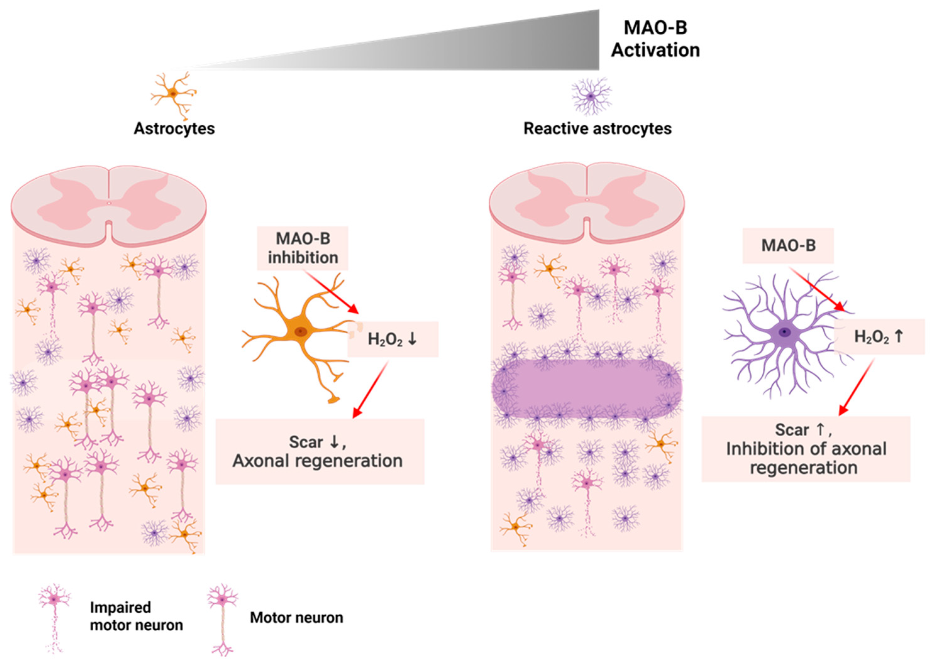
  90. Chun, H.; Lim, J.; Park, K.D.; Lee, C.J. Inhibition of monoamine oxidase B prevents reactive astrogliosis and scar formation in stab wound injury model. Glia 2022, 70, 354–367. [Google Scholar] [CrossRef]
  91. Lee, J.; Kim, J.-G.; Hong, S.; Kim, Y.S.; Ahn, S.; Kim, R.; Chun, H.; Park, K.D.; Jeong, Y.; Kim, D.-E. Intravital imaging of cerebral microinfarct reveals an astrocyte reaction led to glial scar. bioRxiv 2021. [Google Scholar] [CrossRef]
  92. Cho, J.-H.; Tsai, M.-J. The role of BETA2/NeuroD1 in the development of the nervous system. Mol. Neurobiol. 2004, 30, 35–47. [Google Scholar] [CrossRef]
  93. Wang, Y.; Zhang, X.; Chen, F.; Song, N.; Xie, J. In vivo Direct Conversion of Astrocytes to Neurons Maybe a Potential Alternative Strategy for Neurodegenerative Diseases. Front. Aging Neurosci. 2021, 474, 689276. [Google Scholar] [CrossRef]
  94. Liang, X.G.; Tan, C.; Wang, C.K.; Tao, R.R.; Huang, Y.J.; Ma, K.F.; Fukunaga, K.; Huang, M.Z.; Han, F. Myt1l induced direct reprogramming of pericytes into cholinergic neurons. CNS Neurosci. Ther. 2018, 24, 801–809. [Google Scholar] [CrossRef]
  95. Caiazzo, M.; Dell’Anno, M.T.; Dvoretskova, E.; Lazarevic, D.; Taverna, S.; Leo, D.; Sotnikova, T.D.; Menegon, A.; Roncaglia, P.; Colciago, G. Direct generation of functional dopaminergic neurons from mouse and human fibroblasts. Nature 2011, 476, 224–227. [Google Scholar] [CrossRef]
  96. Thiebes, K.P.; Nam, H.; Cambronne, X.A.; Shen, R.; Glasgow, S.M.; Cho, H.-H.; Kwon, J.-S.; Goodman, R.H.; Lee, J.W.; Lee, S. miR-218 is essential to establish motor neuron fate as a downstream effector of Isl1–Lhx3. Nat. Commun. 2015, 6, 7718. [Google Scholar] [CrossRef]
  97. De Gregorio, R.; Pulcrano, S.; De Sanctis, C.; Volpicelli, F.; Guatteo, E.; Von Oerthel, L.; Latagliata, E.C.; Esposito, R.; Piscitelli, R.M.; Perrone-Capano, C. miR-34b/c regulates Wnt1 and enhances mesencephalic dopaminergic neuron differentiation. Stem Cell Rep. 2018, 10, 1237–1250. [Google Scholar] [CrossRef]
  98. Yang, J.; Brown, M.E.; Zhang, H.; Martinez, M.; Zhao, Z.; Bhutani, S.; Yin, S.; Trac, D.; Xi, J.J.; Davis, M.E. High-throughput screening identifies microRNAs that target Nox2 and improve function after acute myocardial infarction. Am. J. Physiol. Heart Circ. Physiol. 2017, 312, H1002–H1012. [Google Scholar] [CrossRef]
  99. Sun, D. The potential of endogenous neurogenesis for brain repair and regeneration following traumatic brain injury. Neural Regen. Res. 2014, 9, 688. [Google Scholar] [CrossRef]
Figure 1. Direct conversion of astrocytes into induced neurons: (a) Depiction of in vivo astrocyte reprogramming into various neuron types, i.e., GABAergic, dopaminergic, or glutamatergic, achieved through the targeted expression of specific transcription factors; (b) Illustration showcasing the potential applications of in vivo direct neuronal reprogramming.
Nanomaterials 13 01680 g001
Figure 2. Efficient iDA neuron direct lineage reprogramming using AuNpRs: (a) Immunostaining analysis of various types of nanomaterials (gold nanorod, gold nanotube, gold nanoporous-rod) induced direct reprogrammed iDA neurons, immunofluorescent staining of induced neuron for neural markers, TH and TUBB3 (Scale bar = 40 µm); Data from Lee et al. 2022, Acta Biomaterialia [29]. Copyright from {2022}Acta Biomaterialia. (b) the dot plots representing the relative expression levels of control fibroblasts vs. APLN transduced fibroblasts (left) and APLN transduced fibroblast vs. APLN transduced fibroblast with AuNpR treating. Red dots, upregulated gene and blue dots, downregulated gene; Data from Lee et al. 2022, Acta Biomaterialia [29]. Copyright from {2022}Acta Biomaterialia.
Nanomaterials 13 01680 g002
Figure 3. Electro mechanism-based nanoparticle fabrication method: (a) The method of fabricating porous structure integrated gold nanorings; Data from Lee et al. 2022, Acta Biomaterialia [29]. Copyright from {2022}Acta Biomaterialia.; (b) the method to fabricate nanosphere hexamer; Data from Lee et al. 2022, Acta Biomaterialia [29]. Copyright from {2022}Acta Biomaterialia.
Nanomaterials 13 01680 g003
Figure 4. Non-viral nanoparticle-based gene delivery system: (a) The schematic illustration of non-viral nanoparticle-based iNs DLR; (b) cell survival ratio in accordance with using various type of positive charged nanoparticle coating materials (cysteamine, PEI, and Mutab); (c) transfection efficiency for using different type of nanoparticle coating materials.
Nanomaterials 13 01680 g004
Figure 5. Schematic illustration showing the consequence of spinal cord injury in neuron and astrocyte.
Nanomaterials 13 01680 g005
Figure 6. Schematic illustration showing the effect of motor neuron DLR on spinal cord injury.
Nanomaterials 13 01680 g006
Table 1. Table showing the various type of DLR accelerators.
SpeciesStarting CellsTarget CellsMaterialsEfficiencyRef.
Ms/HuFibroblastsNeuronsSmall molecules (CHIR, LDN, AA)~35%[22]
Ms/HuFibroblastsNeuronsmiRNA~40%[4]
Ms/huFibroblastsDopaminergic neuronsElectromagnetized
gold nanoparticles
~55%[5]
MsFibroblastsDopaminergic neuronsElectromagnetized
graphene nanosheet
~20%[31]
Ms/HuFibroblastsDopaminergic neuronsElongated nanoporous
gold nanorod
~40%[29]
HuFibroblastsNeuronsPolymer-functionalized Nanodot~40%[46]
Table 2. Table showing the AAV2-based ongoing and completed clinical trials.
No.NameApplicationPhaseStatusIdentifierRef.
1AAV-SMN1Muscular Atrophy, SpinalPhase 4Active, not recruiting, NCT05073133[60]
2AAV2-BDNF Alzheimer’s diseasePhase 1RecruitingNCT05040217[61]
3AAV2-GDNFParkinson’s diseasePhase 1RecruitingNCT04167540[62]
4AAV2-hRPE65v2Inherited retinal dystrophy-Active, not recruitingNCT03602820[58,63]
5AAV2/5-RPGRX-linked retinitis pigmentosaPhase 1/2CompletedNCT03252847[69]
6rAAV2.REP1ChoroideremiaPhase 2CompletedNCT02671539[58]
7AAV2-REP1ChoroideremiaPhase 2CompletedNCT02553135[64]
8AAV2-hAQP1Squamous cell head and neck cancer
Radiation induced xerostomia
Salivary hypofunction
Phase 1RecruitingNCT02446249[65]
9AAV2-hCHMChoroideremiaPhase 1/2Active, not recruitingNCT02341807[66]
10rAAV2.REP1ChoroideremiaPhase 1/2CompletedNCT02077361[58]
11AAV2-GDNFParkinson’s diseasePhase 1CompletedNCT01621581[62]
12AAV2-sFLT01Macular degenerationPhase 1CompletedNCT01024998[67]
13AAV2-NTNParkinson’s diseasePhase 1CompletedNCT00252850[68]
Disclaimer/Publisher’s Note: The statements, opinions and data contained in all publications are solely those of the individual author(s) and contributor(s) and not of MDPI and/or the editor(s). MDPI and/or the editor(s) disclaim responsibility for any injury to people or property resulting from any ideas, methods, instructions or products referred to in the content.

