Communication

Competitive Effects of Oxidation and Quantum Confinement on Modulation of the Photophysical Properties of Metallic-Phase Tungsten Dichalcogenide Quantum Dots

1
Division of Advanced Materials Engineering, Center for Advanced Powder Materials and Parts, Kongju National University, Cheonan 32588, Republic of Korea
2
Department of Advanced Materials Engineering, Chungbuk National University, Chungdae-ro 1, Seowon-gu, Cheongju 34057, Republic of Korea
*
Authors to whom correspondence should be addressed.
These authors contributed equally to this work.
Nanomaterials 2023, 13(14), 2075; https://doi.org/10.3390/nano13142075
Submission received: 13 June 2023 / Revised: 7 July 2023 / Accepted: 11 July 2023 / Published: 15 July 2023

Abstract

:
Metallic-phase transition metal dichalcogenide quantum dots (TMDs-mQDs) have been reported in recent years. However, a dominant mechanism for modulating their intrinsic exciton behaviors has not been determined yet as their size is close to the Bohr radius. Herein, we demonstrate that the oxidation effect prevails over quantum confinement on metallic-phase tungsten dichalcogenide QDs (WX2-mQDs; X = S, Se) when the QD size becomes larger than the exciton Bohr radius. WX2-mQDs with a diameter of ~12 nm show an obvious change in their photophysical properties when the pH of the solution changes from 2 to 11 compared to changing the size from ~3 nm. Meanwhile, we found that quantum confinement is the dominant function for the optical spectroscopic results in the WX2-mQDs with a size of ~3 nm. This is because the oxidation of the larger WX2-mQDs induces sub-energy states, thus enabling excitons to migrate into the lower defect energy states, whereas in WX2-mQDs with a size comparable to the exciton Bohr radius, protonation enhances the quantum confinement.

1. Introduction

Two-dimensional layered transition metal dichalcogenides (TMDs) are known to have two different phases, semiconducting and metallic (1T/1T’), which induce miscellaneous changes in their electronic and optical properties [1,2,3]. Unlike semiconducting TMDs, metallic-phase (1T/1T’) TMDs and their QDs show a feature of high charge transfer efficiency for photocatalytic and electrocatalytic abilities [4,5,6]. Recently, two sizes of metallic-phase TMDs-QDs (TMDs-mQDs) were demonstrated to have the photophysical properties of different charge excitation and decay pathways, which was explained by defect states and valance band splitting [3]. The variation in the optical energy structure in QDs is generally ascribed to quantum confinement and edge functionality [7,8,9]. However, the dominant mechanism that intrinsically controls exciton behaviors as the size and environmental conditions change remains elusive, although this issue is important for utilizing the photophysical properties in practical applications.
Modulating the optical properties of semiconducting TMDs-QDs has been conducted by using size control [3,4,10,11,12], environmental solvents [13,14,15], surface/edge functionalization [16], defect control [17], heterostructures [18], and structural phase control [5]. These modulation techniques have found applications across various fields [19,20]. Their photophysical properties are known to be affected by quantum confinement, surface defects, pH-sensitive edge-functional groups, and structural phases. The semiconducting WS2-QDs with a size of ~2 nm, where a 2–4 nm size is comparable to the exciton Bohr exciton radius [21,22], showed strong emission which was ascribed to quantum confinement [23]. From the excitation-wavelength-dependent PL of WS2-QDs with diameters of 1.5–3.5 nm, the localized states were suggested to be the origin of PL [4]. The high quantum yield (~5.5%) in the dimension- and phase-modulated WS2-QDs with a size of ~2.7 nm was attributed to the suppression of vibrational modes in WS2-QDs [24]. Despite these experimental results showing enhanced PL properties in the TMDs-QDs with a size comparable to the exciton Bohr radius, it is still vague whether quantum confinement is a dominant mechanism in TMDs-QDs with a size larger than the exciton Bohr radius and with an edge structure sensitively varied by pH change and/or functional groups. It was reported that MoSe2-QDs and MoS2-QDs have a pH-dependent PL [12,25], but the pH-independent PL property of WSe2-QDs was also demonstrated [26]. These contrasting results are controversial and have not been explained yet. As the 1T/1T’ metallic-phase structure of TMDs is thermodynamically metastable compared to the stable 2H semiconducting phase structure [6], the optical properties of the TMDs-mQDs remain to be investigated.
In this work, we investigate the difference in the photophysical properties of metallic phase-tungsten dichalcongens QDs (WX2-mQDs) of two sizes (diameters of ~3 nm and ~12 nm) dispersed in water with pH 2 and 11. We measure the change in the optical properties of WX2-mQDs (X = S and Se) depending on the changes in size and the pH of the solution using UV-Vis absorbance, PL, excitation-energy-dependent PL (PLE), and time-resolved PL (TRPL). Based on our comparison between the results, we discuss the primitive function on the photoelectric energy structure and exciton dynamics for WX2-mQDs with sizes varying from ~3 nm to ~12 nm.

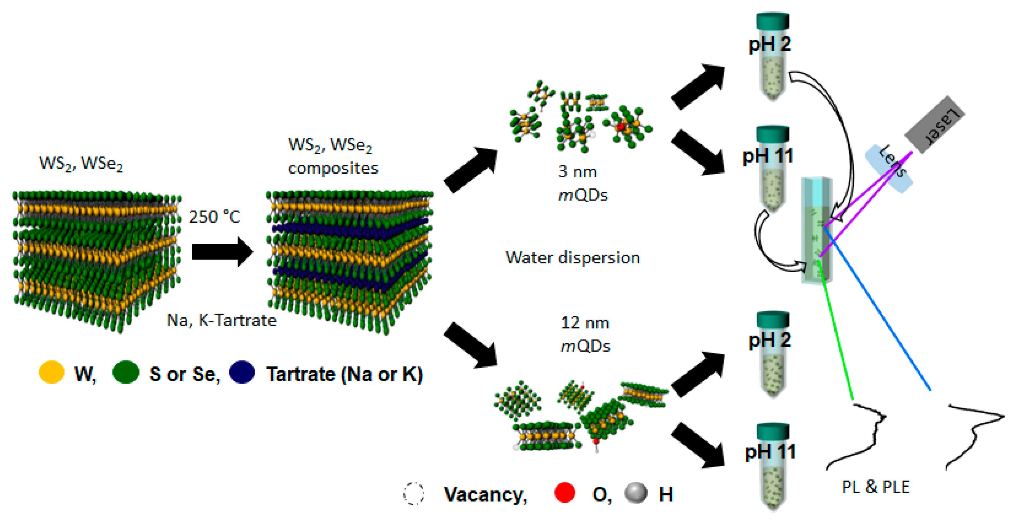
2. Materials and Methods

Potassium sodium tartrate was selected to synthesize WX2-mQDs at low temperature to minimize damage [3,27,28]. To fabricate the WX2-mQDs, we followed the previous process for GQDs and TMD-QDs in Refs. [5,22], and the details can be found there. Briefly, the initial steps started with the mixing and grinding of potassium sodium tartrate (200 mg) with WS2 (WSe2) (20 mg) purchased from Sigma Aldrich in Seoul, Republic of Korea. Then, the ground homogeneous mixtures were reacted in an autoclave vessel at 250 °C for 12 h, and then instantly exfoliated in water with sonication. The sizes of the WX2-mQDs were firstly controlled by AAO (20 nm, Whatman, Merch KGaA, Republic of Korea) filtration, and then dialyzed using dialysis tubing (10,000 and 8000 NMWL, Amicon Ultra-15, Merck KGaA, Seoul, Republic of Korea) to separate them into two sizes, which simultaneously removed the remaining salts. Finally, the WX2-mQDs were obtained as a dispersed solution in water. After drying, the WX2-mQDs were re-dispersed with an appropriately low concentration (<0.1 mg/mL) in 10 mL of water with pH 2 and 11.
The morphologies of the mQDs were analyzed using an AFM (X2-70, Park Systems Corp, Suwon, Republic of Korea) in tapping mode under ambient conditions at the Smart Nature Research Centre. XPS (Sigma Probe, AlKα), UV/Vis spectra (UV-3600, Shimadzu spectrometer, Smart Nature Research Centre, Cheonan, Republic of Korea), fluorescence spectra (Perkin-Elmer LS 55 luminescence spectrometer, PerkinElmer, MA, USA), and transmission electron microscopy (TEM, Titan cubed G2 60-300, FEI, OR, USA) analyses were conducted. PL measurements were carried out using a 325 nm He-Cd continuous-wave laser, a monochromatic light from a 300 W Xenon lamp, and UV spectrometers (Maya2000, Ocean Optics, Orlando, FL, USA) as a PL detector at room temperature.

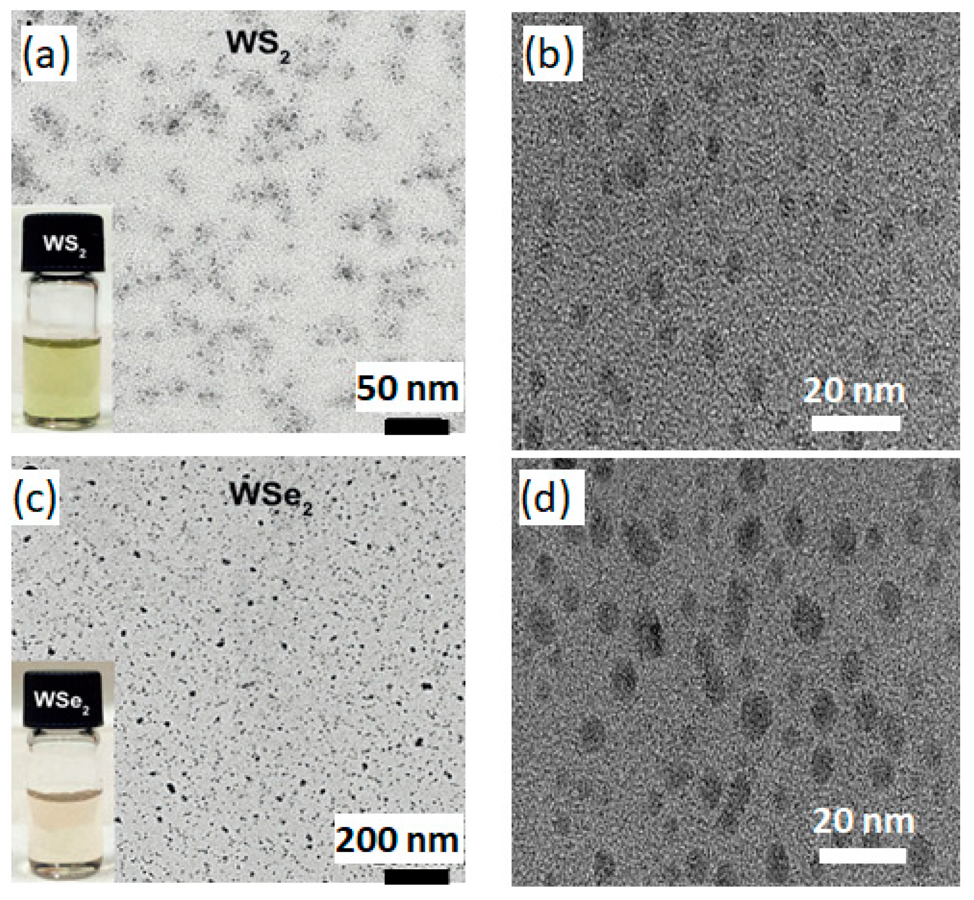
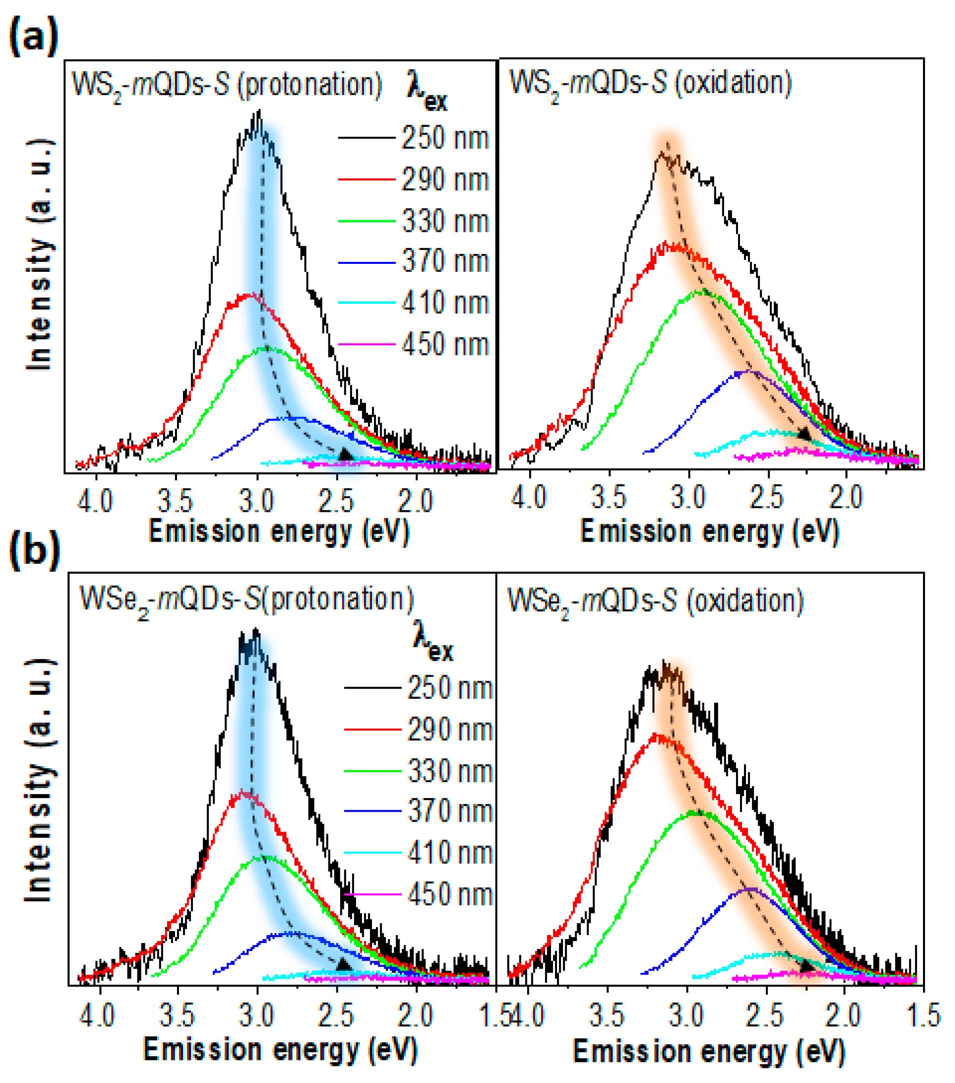
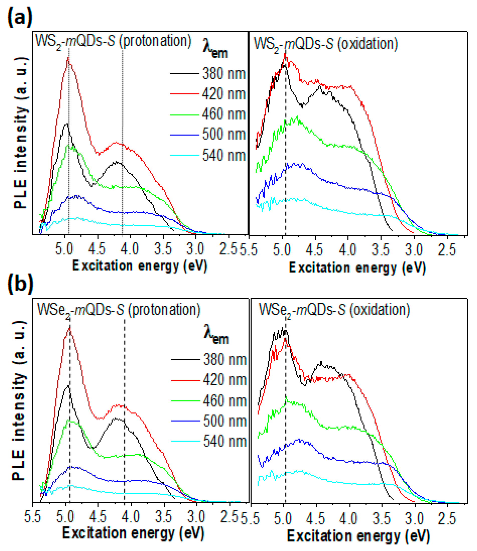
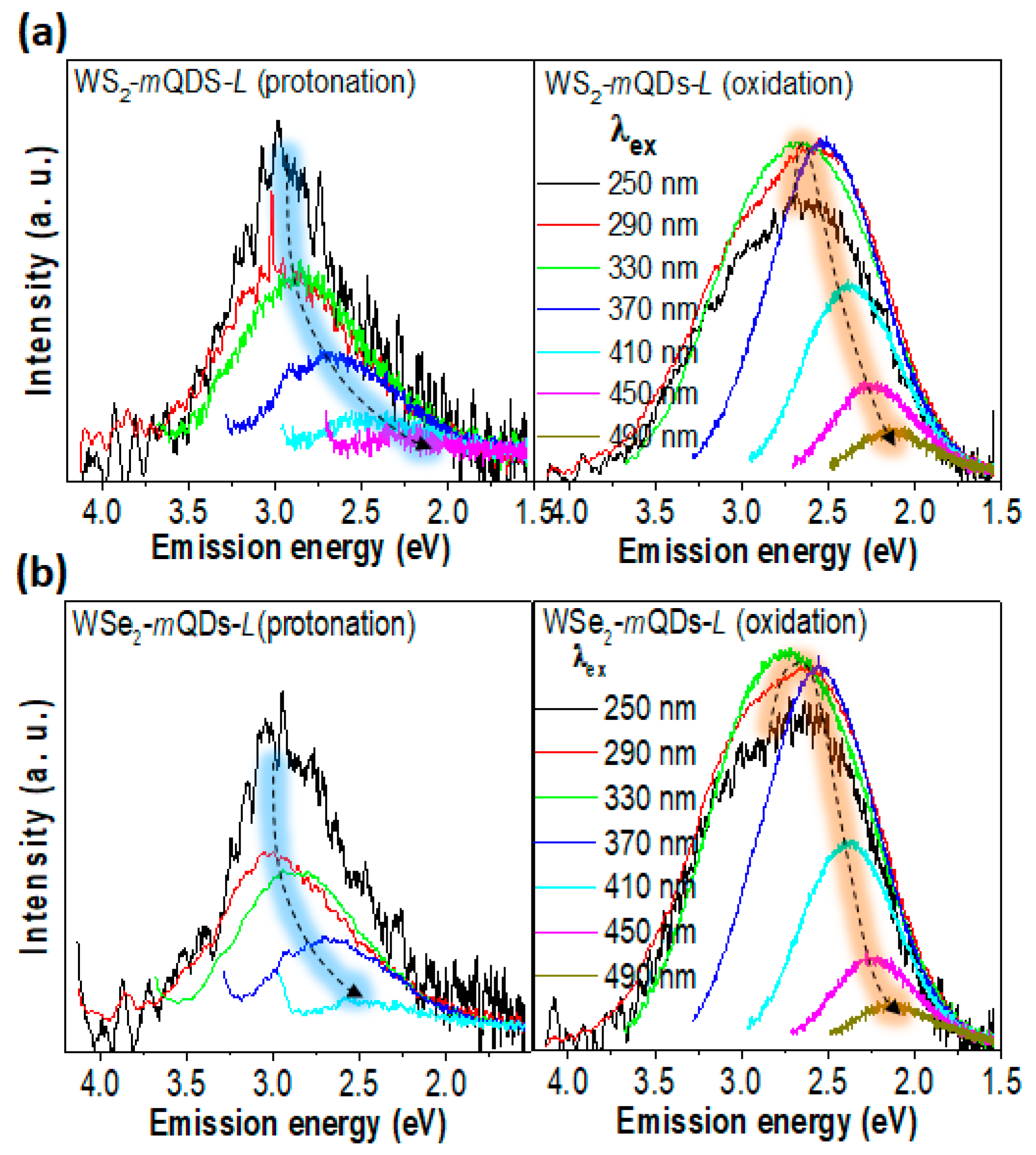
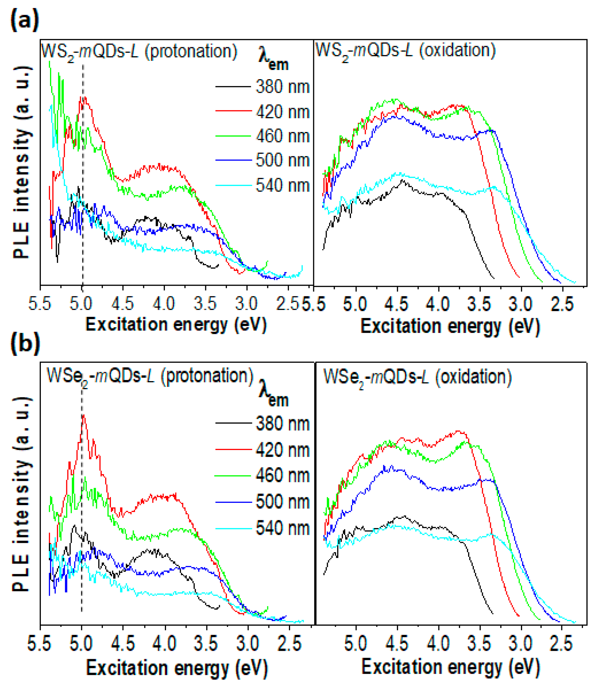
3. Results and Discussion

Figure 1 schematically shows the experimental process: the sample preparation, size separation, and measurement of the optical properties of WX2-mQDs. From the bulk WS2 (WSe2), the Na, K-tartrate intercalated composites were produced using a thermostatic oven and then radically dropped into the water with mild sonication to be broken into QDs with a certain size distribution. After the size separation via filtration and dialysis, the WX2-mQDs were re-dispersed in water with a pH of 2 and 11 for the measurement of their optical properties. More experimental details are provided in the Supporting Information. With the as-prepared WX2-mQDs, the structural properties of WX2-mQDs were examined at pH 7 and are referred to in the previous work [3]. Transmission electron microscopy (TEM) measurements were performed for the WX2-mQDs suspended in pH 7 using a lacy carbon TEM grid (Figure 2). The insets of Figure 2a,c are the digital images of dispersed WS2-mQDs and WSe2-mQDs, respectively. Figure 2b,d are TEM images of WS2-mQDs and WSe2-mQDs, respectively, with a lattice parameter. From TEM (Figure 2 and Ref. [3]) and AFM (Figure S1) analysis, the collected WX2-mQDs are revealed to not only have nano-sized structure, but also have two different sizes (WX2-mQDs-Ss with diameter of ~3 nm similar to the exciton Bohr radius; WX2-mQDs-Ls with diameter of ~12 nm, sufficiently larger than the exciton Bohr radius). The XPS spectra of the tungsten core level (W4f) (Figure S2) show four separated peaks, where the former two peaks at 32 and 34.2 eV correspond to the characteristic peaks of oxygen-free W4f and the latter at 35.2 and 37.5 eV originate from oxidized W4f [29]. The binding energies of the oxygen-free W4f peaks are ~1 eV lower than those from 2H WS2 and WSe2, indicating that the as-prepared WX2-mQDs have metallic phases (1T/1T’) even though the semiconducting phase cannot be totally excluded. The edges of the WX2-mQDs are modified by the hydroxide (OH) and/or trivial tartrate (C4H4O62−) groups during the exfoliation and fracturing process. The WX2-mQDs are observed to be negatively charged in the neutral pH by zeta potential (Figure S4). During the protonation or further oxidation, there is little possibility for further edge-functionalization except for H+ or OH because only NaOH or HCl solutions were used. The WX2-mQDs were well dispersed in the water with pH of 2 and 11 for hours and they were used in the further experiments without any treatment. However, the aggregated WX2-mQDs were also easily re-dispersed by mechanical and ultrasonic agitation even after days.
Figure 3 shows the UV-Vis absorbance spectra of WS2-mQDs-S (a), WS2-mQDs-L (b), WSe2-mQDs-S (c), and WSe2-mQDs-L (d), respectively. The as-prepared TMD-QDs clearly show monotonically decreasing UV-Vis absorbance spectra, indicating the metallic properties of the QDs [3]. In the protonated WX2-mQDs-Ss (black in Figure 3a,c), a new absorption peak clearly appeared at ~4.2 eV (H-transition). This is in contrast to the quasi-continuously increasing absorbance spectra of the oxidized WX2-mQDs-Ss (Figure 3a,c) and WX2-mQDs-Ls (Figure 3b,d).
The H-transition band is much higher than the optical transition bands (~1.9 and ~2.3 eV) between the conduction band and the valance band as shown in the insets of Figure 3a,c [30,31]. Interestingly, the optical transition peak at ~2.1 eV in the protonated WX2-mQDs-Ss is not observed in the WX2-mQDs-Ls (inset Figure 3b,d). It should also be noted that the H-transition is a feature of only protonated WX2-mQDs-Ss. The continuously increasing absorbance in the oxidized WX2-mQDs-Ss and WX2-mQDs-Ls was attributed to the overlap of high-energy excitonic absorptions and strong electron–phonon coupling [22]. However, we postulate in this work that the H-transition originates from the quantum confinement enhanced by the protonation of WX2-mQDs-Ss because the protonation can suppress the edge effect of other functional groups and it supports the formation of highly localized excitonic states [32]. On the other hand, the oxidation induces the sub-energy states around the band edges of the WX2-mQDs-Ss and WX2-mQDs-Ls, resulting in a continuously increasing absorbance [15,33]. The effective band gap (Eeff) in the QDs can be derived by Equation [11]:
E e f f = E g + h 2 8 m r r 2 1.8 e 2 4 ε 0 ε r ,
where Eg is the band gap of bulk material, h is Plank’s constant, mr is the reduced mass of the exciton, r is the radius of the quantum dot, and ε0 (εr) is the permittivity of free space and the relative permittivity of the material. Based on the size distribution of the WX2-mQDs (Figure S1), the bandgap distribution of WX2-mQDs-Ss is 4~6 eV, whereas it is much lower than 2 eV in the WX2-mQDs-Ls. The observed H-transition is located within the bandgap of the WX2-mQDs-S. The weak band around 2 eV is due to the size distribution of the WX2-mQDs-S. The absence of a UV-Vis absorbance peak in the WX2-mQDs-Ls can be explained by the oxidation-induced sub-energy states and an Eeff much lower than 2 eV.
Figure 4, Figure 5 and Figure S4 show the comparison of PL spectroscopic results between the protonation and oxidation of WS2-mQDs-S and WSe2-mQDs-S. In Figure S4a, the protonated WS2-mQDs-S show a harmonic PL (hPL) peak at ~2.95 eV and the full width at half maximum (FWHM) of ~0.78 eV, while in the oxidized WS2-mQDs-S, the hPL spectrum redshifts as much as ~0.03 eV (1%) and broadens the FWHM by about 0.17 eV (21.8%). The WSe2-mQDs-S show a similar trend in the hPL spectra, with a deviation of less than 5% (Figure S4b). The excitation wavelength (λex)-dependent PL (PLE) spectra were measured to examine the change in the optical energy band structure in WX2-mQDs-Ss and WX2-mQDs-Ls according to the protonation and oxidation. Figure 4 shows the PL spectra of WS2-mQDs-S (a) and WSe2-mQDs-S (b), respectively, with a variation in λex from 250 nm to 450 nm. In the protonated WX2-mQDs-Ss, the PL peak at ~2.9 eV radically redshifts to ~2.4 eV when the λex is larger than 370 nm. The intensity sharply decreases from λex ~290 nm (~4.28 eV), which is around the H-transition band. On the other hand, the oxidized WX2-mQDs-Ss show a gradual shift of the PL peak from ~2.9 eV at λex ~250 nm to ~2.25 eV as the λex increases up to 450 nm. Additionally, the spectra have a shoulder on the lower emission energy (~2.5 eV) with a broad FWHM. This indicates that although in the protonated WX2-mQDs-Ss most excitons efficiently occur with high excitation energy decay at ~2.9 eV as the λex is higher than 330 nm, in the oxidized WX2-mQDs-Ss the correlation between the emission energy and the λex becomes strong with the decreasing efficiency and another energy band for exciton decay.
Figure 5 shows the PL intensity of WS2-mQDs-S (a) and WSe2-mQDs-S (b), respectively, at a specific emission wavelength (λem) as a function of the excitation energy. In the PL intensity vs. excitation energy (Eex) of the WX2-mQDs-Ss (Figure 5), two effective Eex bands (~4.96 eV and ~4.2 eV) are observed. Among them, the Eex~4.2 eV looks to be correlated with the H-transition of the UV-Vis spectra. The other Eex~4.96 eV being higher, by as much as ~0.76 eV, than the Eex~4.2 eV is most likely due to the balance band splitting [3,4] and/or the conduction band spin splitting [34]. This trend is similarly observed in the oxidized WX2-mQDs-S. However, the two Eex bands are more broadened and the boundary between the two bands becomes vague. Moreover, the relative intensity at the lower Eex band increases compared to that in the oxidized WX2-mQDs-Ss. This suggests that the oxidation of the WX2-mQDs-Ss considerably affects the exciton behaviors even though it does not overwhelm the quantum confinement. The measured PL properties are listed in Table 1.
Figure 6, Figure 7 and Figure S5 show the PL spectroscopic results from the larger quantum dots, WS2-mQDs-L and WSe2-mQDs-L. The hPL peak and FWHM of protonated WS2-mQDs-L are ~2.83 eV and ~0.89 eV, respectively, which are ~0.12 eV (4%) redshifted and ~0.11 eV (14%) broadened compared with those from the protonated WS2-mQDs-S (Figure S5a). This can be attributed to the size effect. On the other hand, the oxidized WS2-mQDs-L show a more redshifted peak (~2.63 eV) and a broadened FWHM (~1.06 eV). WSe2-mQDs-L shows a similar trend in the hPL spectrum to the WS2-mQDs-L (Figure S5b) (see details in Table 1). Consequently, it is clear that the WX2-mQDs-Ls show redshifts (0.12–0.14 eV) in the peak and broadness (0.09–0.11 eV) of the FWHM as the size increases from ~3 nm to ~12 nm. However, the variance in the hPL peak and the FWHM due to the protonation/oxidation in the WX2-mQDs-Ls is 0.16–0.2 eV (5.6–7.1%) and 0.17–0.21 eV (17–25%), respectively.
This indicates that the pH variation causes more change in the hPL spectrum of the WX2-mQDs-Ls compared to the size increment. From the PLE spectra (Figure 6), the peak in the protonated WX2-mQDs-Ls appears at ~2.9 eV until λex~330 nm and then redshifts as the λex increases, which is similar to that of the WX2-mQDs-Ss. This implies that the intrinsic optical band of the WX2-mQDs is located at ~2.9 eV, demonstrating a large Stokes shift due to the strong exciton binding energy [35]. However, when the WX2-mQDs-Ls are oxidized, the highest emission appears at ~2.5 eV and the emission peak at ~2.9 eV becomes a shoulder in the spectrum. This is in an inverse shape to that shown in the WX2-mQDs-Ss (Figure 4) and the protonated WX2-mQDs-Ls. Moreover, the peak at ~2.9 eV is obviously suppressed when λex ≥ 370 nm. Interestingly, another dramatic change is observed in the PL intensity vs. Eex (Figure 7). In the protonated WX2-mQDs-Ls, the PL intensity varying with a maximum intensity at λex~4.9 eV is similar to that in the WX2-mQDs-Ss as the Eex changes from 5.5 eV to 2.5 eV. On the other hand, when oxidized, the PL intensity becomes flat with curves and the maximum intensity appears at a lower Eex than 4 eV. This indicates that in the oxidized WX2-mQDs-Ls the excited exciton states are spread and the oxidation more critically affects the optical energy band structure of the WX2-mQDs. The origin of the PLE peak shift is generally due to the size distribution of the mQDs or defects [3,4,36]. However, our above hypothesis is that the size increment and oxidation lead to the occurrence of sub-energy states between the conduction band and valance band. The formation of sub-energy states leads to the migration of excited charges into the lower energy states and helps the recombination of excitons. This is indirectly supported by the more redshifted and broadened PL, smoothly decreasing PL intensity, and flat PLE intensity spectra. In addition, the higher quantum yield in the oxidized WX2-mQDs-Ss (4.9 for WSe2 and 4.1 for WSe2) than that (2.8 and 3.3) of the protonated ones directly proves our hypothesis (Table 1).
Figure 8 shows the normalized PL intensity and PL center as a function of measurement time in the protonated and oxidized WX2-mQDs-Ss (Figure 8a,c) and WX2-mQDs-Ls (Figure 8b,d). The total measurement time of the time-resolved PL (TRPL) was 42 ns with λex~266 nm (Figure S6). In the protonated WX2-mQDs-S, the PL intensity sharply decreases to less than 0.2 within 20 ns and the PL center shifts by as much ~0.1 eV from 3.0 eV, whereas the oxidized WX2-mQDs-Ss show a relatively smooth decreasing intensity with a larger center shift (0.3 eV). This suggests that there is more exciton transition into the lower energy states following emissive decay in the oxidized WX2-mQDs-Ss. The protonated WX2-mQDs-Ls show a similar variance in the TRPL spectra to the protonated WX2-mQDs-Ss with a ~0.1 eV lower PL center. However, in the oxidized WX2-mQDs-Ls the PL center starts to appear at ~2.65 eV and then shifts only ~0.1 eV within the measurement time. The PL intensity decreases slightly slower than that of the protonated WX2-mQDs-Ls. It should be noted that the average PL lifetimes of the oxidized WX2-mQDs-Ls are 3.53 ± 0.11 ns for WS2-mQDs-L and 3.42 ± 0.11 ns for WSe2-mQDs-L, which are longer than those (2.99 ± 0.21 ns and 3.16 ± 0.14 ns) of the protonated ones (Figure S7). However, chalcogen dependence is not observed. This indicates that the exciton intraband transition quickly occurs within the broad exciton band above the band edge in the oxidized WX2-mQDs-Ls.
So far, we have analyzed the UV-Vis, PL, PLE, and TRPL results measured on WX2-mQDs-Ss and WX2-mQDs-Ls according to protonation and oxidation. The solution with a pH of 2 can make the edge of WX2-mQDs protonated and even heal the chalcogen vacancy of the crystal structure. This might enhance the exciton localization by quantum confinement and suppress the occurrence of sub-energy states. Moreover, this protonation might lead to energy dissipation through the hydrogen bond interacting with the surrounding solvent molecules, e.g., H2O. On the other hand, oxidation-induced defect states appear around the top valance band and the lowest intrinsic conduction band. Figure 9 is schematically drawn for the optical energy band structures of the protonated WX2-mQDs-Ss and the oxidized WX2-mQDs-Ls. In the protonated WX2-mQDs-Ss, there are discrete energy states due to the enhanced quantum confinement that causes the localization of charges. On the other hand, the oxidized WX2-mQDs-Ls have a broad exciton band above the band edge resulting in exciton delocalization and there are also extra sub-energy states under the conducting band and over the valance band [4]. However, the simple increment in size might not be critical and rather have a negative effect on the quantum yield of WX2-mQDs. The decisive factor for quantum confinement is the exciton Bohr radius (R), which can be deduced by the following equation:
R = ε m 0 m r a 0
where ε is the dielectric constant of the material, m0 is the free electron mass, mr is the reduced mass of the exciton, and a0 ≈ 0.5 Å. Based on the parameters in previous reports [17,18], the R of WX2-mQDs is about 3~4 nm. This suggests that when the size of WX2-mQDs is comparable to the R, the quantum confinement more dominantly affects the PL properties than the environmental effect, whereas when the size >>R (for example, the WX2-mQDs-Ls) the protonation/oxidation plays an important role in the photophysical properties. However, there must be a transition point of the dominant mechanism between the quantum confinement and the environmental effect as the size increases, and this question still remains elusive.

4. Conclusions

We investigated the effect of protonation and oxidation on the photophysical properties of two different size WX2-mQDs. The protonated WX2-mQDs showed relatively sharp harmonic PL spectra with a high emission energy peak, two obvious PLE intensity peaks, short lifetimes, and low quantum yield. On the other hand, the oxidized ones showed relatively broad and lower harmonic PL energy peaks with longer lifetimes and higher QYs. In the WX2-mQDs-Ls, the variation in PL properties was dominantly affected by protonation/oxidation rather than the size effect. This demonstrates that in the WX2-mQDs with a larger size than the exciton Bohr radius, the photophysical properties are more effectively affected by the environmental conditions, whereas in the WX2-mQDs with a size comparable to the exciton Bohr radius, the quantum confinement is a dominant mechanism that explains the optical properties. This work illuminates the transition of the dominant mechanism modulating the exciton dynamics in WX2-mQDs and it is expected to be an important guide for the practical application of TMDs-mQDs. However, we still need to investigate the exact size at which the transition from quantum confinement to the environmental effect occurs.

Supplementary Materials

The following supporting information can be downloaded at https://www.mdpi.com/article/10.3390/nano13142075/s1, Histogram of WX2-mQDs size distribution gathered from AFM images in Ref. [3] of main text. (Figure S1); HRTEM images of WX2-mQDs (Figure S2); XPS spectra of WX2-mQDs (Figure S3); Zeta potential of WX2-mQDs (Figure S4); TRPL of WX2-mQDs (Figure S5); Lifetime of WX2-mQDs (Figure S6) (PDF); Lifetime of WX2-mQDs (Figure S7). References [3,27,28] are cited in the Supplementary Materials.

Author Contributions

The manuscript was written with contributions from all authors. The experiments were conceptualized by K.H.P., S.H.S., and D.L. and the conduction of experiments and organization of data were performed by B.-H.K. and J.Y.Y. All authors contributed to the data analysis and discussion of the results. All authors have read and agreed to the published version of the manuscript.

Funding

This research was supported by the Basic Science Research Program through the National Research Foundation of Korea funded by the Ministry of Education (NRF-2019R1A6A1A03032988). This research was also supported by the Basic Science Research Program through the National Research Foundation of Korea (NRF) founded by the Ministry of Education (NRF-2020R1I1A3071628) and by the Korea Institute for Advancement of Technology (KIAT) and the Ministry of Trade, Industry and Energy (P0018009). This work was further supported by a research grant from Kongju National University in 2022.

Data Availability Statement

Not applicable.

Conflicts of Interest

The authors declare no conflict of interest.

References

  1. Voiry, D.; Mohite, A.; Chhowalla, M. Phase engineering of transition metal dichalcogenides. Chem. Soc. Rev. 2015, 44, 2702–2712. [Google Scholar] [PubMed]
  2. Manzeli, S.; Ovchinnikov, D.; Pasquier, D.; Yazyev, O.V.; Kis, A. 2D transition metal dichalcogenides. Nat. Rev. Mater. 2017, 2, 17033. [Google Scholar]
  3. Kim, B.-H.; Jang, M.-H.; Yoon, H.; Kim, H.J.; Cho, Y.-H.; Jeon, S.; Song, S.-H. Metallic phase transition metal dichalcogenide quantum dots showing different optical charge excitation and decay pathways. NPG Asia Mater. 2021, 13, 41. [Google Scholar]
  4. Caigas, S.P.; Santiago, S.R.M.; Lin, T.-N.; Lin, C.-A.J.; Yuan, C.-T.; Shen, J.-L.; Lin, T.-Y. Origins of excitation-wavelength-dependent photoluminescence in WS2 quantum dots. Appl. Phys. Lett. 2018, 112, 092106. [Google Scholar]
  5. Tan, C.; Luo, Z.; Chaturvedi, A.; Cai, Y.; Du, Y.; Gong, Y.; Huang, Y.; Lai, Z.; Zhang, X.; Zheng, L. Preparation of high-percentage 1T-phase transition metal dichalcogenide nanodots for electrochemical hydrogen evolution. Adv. Mater. 2018, 30, 1705509. [Google Scholar]
  6. Sokolikova, M.S.; Sherrell, P.C.; Palczynski, P.; Bemmer, V.L.; Mattevi, C. Direct solution-phase synthesis of 1T’WSe2 nanosheets. Nat. Commun. 2019, 10, 712. [Google Scholar]
  7. Srivastava, R.R.; Mishra, H.; Singh, V.K.; Vikram, K.; Srivastava, R.K.; Srivastava, S.; Srivastava, A. pH dependent luminescence switching of tin disulfide quantum dots. J. Lumin. 2019, 213, 401–408. [Google Scholar]
  8. Takagahara, T.; Takeda, K. Theory of the quantum confinement effect on excitons in quantum dots of indirect-gap materials. Phys. Rev. B 1992, 46, 15578. [Google Scholar]
  9. Jin, S.H.; Kim, D.H.; Jun, G.H.; Hong, S.H.; Jeon, S. Tuning the photoluminescence of graphene quantum dots through the charge transfer effect of functional groups. ACS Nano 2013, 7, 1239–1245. [Google Scholar]
  10. Jin, H.; Ahn, M.; Jeong, S.; Han, J.H.; Yoo, D.; Son, D.H.; Cheon, J. Colloidal single-layer quantum dots with lateral confinement effects on 2D exciton. J. Am. Chem. Soc. 2016, 138, 13253–13259. [Google Scholar]
  11. Gan, Z.; Liu, L.; Wu, H.; Hao, Y.; Shan, Y.; Wu, X.; Chu, P.K. Quantum confinement effects across two-dimensional planes in MoS2 quantum dots. Appl. Phys. Lett. 2015, 106, 233113. [Google Scholar]
  12. Farzin, M.A.; Abdoos, H. A critical review on quantum dots: From synthesis toward applications in electrochemical biosensors for determination of disease-related biomolecules. Talanta 2021, 224, 121828. [Google Scholar] [PubMed]
  13. Azam, N.; Najabat Ali, M.; Javaid Khan, T. Carbon quantum dots for biomedical applications: Review and analysis. Front. Mater. 2021, 8, 700403. [Google Scholar]
  14. Mishra, H.; Umrao, S.; Singh, J.; Srivastava, R.K.; Ali, R.; Misra, A.; Srivastava, A. pH dependent optical switching and fluorescence modulation of molybdenum sulfide quantum dots. Adv. Opt. Mater. 2017, 5, 1601021. [Google Scholar]
  15. Thomas, A.; Jinesh, K.B. Excitons and Trions in MoS2 Quantum Dots: The Influence of the Dispersing Medium. ACS Omega 2022, 7, 6531–6538. [Google Scholar] [CrossRef]
  16. Loh, G.; Pandey, R.; Yap, Y.K.; Karna, S.P. MoS2 quantum dot: Effects of passivation, additional layer, and h-BN substrate on its stability and electronic properties. J. Phys. Chem. C 2015, 119, 1565–1574. [Google Scholar]
  17. Ding, X.; Peng, F.; Zhou, J.; Gong, W.; Slaven, G.; Loh, K.P.; Lim, C.T.; Leong, D.T. Defect engineered bioactive transition metals dichalcogenides quantum dots. Nat. Commun. 2019, 10, 41. [Google Scholar]
  18. Ren, K.; Sun, M.; Luo, Y.; Wang, S.; Yu, J.; Tang, W. First-principle study of electronic and optical properties of two-dimensional materials-based heterostructures based on transition metal dichalcogenides and boron phosphide. Appl. Surf. Sci. 2019, 476, 70–75. [Google Scholar]
  19. Vitukhnovsky, A.; Zvagelsky, R.; Kolymagin, D.; Pisarenko, A.; Chubich, D. Three-Dimensional Optical Lithography and Nanoscale Optical Connectors. Bull. Russ. Acad. Sci. Phys. 2020, 84, 760–765. [Google Scholar]
  20. Arzhanov, A.; Savostianov, A.; Magaryan, K.; Karimullin, K.; Naumov, A. Photonics of semiconductor quantum dots: Applied aspects. Photonics Russ 2022, 16, 96–112. [Google Scholar]
  21. Chang, J.; Register, L.F.; Banerjee, S.K. Ballistic performance comparison of monolayer transition metal dichalcogenide MX2 (M = Mo, W; X = S, Se, Te) metal-oxide-semiconductor field effect transistors. J. Appl. Phys. 2014, 115, 084506. [Google Scholar] [CrossRef] [Green Version]
  22. Sharma, S.; Bhagat, S.; Singh, J.; Singh, R.C.; Sharma, S. Excitation-dependent photoluminescence from WS2 nanostructures synthesized via top-down approach. J. Mater. Sci. 2017, 52, 11326–11336. [Google Scholar] [CrossRef]
  23. Ghorai, A.; Bayan, S.; Gogurla, N.; Midya, A.; Ray, S.K. Highly luminescent WS2 quantum dots/ZnO heterojunctions for light emitting devices. ACS Aappl. Mater. Interfaces 2017, 9, 558–565. [Google Scholar] [CrossRef]
  24. Kim, M.-J.; Jeon, S.-J.; Kang, T.W.; Ju, J.-M.; Yim, D.; Kim, H.-I.; Park, J.H.; Kim, J.-H. 2H-WS2 Quantum dots produced by modulating the dimension and phase of 1T-nanosheets for antibody-free optical sensing of neurotransmitters. ACS Appl. Mater. Interfaces 2017, 9, 12316–12323. [Google Scholar] [CrossRef]
  25. Ko, B.; Ahn, J.; Song, S.H. pH-Dependent Photophysical Properties of Metallic Phase MoSe2 Quantum Dots. Materials 2022, 15, 4945. [Google Scholar]
  26. Yang, H.; Zhao, Q.; Wang, X.; Wu, Y.; Su, Y.; Wei, W. Facile and highly selective sensing of hypochlorous acid in aqueous solution and living cells by using as-prepared WSe2 quantum dots. Sens. Actuators B Chem. 2021, 337, 129782. [Google Scholar] [CrossRef]
  27. Song, S.H.; Kim, B.H.; Choe, D.H.; Kim, J.; Kim, D.C.; Lee, D.J.; Kim, J.M.; Chang, K.J.; Jeon, S. Bandgap widening of phase quilted, 2D MoS2 by oxidative intercalation. Adv. Mater. 2015, 27, 3152–3158. [Google Scholar] [CrossRef]
  28. Park, K.H.; Yang, J.Y.; Jung, S.; Ko, B.M.; Song, G.; Hong, S.-J.; Kim, N.C.; Lee, D.; Song, S.H. Metallic phase transition metal dichalcogenide quantum dots as promising bio-imaging materials. Nanomaterials 2022, 12, 1645. [Google Scholar]
  29. Zhang, K.; Fu, L.; Zhang, W.; Pan, H.; Sun, Y.; Ge, C.; Du, Y.; Tang, N. Ultrasmall and monolayered tungsten dichalcogenide quantum dots with giant spin–valley coupling and purple luminescence. ACS Omega 2018, 3, 12188–12194. [Google Scholar] [CrossRef] [Green Version]
  30. Zhu, B.; Chen, X.; Cui, X. Exciton binding energy of monolayer WS2. Sci. Rep. 2015, 5, 9218. [Google Scholar] [CrossRef] [Green Version]
  31. Zhao, W.; Ghorannevis, Z.; Amara, K.K.; Pang, J.R.; Toh, M.; Zhang, X.; Kloc, C.; Tan, P.H.; Eda, G. Lattice dynamics in mono-and few-layer sheets of WS2 and WSe2. Nanoscale 2013, 5, 9677–9683. [Google Scholar] [CrossRef] [PubMed] [Green Version]
  32. Lin, K.-Q.; Ong, C.S.; Bange, S.; Faria Junior, P.E.; Peng, B.; Ziegler, J.D.; Zipfel, J.; Bäuml, C.; Paradiso, N.; Watanabe, K. Narrow-band high-lying excitons with negative-mass electrons in monolayer WSe2. Nat. Commun. 2021, 12, 5500. [Google Scholar] [CrossRef] [PubMed]
  33. Zhu, H.; Zan, W.; Chen, W.; Jiang, W.; Ding, X.; Li, B.L.; Mu, Y.; Wang, L.; Garaj, S.; Leong, D.T. Defect-Rich Molybdenum Sulfide Quantum Dots for Amplified Photoluminescence and Photonics-Driven Reactive Oxygen Species Generation. Adv. Mater. 2022, 34, 2200004. [Google Scholar] [CrossRef] [PubMed]
  34. Kośmider, K.; González, J.W.; Fernández-Rossier, J. Large spin splitting in the conduction band of transition metal dichalcogenide monolayers. Phys. Rev. B 2013, 88, 245436. [Google Scholar] [CrossRef] [Green Version]
  35. Hanbicki, A.; Currie, M.; Kioseoglou, G.; Friedman, A.; Jonker, B. Measurement of high exciton binding energy in the monolayer transition-metal dichalcogenides WS2 and WSe2. Solid State Commun. 2015, 203, 16–20. [Google Scholar] [CrossRef] [Green Version]
  36. Lin, L.; Xu, Y.; Zhang, S.; Ross, I.; Ong, A.; Allwood, D. Fabrication of luminescent monolayered tungsten dichalcogenides quantum dots with giant spin-valley coupling. ACS Nano 2013, 7, 8214–8223. [Google Scholar] [CrossRef]
Figure 1. Schematically shown synthesis process of WS2-mQDs-S and WS2-mQDs-L.
Nanomaterials 13 02075 g001
Figure 2. TEM images of WS2-mQDs (a,b) and WSe2-mQDs (c,d).
Nanomaterials 13 02075 g002
Figure 3. UV-Vis absorbance of WX2-mQDs-Ss and WX2-mQDs-Ls. UV-Vis absorbance of WS2-mQDs-S (a), WS2-mQDs-L (b), WSe2-mQDs-S (c), and WSe2-mQDs-L (d). Insets: expansion of lower wavelength range.
Nanomaterials 13 02075 g003
Figure 4. PLE spectra of WX2-mQDs-Ss. PLE spectra of protonated (left) and oxidized (right) WS2-mQDs-S (a) and WSe2-mQDs-S (b) measured at λex from 250 nm to 450 nm.
Nanomaterials 13 02075 g004
Figure 5. PLE intensity vs. excitation energy of WS2-mQDs-S (a) and WSe2-mQDs-S (b) at emission wavelengths from 380 to 540 nm. The left panel is for protonation and the right is for oxidation.
Nanomaterials 13 02075 g005
Figure 6. PLE spectra of WX2-mQDs-Ls. PLE spectra of protonated (left) and oxidized (right) WS2-mQDs-S (a) and WSe2-mQDs-S (b) measured at λex from 250 nm to 450 nm.
Nanomaterials 13 02075 g006
Figure 7. PLE intensity vs. excitation energy of WS2-mQDs-L (a) and WSe2-mQDs-L (b) at emission wavelengths from 380 to 540 nm. The left panel is for protonation and the right is for oxidation.
Nanomaterials 13 02075 g007
Figure 8. Time-resolved PL intensity vs. PL peak center vs. measurement delay time. The long scaling normalized PL intensity and PL peak shift as a function of measurement time delay in WS2-mQD-S (a), WS2-mQD-L (b), WSe2-mQD-S (c), WSe2-mQD-L (d), respectively, with λex~266 nm. Each point has a PL accumulation time of 0–2, 2–6, 6–12, 12–20, 20–30, and 30–42 ns.
Nanomaterials 13 02075 g008
Figure 9. Schematically shown photoelectron dynamics in WX2-mQD-Ss and WX2-mQD-Ls.
Nanomaterials 13 02075 g009
Table 1. Photophysical parameters of WS2 (Se2)-mQDs.
MaterialspHPL Peak (eV)FWHM (eV)QY (%)MaterialspHPL Peak (eV)FWHM (eV)QY (%)
WS2-mQDs-S22.950.782.8WSe2-mQDs-S22.980.783.3
112.920.954.9112.930.974.1
WS2-mQDs-L22.830.89 Wse2-mQDs-L22.840.87
112.631.06 112.681.09
Disclaimer/Publisher’s Note: The statements, opinions and data contained in all publications are solely those of the individual author(s) and contributor(s) and not of MDPI and/or the editor(s). MDPI and/or the editor(s) disclaim responsibility for any injury to people or property resulting from any ideas, methods, instructions or products referred to in the content.

