Communication

Unveiling Oxygen K-Edge and Cobalt L-Edge Electron Energy Loss Spectra of Cobalt Hydroxide and Their Evolution under Electron Beam Irradiation

1
Korea Research Institute of Standard and Science, Daejeon 34113, Republic of Korea
2
Department of Nano Convergence Measurement, Korea University of Science and Technology, Daejeon 34113, Republic of Korea
*
Author to whom correspondence should be addressed.
Nanomaterials 2023, 13(20), 2767; https://doi.org/10.3390/nano13202767
Submission received: 1 September 2023 / Revised: 10 October 2023 / Accepted: 13 October 2023 / Published: 16 October 2023
(This article belongs to the Special Issue NANO KOREA 2023)

Abstract

:
Cobalt hydroxides, Co(OH)2, have attracted considerable attention due to their diverse applications in the fields of energy and the environment. However, probing the electronic structure of Co(OH)2 is challenging, mainly due to its sensitivity to electron beam irradiation. In this study, we report the unperturbed O K-edge and Co L-edge for Co(OH)2 by electron beam damage and investigate the electronic structure transformation of Co(OH)2 under electron beam irradiation, using low current electron energy loss spectroscopy. In particular, the O K-edge pre-peak at 530 eV, which is not found in the undamaged Co(OH)2, begins to appear with an increasing electron beam current. In addition, the Co L-edge peak shifts to a higher energy, close to Co3O4, indicating that the localized phase transition within Co(OH)2 leads to the formation of Co3O4.

1. Introduction

Transition metal hydroxides have attracted interest for various applications, such as batteries, water splitting, and nanomedicine [1,2,3]. Among transition metal hydroxides, cobalt hydroxide, Co(OH)2, has received considerable attention as an electrochemical catalyst for oxygen evolution reaction (OER), because of its electrocatalytic activity and efficient ion intercalation [4,5]. Co(OH)2 exists in two phases depending on its structure: α-Co(OH)2 and β-Co(OH)2 phase. The β-Co(OH)2 phase has a brucite-like arrangement, with hydroxyl ions packed hexagonally and Co(II) ions occupying the octahedral site. The α-Co(OH)2 phase, on the other hand, resembles a hydrotalcite-like structure, where positively charged Co(OH)2-x layers are present. These layers are accompanied by charge-stabilizing anions, such as NO3−, CO32−, Cl, etc., in the interlayer space [6]. The α-Co(OH)2 phase has better electrochemical activity than β-Co(OH)2 due to the expended interlayer spacing and intercalated anions [4,5].
Scanning transmission electron microscopy (STEM)–electron energy loss spectroscopy (EELS) measurements have become increasingly popular in atomic structure and chemical analysis as an effective method for characterizing materials in various fields, including for catalysis, batteries, and biotechnology [7,8,9]. However, one critical drawback of this STEM–EELS technique is electron beam-induced damage to the sample [10,11,12,13]. Electron beam-induced damages manifest through a range of effects, including atomic displacement, electron-beam sputtering, radiolysis, heating, and electrostatic charging [14]. These effects hinder the microscope’s ability to fully observe the original structural properties of materials.
Co(OH)2 is a material that is highly sensitive to electron beam irradiation. Numerous investigations using transmission electron microscopy have been conducted to explore the structural characteristics of Co(OH)2; however, the effect of electron beam irradiation has not been seriously considered. Only recently has a study demonstrated the impact of electron beam irradiation in inducing structural modifications from Co(OH)2 to Co3O4 [15]. However, there is still a lack of EELS results for Co(OH)2, and the reported EELS results vary [16,17], which may be attributed to experimental challenges due to the electron-sensitivity of Co(OH)2. It is worth noting that electron beam damage could be more severe in EELS experiments than in imaging because spectroscopy typically requires a longer dwell time than imaging. Therefore, great caution must be taken with electron beam-sensitive materials for EELS experiments.
In this study, we present the undamaged spectra of the O K-edge and Co L-edge for Co(OH)2, obtained through low-current EELS. In addition, we demonstrate the evolution of the electronic structure of Co(OH)2 under electron beam irradiation in TEM. The low-current EELS experiment (screen current below 30 pA) showed no pre-peak at 530 eV in O K-edge, an observation not reported prior. Previously reported O K-edge spectra for Co(OH)2 displayed a pre-peak at 530 eV, which may have been caused by electron beam damage. The electron beam currents exceeding 30 pA induced changes in the O K-edge and the Co L-edge in Co(OH)2. The Co L-edge underwent an energy shift to the Co3O4 position, which was accompanied by the emergence of the O K pre-edge at 530 eV, indicating a phase transition from Co(OH)2 to Co3O4 under electron beam irradiation.

2. Experimental Methods

2.1. Materials Synthesis

The α-Co(OH)2 and β-Co(OH)2 particles were prepared under ambient conditions following a previous study [18]. To synthesize α-Co(OH)2, a mixture of cobalt chloride hexahydrate (10 mM, CoCl2·6H2O, Sigma-Aldrich, St. Louis, MO, USA), sodium chloride (50 mM, NaCl, DUKSAN, Ansan, Korea), and hexamethylenetetramine (60 mM, C6H12N4, 99.0%, Sigma-Aldrich, St. Louis, MO, USA) was dissolved in 150 mL of a 1:9 blend of ethanol and deionized water. This solution was then heated in an oil bath at 90 ℃ for one hour, resulting in a suspension containing green particles. The suspended product was subsequently centrifuged at 3500 rpm for 15 min, followed by multiple washes using deionized water and anhydrous ethanol. The final green particles were dried completely at room temperature. The synthesis procedure for β-Co(OH)2 mirrored that of α-Co(OH)2, with the exception that sodium chloride (NaCl) was omitted from the initial solution. This modification resulted in the formation of pink powder of β-Co(OH)2. The Co3O4 was synthesized through the annealing of α-Co(OH)2 at 500 °C for a duration of 3 h under atmospheric conditions.

2.2. Electron Microscopy

Scanning transmission electron microscopy (STEM) experiments were conducted using a Thermo Fisher Scientific Spectra 300 operating at an accelerated voltage of 300 kV. EELS experiments were carried out using a Gatan Continuum equipped with a K3 direct electron detector. The electron dose rate for EELS was 4.77 × 10⁷ e/Å2∙s. The energy spread of the zero-loss peak (ZLP) was measured at ~1 eV full-width at half-maximum (FWHM). The dispersion was 0.18 eV/ch, and a convergence angle of 22.7 mrad and a collection angle of 64.2 mrad were used. Automatic ZLP alignment was enabled to correct the energy shift during the acquisition time in dual EELS mode. To minimize electron beam damage, EELS experiments were performed in the low-current condition; the screen current was in the range of 1–100 pA and the pixel time for acquisition was 0.1 s. The EELS area scans were performed at 50 × 50 pixels with a step size of 5 nm. The final spectrum was obtained by averaging 2500 spectra. For the sample, the cobalt hydroxides and cobalt oxide powders were effectively dispersed in ethanol and deposited on a lacey carbon grid.

3. Results and Discussion

Figure 1 illustrates the scanning transmission electron microscopy (STEM) images along with the selected area electron diffraction (SAED) patterns for α-Co(OH)2, β-Co(OH)2, and spinel Co3O4, respectively. In Figure 1a,b, the STEM images show hexagonal microplates of α-Co(OH)2 and β-Co(OH)2, possessing an average dimension of approximately 6 μm. However, the spinel Co3O4 hexagonal microplate, synthesized through the annealing of Co(OH)2, exhibits granular characteristics accompanied by multiple cracks, as evident in Figure 1c. The SAED patterns reveal that both α-Co(OH)2 and β-Co(OH)2 consist of a single crystalline phase, while Co3O4 displays a polycrystalline phase. The combined results from STEM and SAED confirm the successful high-quality synthesis of α-Co(OH)2, β-Co(OH)2, and spinel Co3O4, without the presence of any other phases.
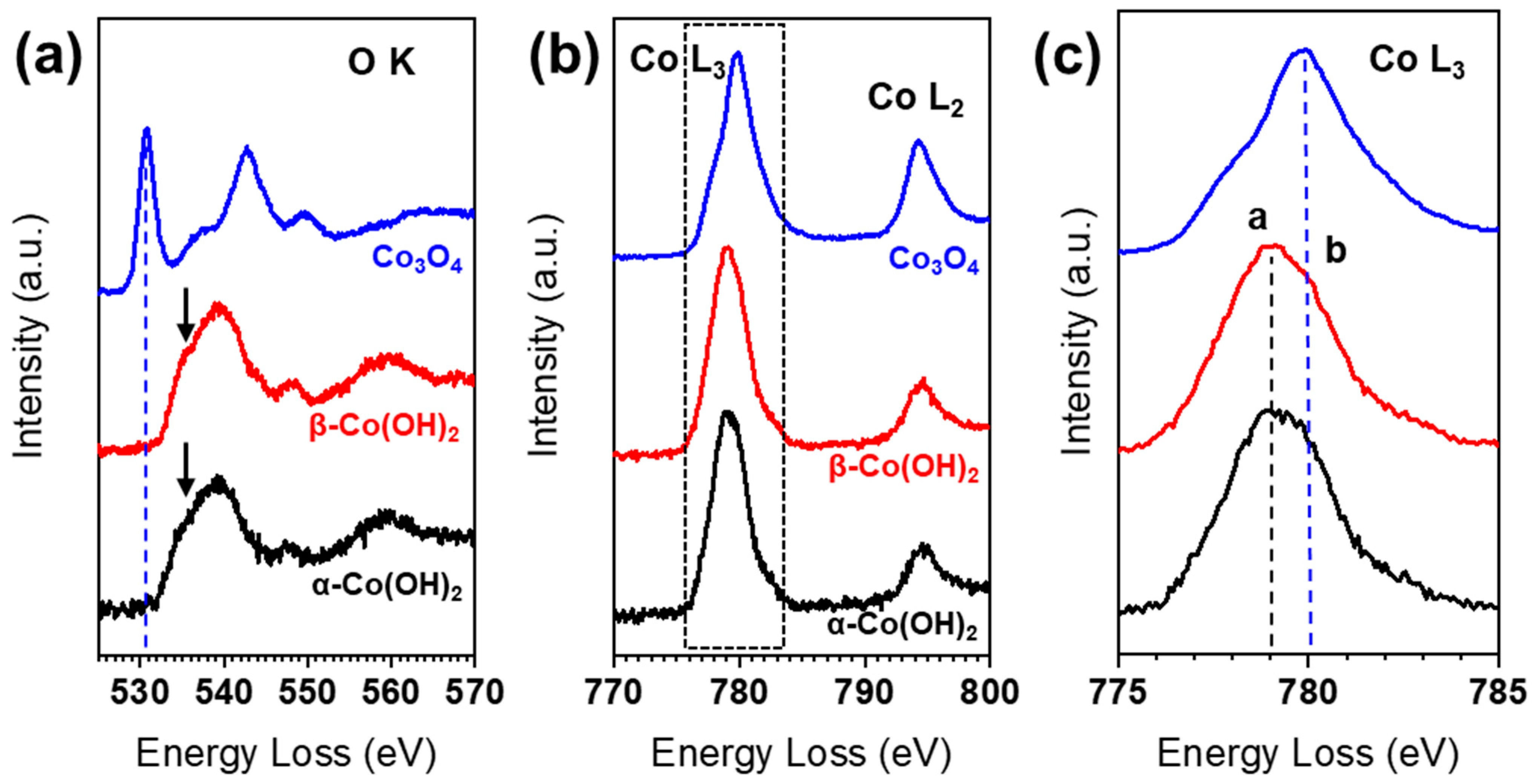
Figure 2 shows the O K-edge and Co L-edge spectra of α-Co(OH)2, β-Co(OH)2, and Co3O4, respectively. The spectra of α-Co(OH)2 and β-Co(OH)2 show distinct features at the O K- and Co L-edges, in contrast to the spectrum of Co3O4. However, the difference between the α- and β-Co(OH)2 spectra was minimal. For Co3O4, the pre-peak of the O K-edge is located at ~530 eV, while the pre-peak of the O K-edge for Co(OH)2 cannot be found, but only a small bump appears at ~534 eV. In the transition metal oxide, the O K-edge pre-peak contributes to the hybridization of O-2p with transition metal 3d states [19]. Previously, several EELS results have been reported for Co(OH)2 in which the O K-edge possesses a pre-peak close to 530 eV, as in the case of Co3O4, indicating the fact that the phase change occurred due to electron beam irradiation [15]. The main peak for the O K-edge is located at ~542 eV in Co3O4, while the main peak is shifted, towards lower energy, to 539 eV in Co(OH)2 marked by the arrows in Figure 2a. The Co L-edge consists of L3 and L2 white lines, which originate from the transitions of 2p3/2 to 3d3/23d5/2 and 2p1/2 to 3d3/2, respectively. These L3 and L2 white lines are affected by the valence states of the transition metal, due to their relationship to unoccupied states in the transition metal 3d band. Similar to the O K-edge, α-Co(OH)2 and β-Co(OH)2 show almost identical Co L-edges, but clearly different peak positions and shapes from Co3O4, as shown in Figure 2b,c. The peak of the Co L3-edge of Co(OH)2 is at ~779 eV (peak a), and the shoulder peak b is found at ~780 eV. Note that the shoulder peak b in Co(OH)2 is almost coincident with the Co L3-edge peak for Co3O4. The white line ratios (L3/L2) are calculated to be 4.5 for Co(OH)2 and 2.6 for Co3O4, in agreement with previously reported values [20].
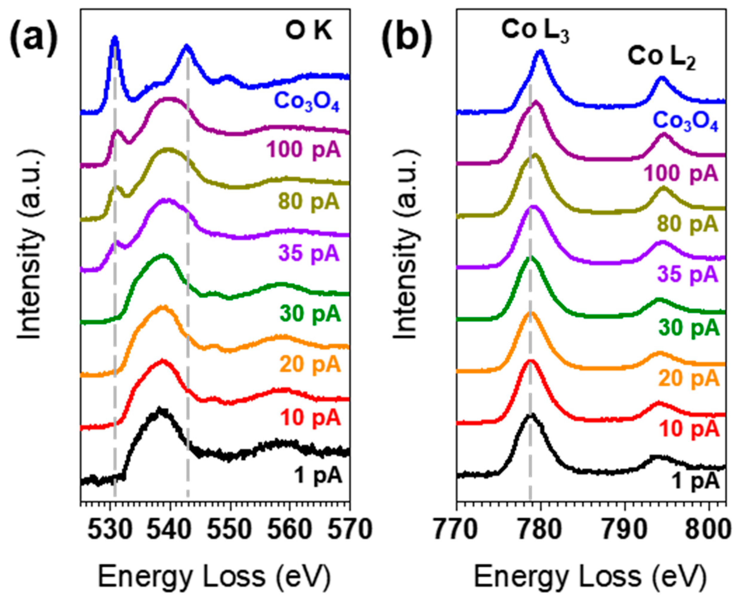
To investigate changes in electronic structure upon electron beam irradiation, we systematically measured the O K- and Co L-edges by varying the beam current. Figure 3 shows the evolution of the O K- and Co L-edges upon electron beam irradiation. At the electron beam current of 1 pA, the pre-peak of the O K-edge is not observed, and this absence persists up to 30 pA. However, at a current exceeding 35 pA, the pre-peak begins to appear at 530 eV. Furthermore, the main peak of the O K-edge at ~538 eV begins to show some spectral weight at 543 eV, which is consistent with the main peak of Co3O4. The Co L-edge shows a similar trend. The position of the L3 peak remains the same up to 30 pA. At currents above 35 pA, the L3 peak shifts towards higher energy, indicating a partial conversion of Co(OH)2 to Co3O4 under high-current electron beam irradiation. When the L3 peak shifts, the L3/L2 ratio also changes. The L3/L2 ratio value decreases from 4.6 at 1 pA to 3.3 at 100 pA, indicating that Co could be oxidized during the EELS experiment.
Finally, we examined the changes in the O K-edge during the phase transition in real time through multiple acquisitions. Figure 4 displays the alterations of the pre-peak in the O K-edge over 20 acquisitions. The scans were conducted within a fixed area of 50 × 50 pixels with a step size of 5 nm. The screen current was 20 pA and each spectrum was scanned for a total of 8 min. In the initial EELS acquisitions, Co(OH)2 did not exhibit a pre-peak in the O K-edge, as previously described. However, after the third acquisition, the O K-edge pre-peak, indicative of spinel Co3O4, began to appear at 530 eV, and its intensity increased with the number of acquisitions. This change suggests that the low-current electron beam also causes phase transition under persistent irradiation.
The phase transition of Co(OH)2 to Co3O4 is observed in the thermal decomposition process, following two steps [21]: (1) Co(OH)2 + 0.25O2 → 2CoOOH + 0.5H2O; (2) 3CoOOH → Co3O4 + 1.5H2O + 0.25O2. However, the phase transition induced by electron beam irradiation could follow a different reaction path, due to the different environment in the TEM chamber. The electron beam could easily kick off the hydrogen atoms in Co(OH)2, resulting in the formation of CoOOH phases, which had been found in an in situ electron diffraction experiment under electron beam irradiation [15]. However, in this study, the existence of CoOOH was not clearly shown in the O K-edge and Co L-edge EELS results, which could be attributed to the relatively fast phase transition from Co(OH)2 to CoOOH, compared to the subsequent phase transition to Co3O4. It is worth noting that the multiple EELS acquisitions in Figure 4 show continuous change of the O K-edge and the Co L-edge over the irradiation time, and no discernible change that could indicate a CoOOH phase is found during these multiple acquisitions. The CoOOH to Co3O4 phase transition is anticipated to follow a reaction similar to the thermal decomposition process, and the water and oxygen molecules could be extracted in the gas phases in TEM.

4. Conclusions

In this study, we reported the original electronic structure of Co(OH)2, and systematically investigated the changes in the electronic structure of Co(OH)2 under an electron beam by changing the electron beam current for the EELS experiment. Employing low-current EELS conditions (with an electron current below 30 pA) revealed the unperturbed O-K edge and Co-L edge spectra for Co(OH)2, thereby contrasting with previous EELS data. However, the electron beam currents exceeding 30 pA induced alterations in the O-K edge and Co-L edge for both α-Co(OH)2 and β-Co(OH)2. The O-K pre-edge at 530 eV, indicative of Co3O4, began to appear with an increasing electron beam current, accompanying the energy shift of the Co-L edge to the position for Co3O4. These observed EELS spectral changes indicate the facile transformation of Co(OH)2 into Co3O4 under electron beam irradiation. Our results emphasize the need for a cautious interpretation of results from EELS experiments and highlight the potential impact of electron beam influence on the electronic structure of materials.

Author Contributions

J.-H.K. conceived the project and designed the experiments; TEM and EELS experiments have been carried out by J.H.S.; data analysis has been done by J.-H.K. and J.H.S.; the manuscript has been written by J.-H.K. and J.H.S. All authors have read and agreed to the published version of the manuscript.

Funding

This work was supported by UST Young Scientist+ Research Program 2022 through the University of Science and Technology (2022YS31).

Data Availability Statement

The datasets used and/or analyzed during the current study are available from the corresponding author on reasonable request.

Acknowledgments

The authors acknowledge the gracious contribution of Co(OH)2 samples from the research group led by Ki Min Nam at Pusan National University.

Conflicts of Interest

The authors declare no conflict of interest.

References

  1. Liu, W.; Yin, R.; Shi, W.; Xu, X.; Shen, X.; Yin, Q.; Xu, L.; Cao, X. Gram-Scale Preparation of 2D Transition Metal Hydroxide/Oxide Assembled Structures for Oxygen Evolution and Zn-Air Battery. ACS Appl. Energy Mater. 2019, 2, 579–586. [Google Scholar] [CrossRef]
  2. Dionigi, F.; Zhu, J.; Zeng, Z.; Merzdorf, T.; Sarodnik, H.; Gliech, M.; Pan, L.; Li, W.; Greeley, J.; Strasser, P. Intrinsic Electrocatalytic Activity for Oxygen Evolution of Crystalline 3d-Transition Metal Layered Double Hydroxides. Angew. Chem. 2021, 133, 14567–14578. [Google Scholar] [CrossRef]
  3. Park, M.-C.; Kim, H.; Park, D.-H.; Yang, J.-H.; Choy, J.-H. Ketoprofen-LDH Nanohybrid for Transdermal Drug Delivery System. Bull. Korean Chem. Soc. 2012, 33, 1827–1828. [Google Scholar] [CrossRef]
  4. Huang, L.; Jiang, J.; Ai, L. Interlayer Expansion of Layered Cobalt Hydroxide Nanobelts to Highly Improve Oxygen Evolution Electrocatalysis. ACS Appl. Mater. Interfaces 2017, 9, 7059–7067. [Google Scholar] [CrossRef]
  5. Liu, P.F.; Yang, S.; Zheng, L.R.; Zhang, B.; Yang, H.G. Electrochemical Etching of α-Cobalt Hydroxide for Improvement of Oxygen Evolution Reaction. J. Mater. Chem. A 2016, 4, 9578–9584. [Google Scholar] [CrossRef]
  6. Hu, Z.-A.; Xie, Y.-L.; Wang, Y.-X.; Xie, L.-J.; Fu, G.-R.; Jin, X.-Q.; Zhang, Z.-Y.; Yang, Y.-Y.; Wu, H.-Y. Synthesis of α-Cobalt Hydroxides with Different Intercalated Anions and Effects of Intercalated Anions on Their Morphology, Basal Plane Spacing, and Capacitive Property. J. Phys. Chem. C 2009, 113, 12502–12508. [Google Scholar] [CrossRef]
  7. Morales, F.; Grandjean, D.; De Groot, F.M.F.; Stephan, O.; Weckhuysen, B.M. Combined EXAFS and STEM-EELS Study of the Electronic State and Location of Mn as Promoter in Co-Based Fischer–Tropsch Catalysts. Phys. Chem. Chem. Phys. 2005, 7, 568–572. [Google Scholar] [CrossRef]
  8. Muto, S.; Yamamoto, Y.; Sakakura, M.; Tian, H.-K.; Tateyama, Y.; Iriyama, Y. STEM-EELS Spectrum Imaging of an Aerosol-Deposited NASICON-Type LATP Solid Electrolyte and LCO Cathode Interface. ACS Appl. Energy Mater. 2022, 5, 98–107. [Google Scholar] [CrossRef]
  9. Cypriano, J.; Werckmann, J.; Vargas, G.; Lopes Dos Santos, A.; Silva, K.T.; Leão, P.; Almeida, F.P.; Bazylinski, D.A.; Farina, M.; Lins, U.; et al. Uptake and Persistence of Bacterial Magnetite Magnetosomes in a Mammalian Cell Line: Implications for Medical and Biotechnological Applications. PLoS ONE 2019, 14, e0215657. [Google Scholar] [CrossRef] [PubMed]
  10. Jiang, N. Electron Beam Damage in Oxides: A Review. Rep. Prog. Phys. 2016, 79, 016501. [Google Scholar] [CrossRef]
  11. Lin, F.; Markus, I.M.; Doeff, M.M.; Xin, H.L. Chemical and Structural Stability of Lithium-Ion Battery Electrode Materials under Electron Beam. Sci. Rep. 2014, 4, 5694. [Google Scholar] [CrossRef]
  12. Okamoto, N.L.; Shimokawa, K.; Tanimura, H.; Ichitsubo, T. Feasible Transformation of MgCo2O4 from Spinel to Defect Rocksalt Structure under Electron Irradiation. Scr. Mater. 2019, 167, 26–30. [Google Scholar] [CrossRef]
  13. Parajuli, P.; Park, H.; Kwon, B.J.; Guo, J.; Key, B.; Vaughey, J.T.; Zapol, P.; Klie, R.F. Direct Observation of Electron Beam-Induced Phase Transition in MgCrMnO4. Chem. Mater. 2020, 32, 10456–10462. [Google Scholar] [CrossRef]
  14. Egerton, R.F.; Li, P.; Malac, M. Radiation Damage in the TEM and SEM. Micron 2004, 35, 399–409. [Google Scholar] [CrossRef]
  15. Seo, J.H.; Park, J.Y.; Kim, Y.-I.; Nam, K.M.; Jang, J.H.; Kwon, J.-H. Real-Time Observation of Phase Transition from Layered to Spinel Phase under Electron Beam Irradiation. J. Anal. Sci. Technol. 2023, 14, 31. [Google Scholar] [CrossRef]
  16. Chen, J.; Li, H.; Chen, S.; Fei, J.; Liu, C.; Yu, Z.; Shin, K.; Liu, Z.; Song, L.; Henkelman, G.; et al. Co–Fe–Cr (Oxy)Hydroxides as Efficient Oxygen Evolution Reaction Catalysts. Adv. Energy Mater. 2021, 11, 2003412. [Google Scholar] [CrossRef]
  17. Wender, H.; Gonçalves, R.V.; Dias, C.S.B.; Zapata, M.J.M.; Zagonel, L.F.; Mendonça, E.C.; Teixeira, S.R.; Garcia, F. Photocatalytic Hydrogen Production of Co(OH)2 Nanoparticle-Coated α-Fe2O3 Nanorings. Nanoscale 2013, 5, 9310. [Google Scholar] [CrossRef]
  18. Park, J.Y.; Kim, H.Y.; Kim, Y.-I.; Jo, S.Y.; Abbas, S.A.; Seo, D.; Ma, A.; Nam, K.M. Chemical and Electrochemical Synthesis of Cobalt Hydroxides: Selective Phase Transformation and Application to Distinct Electrocatalytic Reactions. J. Mater. Chem. A 2022, 10, 12047–12054. [Google Scholar] [CrossRef]
  19. Frati, F.; Hunault, M.O.J.Y.; De Groot, F.M.F. Oxygen K-Edge X-Ray Absorption Spectra. Chem. Rev. 2020, 120, 4056–4110. [Google Scholar] [CrossRef]
  20. Zhao, Y.; Feltes, T.E.; Regalbuto, J.R.; Meyer, R.J.; Klie, R.F. In Situ Electron Energy Loss Spectroscopy Study of Metallic Co and Co Oxides. J. Appl. Phys. 2010, 108, 063704. [Google Scholar] [CrossRef]
  21. Ramesh, T.N. Polytypic Transformations during the Thermal Decomposition of Cobalt Hydroxide and Cobalt Hydroxynitrate. J. Solid State Chem. 2010, 183, 1433–1436. [Google Scholar] [CrossRef]
Figure 1. STEM images and typical selected area electron diffraction patterns of representative (a,d) α-Co(OH)2, (b,e) β-Co(OH)2, and (c,f) spinel Co3O4 microplates, respectively.
Nanomaterials 13 02767 g001
Figure 2. (a) EELS O K-edge and (b) Co L3,2-edge of α-Co(OH)2, β-Co(OH)2, and Co3O4, respectively. (c) Enlarged spectra for the Co L3-edge from the dotted box in (b). Each spectrum is shifted vertically for better comparison.
Nanomaterials 13 02767 g002
Figure 3. Changes in (a) the O K-edge and (b) the Co L-edge with increasing electron beam current.
Nanomaterials 13 02767 g003
Figure 4. Changes in O K-edge of Co(OH)2 over a number of multiple EELS acquisitions.
Nanomaterials 13 02767 g004
Disclaimer/Publisher’s Note: The statements, opinions and data contained in all publications are solely those of the individual author(s) and contributor(s) and not of MDPI and/or the editor(s). MDPI and/or the editor(s) disclaim responsibility for any injury to people or property resulting from any ideas, methods, instructions or products referred to in the content.