Article

Zero- to One-Dimensional Zn24 Supraclusters: Synthesis, Structures and Detection Wavelength

1
Guangxi Key Laboratory of Electrochemical and Magnetochemical Functional Materials, College of Materials Science and Engineering, Guilin University of Technology, Guilin 541004, China
2
College of Chemistry, Guangdong University of Petrochemical Technology, Maoming 525000, China
*
Authors to whom correspondence should be addressed.
Nanomaterials 2023, 13(23), 3058; https://doi.org/10.3390/nano13233058
Submission received: 3 November 2023 / Revised: 27 November 2023 / Accepted: 27 November 2023 / Published: 30 November 2023

Abstract

:
A zinc supracluster [Zn24(ATZ)18(AcO)30(H2O)1.5]·(H2O)3.5 (Zn24), and a 1D zinc supracluster chain {[Zn24(ATZ)18(AcO)30(C2H5OH)2(H2O)3]·(H2O)2.5}n (1-D⊂Zn24) with molecular diameters of 2 nm were synthesized under regulatory solvothermal conditions or the micro bottle method. In an N,N-dimethylformamide solution of Zn24, Fe3+, Ni2+, Cu2+, Cr2+ and Co2+ ions exhibited fluorescence-quenching effects, while the rare earth ions Ce3+, Dy3+, Er3+, Eu3+, Gd3+, Ho3+, La3+, Nd3+, Sm3+, and Tb3+showed no obvious fluorescence quenching. In ethanol solution, the Zn24 supracluster can be used to selectively detect Ce3+ ions with excellent efficiency (limit of detection (LOD) = 8.51 × 10⁻⁷ mol/L). The Zn24 supracluster can also detect wavelengths between 302 and 332 nm using the intensity of the emitted light.

1. Introduction

In recent years, polynuclear metal complexes have received considerable attention due to their functional applications in science and technology, exhibiting magnetic [1,2,3,4], fluorescence [5,6,7,8], optical [9,10,11,12], electronic [13], optoelectronic [14] and catalytic properties [15,16,17]. In addition, they are used in the treatment and diagnosis of various diseases [18,19], as well as in the components of sensors [20,21,22]. Among the diverse transition-metal polynuclear complexes, zinc complexes show unique properties and variable structural fluorescence [22] and catalytic [15,17] properties. These properties are due to the d10 electron shell structure of Zinc(II) that has ideal flexible coordination modes and various coordination numbers. In addition, many aspects of the synthesis affect the structure and nuclear number of transition-metal polynuclear complexes, including the metal ions, ligands, concentrations, counter-ions, templates, solvents, temperatures, and pH [23,24,25].
The careful selection of an appropriate organic ligand with specific characteristics, such as variable bonding modes and the ability to engage in supramolecular interactions, can facilitate the tailoring and construction of clusters with desirable properties [26,27,28]. Based on the advantages of abundant coordination modes (Scheme 1), multiple coordination sites, strong binding ability, and a particular orientation [29], tetrazole is desirable for the preparation of metal cluster/cage coordination polymers (CPs) with rich node–linker connectivity, diverse one- through three-dimensionality, specific topological structures, and superior physicochemical properties [30,31,32,33]. Among these compounds, nonadecanuclear sliver cluster-based CPs have the highest nuclear number of tetrazolium and its derivatives [34]. Using 5-amino-1,2,3,4-tetrazole (Hatz), we synthesized a zinc supracluster [Zn24(ATZ)18(AcO)30(H2O)1.5]·(H2O)3.5 (Zn24), and a zinc supracluster chain {[Zn24(ATZ)18(AcO)30(C2H5OH)2(H2O)3]·(H2O)3}n (1-D⸦Zn24). To the best of our knowledge, both Zn24 and 1-D⸦Zn24 are the largest cluster or cluster-based CPs constructed using tetrazole and its derivatives. In particular, the Zn24 supracluster can detect wavelengths of light in the range of 300–340 nm.

2. Experimental Methodology

2.1. Materials and Physical Measurements

All chemicals were bought commercially and used directly after receipt. Elemental analyses were performed using a Perkin-Elmer 240 elemental analyzer (CHN). The FT-IR spectra were captured in the 4000–400 cm−1 region from KBr pellets on a Bio-Rad FTS-7 spectrometer. The SHELXL crystallographic program for molecular structures was used to determine the X-ray crystal structures using an Agilent G8910A CCD diffractometer. Photoluminescence experiments were performed using a Hitachi F-4600 fluorescence spectrophotometer. The power X-ray diffraction (PXRD) patterns were determined using a PANalytical X’Pert3 power diffractometer (operating at 40 kV and 40 mA) with graphite-monochromatized Cu Kα radiation (λ = 1.54056 Å). 1 H NMR spectra were recorded on Bruker AVANCE III 500 instruments.

2.2. Synthesis of L1H2–L5H2

A mixture of 5-amino-1,2,3,4-tetrazole (Hatz) (10 mmol), salicylaldehyde derivatives (10 mmol), and ethanol (20 mL) was refluxed at 353 K for 1 h in a 100 mL flask. A beige precipitate of LnH2 formed, and it was then rinsed three times with fresh ethanol (10 mL × 3) and dried at 50 °C for 24 h (refer to the ESI† for details).

2.3. Synthesis of Zn24

A mixture of H2L1 (0.5 mmol, 0.1340 g), Zn(CH3COO)2·2H2O (0.5 mmol, 0.1048 g), and ethanol (10 mL) was stirred for 30 min, with the pH adjusted to 6 through the addition of triethylamine. The mixture was then sealed in a 15 mL Teflon-lined stainless-steel vessel, and heated at 353 K for 48 h in an oven, followed by slow cooling to room temperature. Four-cornered golden yellow crystals in a double-cone shape were collected, washed with ethanol, and dried in air. Phase-pure Zn24 crystals were obtained through manual separation (yield: 66.5 mg, ca. 64.33% based on Zn(II)). Anal. Calc. for Zn24: C78H138N90O66Zn24 (Mr = 4961.66), calc.: C, 18.88; H, 2.80; N, 25.39%. Found: C, 18.79; H, 2.87; N, 25.46%. The FT-IR data for Zn24 (Figure S1, KBr, cm−1) were as follows: 3445 s, 1578 m, 1400 w, 1165 w, 1101 m, 941 w, 758 w, 685 w, 616 w, and 483 w.

2.4. Synthesis of 1-D⊂Zn24

A mixture of H2L1 (0.5 mmol, 0.1340 g), Zn(CH3COO)2·2H2O (0.5 mmol, 0.1048 g), ethanol (10 mL) and acetonitrile (2 mL) was stirred for 30 min with the pH adjusted to 6 through the addition of triethylamine. The mixture was then sealed in a 20-mL micro bottle capable of autonomously adjusting the reaction pressure and heated at 343 K for 48 h. Subsequently, the micro bottle was slowly cooled to room temperature. Four-cornered golden yellow crystals with a double-cone shape were collected, washed with ethanol and dried in air. Phase-pure crystals of 1-D⊂Zn24 were obtained through manual separation (yield: 70.2 mg, ca. 66.67% based on Zn(II)). Anal. Calc. for 1-D⊂Zn24: C82H150N90O68Zn24 (Mr = 5053.79), calc.: C, 19.01; H, 2.99; N, 24.93%. Found: C, 18.92; H, 3.06; N, 25.04%. The FT-IR data for Zn24 (Figure S1, KBr, cm−1) were as follows: 3445 s, 1578 m, 1400 w, 1165 w, 1101 m, 941 w, 758 w, 685 w, 616 w, and 483 w.

2.5. Single-Crystal X-ray Diffraction

The single-crystal data of the Zn24 and 1-D⸦Zn24 complexes were collected using a SuperNova (single source at offset) Eos with graphite monochromatic Mo-Kα radiation (λ = 0.71073 Å) in the ω scan mode in the ranges of 3.16° ≤ θ ≤ 25.01° and 3.30° ≤ θ ≤ 25.01°, respectively. Raw frame data were integrated using the SAINT program [35]. The Zn24 and 1-D⸦Zn24 structures were solved with direct methods using SHELXS [35] and refined with full-matrix least-squares on F2 using SHELXL-2018 within the Olex2 GUI [36]. Empirical absorption correction using spherical harmonics was implemented in the SCALE3 ABSPACK scaling algorithm. All non-hydrogen atoms were refined anisotropically. All hydrogen atoms to carbon atoms were positioned geometrically and refined as riding atoms. Calculations and graphics were performed with SHELXTL [35]. The computer programs used in this study were CrysAlis PRO (Agilent Technologies, Version 1.171.37.35 released 13-08-2014 CrysAlis171.NET compiled 13 August 2014), SHELXL [35], and Olex2 [36]. The crystallographic details of Zn24 and 1-D⸦Zn24 are provided in Table 1. Selected bond lengths and angles for Zn24 and 1-D⸦Zn24 are listed in Tables S1 and S2.

3. Results and Discussion

3.1. Structural and Synthetic Details

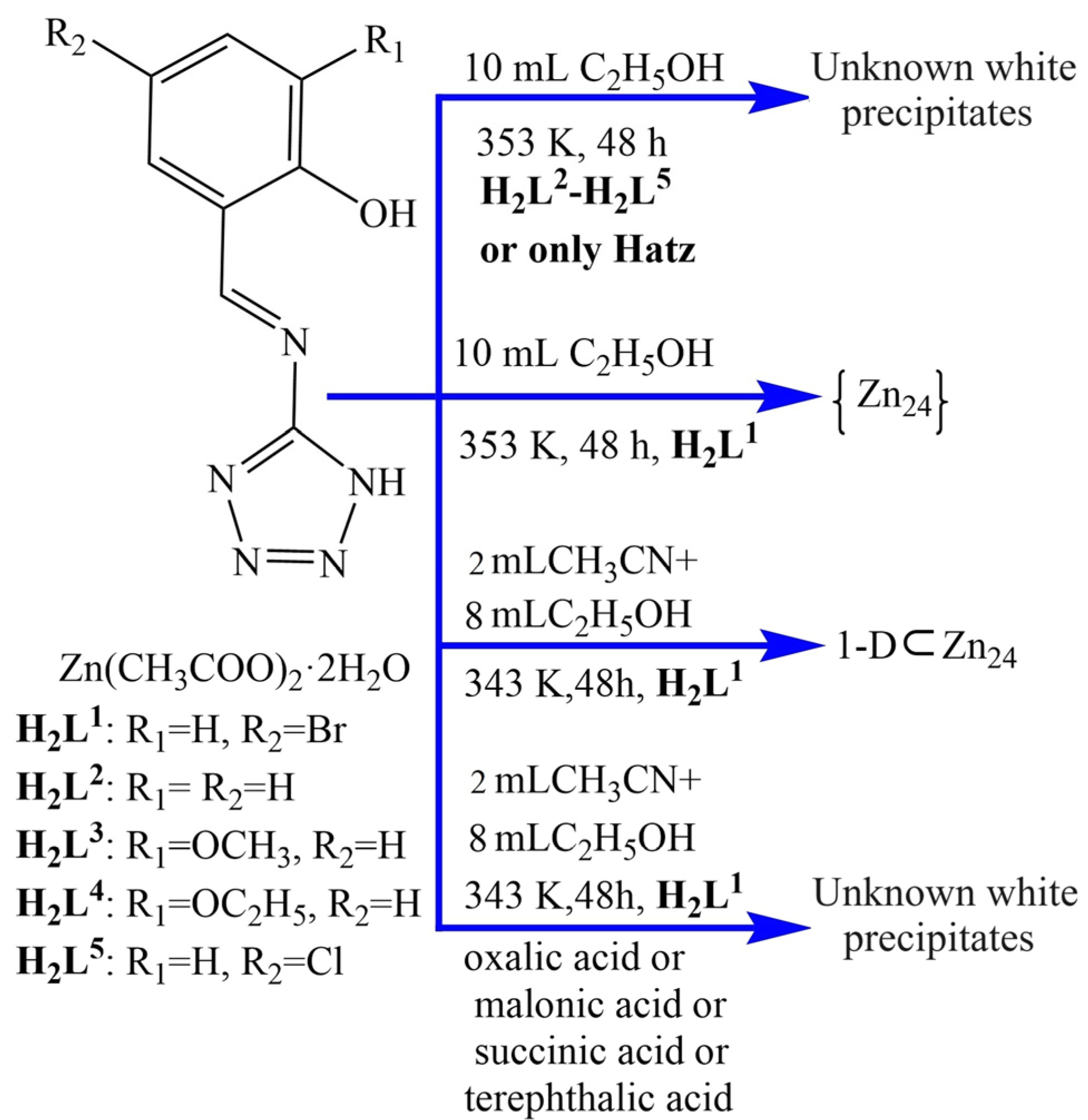
Herein, we investigated the effects of ligand, reaction temperature, counterbalance anion, ligand/metal ion molar ratio, solvent, and synthetic method on the self-assembly of supraclusters (Scheme 2). The synthetic strategy for the Hatz system is depicted in Scheme 2. First, a mixture of Zn(OAc)2·2H2O (0.2 mmol), 4-bromo-2-[(1H-tetrazol-5-ylimino)-methyl]-phenol (H2L1, 0.2 mmol), and anhydrous ethanol (10 mL) was poured into a Teflon-lined autoclave (20 mL). The autoclave was cooled slowly to room temperature after heating at 80 °C for 2 days. Four-cornered golden yellow Zn24 crystals in a double-cone shape were collected via filtration. In the Zn24 supracluster, Hatz is produced by the decomposition of H2L1. To understand the role of H2L1 in the synthesis, we used various salicylaldehyde-derived Schiff bases (H2L2-H2L5) instead of 4-bromo-2-[(1H-tetrazol-5-ylimino)-methyl]-phenol (H2L1) to conduct the same experiment, but we could not obtain the Zn24 supercluster or analog. Similarly, if only H2L1 was replaced by Hatz, the Zn24 supercluster or analog was not obtained. Through previous experiments, we can draw the conclusion that although 5-bromosalicylicaldehyde does not participate in coordination in the 24-atom Zn cluster, 5-bromosalicylicaldehyde is an essential raw material for the synthesis of the Zn24 cluster. Thus, we speculate that 5-bromosalicylicaldehyde may act as a template.
According to the ring structure of Zn24, the Zn24 cluster can form a 1D chain or 2D network through bridging ligands. H2L1 and Zn(CH3COO)2·2H2O were selected as the starting materials. By tuning the reaction temperature, solvent, ligand/metal salt molar ratio, and synthetic methods, the optimal synthesis conditions for the 1D Zn supracluster chain 1-D⸦Zn24 were determined as follows: reaction temperature, 70 °C; solvent, anhydrous ethanol (8 mL), and acetonitrile (2 mL); H2L1/Zn(CH3COO)2·2H2O molar ratio, 2:1; and reactor, micro bottle (autonomously adjusting the reaction pressure).
To obtain a 2D supracluster network, we replaced acetic acid with various carboxylic acids such as oxalic acid, malonic acid, succinic acid, and terephthalic acid. However, these experiments were unsuccessful.

3.2. Crystal Structures of Zn24 and 1-D⸦Zn24

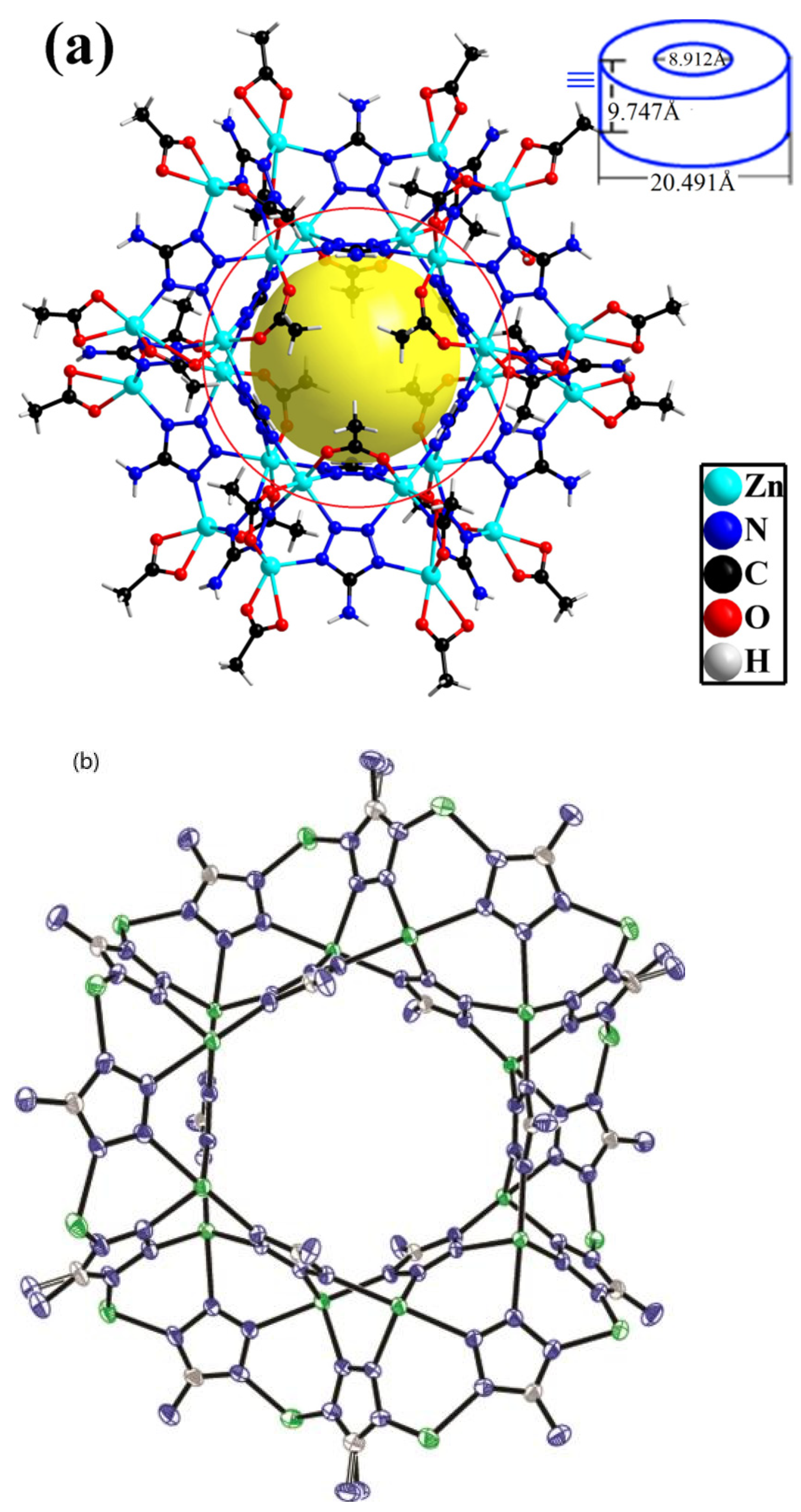
Single crystal analysis confirmed Zn24 to have a 0D wheel-like coordination supracluster of the monoclinic crystal system with P21/n space group consisting of 24 ZnII atoms, 30 acetate groups, 1.5 coordinated water molecules, 3.5 lattice water molecules and 18 Atz ligands derived from H2L1 (Figure 1a). The Zn24 cluster was stabilized by 5-amino-1,2,3,4-tetrazole, which binds along the wheel of the cluster core (Figure 1b), bridging the four neighboring zinc atoms. Acetate groups further stabilized the cluster through 18 μ2:ƞ1:ƞ1-acetate bridging two zinc atoms. The 12 Zn ions in the inner ring of the wheel (inner red ring shown in Figure 1a) coordinated with four N atoms from four different Atz ligands and two O atoms from two syn-syn-μ2:ƞ1:ƞ1-acetate bridging groups to form a distorted octahedral geometry. By contrast, the 12 Zn atoms in the outer ring (outer red ring shown in Figure 1a) coordinated with two N atoms from two different Atz ligands. They also coordinated with two or three O atoms from one syn-syn-μ2:ƞ1:ƞ1-acetate bridging group and one μ1:ƞ1:ƞ1-acetate terminal group or one μ1:ƞ1-acetate ligand, as well as one coordinated water molecule, to form a distorted tetragonal pyramidal, or a trigonal bipyramidal or an octahedral geometry. Close inspection of the nanosized wheel-like conformation showed approximate wheel dimensions of 8.912 × 20.491 × 9.747 Å (inner ring diameter × outer ring diameter × wheel thickness), where the inner ring diameter is the distance between tetrazole planes (i.e., planes (N31,N32,N33,N34,C27) and (N31, N32, N33, N34, C27)i, symmetry code: (i)-x,-y,-z); the outer ring diameter is N30····N30i; and the wheel thickness is the distance between the C13-C29i-C26i -plane and the C13i-C29-C26-plane.
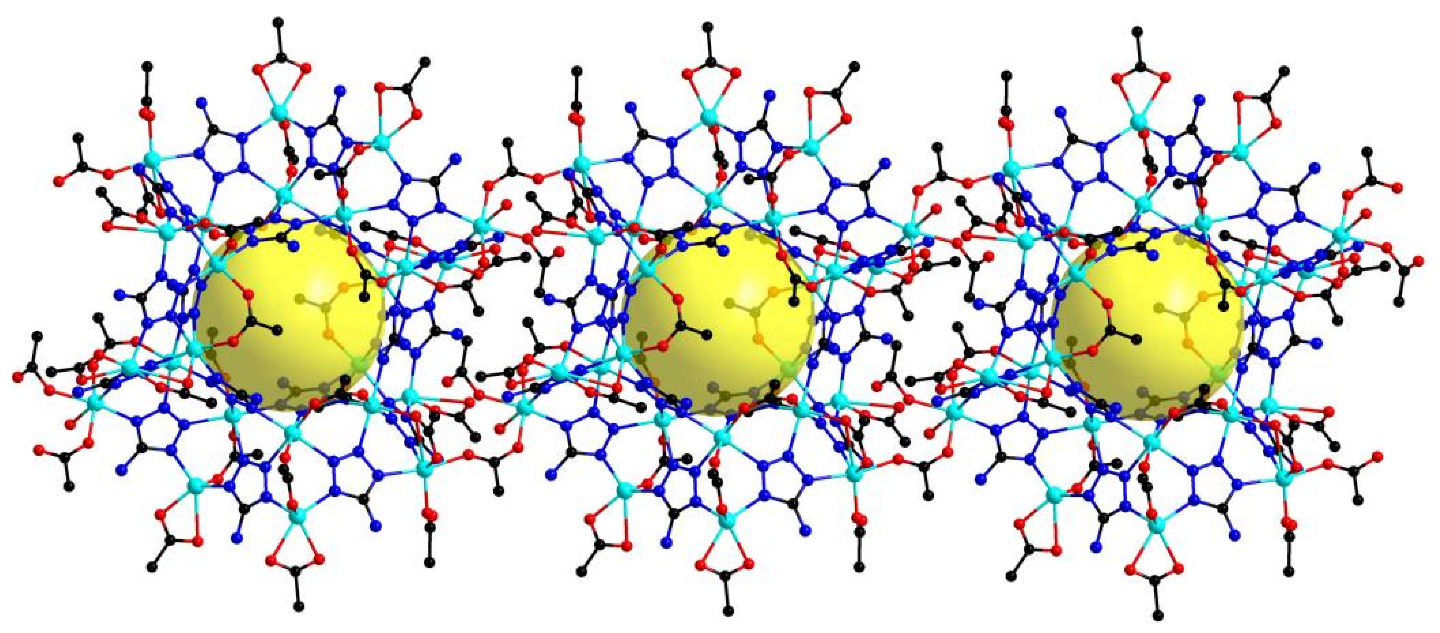
Complexes 1-D⸦Zn24 and Zn24 have similar basic structures, i.e., the Zn24 supracluster. Complex 1-D⸦Zn24 was constructed as a 1D Zn24 suprachain through the double syn-anti- μ2:ƞ1:ƞ1-bridging acetic group linking of Zn24 (Figure 2).

3.3. Luminescent Properties

Numerous studies have demonstrated the good fluorescence of clusters, especially those of Zn(II) and Cd(II) ions with closed d subshells [37,38]. In recent years, Zn(II) clusters have been widely used in luminescent probes due to their desirable advantages in detection and promising applications in biological and environmental systems [39]. Luminescent probes and complexes can selectively detect various sizes of molecules or ions through their adjustable porosity [40].
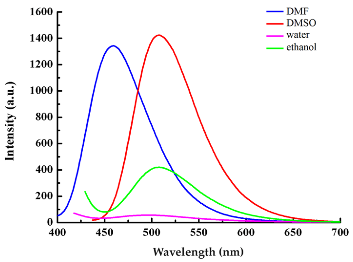
In this paper, the luminescent properties (the phase purity of Zn24 has been checked using PXRD patterns, Figure S7) of Zn24 were investigated in different solvents with concentrations of 1 × 10⁻⁶ mol·L−1 (Figure 3). Upon photoexcitation at 404, 382, 426 and 418 nm in water, N,N-dimethylformamide (DMF), DMSO, and ethanol solvent, Zn24 exhibited green, blue, green and green luminescent emission bands with fluorescence maxima at 496, 459, 507, and 508 nm, respectively. These results predominantly originated from the metal-to-ligand charge-transfer excited state [41,42]. Furthermore, Zn24 exhibited a qualitative change in its luminescence due to the interaction between metal ions and ligands, and it had a stronger fluorescence intensity in DMF and ethanol solutions. Although Zn24 also had a stronger fluorescence intensity in DMSO, we did not consider DSMO for Zn24 luminescent probes due to its high toxicity. Thus, we discussed the Zn24 complex as a luminescent probe for highly selective sensing in DMF and ethanol.
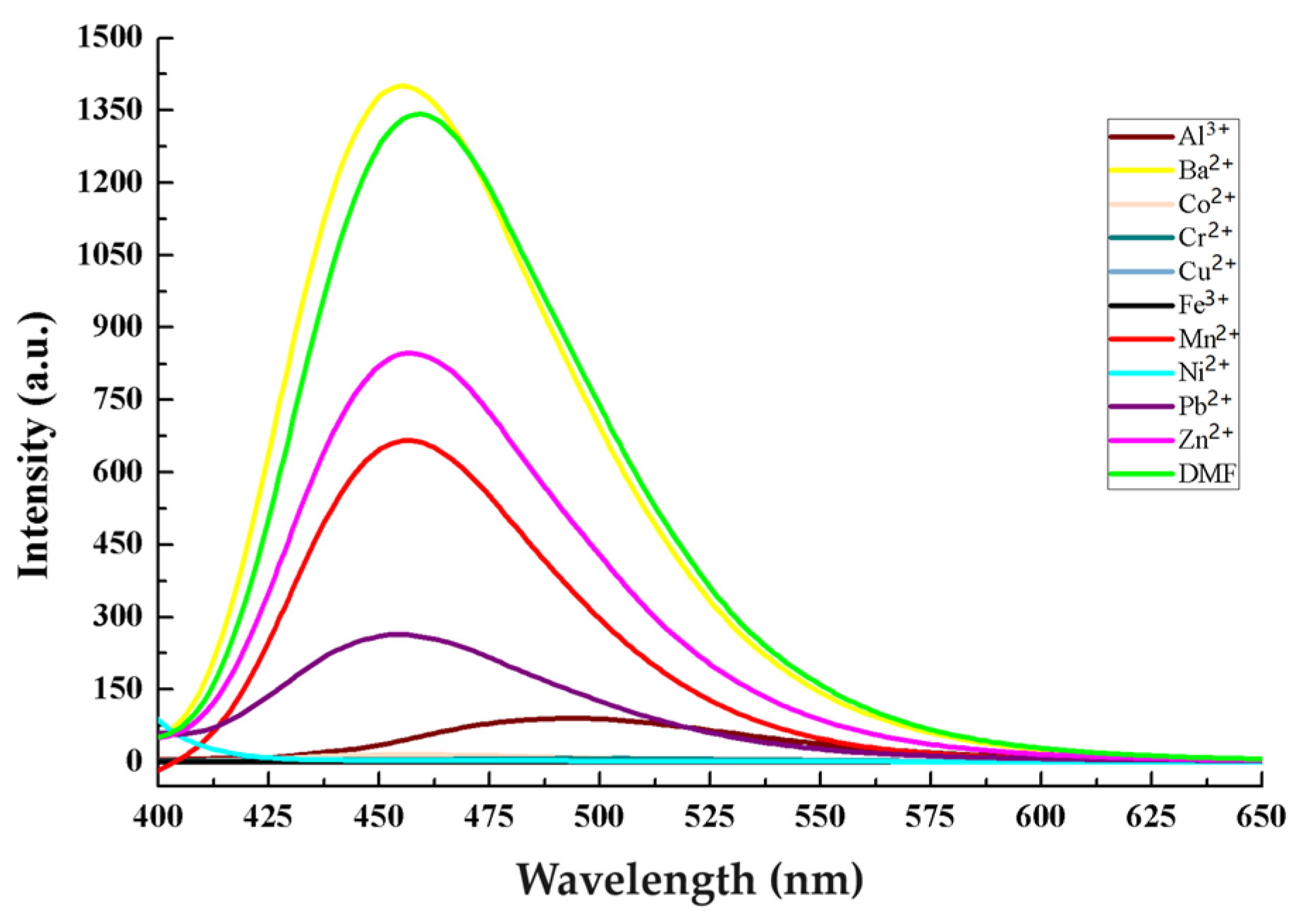
Zn24 (1 mg) was immersed in 10 mL of DMF solutions containing MCln (M = Al3+, Ba2+, Co2+, Cr2+, Cu2+, Fe3+, Mn2+, Ni2+, Pb2+, or Zn2+) to form complex suspensions incorporating various metal ions for luminescence studies, and Fe3+, Ni2+, Cu2+, Cr2+, and Co2+ ions all demonstrated a fluorescence-quenching effect (Figure 4), indicating that Zn24 was not selective toward ions in DMF solution. At the same time, the rare earth ions (Ce3+, Dy3+, Er3+, Eu3+, Gd3+, Ho3+, La3+, Nd3+, Sm3+, and Tb3+) have no clear fluorescence-quenching effects (Figure S3). However, Zn24 showed high selectivity in ethanol solution. The luminescence intensity of Zn24 revealed that the addition of Ce3+ can lead to complete quenching in ethanol solution compared with other metal ions (Figure 5).
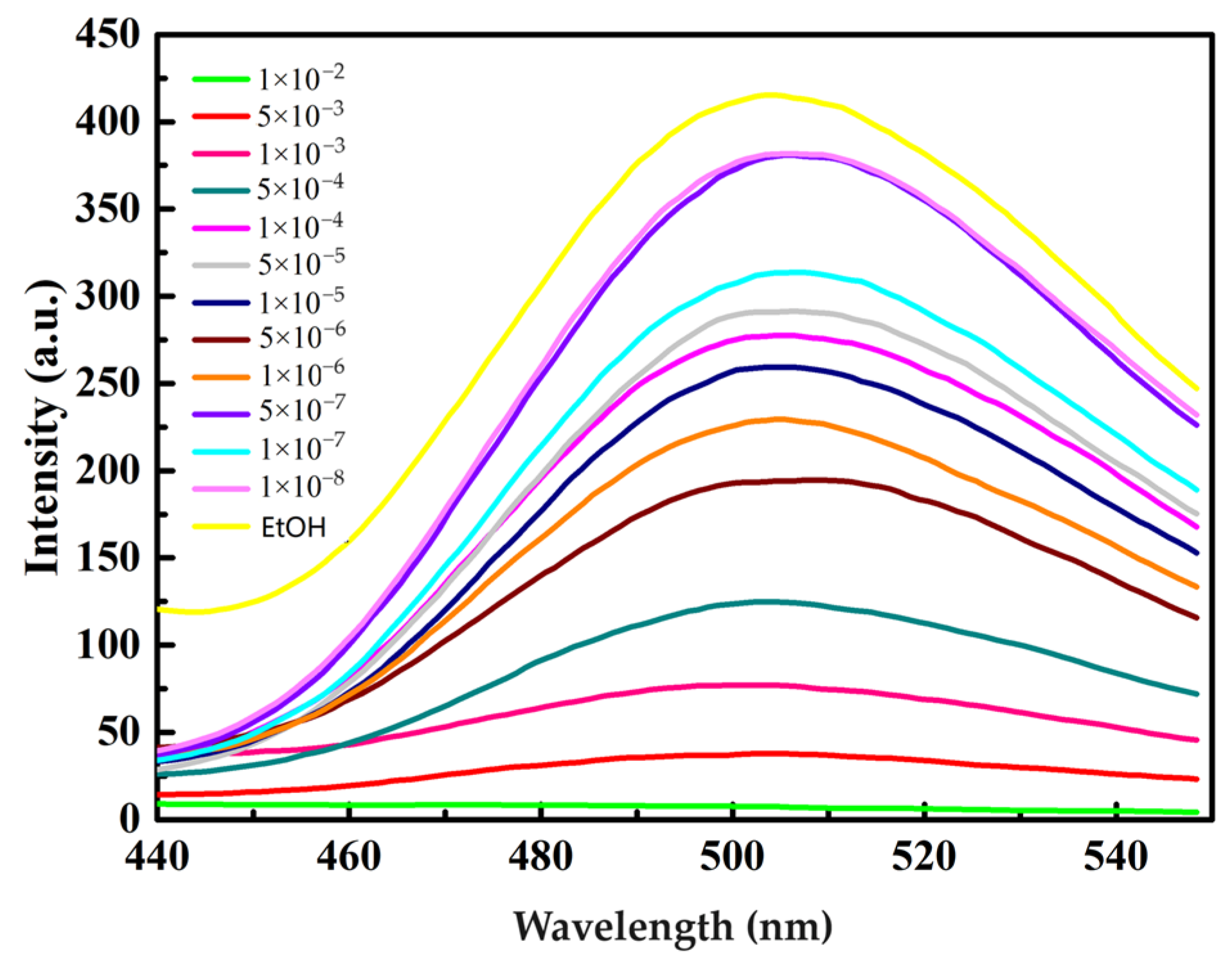
The Zn24 (1 mg, with a final concentration of 1 mg/mL in ethanol) and different concentrations of Ce3+ (from 1 × 10⁻² to 1 × 10⁻⁸ mol/L) were added to the sample tube at room temperature. The fluorescence spectrum was taken at its excitation wavelength (λ = 402 nm).
The fluorescence spectra of the Zn24-Ce3+ system for various concentrations of Ce3+ are shown in Figure 6. The fluorescence intensity at 504 nm progressively increased as the concentration of Ce3+ decreased. In addition, we quantitatively analyzed the quenching efficiency through the Stern–Volmer equation: Io/I = Ksv[C] + l, where Io and I are the respective emission intensities before and after adding Ce3+, while C is the concentration of Ce3+ in ethanol solution. The quenching efficiency, of Zn24 was −1.68 × 10 M−1 (Figure S5). According to the limit of detection (LOD) = 3δ/Ksv (Figure S6), we calculated an LOD of 8.51 × 10⁻⁷ mol/L, which was lower than the reported LOD of the Ln-MOF [41].

3.4. Detection Wavelength

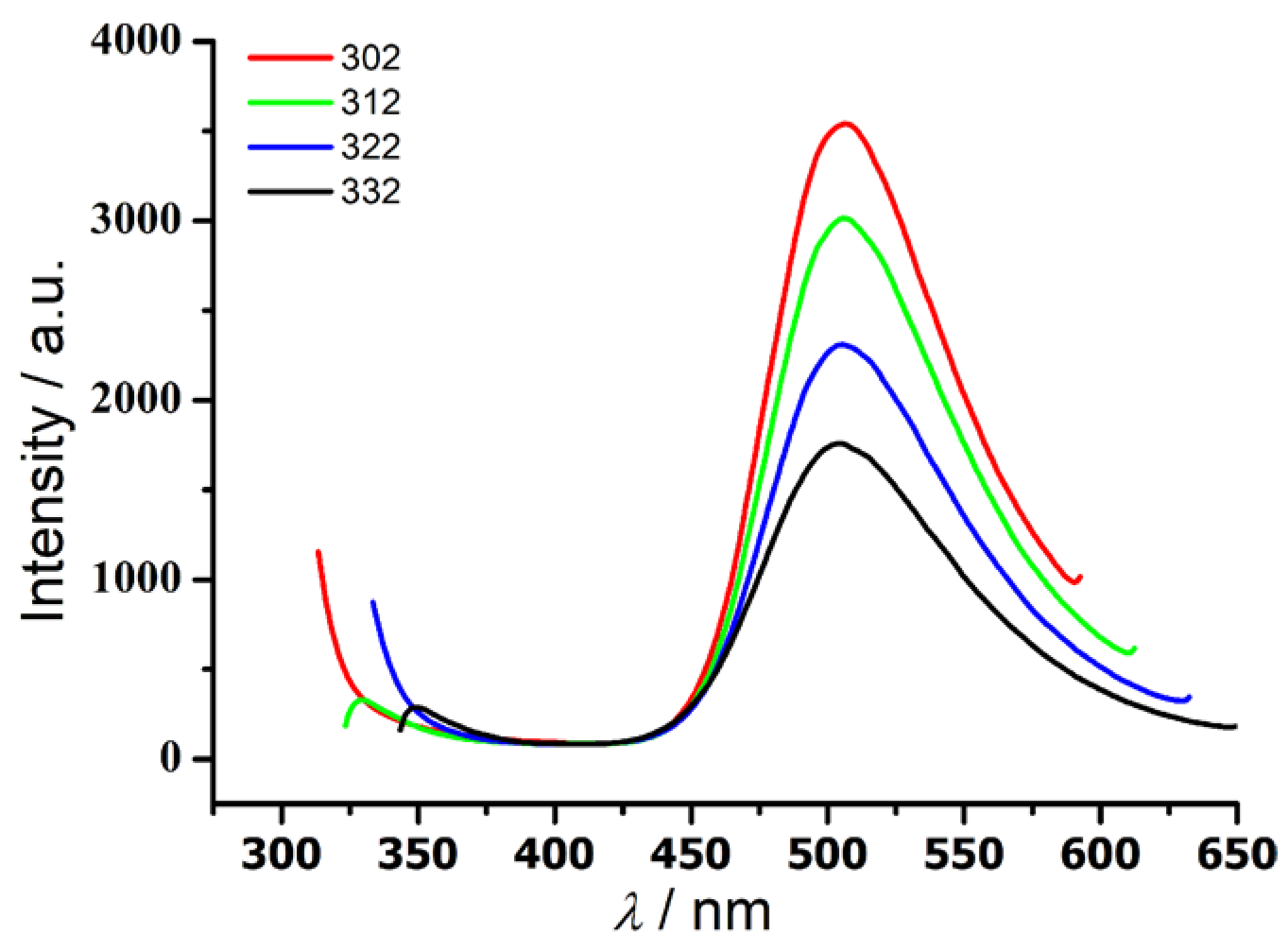
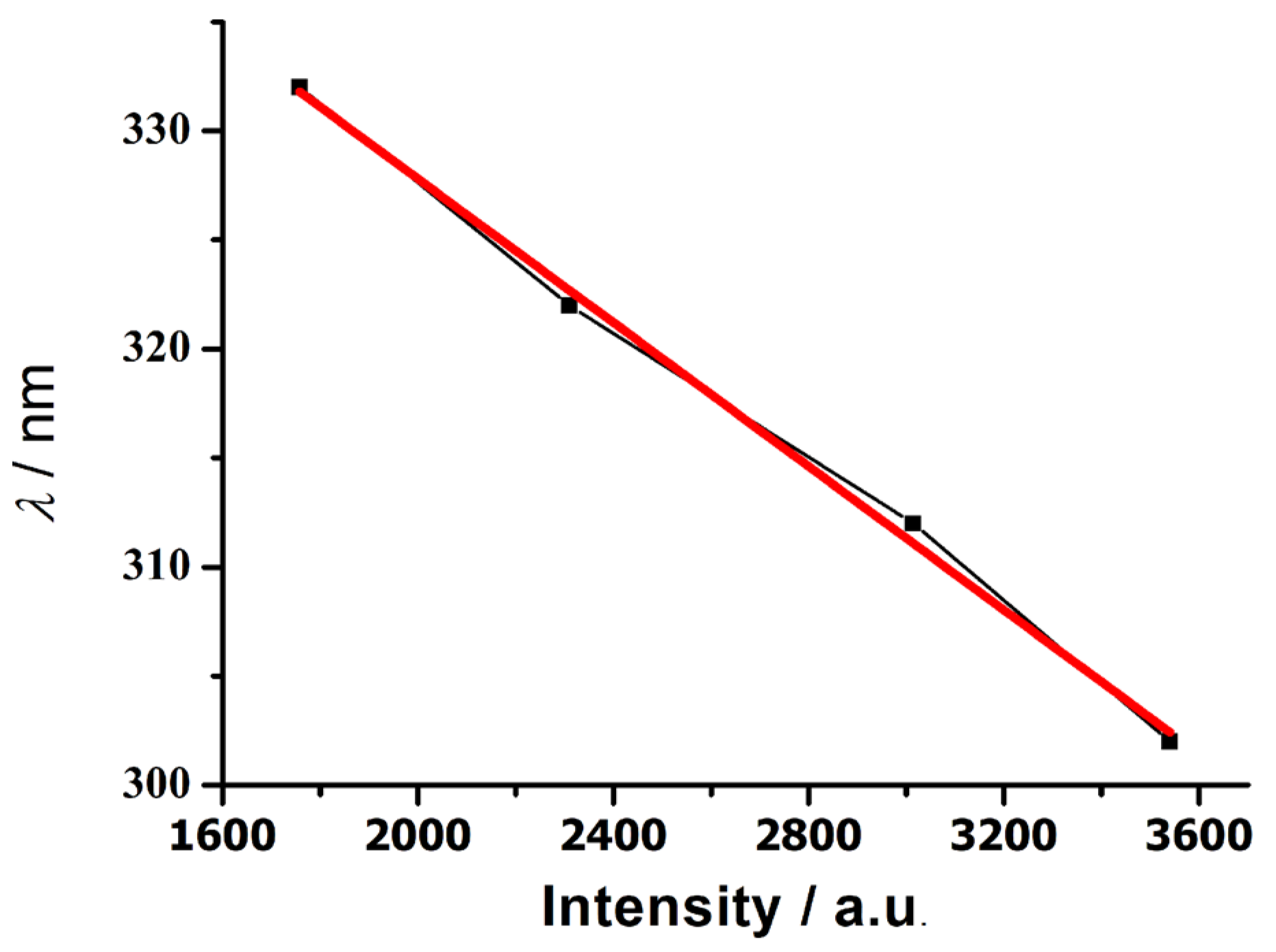
The solid-state fluorescence spectra of Hatz were obtained at a slit width of 5 nm and an excitation wavelength of 402 nm, while the solid-state fluorescence spectra of Zn24 were obtained at an excitation wavelength of 302–332 nm (Figure 7 and Figure S4). Under the same test conditions, the fluorescence spectrum of Hatz peaked at 615 nm, while that of Zn24 peaked at 502 nm. The luminous color changed from red to blue–green, and the fluorescence intensity of Zn24 was more than 200 times that of Hatz. The full-type Zn2+ metal ion in Zn24 has an extra-nuclear d10 electron, and did not undergo a d–d transition, leading to a significant enhancement in luminous intensity. At the same time, the deprotonated tetrazolium ring is an electron-deficient conjugated ring that causes the electron migration (M→L) of zinc ions to the tetrazolium ring [42,43]. As a result, the luminous color changed from red to blue–green, and the fluorescence intensity of Zn24 was more than 200 times that of Hatz. The fluorescence intensity of Zn24 decreased linearly with the increase in excitation wavelength (Figure 7 and Figure 8). Between 302 and 332 nm, the wavelength of the excitation light can be determined by detecting the intensity of the emitted light.

3.5. Hirshfeld Surface Analysis of the Complex Zn24

Hirshfeld surface analysis [44] is a useful tool for describing the surface characteristics of molecules, and was performed to visualize the different intermolecular interactions in crystal structures by employing 3D molecular surface contours. Figure 9 displays the findings of the Zn24 Hirshfeld surface study. The middle shape index ranges from −1.000 to 1.000 Å, whereas the range of the dnorm surface on the left is −1.238 to 1.570 Å. The range of the curvature curvedness was −4.000 to 0.400 Å. In Figure 9, the dnorm surface map of Zn24 is colored from light to dark red spots to represent the interaction force of the complex Zn24 from weak to strong.
One useful supplement for Hirshfeld surface analysis is the 2-D fingerprint plot [45]. It quantitatively analyses the nature and type of intermolecular interaction between the molecules inside the crystals. The fingerprint plots can be decomposed to highlight particularly close contacts between the elements (Figure 10). The H···H interaction is one of the most significant contacts for the Zn24 complex.
The main intermolecular interaction of Zn24 is H···H contact, which is reflected in the middle of the scattered points of the 2-D fingerprint plots (the percentage of H···H contacts of Zn24 is 40.4%). Another main intermolecular interaction of Zn24 is O···H interaction, which is represented by double spikes in the bottom left (acceptor and donor) region of the fingerprint plots. Accordingly, we can infer that there are significant N-H···O hydrogen bonds (Table S3) observed in Zn24 (the percentage of O···H contacts of Zn24 is 22.4%). Also, the N···H contacts play important roles for Zn24. The percentage of N···H contacts of Zn24 is 10.0%. In addition to those above, the presence of C···H, C···O, and N···O contacts were also observed. These three forces accounted for 4.0%, 1.4% and 1.3% of the total Hirshfeld surface force, respectively.

4. Conclusions

A supracluster zinc, Zn24, and a zinc supracluster 1D chain, 1-D⊂Zn24, were synthesized through regulatory solvothermal reactions. In an ethanol solution, Zn24 was synthesized at 353 K using a solvothermal method, whereas 1-D⊂Zn24 was formed when a mixed solution (2 mL acetonitrile + 8 mL ethanol) was used at 343 K using a micro bottle. The Zn24 supracluster can be used to selectively detect Ce3+ with excellent efficiency (LOD = 8.51 × 10⁻⁷ mol/L), and can be used as a potential sensor for their detection. In addition, the Zn24 supracluster can detect wavelengths between 302 and 332 nm, using the intensity of emitted light. Thus, the Zn24 supracluster can potentially be used as a spectral detection material to prepare optical wave detectors.

Supplementary Materials

The following supporting information can be downloaded at: https://www.mdpi.com/article/10.3390/nano13233058/s1, Figure S1: IR of H2L1-H2L5; Figure S2: IR of Zn24; Figure S3: The liquid-state fluorescence behaviors of Zn24 in DMF; Figure S4: Emission spectra of HATZ in a solid state at 402 nm excitation wavelength at room temperature; Figure S5: The fitting curve of the luminescence intensity of Zn24 at different Ce3+ concentration; Figure S6: The fluorescence spectra of blank Zn24 (1 mg·mL−1) at different measurements; Figure S7: XRD of the complex Zn24; Table S1: Selected bond lengths (Å) and angles (°) for Zn24; Figure S8: 1H NMR (400 MHz, DMSO-d6) for L1H2; Figure S9: 1H NMR (400 MHz, DMSO-d6) for L2H2; Figure S10: 1H NMR (400 MHz, DMSO-d6) for L3H2; Figure S11: 1H NMR (400 MHz, DMSO-d6) for L4H2; Figure S12: 1H NMR (400 MHz, DMSO-d6) for L5H2; Table S2: Selected bond lengths (Å) and angles (°) for 1-D⸦Zn24; Table S3: Hydrogen bond lengths (Å) and angles (°) for Zn24.

Author Contributions

Y.C., Z.C. and J.W.: designed and supervised the project and experiments; Y.C., Z.C., J.W., X.M. and L.Y. performed the data analysis and prepared the manuscript; S.Z., and F.T.: provided resources and acquired funding; and S.Z. and F.T. provided academic guidance. All authors have read and agreed to the published version of the manuscript.

Funding

This research was funded by the National Natural Science Foundation of China (No. 21861014), and by the Talent Introduction Program of Guangdong Institute of Petrochemical Technology (No. 2020RC033).

Data Availability Statement

The data presented in this study are available in this article and Supplementary Material.

Conflicts of Interest

The authors declare that they have no conflict of interest to report regarding the present study.

References

  1. Bao, S.S.; Zheng, L.M. Magnetic materials based on 3d metal phosphonates. Coord. Chem. Rev. 2016, 319, 63–85. [Google Scholar] [CrossRef]
  2. Han, S.D.; Zhao, J.P.; Liu, S.J.; Bu, X.H. Hydro(solvo)thermal synthetic strategy towards azido/formato-mediated molecular magnetic materials. Coord. Chem. Rev. 2016, 289–290, 32–48. [Google Scholar]
  3. Kitchen, J.A. Lanthanide-based self-assemblies of 2, 6-pyridyldicarboxamide ligands: Recent advances and applications as next-generation luminescent and magnetic materials. Coord. Chem. Rev. 2017, 340, 232–246. [Google Scholar] [CrossRef]
  4. Ashebr, T.G.; Li, H.; Ying, X.; Li, X.-L.; Zhao, C.; Liu, S.; Tang, J. Emerging trends on designing high-performance dysprosium (III) single-molecule magnets. ACS Mater. Lett. 2022, 4, 307–319. [Google Scholar] [CrossRef]
  5. Liu, J.Q.; Luo, Z.-D.; Pan, Y.; Singh, A.K.; Trivedi, M.; Kumar, A. Recent developments in luminescent coordination polymers: Designing strategies, sensing application and theoretical evidences. Coord. Chem. Rev. 2020, 406, 213145. [Google Scholar] [CrossRef]
  6. Zhou, Q.; Ren, G.; Yang, Y.; Wang, C.; Che, G.; Li, M.; Yu, M.-H.; Li, J.; Pan, Q. Fluorescence Thermometers Involving Two Ranges of Temperature: Coordination Polymer and DMSP Embedding. Inorg. Chem. 2023, 62, 16652–16658. [Google Scholar] [CrossRef] [PubMed]
  7. Yang, Z.; Xu, T.; Li, H.; She, M.; Chen, J.; Wang, Z.; Zhang, S.; Li, J. Zero-Dimensional Carbon Nanomaterials for Fluorescent Sensing and Imaging. Chem. Rev. 2023, 123, 11047–11136. [Google Scholar] [CrossRef]
  8. Zhang, C.; Qin, Y.; Ke, Z.; Yin, L.; Xiao, Y.; Zhang, S. Highly efficient and facile removal of As (V) from water by using Pb-MOF with higher stable and fluorescence. Appl. Organomet. Chem. 2023, 37, e7066. [Google Scholar] [CrossRef]
  9. Suess, R.J.; Winnerl, S.; Schneider, H.; Helm, M.; Berger, C.; De Heer, W.A.; Murphy, T.E.; Mittendorff, M. Role of transient reflection in graphene nonlinear infrared optics. Acs Photonics 2016, 3, 1069–1075. [Google Scholar] [CrossRef]
  10. Gwo, S.; Sun, L.; Li, X.; Chen, H.-Y.; Lin, M.-H. Nanomanipulation and controlled self-assembly of metal nanoparticles and nanocrystals for plasmonics. Chem. Soc. Rev. 2016, 45, 5672–5716. [Google Scholar] [CrossRef]
  11. Gwo, S.; Wang, C.Y.; Chen, H.Y.; Lin, M.H.; Sun, L.; Li, X.; Chen, W.L.; Chang, Y.M.; Ahn, H. Plasmonic metasurfaces for nonlinear optics and quantitative SERS. ACS Photonics 2016, 3, 1371–1384. [Google Scholar] [CrossRef]
  12. Yang, X.G.; Zhai, Z.M.; Lu, X.M.; Ma, L.F.; Yan, D. Fast crystallization-deposition of orderly molecule level heterojunction thin films showing tunable up-conversion and ultrahigh photoelectric response. ACS Cent. Sci. 2020, 6, 1169–1178. [Google Scholar] [CrossRef] [PubMed]
  13. Lee, S.W.; Choi, B.J.; Eom, T.; Han, J.H.; Kim, S.K.; Song, S.J.; Lee, W.; Hwang, C.S. Influences of metal, non-metal precursors, and substrates on atomic layer deposition processes for the growth of selected functional electronic materials. Coord. Chem. Rev. 2013, 257, 3154–3176. [Google Scholar] [CrossRef]
  14. Zhao, Y.X.; Yang, G.; Lu, X.M.; Yang, C.D.; Fan, N.N.; Yang, Z.T.; Wang, L.Y.; Ma, L.F. {Zn6} Cluster based metal–organic framework with enhanced room-temperature phosphorescence and optoelectronic performances. Inorg. Chem. 2019, 58, 6215–6221. [Google Scholar] [CrossRef] [PubMed]
  15. Pang, J.Y.; Gao, Q.; Yin, L.; Zhang, S.H. Synthesis and catalytic performance of banana cellulose nanofibres grafted with poly (ε-caprolactone) in a novel two-dimensional zinc (II) metal-organic framework. Int. J. Biol. Macromol. 2023, 224, 568–577. [Google Scholar] [CrossRef] [PubMed]
  16. Liu, J.; Yang, G.P.; Jin, J.; Wu, D.; Ma, L.F.; Wang, Y.Y. A first new porous d–p HMOF material with multiple active sites for excellent CO2 capture and catalysis. Chem. Commun. 2020, 56, 2395–2398. [Google Scholar] [CrossRef] [PubMed]
  17. Wu, Y.P.; Tian, J.W.; Liu, S.; Li, B.; Zhao, J.; Ma, L.F.; Li, D.S.; Lan, Y.Q.; Bu, X. Bi-Microporous metal–organic frameworks with cubane [M4 (OH) 4](M= Ni, Co) clusters and pore-space partition for electrocatalytic methanol oxidation reaction. Angew. Chem. Int. Ed. 2019, 58, 12185–12189. [Google Scholar] [CrossRef]
  18. Baig, M.M.F.A.; Ma, J.; Gao, X.; Khan, M.A.; Ali, A.; Farid, A.; Zia, A.W.; Noreen, S.; Wu, H. Exploring the robustness of DNA nanotubes framework for anticancer theranostics toward the 2D/3D clusters of hypopharyngeal respiratory tumor cells. Int. J. Biol. Macromol. 2023, 236, 123988. [Google Scholar] [CrossRef]
  19. Zhivkova, T.; Culita, D.C.; Abudalleh, A.; Dyakova, L.; Mocanu, T.; Madalan, A.M.; Georgieva, M.; Miloshev, G.; Hanganu, A.; Marinescu, G.; et al. Homo-and heterometallic complexes of Zn (ii),{Zn (ii) Au (i)}, and {Zn (ii) Ag (i)} with pentadentate Schiff base ligands as promising anticancer agents. Dalton Trans. 2023, 52, 12282–12295. [Google Scholar] [CrossRef]
  20. Zhang, Y.; Yuan, S.; Day, G.; Wang, X.; Yang, X.; Zhou, H.C. Luminescent sensors based on metal-organic frameworks. Coord. Chem. Rev. 2018, 354, 28–45. [Google Scholar] [CrossRef]
  21. Yu, C.X.; Hu, F.L.; Song, J.G.; Zhang, J.L.; Liu, S.S.; Wang, B.X.; Meng, H.; Liu, L.; Ma, L.F. Ultrathin two-dimensional metal-organic framework nanosheets decorated with tetra-pyridyl calix [4] arene: Design, synthesis and application in pesticide detection. Sens. Actuat. B-Chem. 2020, 310, 127819. [Google Scholar] [CrossRef]
  22. Wang, S.T.; Zheng, X.; Zhang, S.H.; Li, G.; Xiao, Y. A study of GUPT-2, a water-stable zinc-based metal–organic framework as a highly selective and sensitive fluorescent sensor in the detection of Al3+ and Fe3+ ions. CrystEngComm 2021, 23, 4059–4068. [Google Scholar] [CrossRef]
  23. Avila, R.J.; Emery, D.J.; Pellin, M.J.; Martinson, B.F.A.; Farha, K.O.; Hupp, T.J. Porphyrins as Templates for Site-Selective Atomic Layer Deposition: Vapor Metalation and in Situ Monitoring of Island Growth. ACS Appl. Mater Inter. 2016, 8, 19853–19859. [Google Scholar] [CrossRef] [PubMed]
  24. Gao, P.; Ma, H.; Wu, Q.; Qiao, L.; Volinsky, A.A.; Su, Y.J. Size-dependent vacancy concentration in nickel, copper, gold, and platinum nanoparticles. Phys. Chem. C 2016, 120, 17613–17619. [Google Scholar] [CrossRef]
  25. Kumar, R.; Kaur, N.; Kaur, R.; Kaur, N.; Sahoo, S.C.; Nanda, P.K. Temperature controlled synthesis and transformation of dinuclear to hexanuclear zinc complexes of a benzothiazole based ligand: Coordination induced fluorescence enhancement and quenching. J. Mol. Struct. 2022, 1265, 133300. [Google Scholar] [CrossRef]
  26. Islam, S.; Tripathi, S.; Hossain, A.; Seth, S.K.; Mukhopadhyay, S.J. pH-induced structural variations of two new Mg (II)-PDA complexes: Experimental and theoretical studies. J. Mol. Struct. 2022, 1265, 133373. [Google Scholar] [CrossRef]
  27. Feng, C.; Zhu, Y.Q.; Huang, H.H.; Zhao, H.J. A Triazolate-Supported Fe33-O) Core: Crystal Structure, Fluorescence, and Hirshfeld Surface Analysis. J. Clust. Sci. 2016, 27, 1181–1190. [Google Scholar] [CrossRef]
  28. Sebastian, S.; Tim, S.; Mariam, B.; Jan, V.L.; Arif, N.M.; Paul, K.G. A planar decanuclear cobalt (II) coordination cluster. Inorg. Chim. Acta 2018, 482, 522–525. [Google Scholar]
  29. Gaponik, P.N.; Voitekhovich, S.V.; Ivashkevich, O.A. Metal derivatives of tetrazoles. Russ. Chem. Rev. 2010, 37, 507–603. [Google Scholar]
  30. Li, J.R.; Yu, Q.; Sañudo, E.C.; Tao, Y.; Bu, X.H. An azido–CuII–triazolate complex with utp-type topological network, showing spin-canted antiferromagnetism. Chem. Commun. 2007, 25, 2602–2604. [Google Scholar] [CrossRef]
  31. Tong, X.L.; Hu, T.L.; Zhao, J.P.; Wang, Y.K.; Zhang, H.; Bu, X.H. Chiral magnetic metal–organic frameworks of MnII with achiral tetrazolate-based ligands by spontaneous resolution. Chem. Commun. 2010, 46, 8543–8545. [Google Scholar] [CrossRef]
  32. Bai, Y.; Dou, Y.; Xie, L.H.; Rutledge, W.; Li, J.R.; Zhou, H.C. Zr-based metal–organic frameworks: Design, synthesis, structure, and applications. Chem. Soc. Rev. 2016, 45, 2327. [Google Scholar] [CrossRef] [PubMed]
  33. Kang, X.M.; Tang, M.H.; Yang, G.L.; Zhao, B. Cluster/cage-based coordination polymers with tetrazole derivatives. Coord. Chem. Rev. 2020, 422, 213424. [Google Scholar] [CrossRef]
  34. Liang, M.X.; Ruan, C.Z.; Sun, D.; Kong, X.J.; Ren, Y.P.; Long, L.S.; Huang, R.B.; Zheng, L.S. Solvothermal synthesis of four polyoxometalate-based coordination polymers including diverse Ag (I) · π interactions. Inorg. Chem. 2014, 53, 897–902. [Google Scholar] [CrossRef] [PubMed]
  35. Sheldrick, G.M. Crystal structure refinement withSHELXL. Acta Cryst. 2015, A71, 3–8. [Google Scholar] [CrossRef] [PubMed]
  36. Dolomanov, O.V.; Bourhis, L.J.; Gildea, R.J.; Howard, J.A.K.; Puschmann, H.J. OLEX2: A complete structure solution, refinement and analysis program. Appl. Cryst. 2009, 42, 339–341. [Google Scholar] [CrossRef]
  37. Wei, G.H.; Yang, J.; Ma, J.F.; Liu, Y.Y.; Li, S.L.; Zhang, L.P. Syntheses, structures and luminescent properties of zinc (II) and cadmium (II) coordination complexes based on new bis (imidazolyl) ether and different carboxylate ligands. Dalton Trans. 2008, 23, 3080–3092. [Google Scholar] [CrossRef]
  38. Su, Z.; Fan, J.; Okamura, T.A.; Chen, M.S.; Chen, S.S.; Sun, W.Y. Interpenetrating and self-penetrating zinc (II) complexes with rigid tripodal imidazole-containing ligand and benzenedicarboxylate. Cryst. Growth Des. 2010, 10, 1911–1922. [Google Scholar] [CrossRef]
  39. Tian, Y.; Wang, Y.; Xu, Y.; Liu, Y.; Li, D.; Fan, C. A highly sensitive chemiluminescence sensor for detecting mercury (II) ions: A combination of Exonuclease III-aided signal amplification and graphene oxide-assisted background reduction. Sci. China Chem. 2015, 58, 514–518. [Google Scholar] [CrossRef]
  40. Gao, W.Y.; Chrzanowski, M.; Ma, S. Metal–metalloporphyrin frameworks: A resurging class of functional materials. Chem. Soc. Rev. 2015, 45, 1602–1608. [Google Scholar] [CrossRef]
  41. Zhang, Q.; Wang, J.; Kirillov, A.M.; Dou, W.; Xu, C.; Xu, C.; Yang, L.; Fang, R.; Liu, W. Multifunctional Ln–MOF luminescent probe for efficient sensing of Fe3+, Ce3+, and acetone. ACS Appl. Mater. Inter. 2018, 10, 23976–23986. [Google Scholar] [CrossRef]
  42. Chen, Y.; Yang, T.; Pan, H.; Yuan, Y.; Chen, L.J. Photoemission mechanism of water-soluble silver nanoclusters: Ligand-to-metal–metal charge transfer vs strong coupling between surface plasmon and emitters. J. Am. Chem. Soc. 2014, 136, 1686–1689. [Google Scholar] [CrossRef]
  43. Huo, P.; Hou, J.; Lei, Z.; Chen, Q.-Y.; Dai, T. Ligand-to-ligand charge transfer within metal–organic frameworks based on manganese coordination polymers with tetrathiafulvalene-bicarboxylate and bipyridine ligands. Inorg. Chem. 2016, 55, 6496–6503. [Google Scholar] [CrossRef] [PubMed]
  44. Spackman, M.A.; Jayatilaka, D. Hirshfeld surface analysis. CrystEngComm 2009, 11, 19–32. [Google Scholar] [CrossRef]
  45. Sama, F.; Ansari, L.A.; Raizada, M.; Ahmad, M.; Nagaraja, C.M.; Shahid, M.; Kumar, A.; Khan, K.; Siddiqi, Z.A. Design, structures and study of non-covalent interactions of mono-, di-, and tetranuclear complexes of a bifurcated quadridentate tripod ligand, N-(aminopropyl)-diethanolamine. New. J. Chem. 2017, 41, 1959–1972. [Google Scholar] [CrossRef]
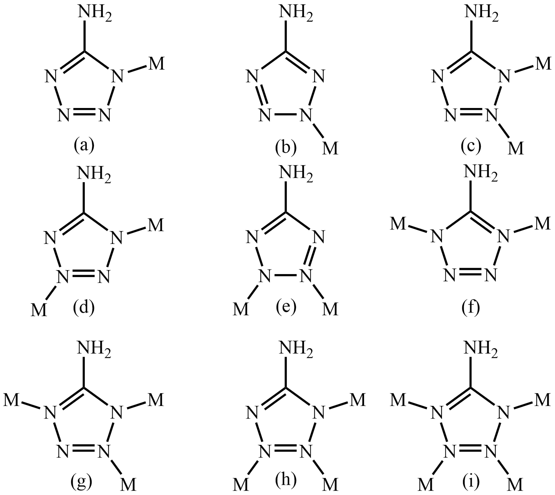
Scheme 1. Coordination modes of Hatz. (a) μ1:1η1; (b) μ1:1η2; (c) μ2:1η1:2η1; (d) μ2:1η1:3η1; (e) μ2:2η1:3η1; (f) μ2:1η1:4η1; (g) μ3:1η1:2η1:4η1; (h) μ3:1η1:2η1:3η1; (i) μ4:1η1:2η1:3η1:4η1.
Nanomaterials 13 03058 sch001
Scheme 2. Regulatory process of Zn24 and 1-D⸦Zn24.
Nanomaterials 13 03058 sch002
Figure 1. (a) Crystal structure of Zn24. (b) Zinc Atz core.
Nanomaterials 13 03058 g001
Figure 2. 1D chain of 1-D⸦Zn24.
Nanomaterials 13 03058 g002
Figure 3. Luminescence of Zn24 in different solutions.
Nanomaterials 13 03058 g003
Figure 4. Liquid-state fluorescence behavior of Zn24 in DMF.
Nanomaterials 13 03058 g004
Figure 5. Liquid-state fluorescence behavior of Zn24 in ethanol.
Nanomaterials 13 03058 g005
Figure 6. Liquid-state fluorescence behavior of Zn24 for different concentrations of Ce3+ in ethanol.
Nanomaterials 13 03058 g006
Figure 7. Solid-state emission spectra of Zn24 under 302, 312, 322 and 332 nm excitation at room temperature.
Nanomaterials 13 03058 g007
Figure 8. Fluorescence intensity (a.u.) vs. wavelength of excitation light (nm).
Nanomaterials 13 03058 g008
Figure 9. Hirshfeld surface mapped with dnorm (left), shape index (middle), and curvedness (right) for complex Zn24.
Nanomaterials 13 03058 g009
Figure 10. Fingerprint plots of complex Zn24.
Nanomaterials 13 03058 g010
Table 1. Crystallographic data for Zn24 and 1-DZn24.
ComplexesZn241-D⊂Zn24
Formula C78H138N90O66Zn24C82H150N90O68 Zn24
Formula weight4961.665053.79
Crystal systemMonoclinic triclinic
Crystal size (mm)0.25 × 0.21 × 0.160.21 × 0.18 × 0.13
Space groupP21/nPī
a (Å)20.264(1)16.718(1)
b (Å)23.996(1)17.449(1)
c (Å)21.663(1)19.054(1)
α (°)90.0070.651(3)
β (°)108.738(4)80.304(3)
γ (°)90.0070.651(3)
V3)9975.3(6)5062.8(3)
F(000)49682536
Z21
Dc (g cm–3)1.6461.658
μ (mm–1)2.9172.877
θ range (°)3.16, 25.013.30,25.01
Ref. meas./indep.74,768, 17,43339,347, 17,765
Obs. ref. [I > 2σ(I)]11,95311,553
Rint0.05200.0473
R1 [I ≥ 2σ (I)] a0.06000.0673
ωR2(all data) b0.19340.2037
Goof1.0391.041
Δρ(max, min) (e Å−3)1.001, −0.8671.126, −0.694
a R1 = Σ||Fo| − |Fc||/Σ|Fo|. b wR2 = [Σw(|Fo2|–|Fc2|)2w(|Fo2|)2]1/2.
Disclaimer/Publisher’s Note: The statements, opinions and data contained in all publications are solely those of the individual author(s) and contributor(s) and not of MDPI and/or the editor(s). MDPI and/or the editor(s) disclaim responsibility for any injury to people or property resulting from any ideas, methods, instructions or products referred to in the content.