Metrological Protocols for Reaching Reliable and SI-Traceable Size Results for Multi-Modal and Complexly Shaped Reference Nanoparticles
Abstract
:1. Introduction
- i
- Sample preparation: The protocol must be optimized to minimize additional NP agglomeration. The presence of well-dispersed homogeneous isolated NPs on the substrate facilitates accurate and reliable NP size results. Moreover, automated analysis is thus enabled.
- ii
- Calibration and metrological qualification of the instrument: Calibrating the instrument means creating a correlation between the obtained measurement result and the definition of the unit concerned in the SI (Système International). This periodically performed action makes the measurement result reliable and comparable with other laboratories with the same or other techniques. The calibration process can be carried out with certified reference materials. The metrological qualification of the instrument allows the operator to identify all error sources affecting the measurement result and hence quantitatively assess an uncertainty budget associated with the measurement.
- iii
- Data acquisition: The term “measurand” is linked to the quantity to be measured and must match the dimensional descriptors (particle shape and particle size) [2] of the nano-object to be determined as accurately as possible, describing its geometry as completely as possible. For this study, the near-spherical NPs can be defined by a single parameter, but the more complex geometries will need at least two parameters. Ideally, the measurand’s definition must be as specific as possible including the terms: mean, median, or mode. In this paper, all the reported results are the mean values of the respective measurands.
- iv
- Microscopy image and data analysis process: This is a key step because the size measurand value is extracted from the image through software provided with mathematical tools. Several software packages suitable for NP size exist on the market (e.g., MoutainsMap® and Digital Surf) or as open access software (e.g., ImageJ and Gwyddion). However, algorithms determining the NP size from the EM raw signal are considered as a black box and the common “watershed” algorithms capable of identifying the NP boundaries within agglomerates are often unsatisfactory for reliable segmentation. That is the reason why we have decided for the microscopy techniques in this study to handle measurements only on isolated NPs. Furthermore, image analysis software, Platypus ®, was specially designed by Pollen Metrology for the nPSize project and adapted for EM and AFM images.
2. Materials and Methods
2.1. Material Preparation
2.2. Measurement Techniques
2.2.1. Transmission Electron Microscopy (TEM)
2.2.2. Scanning Electron Microscopy (SEM)
2.2.3. Scanning Electron Microscopy in Transmission Mode (TSEM)
2.2.4. Atomic Force Microscopy (AFM)
2.2.5. Small Angle X-ray Scattering (SAXS)
2.3. Sample Preparation for Microscopy-Based Techniques
2.3.1. Deposition on the Copper Grid (TEM and TSEM)
2.3.2. Deposition onto Mica or a Silicon Wafer (SEM and AFM)
3. Instrument Calibration and Measurement Procedures
3.1. TEM Measurement Traceability
3.2. SEM Measurement Traceability
3.3. TSEM Measurement Traceability
3.4. AFM Measurement Traceability
3.5. SAXS Measurement Traceability
3.6. Measurands and Descriptors
4. Results and Discussion
4.1. Near-Spheroidal Nanoparticle Bimodal Populations
4.2. Bipyramidal Titania Nanoparticles
4.3. Gold Nanocubes
4.4. Gold Nanorods
4.5. Outcome of the ILC
5. Conclusions
Supplementary Materials
Author Contributions
Funding
Institutional Review Board Statement
Informed Consent Statement
Data Availability Statement
Conflicts of Interest
References
- Deumer, J.; Pauw, B.R.; Marguet, S.; Skroblin, D.; Taché, O.; Kumrey, M.; Gollwitzer, C. Small-angle X-ray scattering: Characterization of cubic Au nanoparticles using Debye’s scattering formula. J. Appl. Cryst. 2022, 55, 993–1001. [Google Scholar] [CrossRef]
- Standard ISO 19749; 2021 Nanotechnologies—Measurements of Particle Size and Shape Distributions by Scanning Electron Microscopy. ISO: Geneva, Switzerland, 2021.
- Standard ISO ISO 21363; 2020 Nanotechnologies—Measurements of Particle Size and Shape Distributions by Transmission Electron Microscopy. ISO: Geneva, Switzerland, 2020.
- Kestens, V.; Roebben, G.; Herrmann, J.; Jämting, Å.; Coleman, V.; Minelli, C.; Clifford, C.; De Temmerman, P.-J.; Mast, J.; Junjie, L.; et al. Challenges in the size analysis of a silica nanoparticle mixture as candidate certified reference material. J. Nanopart. Res. 2016, 18, 171. [Google Scholar] [CrossRef] [Green Version]
- Rice, S.B.; Chan, C.; Brown, S.; Eschbach, P.; Han, L.; Ensor, D.S.; Stefaniak, A.B.; Bonevich, J.E.; Vladár, A.E.; Walker, A.R.H.; et al. Particle size distributions by transmission electron microscopy: An interlaboratory comparison case study. Metrologia 2013, 50, 663. [Google Scholar] [CrossRef] [PubMed] [Green Version]
- Wang, C.Y.; Fu, W.E.; Lin, H.L.; Peng, G.S. Preliminary study on nanoparticle sizes under the APEC technology cooperative framework. Meas. Sci. Technol. 2007, 18, 487. [Google Scholar] [CrossRef]
- Lamberty, A.; Franks, K.; Braun, A.; Kestens, V.; Roebben, G.; Linsinger, T.P.J. Interlaboratory comparison for the measurement of particle size and zeta potential of silica nanoparticles in an aqueous suspension. J. Nanopart. Res. 2011, 13, 7317. [Google Scholar] [CrossRef]
- Meli, F.; Klein, T.; Buhr, E.; Frase, C.G.; Gleber, G.; Krumrey, M.; Duta, A.; Duta, S.; Korpelainen, V.; Bellotti, R.; et al. Traceable size determination of nanoparticles, a comparison among European metrology institutes. Meas. Sci. Technol. 2012, 23, 125005. [Google Scholar] [CrossRef]
- Lin, H.-L.; Fu, W.-E.; Weng, H.-F.; Misumi, I.; Sugawara, K.; Gonda, S.; Takahashi, K.; Takahata, K.; Ehara, K.; Takatsuji, T. Nanoparticle Characterization - Supplementary Comparison on Nanoparticle Size. Metrologia 2019, 56, 4004. [Google Scholar] [CrossRef] [Green Version]
- Grulke, E.A.; Rice, S.B.; Xiong, J.; Yamamoto, K.; Yoon, T.H.; Thomson, K.; Saffaripour, M.; Smallwood, G.J.; Lambert, J.W.; Stromberg, A.J.; et al. Size and shape distributions of carbon black aggregates by transmission electron microscopy. Carbon 2018, 130, 822. [Google Scholar] [CrossRef]
- Grulke, E.A.; Wu, X.; Ji, Y.; Buhr, E.; Yamamoto, K.; Song, N.W.; Stefaniak, A.B.; Schwegler-Berry, D.; Burchett, W.W.; Lambert, J.; et al. Differentiating gold nanorod samples using particle size and shape distributions from transmission electron microscope images. Metrologia 2018, 55, 254–267. [Google Scholar] [CrossRef] [PubMed]
- Grulke, E.A.; Yamamoto, K.; Kumagai, K.; Häusler, I.; Österle, W.; Ortel, E.; Hodoroaba, V.-D.; Brown, S.C.; Chan, C.; Zheng, J.; et al. Size and shape distributions of primary crystallites in titania aggregates. Adv. Powder Technol. 2017, 28, 1647. [Google Scholar] [CrossRef] [Green Version]
- Meija, J.; Bushell, M.; Couillard, M.; Beck, S.; Bonevich, J.; Cui, K.; Foster, J.; Will, J.; Fox, D.; Cho, W.; et al. Particle Size Distributions for Cellulose Nanocrystals Measured by Transmission Electron Microscopy: An Interlaboratory Comparison. Anal. Chem. 2020, 92, 13434–13442. [Google Scholar] [CrossRef]
- Meija, J.; Bushell, M.; Couillard, M.; Beck, S.; Bonevich, J.; Cui, K.; Foster, J.; Will, J.; Fox, D.; Cho, W.; et al. Particle Size Distributions for Cellulose Nanocrystals Measured by Atomic 1 Force Microscopy: An Interlaboratory Comparison. Cellulose 2021, 28, 1387–1403. [Google Scholar]
- Standard ISO/TS 19590; Nanotechnologies–Size Distribution and Concentration of Inorganic Nanoparticles in Aqueous Media Via Single Particle Inductively Coupled Plasma Mass Spectrometry. ISO: Geneva, Switzerland, 2017.
- Stöber, W.; Fink, A.; Bohn, E. Controlled growth of monodisperse silica spheres in the micron size range. J. Colloid Interface Sci. 1968, 26, 62–69. [Google Scholar] [CrossRef]
- Geertsen, V.; Barruet, E.; Gobeaux, F.; Lacour, J.-L.; Taché, O. Contribution to Accurate Spherical Gold Nanoparticle Size Determination by Single-Particle Inductively Coupled Mass Spectrometry: A Comparison with Small-Angle X-ray Scattering. Anal. Chem. 2018, 90, 9742–9750. [Google Scholar] [CrossRef] [Green Version]
- Lavric, V.; Isopescu, R.; Maurino, V.; Pellegrino, F.; Pellutiè, L.; Ortel, E.; Hodoroaba, V.-D. New Model for Nano-TiO2 Crystal Birth and Growth in Hydrothermal Treatment Using an Oriented Attachment Approach. Cryst. Growth Des. 2017, 17, 5640–5651. [Google Scholar] [CrossRef]
- Pellegrino, F.; Raluca, I.; Pellutiè, L.; Sordello, F.; Rossi, A.M.; Ortel, E.; Martra, G.; Hodoroaba, V.-D.; Maurino, V. Machine learning approach for elucidating and predicting the role of synthesis parameters on the shape and size of TiO2 nanoparticles. Sci. Rep. 2020, 10, 18910. [Google Scholar] [CrossRef]
- Haggui, M.; Dridi, M.; Plain, J.; Marguet, S.; Perez, H.; Schatz, G.C.; Wiederrecht, G.P.; Gray, S.K.; Bachelot, R. Spatial Con-finement of Electromagnetic Hot and Cold Spots in Gold Nanocubes. ACS Nano 2012, 6, 1299–1307. [Google Scholar] [CrossRef]
- Gómez-Graña, S.; Hubert, F.; Testard, F.; Guerrero-Martínez, A.; Grillo, I.; Liz-Marzán, L.M.; Spalla, O. Surfactant (bi) layers on gold Nanorods. Langmuir 2012, 28, 1453–1459. [Google Scholar] [CrossRef]
- Klein, T.; Buhr, E.; Frase, C.G. TSEM: A review of scanning electron microscopy in transmission mode and its applications. In Advances in Imaging and Electron Physics; Hawkes, P., Ed.; Elsevier: Amsterdam, The Netherlands, 2012; Volume 171, pp. 297–356. [Google Scholar]
- Hodoroaba, V.-D.; Rades, S.; Unger, W.E.S. Inspection of morphology and elemental imaging of single nanoparticles by highresolution SEM/EDX in transmission mode. Surf. Interface Anal. 2014, 46, 945–948. [Google Scholar] [CrossRef]
- Klein, T.; Buhr, E.; Johnsen, K.-P.; Frase, C.G. Traceable measurement of nanoparticle size using a scanning electron microscope in transmission mode (TSEM). Meas. Sci. Technol. 2011, 22, 094002. [Google Scholar] [CrossRef]
- Hodoroaba, V.-D.; Motzkus, C.; Macé, T.; Vaslin-Reimann, S. Performance of High-Resolution SEM/EDX Systems Equipped with Transmission Mode (TSEM) for Imaging and Measurement of Size and Size Distribution of Spherical Nanoparticles. Microsc. Microanal. 2014, 20, 602–612. [Google Scholar] [CrossRef]
- Krumrey, M.; Ulm, G. High-accuracy detector calibration at the PTB four-crystal monochromator beamline. Nucl. Instrum. Methods Phys. Res. Sect. A Accel. Spectrometers Detect. Assoc. Equip. 2001, 467–468, 1175–1178. [Google Scholar] [CrossRef]
- Schavkan, A.; Gollwitzer, C.; Garcia-Diez, R.; Krumrey, M.; Minelli, C.; Bartczak, D.; Cuello-Nuñez, S.; Goenaga-Infante, H.; Rissler, J.; Sjöström, E.; et al. Number Concentration of Gold Nanoparticles in Suspension: SAXS and spICPMS as Traceable Methods Compared to Laboratory Methods. Nanomaterials 2019, 9, 502. [Google Scholar] [CrossRef] [Green Version]
- Crouzier, L.; Delvallée, A.; Ducourtieux, S.; Devoille, L.; Noircler, G.; Ulysse, C.; Taché, O.; Barruet, E.; Tromas, C.; Feltin, N. Development of a new hybrid approach combining AFM and SEM for the nanoparticle dimensional metrology. Beilstein J. Nanotechnol. 2019, 10, 1523–1536. [Google Scholar] [CrossRef] [Green Version]
- Crouzier, L.; Delvallée, A.; Devoille, L.; Artous, S.; Saint-Antonin, F.; Feltin, N. Influence of electron landing energy on the measurement of the dimensional properties of nanoparticle populations imaged by SEM. Ultramicroscopy 2021, 226, 113300. [Google Scholar] [CrossRef]
- Petry, J.; De Boeck, B.; Sebaïhi, N.; Coenegrachts, M.; Caebergs, T.; Dobre, M. Uncertainty evaluation in Atomic Force Microscopy measurement of nanoparticles based on statistical mixed model in a Bayesian framework. Meas. Sci. Technol. 2021, 32, 085008. [Google Scholar] [CrossRef]
- Standard ISO 17867; 2020 Particle Size Analysis—Small Angle X-ray Scattering (SAXS). ISO: Geneva, Switzerland, 2020.
- Crouzier, L.; Feltin, N.; Delvallée, A.; Pellegrino, F.; Maurino, V.; Cios, G.; Tokarski, T.; Salzmann, C.; Deumer, J.; Gollwitzer, C.; et al. Correlative Analysis of the Dimensional Properties of Bipyramidal Titania Nanoparticles by Complementing Electron Microscopy with Other Methods. Nanomaterials 2021, 11, 3359. [Google Scholar] [CrossRef]
- Compton, O.; Osterloh, F. Evolution of size and shape in the colloidal crystallisation of gold nanoparticles. J. Am. Chem. Soc. 2007, 129, 7793. [Google Scholar] [CrossRef]
- Gaillard, C.; Mech, A.; Rauscher, H. The EU FP7 NanoDefine Project, Development of an integrated approach based on validated and standardized methods to support the implementation of the EC recommendation for a definition of nanomaterial. In The NanoDefine Methods Manual NanoDefine; Technical Report D7.6; NanoDefine Consortium: Wageningen, The Netherlands, 2015. [Google Scholar]




| Simple Shape Isotropic | Complex Shape Anisotropic | ||||
|---|---|---|---|---|---|
| Sample Name | nPSize01 | nPSize12 | nPSize03 | nPSize04 | nPSize07 |
| Description | Bimodal gold | Bimodal silica | Bipyramidal titania | Gold nanocubes | Gold nanorods |
| Shape | Spheroid | Sphere | Bipyramid | Cube | Rod |
| Number of modes | 2 | 2 | 1 | 1 | 1 |
| Nominal size | 30 nm/60 nm | 30 nm/60 nm | 45 nm/60 nm | 60 nm | 15 nm/40 nm |
| Polydispersity | Shape/size | Size | - | - | - |
| Expected truncation * | Yes | No | Yes | Yes | No |
| Source | Contribution |
|---|---|
| Sample preparation | Significant (<3 nm) |
| Repeatability | Medium for complex sample (<1.5 nm) Minor for spherical and monomodal populations (<1 nm) |
| Statistics | Minor (<1 nm) |
| Thresholding | Minor (<1 nm) |
| Boundary determination | Minor (<1 nm) |
| Pixel size | Minor (<0.5 nm) |
| Calibration | Minor (<0.4 nm) |
| Source | Contribution |
|---|---|
| e-beam size | Significant (<2 nm) |
| Sample preparation | Significant (depending on the sample) |
| Thresholding | Significant (<2 nm) |
| Repeatability | Medium (<0.5 nm) |
| Magnification/pixel size | Medium (<0.5 nm) |
| Contamination | Medium (<1–2 nm) |
| Beam damage | Minor (when taking precautions) |
| Orientation/adhesion on the surface | Minor (if near-spherical NPs) |
| Source | Contribution |
|---|---|
| Thresholding | Significant (1–7 nm) |
| Sample preparation | Significant (depending on the sample) |
| Selection of particles and statistics | Medium (<1.5 nm) |
| Effects of TSEM imaging | Medium (<1 nm) |
| Determination of pixel size | Minor (<0.2 nm) |
| Source | Contribution |
|---|---|
| Repeatability | Significant (≈1 nm) |
| Amplitude set point * | Significant (≈1 nm) |
| Calibration * | Medium (<1 nm) |
| Operator * | Medium (<1 nm) |
| Scan speed * | Medium (<1 nm) |
| Temperature drift * | Medium (<1 nm) |
| Image analysis * | Medium (<1 nm) |
| Baseline roughness | Medium (<1 nm) |
| Resolution limit along the Z-axis | Minor (<0.1 nm) |
| XY contributors (pixel size, resolution limit noise along XY-axis) | Minor (<0.1 nm) |
| Source | Contribution |
|---|---|
| Model fitting | Significant (a few % depending on the sample polydispersity) |
| Detector pixel size | Minor (10−3) |
| Distance sample–detector | Minor (2. × 10−4) |
| Photon energy | Minor (10−4) |
| Beam center | Minor (10−4) |
| Sample | Shape | Parameters | Measurands | ||
|---|---|---|---|---|---|
| EM | AFM | SAXS | |||
| nPSize01 |  | D = equivalent circular diameter (ECD) | Deq | hAFM | DSAXS |
| nPSize12 |  | D = sphere diameter | Deq | hAFM | DSAXS |
| nPSize03 |  | L = length (major axis) s = side of the square base | DMinFeret, DMaxFeret * | hAFM | sSAXS |
| nPSize04 |  | s = side | DMinFeret, DMaxFeret * | hAFM | sSAXS |
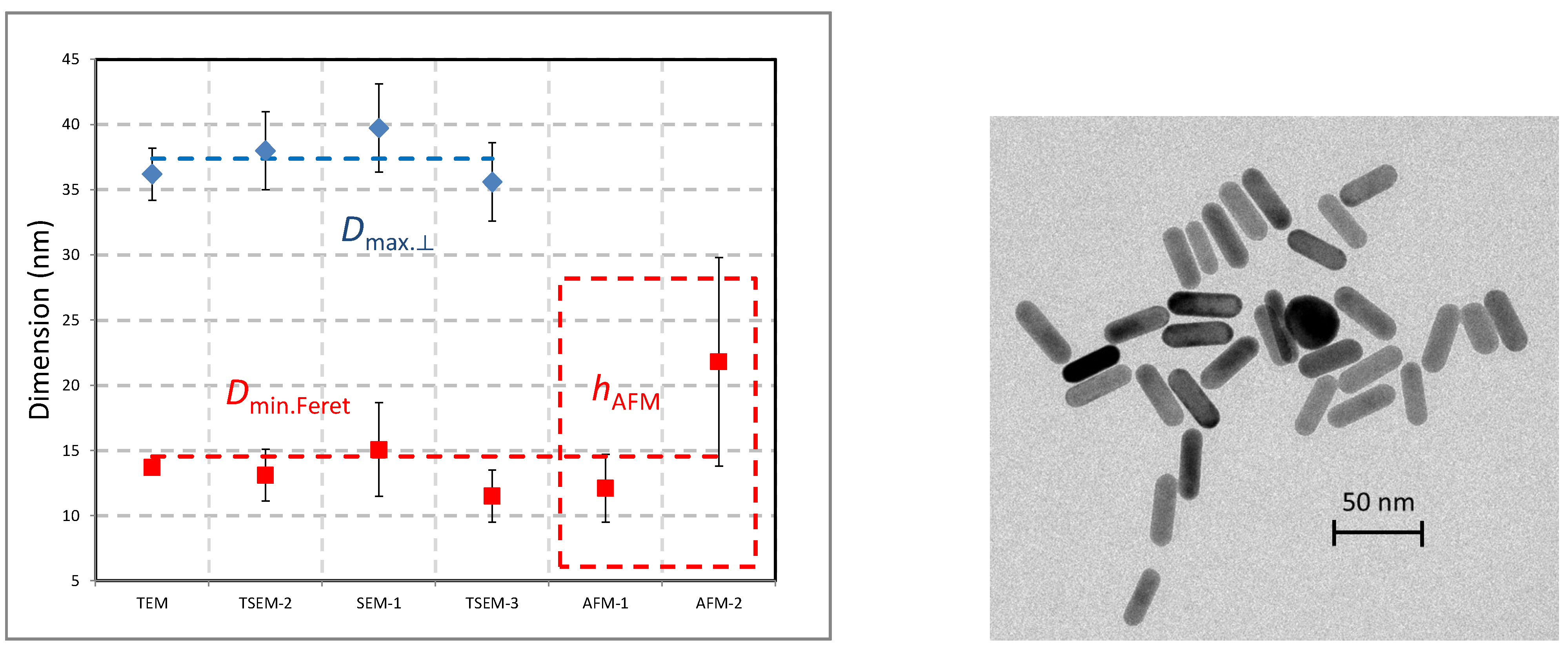
| nPSize07 |  | L = length D = section diameter | DMinFeret, DMaxFeret * | hAFM | - |
| Mesurand | Perfect Nanocubes | Total Population |
|---|---|---|
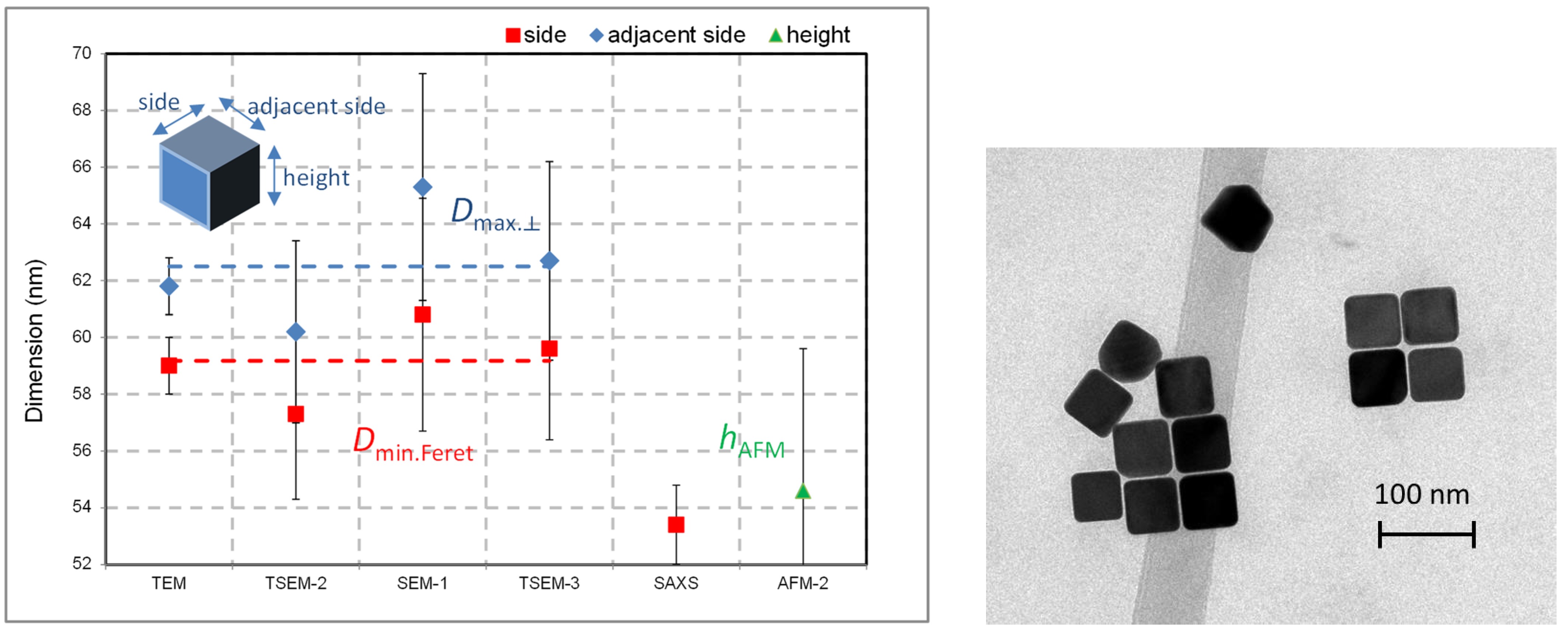
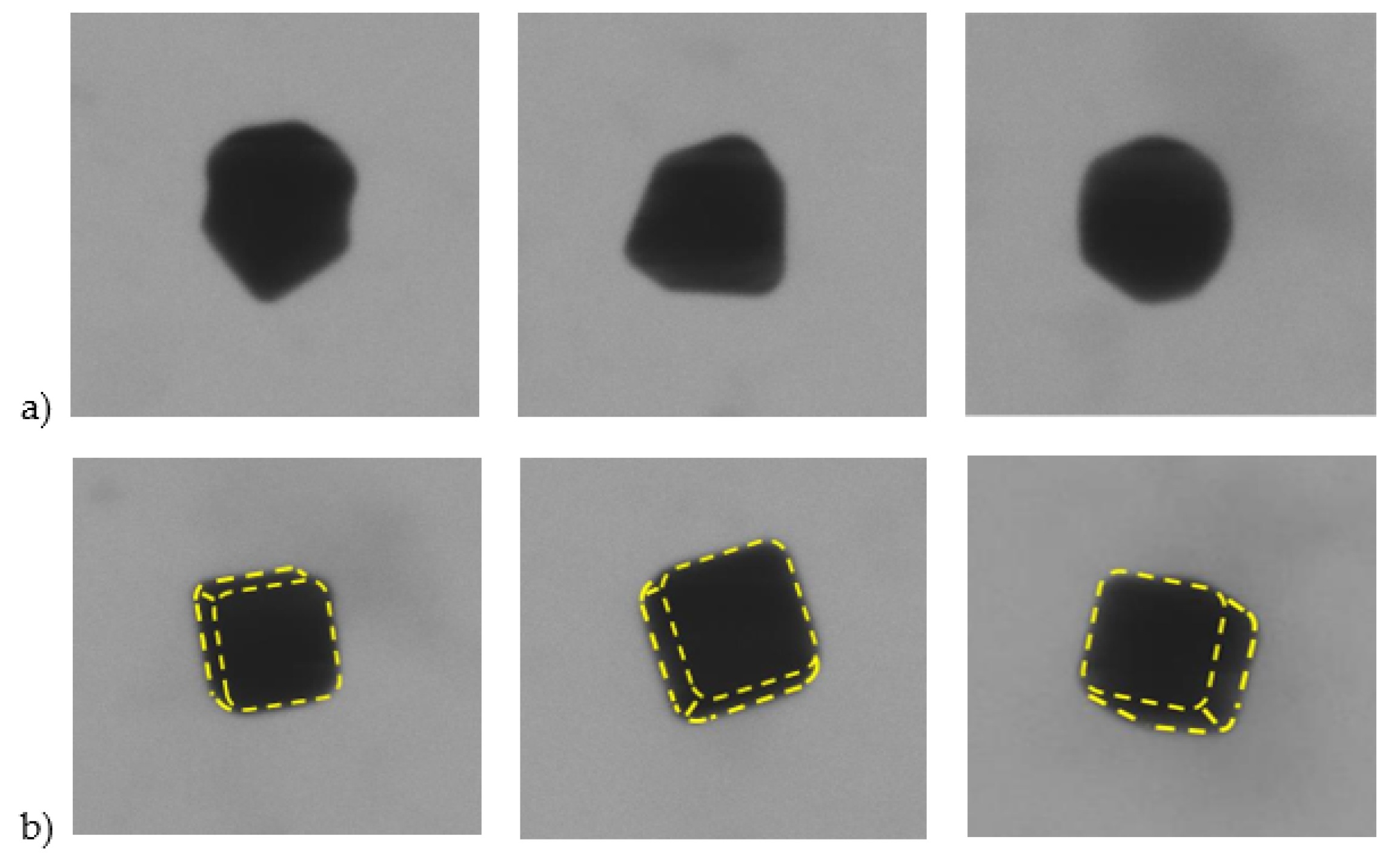
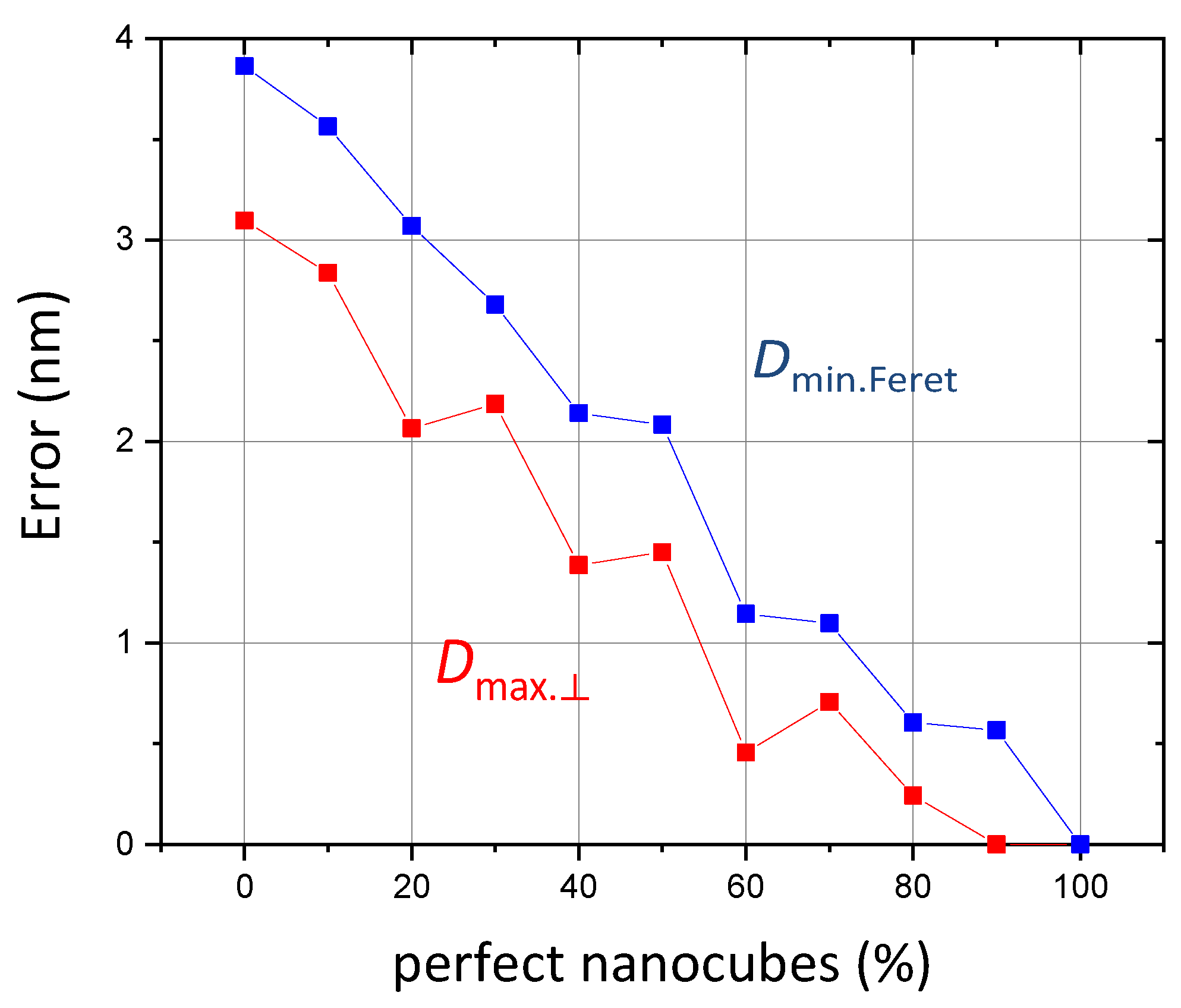
| Minimum Feret diameter | (59.9 ± 2.0) nm | (60.8 ± 2.1) nm |
| Maximum Feret diameter | (63.3 ± 1.9) nm | (65.3 ± 1.9) nm |
| Type of the Nanoparticulate Material | EM | EM + AFM | EM + AFM + SAXS | |||
|---|---|---|---|---|---|---|
| Deq | DMinFeret | DMaxFeret | Deq/hAFM DMinFeret/hAFM | DMinFeret/DSAXS Deq/DSAXS/hAFM | ||
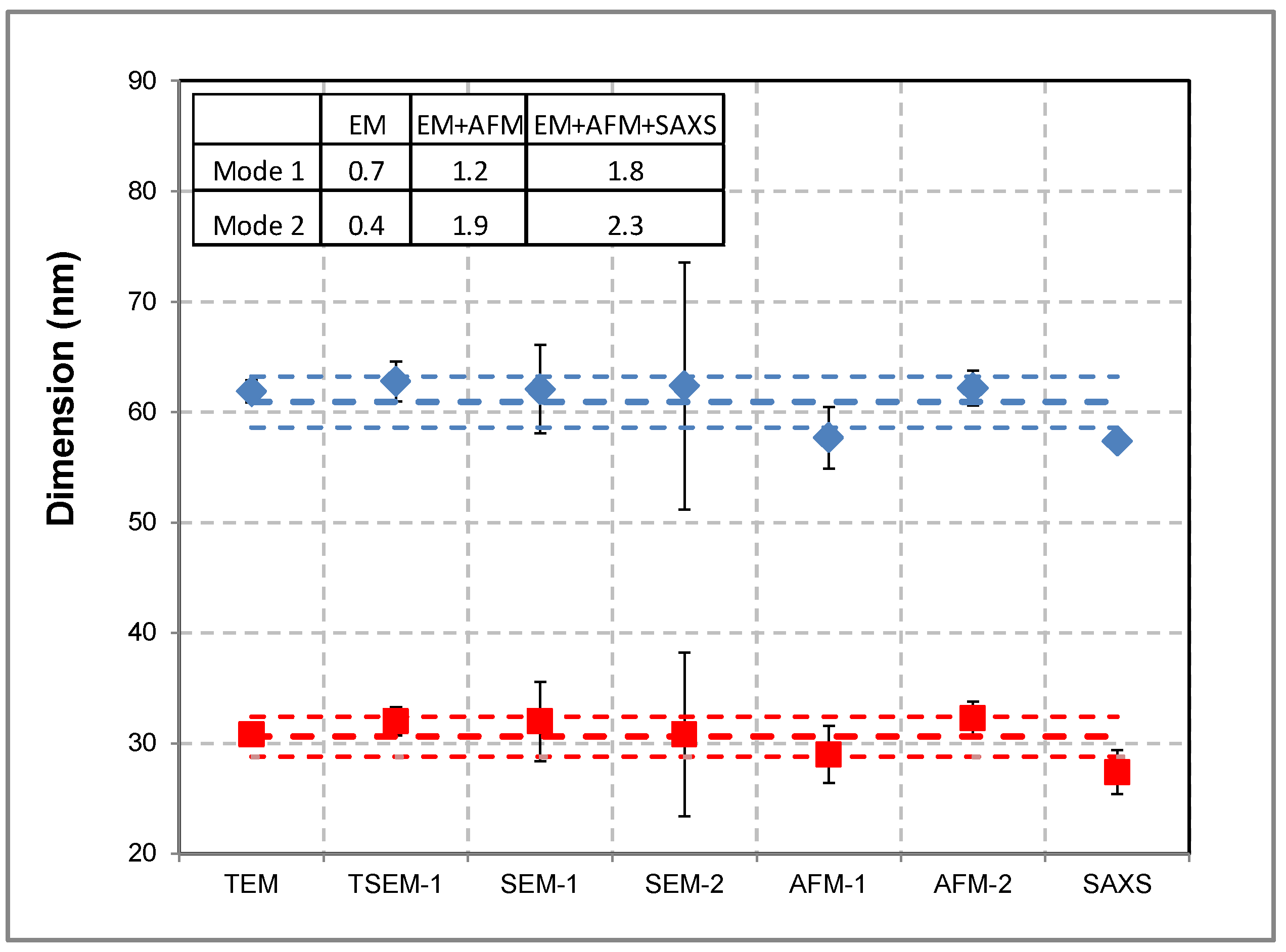
| Spherical shape | Bimodal population | <1 nm | - | - | <2 nm | ≈2 nm |
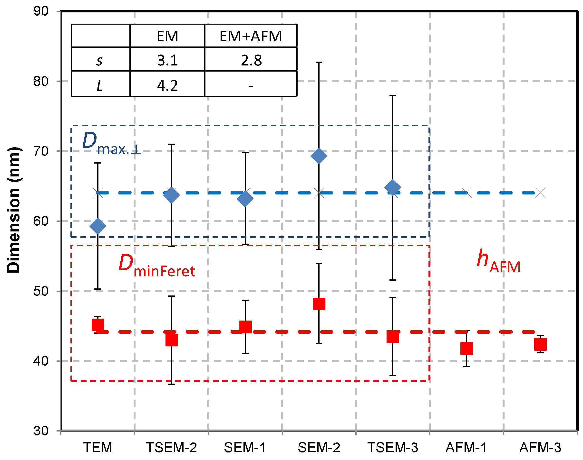
| Complex shape | Bipyramids | - | ≈2.5 nm | ≈5 nm (depends on the orientation) | ≈2.5 nm (for s) | ≈2.5 nm (for s) and ≈5 nm (for L) |
| Nanocubes | - | <2 nm | < 2 nm | <2.5 nm | - | |
| Nanorods | - | <2 nm | ≈2 nm | ≈4 nm | - | |
Disclaimer/Publisher’s Note: The statements, opinions and data contained in all publications are solely those of the individual author(s) and contributor(s) and not of MDPI and/or the editor(s). MDPI and/or the editor(s) disclaim responsibility for any injury to people or property resulting from any ideas, methods, instructions or products referred to in the content. |
© 2023 by the authors. Licensee MDPI, Basel, Switzerland. This article is an open access article distributed under the terms and conditions of the Creative Commons Attribution (CC BY) license (https://creativecommons.org/licenses/by/4.0/).
Share and Cite
Feltin, N.; Crouzier, L.; Delvallée, A.; Pellegrino, F.; Maurino, V.; Bartczak, D.; Goenaga-Infante, H.; Taché, O.; Marguet, S.; Testard, F.; et al. Metrological Protocols for Reaching Reliable and SI-Traceable Size Results for Multi-Modal and Complexly Shaped Reference Nanoparticles. Nanomaterials 2023, 13, 993. https://doi.org/10.3390/nano13060993
Feltin N, Crouzier L, Delvallée A, Pellegrino F, Maurino V, Bartczak D, Goenaga-Infante H, Taché O, Marguet S, Testard F, et al. Metrological Protocols for Reaching Reliable and SI-Traceable Size Results for Multi-Modal and Complexly Shaped Reference Nanoparticles. Nanomaterials. 2023; 13(6):993. https://doi.org/10.3390/nano13060993
Chicago/Turabian StyleFeltin, Nicolas, Loïc Crouzier, Alexandra Delvallée, Francesco Pellegrino, Valter Maurino, Dorota Bartczak, Heidi Goenaga-Infante, Olivier Taché, Sylvie Marguet, Fabienne Testard, and et al. 2023. "Metrological Protocols for Reaching Reliable and SI-Traceable Size Results for Multi-Modal and Complexly Shaped Reference Nanoparticles" Nanomaterials 13, no. 6: 993. https://doi.org/10.3390/nano13060993
APA StyleFeltin, N., Crouzier, L., Delvallée, A., Pellegrino, F., Maurino, V., Bartczak, D., Goenaga-Infante, H., Taché, O., Marguet, S., Testard, F., Artous, S., Saint-Antonin, F., Salzmann, C., Deumer, J., Gollwitzer, C., Koops, R., Sebaïhi, N., Fontanges, R., Neuwirth, M., ... Hodoroaba, V.-D. (2023). Metrological Protocols for Reaching Reliable and SI-Traceable Size Results for Multi-Modal and Complexly Shaped Reference Nanoparticles. Nanomaterials, 13(6), 993. https://doi.org/10.3390/nano13060993


