
3D-ZnO Superstructure Decorated with Carbon-Based Material for Efficient Photoelectrochemical Water-Splitting under Visible-Light Irradiation

 , , ,
, , ,
Abstract
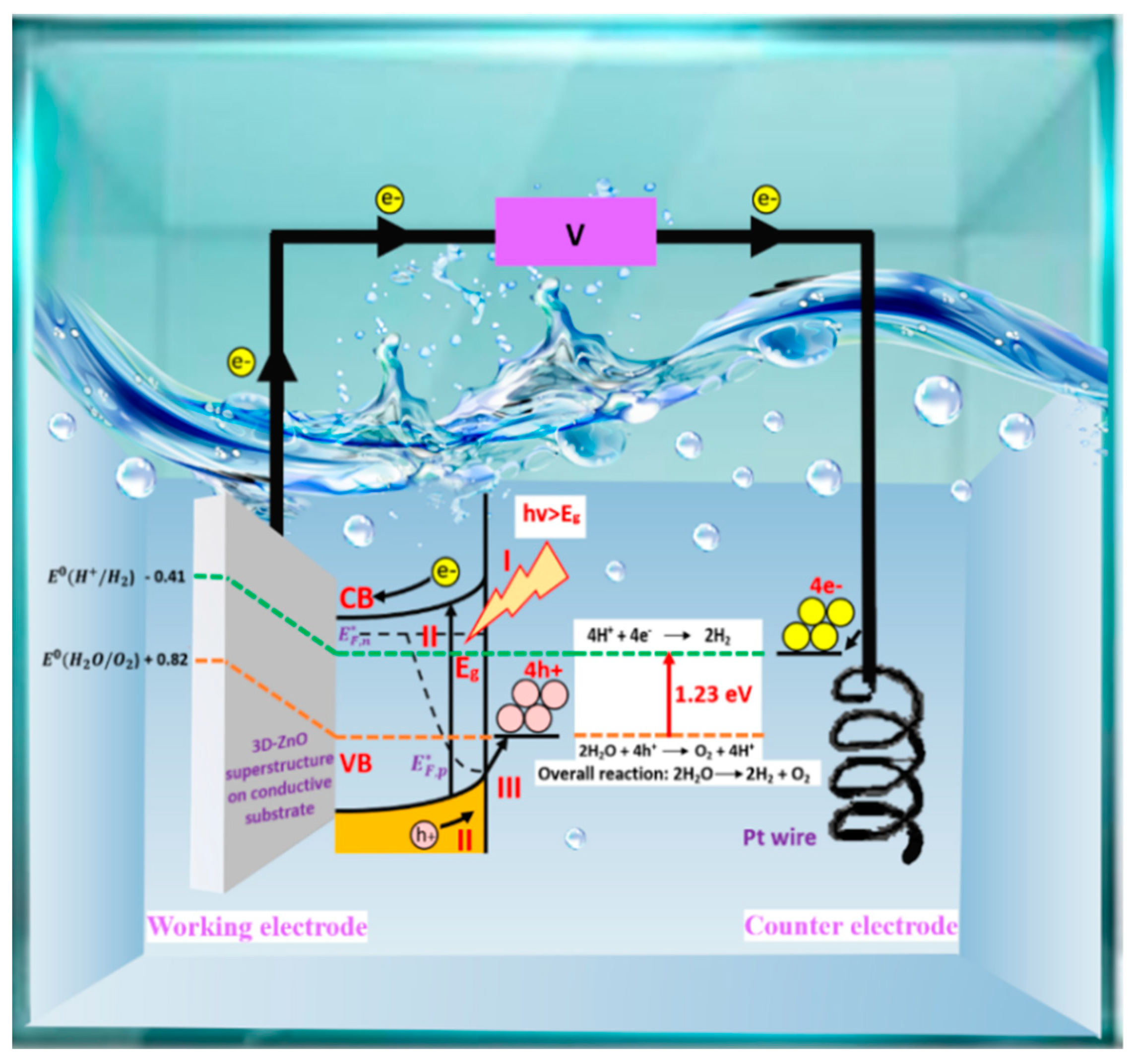
:1. Introduction
2. 3D-ZnO Superstructures’ Synthesis and Their PEC Performance
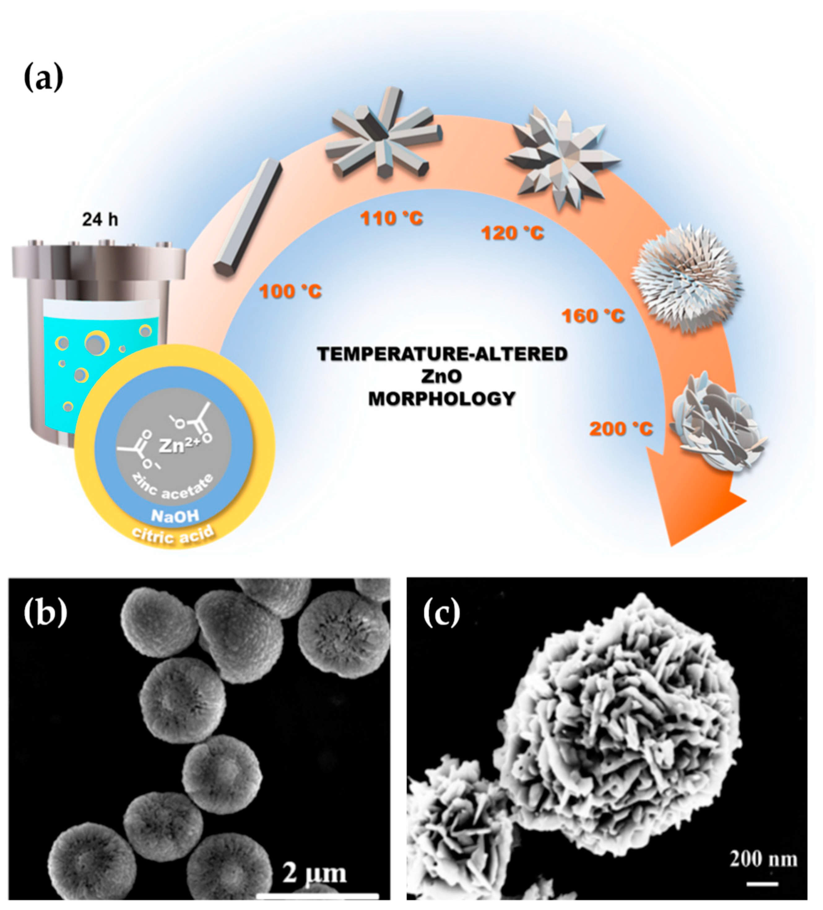
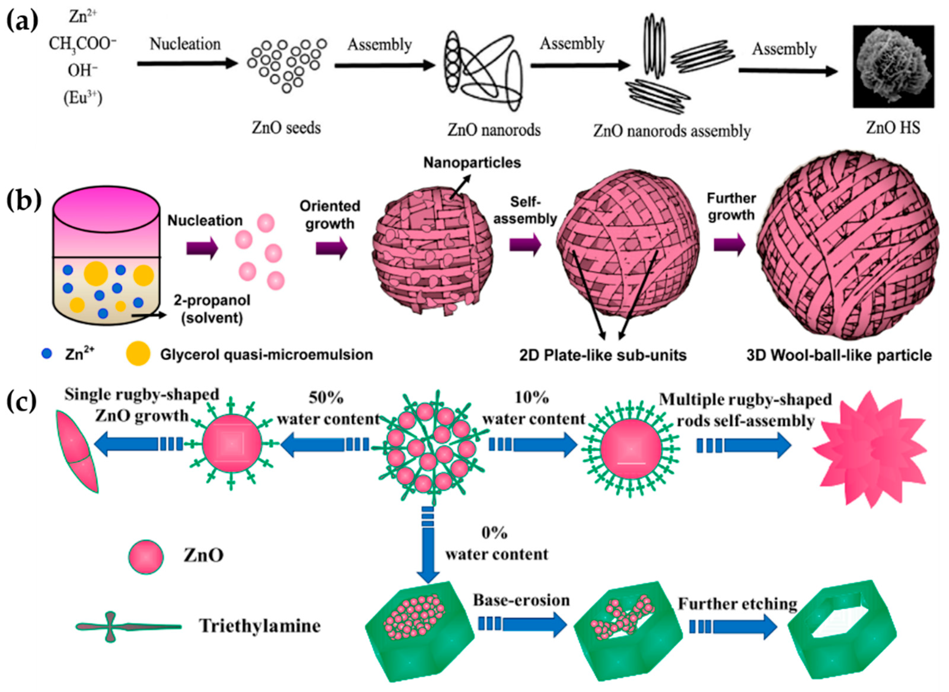
2.1. Nanoflower-like Morphology
2.2. 3D Wool Ball-like
2.3. Hexagonal Ring-like
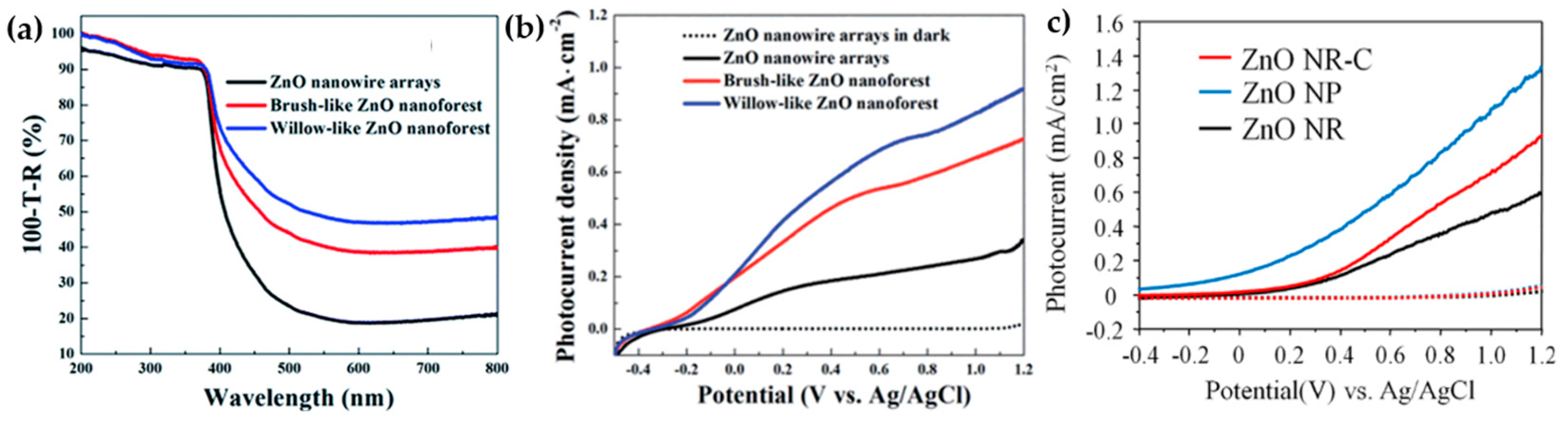
3. Morphological Effect of 3D-ZnO Superstructures on PEC Water-Splitting Performances
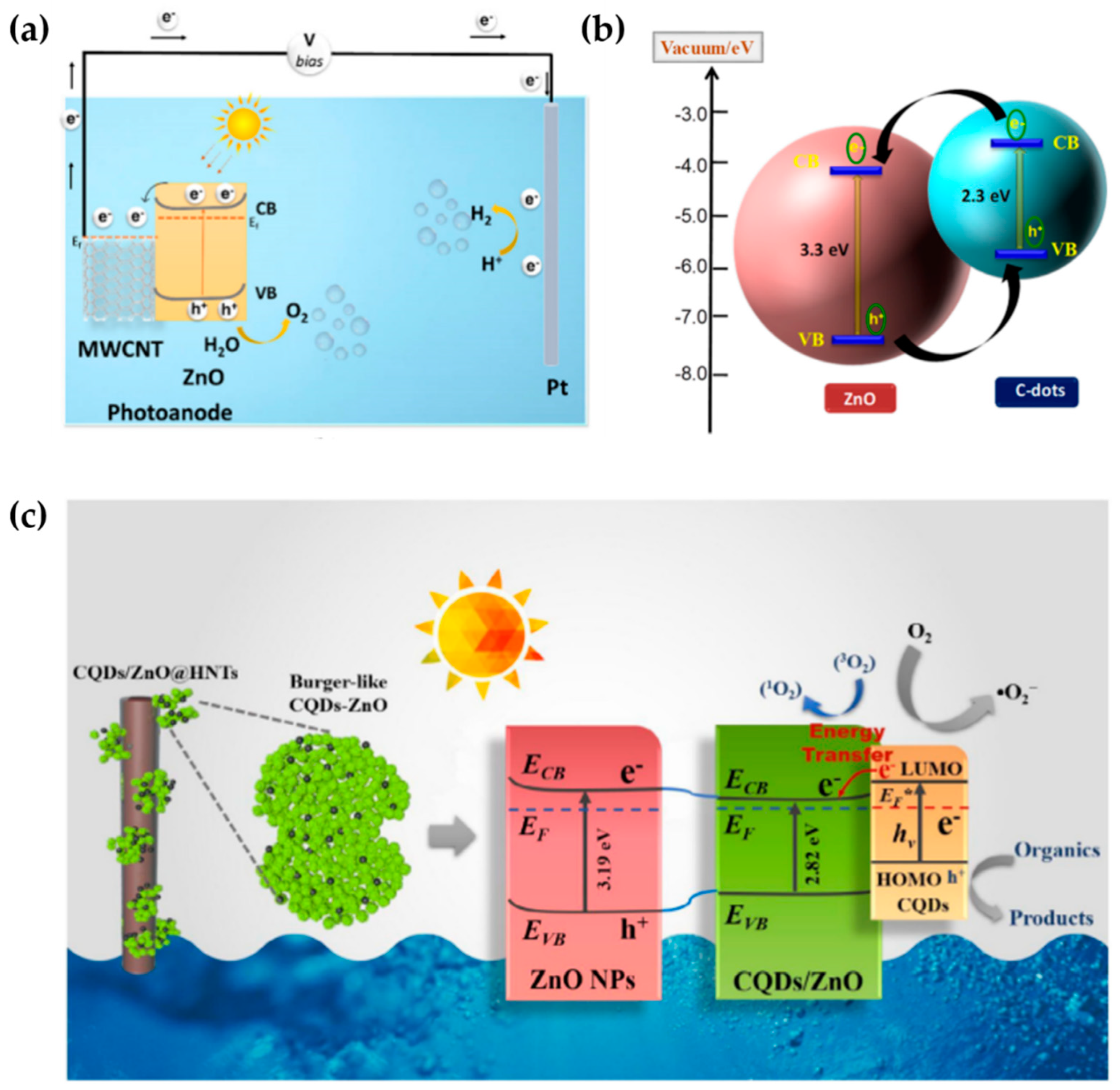
4. A Recent Modification of 3D-ZnO Superstructures by Carbon-Based Material
5. Conclusions and Future Perspectives
Author Contributions
Funding
Data Availability Statement
Acknowledgments
Conflicts of Interest
References
- Joy, J.; Mathew, J.; George, S.C. Nanomaterials for Photoelectrochemical Water Splitting—Review. Int. J. Hydrogen Energy 2018, 43, 4804–4817. [Google Scholar] [CrossRef]
- Mahala, C.; Sharma, M.D.; Basu, M. Type-II Heterostructure of ZnO and Carbon Dots Demonstrates Enhanced Photoanodic Performance in Photoelectrochemical Water Splitting. Inorg. Chem. 2020, 59, 6988–6999. [Google Scholar] [CrossRef] [PubMed]
- Dincer, I.; Acar, C. Review and Evaluation of Hydrogen Production Methods for Better Sustainability. Int. J. Hydrogen Energy 2015, 40, 11094–11111. [Google Scholar] [CrossRef]
- Le Caër, S. Water Radiolysis: Influence of Oxide Surfaces on H2 Production under Ionizing Radiation. Water 2011, 3, 235–253. [Google Scholar] [CrossRef]
- Keipi, T.; Tolvanen, H.; Konttinen, J. Economic Analysis of Hydrogen Production by Methane Thermal Decomposition: Comparison to Competing Technologies. Energy Convers. Manag. 2018, 159, 264–273. [Google Scholar] [CrossRef]
- Touloupakis, E.; Faraloni, C.; Benavides, A.M.S.; Torzillo, G. Recent Achievements in Microalgal Photobiological Hydrogen Production. Energies 2021, 14, 7170. [Google Scholar] [CrossRef]
- Fereidooni, M.; Mostafaeipour, A.; Kalantar, V.; Goudarzi, H. A Comprehensive Evaluation of Hydrogen Production from Photovoltaic Power Station. Renew. Sustain. Energy Rev. 2018, 82, 415–423. [Google Scholar] [CrossRef]
- Gupta, B.; Melvin, A.A.; Matthews, T.; Dash, S.; Tyagi, A.K. Facile Gamma Radiolytic Synthesis of Synergistic Co3O4-RGO Nanocomposite: Direct Use in Photocatalytic Water Splitting. Mater. Res. Express 2014, 1, 045507. [Google Scholar] [CrossRef]
- Jiang, C.; Moniz, S.J.A.; Wang, A.; Zhang, T.; Tang, J. Photoelectrochemical Devices for Solar Water Splitting-Materials and Challenges. Chem. Soc. Rev. 2017, 46, 4645–4660. [Google Scholar] [CrossRef]
- Peerakiatkhajohn, P.; Yun, J.-H.; Wang, S.; Wang, L. Review of Recent Progress in Unassisted Photoelectrochemical Water Splitting: From Material Modification to Configuration Design. J. Photonics Energy 2016, 7, 012006–012027. [Google Scholar] [CrossRef]
- Wang, Y.; Suzuki, H.; Xie, J.; Tomita, O.; Martin, D.J.; Higashi, M.; Kong, D.; Abe, R.; Tang, J. Mimicking Natural Photosynthesis: Solar to Renewable H2 Fuel Synthesis by Z-Scheme Water Splitting Systems. Chem. Rev. 2018, 118, 5201–5241. [Google Scholar] [CrossRef]
- Ng, B.J.; Putri, L.K.; Kong, X.Y.; Teh, Y.W.; Pasbakhsh, P.; Chai, S.P. Z-Scheme Photocatalytic Systems for Solar Water Splitting. Adv. Sci. 2020, 7, 1903171–1903213. [Google Scholar] [CrossRef] [PubMed]
- Kaur, G.; Divya; Satsangi, V.R.; Dass, S.; Shrivastav, R. 3D-Nano-Hetero-Structured n/n Junction, CuO/Ru–ZnO Thin Films, for Hydrogen Generation with Enhanced Photoelectrochemical Performances. Int. J. Hydrogen Energy 2020, 45, 21051–21067. [Google Scholar] [CrossRef]
- Pan, H.; Zhu, S.; Lou, X.; Mao, L.; Lin, J.; Tian, F.; Zhang, D. Graphene-Based Photocatalysts for Oxygen Evolution from Water. RSC Adv. 2015, 5, 6543–6552. [Google Scholar] [CrossRef]
- Sohila, S.; Rajendran, R.; Yaakob, Z.; Teridi, M.A.M.; Sopian, K. Photoelectrochemical Water Splitting Performance of Flower-like ZnO Nanostructures Synthesized by a Novel Chemical Method. J. Mater. Sci. Mater. Electron. 2016, 27, 2846–2851. [Google Scholar] [CrossRef]
- Lv, R.; Wang, T.; Su, F.; Zhang, P.; Li, C.; Gong, J. Facile Synthesis of ZnO Nanopencil Arrays for Photoelectrochemical Water Splitting. Nano Energy 2014, 7, 143–150. [Google Scholar] [CrossRef]
- Guo, C.; Xie, J.; Yang, H.; Science, C.L.-A. Au@CdSCore–Shell Nanoparticles-Modified ZnO Nanowires Photoanode for Efficient Photoelectrochemical Water Splitting. Wiley Online Libr. 2015, 2, 1500135. [Google Scholar] [CrossRef]
- Pratomo, U.; Adhia, R.; Pramudya, A.; Kadja, G.T.M.; Khalil, M.; Primadona, I. Enhancement of the ZnO Nanotube Photoelectrochemical Performance by MXene Layer. Mater. Lett. 2023, 337, 133932. [Google Scholar] [CrossRef]
- Giasari, A.S.; Maharani Muharam, A.P.; Syampurwadi, A.; Dedi; Eddy, D.R.; Primadona, I. Morphological Effect of One-Dimensional ZnO Nanostructures on the Photocatalytic Activity. J. Phys. Chem. Solids 2023, 176, 111259. [Google Scholar] [CrossRef]
- Sun, X.; Li, Q.; Jiang, J.; Mao, Y. Nanoscale Morphology-Tunable Synthesis of ZnO Nanoforest and Its Photoelectrochemical Performance. Nanoscale 2014, 6, 8769–8780. [Google Scholar] [CrossRef]
- Desai, M.A.; Vyas, A.N.; Saratale, G.D.; Sartale, S.D. Zinc Oxide Superstructures: Recent Synthesis Approaches and Application for Hydrogen Production via Photoelectrochemical Water Splitting. Int. J. Hydrogen Energy 2019, 44, 2091–2127. [Google Scholar] [CrossRef]
- Ma, J.; Liu, J.; Bao, Y.; Zhu, Z.; Wang, X.; Zhang, J. Synthesis of Large-Scale Uniform Mulberry-like ZnO Particles with Microwave Hydrothermal Method and Its Antibacterial Property. Ceram. Int. 2013, 39, 2803–2810. [Google Scholar] [CrossRef]
- Liu, H.R.; Shao, G.X.; Zhao, J.F.; Zhang, Z.X.; Zhang, Y.; Liang, J.; Liu, X.G.; Jia, H.S.; Xu, B.S. Worm-like Ag/ZnO Core-Shell Heterostructural Composites: Fabrication, Characterization, and Photocatalysis. J. Phys. Chem. C 2012, 116, 16182–16190. [Google Scholar] [CrossRef]
- Singh, S.; Srivastava, V.C.; Lo, S.L.; Mandal, T.K.; Naresh, G. Morphology-Controlled Green Approach for Synthesizing the Hierarchical Self-Assembled 3D Porous ZnO Superstructure with Excellent Catalytic Activity. Microporous Mesoporous Mater. 2017, 239, 296–309. [Google Scholar] [CrossRef]
- Yang, S.; Wang, J.; Li, X.; Zhai, H.; Han, D.; Wei, B.; Wang, D.; Yang, J. One-Step Synthesis of Bird Cage-like ZnO and Other Controlled Morphologies: Structural, Growth Mechanism and Photocatalytic Properties. Appl. Surf. Sci. 2014, 319, 211–215. [Google Scholar] [CrossRef]
- Qu, J.; Yang, Y.; Wu, Q.; Coxon, P.R.; Liu, Y.; He, X.; Xi, K.; Yuan, N.; Ding, J. Hedgehog-like Hierarchical ZnO Needle-Clusters with Superior Electron Transfer Kinetics for Dye-Sensitized Solar Cells. RSC Adv. 2014, 4, 11430–11437. [Google Scholar] [CrossRef]
- Ma, W.; Gu, Z.; Nan, H.; Geng, B.; Zhang, X. Morphology-Controllable Synthesis of 3D Firecracker-like ZnO Nanoarchitectures for High Catalytic Performance. CrystEngComm 2015, 17, 1121–1128. [Google Scholar] [CrossRef]
- Pan, X.; Liu, X.; Bermak, A.; Fan, Z. Self-Gating Effect Induced Large Performance Improvement of ZnO Nanocomb Gas Sensors. ACS Nano 2013, 7, 9318–9324. [Google Scholar] [CrossRef]
- Yao, Y.F.; Tu, C.G.; Chang, T.W.; Chen, H.T.; Weng, C.M.; Su, C.Y.; Hsieh, C.; Liao, C.H.; Kiang, Y.W.; Yang, C.C. Growth of Highly Conductive Ga-Doped ZnO Nanoneedles. ACS Appl. Mater. Interfaces 2015, 7, 10525–10533. [Google Scholar] [CrossRef]
- Permatasari, F.A.; Irham, M.A.; Bisri, S.Z.; Iskandar, F. Carbon-Based Quantum Dots for Supercapacitors: Recent Advances and Future Challenges. Nanomaterials 2021, 11, 91. [Google Scholar] [CrossRef]
- Olak-Kucharczyk, M.; Szczepańska, G.; Kudzin, M.H.; Pisarek, M. The Photocatalytical Properties of RgO/TiO2 Coated Fabrics. Coatings 2020, 10, 1041. [Google Scholar] [CrossRef]
- Wen, P.; Sun, Y.; Li, H.; Liang, Z.; Wu, H.; Zhang, J.; Zeng, H.; Geyer, S.M.; Jiang, L. A Highly Active Three-Dimensional Z-Scheme ZnO/Au/g-C3N4 Photocathode for Efficient Photoelectrochemical Water Splitting. Appl. Catal. B Environ. 2020, 263, 118180. [Google Scholar] [CrossRef]
- Chen, T.W.; Kalimuthu, P.; Veerakumar, P.; Lin, K.C.; Chen, S.M.; Ramachandran, R.; Mariyappan, V.; Chitra, S. Recent Developments in Carbon-Based Nanocomposites for Fuel Cell Applications: A Review. Molecules 2022, 27, 761. [Google Scholar] [CrossRef] [PubMed]
- Facure, M.H.M.; Schneider, R.; Mercante, L.A.; Correa, D.S. A Review on Graphene Quantum Dots and Their Nanocomposites: From Laboratory Synthesis towards Agricultural and Environmental Applications. Environ. Sci. Nano 2020, 7, 3710–3734. [Google Scholar] [CrossRef]
- Xia, Y.; Wang, J.; Chen, R.; Zhou, D.; Xiang, L. A Review on the Fabrication of Hierarchical Zno Nanostructures for Photocatalysis Application. Crystals 2016, 6, 148. [Google Scholar] [CrossRef]
- Carminati, S.A.; Rodríguez-Gutiérrez, I.; De Morais, A.; Da Silva, B.L.; Melo, M.A.; Souza, F.L.; Nogueira, A.F. Challenges and Prospects about the Graphene Role in the Design of Photoelectrodes for Sunlight-Driven Water Splitting. RSC Adv. 2021, 11, 14374–14398. [Google Scholar] [CrossRef] [PubMed]
- Li, D.; Li, Y.; Zhang, Y.; Chang, F. Facile Synthesis of Three-Dimensional ZnO Hierarchical Microspheres Composed of Well-Ordered Nanorods by Hydrothermal Method. Results Phys. 2019, 12, 953–958. [Google Scholar] [CrossRef]
- Wang, X.; Ahmad, M.; Sun, H. Three-Dimensional ZnO Hierarchical Nanostructures: Solution Phase Synthesis and Applications. Materials 2017, 10, 1304–1326. [Google Scholar] [CrossRef] [PubMed]
- Fan, F.; Tang, P.; Wang, Y.; Feng, Y.; Chen, A.; Luo, R.; Li, D. Facile Synthesis and Gas Sensing Properties of Tubular Hierarchical ZnO Self-Assembled by Porous Nanosheets. Sens. Actuators B Chem. 2015, 215, 231–240. [Google Scholar] [CrossRef]
- Strano, V.; Greco, M.G.; Ciliberto, E.; Mirabella, S. Zno Microflowers Grown by Chemical Bath Deposition: A Low-Cost Approach for Massive Production of Functional Nanostructures. Chemosensors 2019, 7, 62–70. [Google Scholar] [CrossRef]
- Khan, M.F.; Ansari, A.H.; Hameedullah, M.; Ahmad, E.; Husain, F.M.; Zia, Q.; Baig, U.; Zaheer, M.R.; Alam, M.M.; Khan, A.M.; et al. Sol-Gel Synthesis of Thorn-like ZnO Nanoparticles Endorsing Mechanical Stirring Effect and Their Antimicrobial Activities: Potential Role as Nano-Antibiotics. Sci. Rep. 2016, 6, 27689. [Google Scholar] [CrossRef] [PubMed]
- Padvi, M.N.; Harale, N.S.; Patil, P.S.; Dhas, S.D.; Moholkar, A.V. Hydrothermal Synthesis of NO2 Gas-Sensitive and Hydrophobic Zinc Oxide Thin Films. J. Mater. Sci. Mater. Electron. 2021, 32, 3140–3154. [Google Scholar] [CrossRef]
- Zhu, L.; Li, Y.; Zeng, W. Hydrothermal Synthesis of Hierarchical Flower-like ZnO Nanostructure and Its Enhanced Ethanol Gas-Sensing Properties. Appl. Surf. Sci. 2018, 427, 281–287. [Google Scholar] [CrossRef]
- Ansari, M.S.; Maragani, R.; Banik, A.; Misra, R.; Qureshi, M. Enhanced Photovoltaic Performance Using Biomass Derived Nano 3D ZnO Hierarchical Superstructures and a D–A Type CS-Symmetric Triphenylamine Linked Bisthiazole. Electrochim. Acta 2018, 259, 262–275. [Google Scholar] [CrossRef]
- Ma, Q.; Fang, Y.; Liu, Y.; Song, J.; Fu, X.; Li, H.; Chu, S.; Chen, Y. Facile Synthesis of ZnO Morphological Evolution with Tunable Growth Habits: Achieving Superior Gas-Sensing Properties of Hemispherical ZnO/Au Heterostructures for Triethylamine. Phys. E Low-Dimens. Syst. Nanostruct. 2019, 106, 180–186. [Google Scholar] [CrossRef]
- Qu, X.; Yang, R.; Tong, F.; Zhao, Y.; Wang, M.H. Hierarchical ZnO Microstructures Decorated with Au Nanoparticles for Enhanced Gas Sensing and Photocatalytic Properties. Powder Technol. 2018, 330, 259–265. [Google Scholar] [CrossRef]
- Basnet, P.; Chatterjee, S. Structure-Directing Property and Growth Mechanism Induced by Capping Agents in Nanostructured ZnO during Hydrothermal Synthesis—A Systematic Review. Nano-Struct. Nano-Objects 2020, 22, 100426. [Google Scholar] [CrossRef]
- Ejsmont, A.; Goscianska, J. Hydrothermal Synthesis of ZnO Superstructures with Controlled Morphology via Temperature and PH Optimization. Materials 2023, 16, 1641. [Google Scholar] [CrossRef]
- Pérez Velasco, E.A.; Galindo, R.B.; Valdez Aguilar, L.A.; González Fuentes, J.A.; Puente Urbina, B.A.; Lozano Morales, S.A.; Valdés, S.S. Effects of the Morphology, Surface Modification and Application Methods of ZnO-NPs on the Growth and Biomass of Tomato Plants. Molecules 2020, 25, 1282–1292. [Google Scholar] [CrossRef]
- Ramya, M.; Nideep, T.K.; Nampoori, V.P.N.; Kailasnath, M. Solvent Assisted Evolution and Growth Mechanism of Zero to Three Dimensional ZnO Nanostructures for Dye-Sensitized Solar Cell Applications. Sci. Rep. 2021, 11, 6159. [Google Scholar] [CrossRef]
- Ghosh, S.; Sartape, R.; Chakraborty, J. Role of Dye-Induced Corrosion in Determining the Efficiency of ZnO-Based DSSC: The Case of ZnO Nanoforest in N719. J. Mater. Sci. Mater. Electron. 2020, 31, 2202–2220. [Google Scholar] [CrossRef]
- Babayevska, N.; Iatsunskyi, I.; Florczak, P.; Jarek, M.; Peplińska, B.; Jurga, S. Enhanced Photodegradation Activity of ZnO:Eu3+ and ZnO:Eu3+@Au 3D Hierarchical Structures. J. Rare Earths 2019, 38, 21–28. [Google Scholar] [CrossRef]
- Bao, Y.; Gao, L.; Feng, C.; Ma, J.; Liu, C.; Zhang, W. A Solvent-Dependent Fabrication of Flower-like and Hexagonally Ring-like ZnO Architectures in One Minute. Arab. J. Chem. 2019, 13, 4035–4042. [Google Scholar] [CrossRef]
- Kakarndee, S.; Nanan, S. SDS Capped and PVA Capped ZnO Nanostructures with High Photocatalytic Performance toward Photodegradation of Reactive Red (RR141) Azo Dye. J. Environ. Chem. Eng. 2018, 6, 74–94. [Google Scholar] [CrossRef]
- Li, M.; Bao, L.Z.; Yin, Z.C.; Ji, X.M. Heparin-Mediated Growth of Self-Organized ZnO Quasi-Microspheres with Twinned Donut-Like Hierarchical Structures. ChemistrySelect 2019, 4, 7805–7810. [Google Scholar] [CrossRef]
- Septiani, N.L.W.; Kaneti, Y.V.; Yuliarto, B.; Nugraha; Dipojono, H.K.; Takei, T.; You, J.; Yamauchi, Y. Hybrid Nanoarchitecturing of Hierarchical Zinc Oxide Wool-Ball-like Nanostructures with Multi-Walled Carbon Nanotubes for Achieving Sensitive and Selective Detection of Sulfur Dioxide. Sens. Actuators B Chem. 2018, 261, 241–251. [Google Scholar] [CrossRef]
- Odoom-Wubah, T.; Li, Q.; Wang, Q.; Rukhsana Usha, M.Z.; Huang, J.; Li, Q. Template-Free Synthesis of Carbon Self-Doped ZnO Superstructures as Efficient Support for Ultra Fine Pd Nanoparticles and Their Catalytic Activity towards Benzene Oxidation. Mol. Catal. 2019, 469, 118–130. [Google Scholar] [CrossRef]
- Begum, S.; Ahmaruzzaman, M.; Adhikari, P.P. Ecofriendly Bio-Synthetic Route to Synthesize ZnO Nanoparticles Using Eryngium Foetidum L. and Their Activity against Pathogenic Bacteria. Mater. Lett. 2018, 228, 37–41. [Google Scholar] [CrossRef]
- Jiang, X.H.; Ma, S.Y.; Li, W.Q.; Wang, T.T.; Jin, W.X.; Luo, J.; Cheng, L.; Mao, Y.Z.; Zhang, M. Synthesis of Hierarchical ZnO Nanostructure Assembled by Nanorods and Their Performance for Gas Sensing. Mater. Lett. 2015, 142, 299–303. [Google Scholar] [CrossRef]
- Shi, W.; Shang, Y.; Maruf Ahmed, M.; Zhao, R.; Li, S.; Du, J.; Li, J. A Facile Controllable Self-Assembly of 3D Elliptical ZnO Microspheres from 1D Nanowires for Effective Detection of Acetone. Mater. Lett. 2020, 270, 127706. [Google Scholar] [CrossRef]
- Guo, S.; Zhao, X.; Zhang, W.; Wang, W. Optimization of Electrolyte to Significantly Improve Photoelectrochemical Water Splitting Performance of ZnO Nanowire Arrays. Mater. Sci. Eng. B 2018, 227, 129–135. [Google Scholar] [CrossRef]
- Liu, Z.; Cai, Q.; Ma, C.; Zhang, J.; Liu, J. Photoelectrochemical Properties and Growth Mechanism of Varied ZnO Nanostructures. New J. Chem. 2017, 41, 7947–7952. [Google Scholar] [CrossRef]
- Niu, H.; Zhou, D.; Yang, X.; Li, X.; Wang, Q.; Qu, F. Towards Three-Dimensional Hierarchical ZnO Nanofiber@Ni(OH)2 NanoflakeCore–Shell Heterostructures for High-Performance Asymmetric Supercapacitors. J. Mater. Chem. 2015, 3, 18413–18421. [Google Scholar] [CrossRef]
- Yuan, C.; Lin, H.; Lu, H.; Xing, E.; Zhang, Y.; Xie, B. Electrodeposition of Three-Dimensional ZnO@ MnO2 Core–Shell Nanocables as High-Performance Electrode Material for Supercapacitors. Energy 2015, 93, 1259–1266. [Google Scholar] [CrossRef]
- Chen, X.; Lin, P.; Yan, X.; Bai, Z.; Yuan, H.; Shen, Y.; Liu, Y.; Zhang, G.; Zhang, Z.; Zhang, Y. Three-Dimensional Ordered ZnO/Cu2O Nanoheterojunctions for Efficient Metal–Oxide Solar Cells. ACS Appl. Mater. Interfaces 2015, 7, 3216–3223. [Google Scholar] [CrossRef] [PubMed]
- Men, X.; Chen, H.; Chang, K.; Fang, X.; Wu, C.; Qin, W.; Yin, S. Three-Dimensional Free-Standing ZnO/Graphene Composite Foam for Photocurrent Generation and Photocatalytic Activity. Appl. Catal. B Environ. 2016, 187, 367–374. [Google Scholar] [CrossRef]
- Sun, M.; Xu, Z.; Yin, M.; Lin, Q.; Lu, L.; Xue, X.; Zhu, X.; Cui, Y.; Fan, Z.; Ding, Y.; et al. Broad-Band Three-Dimensional Nanocave ZnO Thin Film Photodetectors Enhanced by Au Surface Plasmon Resonance. Nanoscale 2016, 8, 8924–8930. [Google Scholar] [CrossRef]
- Ikhmayies, S.J. Production of Three-Dimensional ZnO Multilayered Structures from Self-Assembled ZnO Microdiscs. JOM 2020, 72, 628–634. [Google Scholar] [CrossRef]
- Hmar, J.J.L.; Majumder, T.; Dhar, S.; Mondal, S.P. Sulfur and Nitrogen Co-Doped Graphene Quantum Dot Decorated ZnO Nanorod/Polymer Hybrid Flexible Device for Photosensing Applications. Thin Solid Film. 2016, 612, 274–283. [Google Scholar] [CrossRef]
- Indriyati; Primadona, I.; Permatasari, F.A.; Irham, M.A.; Nasir, M.; Iskandar, F. Recent Advances and Rational Design Strategies of Carbon Dots towards Highly Efficient Solar Evaporation. Nanoscale 2021, 13, 7523–7532. [Google Scholar] [CrossRef]
- Liu, C.; Qiu, Y.; Wang, F.; Li, L.; Liang, Q.; Chen, Z. Electrodeposition of ZnO Nanoflake-Based Photoanode Sensitized by Carbon Quantum Dots for Photoelectrochemical Water Oxidation. Ceram. Int. 2017, 43, 5329–5333. [Google Scholar] [CrossRef]
- Mansuriya, B.D.; Altintas, Z. Carbon Dots: Classification, Properties, Synthesis, Characterization, and Applications in Health Care-an Updated Review (2018–2021). Nanomaterials 2021, 11, 2525. [Google Scholar] [CrossRef] [PubMed]
- Tayyebi, A.; Soltani, T.; Lee, B.K.; Outokesh, M.; Tayebi, M. Novel Visible Light Photocatalytic and Photoelectrochemical (PEC) Activity of Carbon-Doped Zinc Oxide/Reduced Graphene Oxide: Supercritical Methanol Synthesis with Enhanced Photocorrosion Suppression. J. Alloys Compd. 2017, 723, 1001–1010. [Google Scholar] [CrossRef]
- Kim, J.K.; Bae, S.; Kim, W.; Jeong, M.J.; Lee, S.H.; Lee, C.L.; Choi, W.K.; Hwang, J.Y.; Park, J.H.; Son, D.I. Nano Carbon Conformal Coating Strategy for Enhanced Photoelectrochemical Responses and Long-Term Stability of ZnO Quantum Dots. Nano Energy 2015, 13, 258–266. [Google Scholar] [CrossRef]
- Shim, J.; Kim, J.K.; Lee, K.S.; Lee, C.L.; Ma, M.; Choi, W.K.; Hwang, J.Y.; Yang, H.Y.; Angadi, B.; Park, J.H.; et al. A Facile Chemical Synthesis of ZnO@multilayer Graphene Nanoparticles with Fast Charge Separation and Enhanced Performance for Application in Solar Energy Conversion. Nano Energy 2016, 25, 9–17. [Google Scholar] [CrossRef]
- Prasadam, V.P.; Flores, A.M.H.; Audinot, J.N.; Bahlawane, N. CNT-ZnO Core-Shell Photoanodes for Photoelectrochemical Water Splitting. Coatings 2022, 12, 47. [Google Scholar] [CrossRef]
- Pawar, A.U.; Kim, C.W.; Kang, M.J.; Kang, Y.S. Crystal Facet Engineering of ZnO Photoanode for the Higher Water Splitting Efficiency with Proton Transferable Nafion Film. Nano Energy 2016, 20, 156–167. [Google Scholar] [CrossRef]
- Li, J.; Liu, K.; Xue, J.; Xue, G.; Sheng, X.; Wang, H.; Huo, P.; Yan, Y. CQDS Preluded Carbon-Incorporated 3D Burger-like Hybrid ZnO Enhanced Visible-Light-Driven Photocatalytic Activity and Mechanism Implication. J. Catal. 2019, 369, 450–461. [Google Scholar] [CrossRef]
- Alam, S.; Sahu, T.K.; Gogoi, D.; Peela, N.R.; Qureshi, M. Bio-Template Assisted Hierarchical ZnO Superstructures Coupled with Graphene Quantum Dots for Enhanced Water Oxidation Kinetics. Sol. Energy 2020, 199, 39–46. [Google Scholar] [CrossRef]
- Wang, J.; Wang, G.; Jiang, J.; Wan, Z.; Su, Y.; Tang, H. Insight into Charge Carrier Separation and Solar-Light Utilization: RGO Decorated 3D ZnO Hollow Microspheres for Enhanced Photocatalytic Hydrogen Evolution. J. Colloid Interface Sci. 2020, 564, 322–332. [Google Scholar] [CrossRef]
- Ahmed, G.; Hanif, M.; Khan, A.J.; Zhao, L.; Zhang, J.; Liu, Z. ZnO Flowers and Graphene Oxide Hybridization for Efficient Photocatalytic Degradation of O-Xylene in Water. Mater. Chem. Phys. 2018, 212, 479–489. [Google Scholar] [CrossRef]
- Mahala, C.; Sharma, M.D.; Basu, M. ZnO Nanosheets Decorated with Graphite-Like Carbon Nitride Quantum Dots as Photoanodes in Photoelectrochemical Water Splitting. ACS Appl. Nano Mater. 2020, 3, 1999–2007. [Google Scholar] [CrossRef]
- Chandrasekaran, S.; Chung, J.S.; Kim, E.J.; Hur, S.H. Exploring Complex Structural Evolution of Graphene Oxide/ZnO Triangles and Its Impact on Photoelectrochemical Water Splitting. Chem. Eng. J. 2016, 290, 465–476. [Google Scholar] [CrossRef]
- Zhang, X.; Shakeel, M.; Li, B.; Arif, M.; Kong, X.; Liu, J.; Zuo, S. Fabrication of 3-D ZnO/CN Nanorods for Photo-/Electrocatalytic Water Splitting: An Efficient Morphology for Charge Carriers Transportation. Int. J. Hydrogen Energy 2019, 44, 21821–21836. [Google Scholar] [CrossRef]

| Method | Advantages | Limitations |
|---|---|---|
| Chemical bath deposition | Various substrates can be used to deposit samples and low cost | Solution waste is produced after each deposition |
| Electrochemical deposition | Easy to control the morphology and structure by adjusting electrochemical parameters | The growth substrate should be conductive |
| Hydrothermal | Low cost, green synthesis, simple equipment, and homogeneous production across a vast region | Higher temperature and reaction pressure |
| Co-precipitation | Rapid and low-cost | The rapid process results in the simultaneous nucleation and growth of ZnO, making it difficult to understand the specific growth process |
| Sol-gel | Mild synthesis condition, not expensive, and simple | Needs additional purification due to impurities from sol-gel matrix components |
| Growth Modifier | Role of Growth Modifier | Resultant Morphology | Ref. |
|---|---|---|---|
| Ammonia | OH− ions source and etching agent | Nanoforest | [51] |
| Trisodium citrate dihydrate | Directing agent | Nanoflower | [52] |
| Triethylamine | Directing agent | Hexagonal ring-like and flower-like | [53] |
| Sodium dodecyl sulfate | Capping agent | Plate-like | [54] |
| Polygalacturonic acid | Templating and directing agent | Multi-cage like | [44] |
| CTAB | Surfactant | Flower-like | [43] |
| Heparin | Biotemplate and chelating agent | Quasi-microsphere and twinned donut-like hemispheres | [55] |
| Glycol | Directing agent | Hemi-spherical | [45] |
| Glycerol | Crystal growth director | Wool-ball-like | [56] |
| Cinnamon champora leaf | Bio-template | Spherical-like | [57] |
| Eryngium foetidum L. | Bio-template | Spherical-like | [58] |
| Material | Electrolyte & PEC Conditions | Results | Ref. |
|---|---|---|---|
| rGO-modified 3D-ZnO hollow microsphere | Na2SO3 (0.25 M)-Na2S (0.35 M), I = 100 mW/cm2 from 300 W xenon lamp | j = 0.1 mA/cm2 at 1 V vs. Ag/AgCl (2.7 times better than bare ZnO) | [80] |
| GO/ZnO flower-like hybrid composites | Na2SO4 (0.1 M), I = 100 mW/cm2 from 300 W xenon lamp | j = 0.09 mA/cm2 at 0.65 V vs. RHE (8 times better than bare ZnO) | [81] |
| g-C3N4 QDs-decorated ZnO nanosheets | Na2SO4 (0.5 M), I = 100 mW/cm2 from 300 W xenon lamp | j = 1.68 mA/cm2 at 1.2 V vs. RHE (1.3 times better than bare ZnO) | [82] |
| CNTs/ZnO core–shell nanocomposites | NaOH (0.1 M), I = 100 mW/cm2 from 300 W xenon lamp | j = 0.55 mA/cm2 at 1.23 V vs. RHE (458% better than bare ZnO) | [76] |
| GO-modified ZnO triangles | NaOH (1.0 M), UV light ~360 nm | j = 1.52 mA/cm2 at 1.45 V vs. RHE (2.08 times better than bare ZnO) | [83] |
| GQDs/bio-template ZnO superstructures composites | NaOH (1.0 M), I = 100 mW/cm2 from 300 W tungsten halogen lamp | j = 0.61 mA/cm2 at 1.23 V vs. RHE (77% better than bare ZnO) | [79] |
| rGO/carbon-doped flower-like ZnO MRs | Na2SO4 (0.1 M), A 250 W Xe lamp (Oriel) with a 420 nm cut-off filter was used for excitation | j = 0.1 mA/cm2 at 1 V vs. RHE (20 times better than bare ZnO) | [73] |
| C60 (fullerene)/ZnO core–shell QDs | NaClO4 (0.5 M), I = 100 mW/cm2 from 300 W xenon lamp | j = 0.235 mA/cm2 at 1.23 V vs. RHE (6 times better than bare ZnO) | [74] |
| MLG/ZnO core nanoparticle | NaClO4 (0.5 M), I = 100 mW/cm2 from 300 W xenon lamp | j = 0.13 mA/cm2 at 0.2 V vs. Ag/AgCl (4.3 times better than bare ZnO) | [75] |
Disclaimer/Publisher’s Note: The statements, opinions and data contained in all publications are solely those of the individual author(s) and contributor(s) and not of MDPI and/or the editor(s). MDPI and/or the editor(s) disclaim responsibility for any injury to people or property resulting from any ideas, methods, instructions or products referred to in the content. |
© 2023 by the authors. Licensee MDPI, Basel, Switzerland. This article is an open access article distributed under the terms and conditions of the Creative Commons Attribution (CC BY) license (https://creativecommons.org/licenses/by/4.0/).
Share and Cite
Pratomo, U.; Pratama, R.A.; Irkham, I.; Sulaeman, A.P.; Mulyana, J.Y.; Primadona, I. 3D-ZnO Superstructure Decorated with Carbon-Based Material for Efficient Photoelectrochemical Water-Splitting under Visible-Light Irradiation. Nanomaterials 2023, 13, 1380. https://doi.org/10.3390/nano13081380
Pratomo U, Pratama RA, Irkham I, Sulaeman AP, Mulyana JY, Primadona I. 3D-ZnO Superstructure Decorated with Carbon-Based Material for Efficient Photoelectrochemical Water-Splitting under Visible-Light Irradiation. Nanomaterials. 2023; 13(8):1380. https://doi.org/10.3390/nano13081380
Chicago/Turabian StylePratomo, Uji, Rifky Adhia Pratama, Irkham Irkham, Allyn Pramudya Sulaeman, Jacob Yan Mulyana, and Indah Primadona. 2023. "3D-ZnO Superstructure Decorated with Carbon-Based Material for Efficient Photoelectrochemical Water-Splitting under Visible-Light Irradiation" Nanomaterials 13, no. 8: 1380. https://doi.org/10.3390/nano13081380