Review

Recent Advances in ZnO Nanomaterial-Mediated Biological Applications and Action Mechanisms

1
College of Food and Biological Engineering, Chengdu University, Chengdu 610106, China
2
CAS Key Laboratory for Biomedical Effects of Nanomaterials and Nanosafety, Institute of High Energy Physics, Chinese Academy of Sciences, Beijing 100049, China
3
Clinical Medical College, Chengdu University, Chengdu 610106, China
*
Author to whom correspondence should be addressed.
Nanomaterials 2023, 13(9), 1500; https://doi.org/10.3390/nano13091500
Submission received: 28 March 2023 / Revised: 19 April 2023 / Accepted: 24 April 2023 / Published: 27 April 2023

Abstract

:
In recent years, with the deepening research, metal zinc oxide (ZnO) nanomaterials have become a popular research object in the biological field, particularly in biomedicine and food safety, which is attributed to their unique physicochemical properties such as high surface area and volume ratio, luminescence effect, surface characteristics and biological activities. Herein, this review provides a detailed overview of the ZnO nanomaterial-mediated biological applications that involve anti-bacterial, anti-tumor, anti-inflammation, skin care, biological imaging and food packaging applications. Importantly, the corresponding action mechanisms of ZnO nanomaterials are pointed. Additionally, the structure and structure-dependent physicochemical properties, the common synthesis methods and the biosafety of ZnO nanoparticles are revealed in brief. Finally, the significance and future challenges of ZnO nanomaterial applications are concluded.

1. Introduction

Nanoparticles are a kind of tiny particle with a particle size between 1 and 100 nm, including zero-dimensional structures (quantum dots and nanoclusters), one-dimensional structures (nanotubes and nanorods), two-dimensional structures (thin films) and three-dimensional structures (composites and nanofibers, etc.) [1]. The size of semiconductor inorganic particles down to the nanoscale significantly affects the physical, chemical and biological activity of the particles. ZnO is a multifunctional semiconductor material with a wide bandgap (3.37 eV) and large excitation binding energy (60 mV) at room temperature [2]. As more and more functions of ZnO nanomaterials are discovered, their main applications have expanded from industrial manufacturing to the biological field. In the background of antibiotic abuse and increased bacterial resistance nowadays, the multifunctional characteristics of ZnO nanoparticles, such as high levels of reactive oxygen species (ROS) production, inhibition of inflammatory factor release and inhibition of mast cell degranulation, make them a new promising candidate for anti-bacterial and anti-inflammatory drug development [3,4,5,6]. Meanwhile, their good antibacterial efficacy means they can be effectively extended to food packaging applications, and they are a well-documented Food and Drug Administration (FDA)-approved material [1]. Next, the large specific surface area, high drug loading rate, high selectivity and low toxicity of ZnO nanoparticles make them ideal drug delivery systems for targeted therapy, particularly for cancer therapy. Additionally, ZnO nanoparticles can absorb a large amount of UV light and thus have been widely used in sunscreen products. Furthermore, owing to their photoluminescence performance, ZnO nanoparticles have been widely used in bioimaging, which provides new tools for biomedical diagnosis. Thus, it seems that ZnO nanoparticles are already everywhere in daily life. Nowadays, studies on the biological applications of these ZnO nanomaterials are common, but a more comprehensive overview of them with a systematic mechanism summarization is still rare. Therefore, this review provides an overview of ZnO-based nanomaterials from action mechanisms to applications in biological fields. Firstly, the structure, as well as structure-dependent physicochemical properties and the common synthesis methods of ZnO nanoparticles, are revealed in brief. Then, the biological applications and action mechanisms of ZnO nanoparticles are summarized in detail, which involve anti-bacterial, anti-tumor, anti-inflammatory, bioimaging, skin care and food packaging applications (Figure 1). In addition, the biosafety of ZnO nanoparticles is mentioned. The hope is that such a review can help researchers to fully understand the progress and status of the field so as to promote future developments.

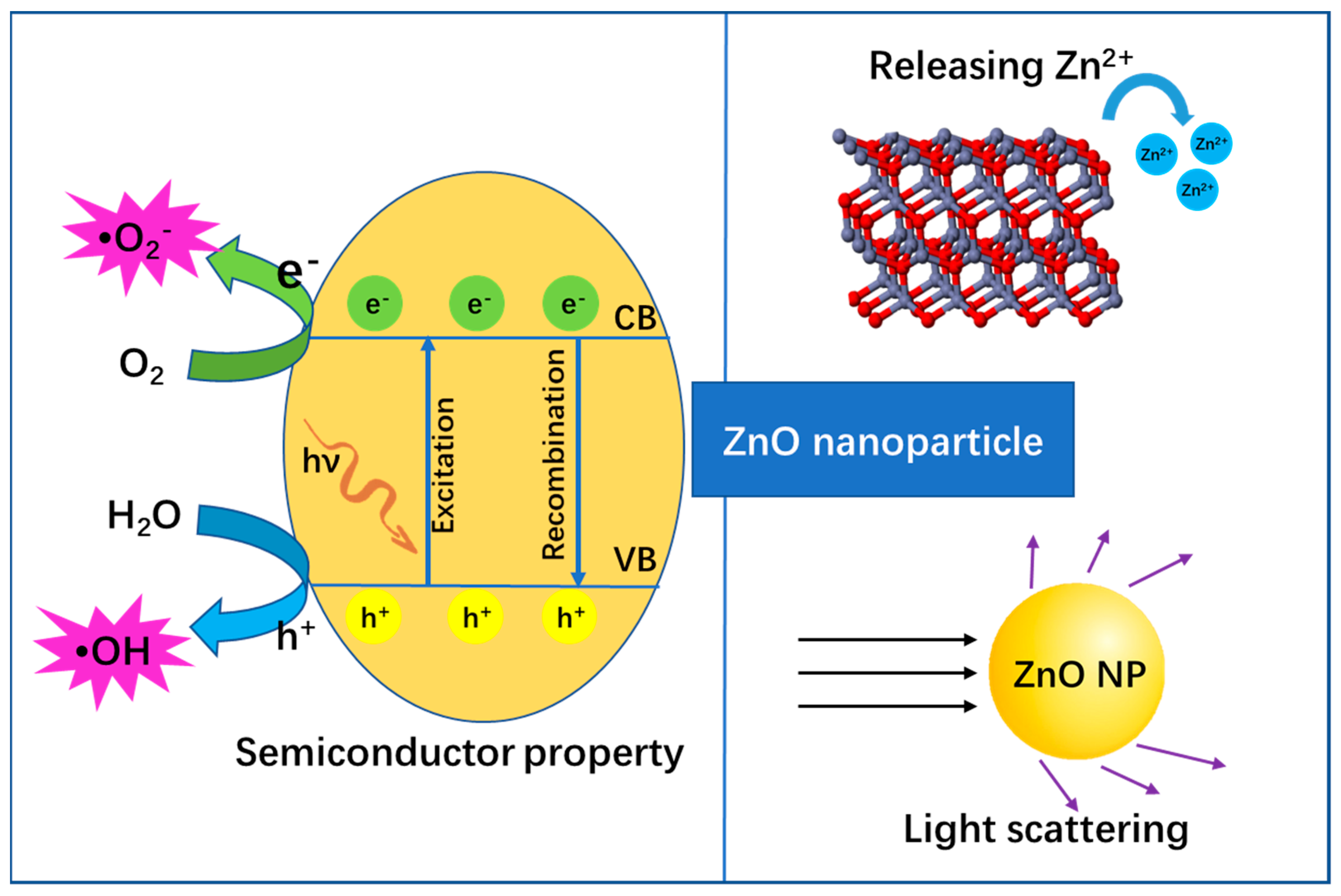
2. The Structure and Structure-Dependent Properties of ZnO Nanoparticles

It is well-known that ZnO is a typical semiconductor with a wurtzite crystal structure, which is the basis for its unique properties such as catalytic, electrical and optical properties [13]. For example, as seen in Figure 2, when ZnO is excited by light (e.g., ultra-violet (UV)), electron(e)-hole(h+) pairs are generated, where the excited e will jump into the conduction band (CB) and h+ is left in the valence band (VB). Subsequently, free radicals continuously generate from both the reductive pathway (e) and oxidative pathway (h+), where the e transfers to an acceptor (often O2) to produce •O2 and h+ transfers to a donor (often H2O) to produce •OH [14]. Both •O2 and •OH are physiologically relevant toxic ROS, which can attack biomacromolecules (e.g., DNA) and cell components [15]. Herein, this property endows ZnO nanoparticles with the ability for anti-bacterial and anti-cancer biomedical applications, etc. On the contrary, recombination of the e-–h+ pair emits photons, which allows ZnO nanoparticles to be applied in bio-imaging [14]. Next, as a metal oxide, ZnO can slowly release the metal ion of Zn2+ in a complex external environment. Such a metal ion can interact with protein, and thus can possibly inhibit enzyme activity to cause physiological dysfunction and eventually lead to cell death. Meanwhile, structural characteristics such as suitable size, surface charge effect, abundant surface modification and high specific surface area endow ZnO nanoparticles with multiple functions such as ready cell ingestion, target identification and efficient drug delivery. Therefore, these properties are also beneficial to a variety of biotherapy applications (e.g., anti-bacterial, anti-cancer, anti-inflammatory). In addition, the nano-sized ZnO possesses a light scattering and reflecting function, which is closely related to its sunscreen applications. In a word, the unique structural characteristics of ZnO nanoparticles determine its multifunctional performance and ultimately give its potential for rich biological applications.

3. The Common Synthesis Methods of ZnO Nanoparticles

Considering the large demand for ZnO nanoparticles for industry and living applications, the development of new synthesis methods to prepare uniform nano-sized ZnO is of great significance for both fundamental study and practical application. To date, a wide variety of ZnO nanoparticle preparation methods have been put forward, from physicochemical synthesis to green biogenic synthesis. Initially, physicochemical synthesis was the common preparation means, which mainly includes the methods of precipitation [16,17,18,19,20,21], electrochemical deposition [22,23,24], hydrothermal synthesis [25,26,27,28,29,30,31,32], sol-gel process [33,34], solid state reaction [35,36], chemical vapor synthesis [37], gel combustion [38], microemulsion [39], solvothermal synthesis [40,41,42], spray pyrolysis [43], sputtering [44,45,46], sonication [47,48], microwave [49], thermal evaporation [50,51,52] and so on. Each of these methods has its own advantages. Typically, the precipitation method is synthetically controlled. Additionally, it does not require complicated equipment and expensive raw materials, and thus is low cost and suitable for large-scale production [16,47]. Spray pyrolysis is suitable for large area preparation in a short span of time and its instrumentation is simple [43]. The solvothermal method is low-cost, simple, catalyst-free and seed-free [42]. The microwave approach has a high reaction selectivity, short reaction time, rapid volume heating and is energy-efficient [49]. The sol-gel way has the superiority of simplicity, repeatability and reliability. Importantly, the products of the sol-gel method exhibit the characteristics of high purity, low particle size and high specific surface area [21,53]. Hydrothermal synthesis is simple and can operate in a controlled manner, which is helpful to tailor the particles’ morphology via controlling the reaction rates of hydrolysis and condensation [53]. Moreover, the electrochemical method also is cost-effective and has a high purity of the final products. In particular, it is a low temperature operation process [22,23]. It is worth mentioning that in addition to their advantages, disadvantages also exist among these methods. For instance, the common physical methods of sputtering and thermal evaporation are expensive and consume high amounts of energy [19]. Traditional chemogenic syntheses such as hydrothermal synthesis, sol-gel process, chemical vapor synthesis, gel combustion and solvothermal synthesis usually need a high temperature atmosphere, and some of them must even be carried out in an expensive inertia environment. More seriously, most of them need toxic reagents or additives in the reaction process. As a result, the high temperature, complexity and toxicity of these methods limit their widespread application [17,36]. In this respect, our member once employed a catalyst-free and low-temperature oxidization method to prepared ZnO nanorod arrays [54]. The ZnO nanorod arrays were controlled growth on metal zinc foils and did not affect the conductive function of the zinc foils. The control of oxygen introduction played a key role in the success of this synthesis. This work provides great opportunities to investigate the effects of the material structure of these properties. Recently, an emerging concept named green synthesis has been highly preferred owing to its advantages of biocompatibility, biosafety, eco-friendliness, cost effectiveness and easiness to large-scale synthesis as compared with physicochemical methods [55,56]. The green synthesis of ZnO nanoparticles uses biological components as raw materials, such as plants extract, microorganisms and other bio-regenerable materials, which are regarded as excellent renewable sources. Additionally, the key mechanism of them is that the biomolecules derived from biological components are active compounds that serve as reducing and capping agents [55,57]. Currently, with cutting-edge developments in science and technology such as artificial intelligence, we believe that the potential of green synthesis can be more fully developed, and more and more new synthesis methods will emerge in the near future.

4. Bio-Application and Action Mechanisms of ZnO Nanomaterials

4.1. ZnO Nanomaterials for Anti-Bacterial and Anti-Fungal Applications

Both bacteria and fungi are microorganisms. The difference is, bacteria have no nucleus surrounded by nuclear membrane, which means they belong to prokaryotes, and fungi do have this, and belong to eukaryotes. The development of efficient bactericide and fungicide is of great significance in many fields such as oral care and cleansing, medical bandages, anti-infection, wound healing and plant pathogen-induced crop loss treatment. Herein, this section will introduce in detail the potential of ZnO nanoparticles for anti-bacterial and anti-fungal applications. It is worth mentioning that the anti-bacterial and anti-fungal mechanisms of ZnO nanoparticles are similar; we thus just introduce the anti-bacterial mechanism here.

4.1.1. Anti-Bacterial Mechanisms

ZnO is currently considered one of the most promising inorganic antimicrobial agents due to its low cytotoxicity, high selectivity and good heat resistance [3]. Although it has certain antibacterial activities, its antibacterial mechanism is not fully understood. To date, the existing view is that ZnO can cause bacterial death through direct damage to cells or indirect generation of ROS to inhibit protein function, destruct nucleic acid and produce harmful lipid peroxidation in bacteria [58]. By searching the literature, we summarize the main antibacterial mechanisms of ZnO nanoparticles as follows (Figure 3): (1) The direct interaction of nanoparticles with bacterial membrane leads to the destruction and rupture of the membrane’s integrity [59]. (2) Generating ROS in the cell to destroy the bacteria [4]. (3) Release of toxic Zn2+ ions to kill bacteria. Zn2+ has a special affinity to bacterial sulfhydryl groups. It can bind and oxidize the groups to inhibit glycolytic enzyme activity, and thereby inhibit the glycolytic pathway [60]. Thus, it can inhibit the action of bacterial respiratory enzymes and eventually lead to cell death. What is noteworthy is that for different types of bacteria, ZnO nanomaterials enter the bacteria in different ways. For Gram-positive bacteria, the Zn2+ in ZnO nanomaterials can be chelated by teichoic acid and lipoteichoic acid of the cell and translocate into cell. For Gram-negative bacteria, it has the porins that act as ion channels to promote passive diffusion into cell. (4) Disruption of the efflux pump and membrane potential on the cell membrane [61]. (5) Internalization of ZnO leads to intracellular DNA damage.

4.1.2. Anti-Bacterial and Anti-Fungal Applications

The size, morphology and even synthetic method of the nanoparticles will affect their antibacterial properties. Typically, the smaller particle size endows ZnO nanoparticles with the ability to more easily penetrate bacteria, and thus their antibacterial activity is stronger [3]. Based on this characteristic, many researchers have controlled the size of ZnO nanoparticles using various methods to improve their antibacterial activity. Lallo da Silva et al. obtained ZnO nanoparticles using the sol-gel method and used (3-glycidyloxypropyl) trimethoxysilane (GPTMS) as surface modifier [3]. The experimental results showed that the particle size was smaller and the antimicrobial performance of the material was stronger. Additionally, ZnO nanoparticles with a size of about 5 nm exhibited excellent anti-microbial activity. Verma et al. produced ZnO nanocrystalline via high energy ball milling (HEBM) and investigated the effect of size-dependent core–shell intrinsic defects to antibacterial activity [62]. The experimental results showed that the core shrunk in the ZnO core–shell structure with a decrease in size. Meanwhile, the antibacterial activity had a negative correlation between bacterial viability and the size of ZnO. Additionally, the morphology of the nanoparticles also affected their antibacterial activity. For example, Sharma et al. synthesized ZnO nanoparticles with various morphologies using an eco-friendly bio-mediated solution combustion method using different doses of Carica papaya milk (CPM) latex as fuel[59]. Interestingly, ZnO nanoparticles with the morphology of nano-flowers exhibit excellent photocatalytic activity over the other ZnO nanoparticles. As a result, ZnO nano-flowers manifest powerfully antibacterial activity against Staphylococcus aureus and Pseudomonas aeruginosa. Similarly, like the eco-friendly bio-mediated synthetic method above, researchers found that the synthesis of ZnO nanoparticles associated with plant extracts can obtain materials with ideal antibacterial effects, where plant-mediated ZnO green syntheses such as Psidium guajava leaf extract [63], Pedalium murex plant extract [64], Artemisia haussknechti leaf extract [65], Parthenium hysterophorus leaf extract [66], Gymnema sylvestre leaf extract [67], Aloe vera and Hibiscus sabdariffa plant extracts [68] have been reported. In addition, the synthetic method of ZnO nanoparticles is also closely related with their antibacterial activity. A typical example is that Mahendiran et al. proved that among all the ZnO nanoparticles they prepared, the best bactericidal activity was obtained with ZnO nanoparticles synthesized with biological methods rather than chemical methods [68].
Recently, researchers found that the applications of ZnO nanoparticles are limited due to their wide energy band gap and the high complexation of photogenerated electron-hole pairs. Fortunately, metals’ hybridization to form a complex, such as a combination of silver, gold, or palladium with metal oxides, can reduce their band gap [69]. Nguyen et al. synthesized silver-decorated ZnO nanomaterials, which made the absorption edge shift to the lower energy region of oxide nanoparticles and improved the antibacterial performance of the materials against Staphylococcus aureus (Gram-positive) and Escherichia coli (Gram-negative) bacteria [69]. In addition to the doping of other metal ions, ZnO nanomaterials are also widely used when compounded with other non-metal inorganic materials, such as ZnO/graphene oxide composites [2]. Compared with individual ZnO nanomaterial or graphene oxide alone as an antibacterial agent, the electrons in the ZnO/graphene oxide composite can be rapidly transferred between ZnO particles and graphene oxide, which facilitates the formation of ROS to disrupt the bacterial cell membrane, and thus enhances the antibacterial properties. Furthermore, ZnO nanomaterials can also be compounded with organic materials, such as Chitosan [70]. Chitosan is more widely used as a stabilizer due to its good biocompatibility and low toxicity. More importantly, the chitosan amine group possesses a positive charge, which promotes the complexation of chitosan-stabilized ZnO nanoparticles with the cell surface (negative charge), and thus produces a strengthened antibacterial effect.
In addition to anti-bacterial applications, research also shows that ZnO nanoparticles can be used as a fungicide [71,72,73,74,75,76,77,78,79,80,81,82]. For example, Khan et al. reported thorn-like ZnO nanoparticles via sol-gel synthesis, which showed an effective antifungal effect to Candida albicans [75]. Sharma et al. prepared ZnO nanoparticles by a simple homogeneous precipitation method without adding any chelating, surfactant or gelating agents. It was found that the antifungal activity against Candida albicans was enhanced with the increased concentration of ZnO nanoparticles [80]. Krishnamoorthy et al. prepared ZnO nanoparticles using Citrus hystrix leaf extract. The nanoparticles exhibited good antifungal activity against Candida albicans and Aspergillus niger at their higher concentration of 5 μg/mL [76]. Moreover, Ahmed et al. prepared ZnO nanoparticles using Azadirachta indica leaf extract for biocontrol of diseased lychee fruits. The disease-causing pathogens were identified as Aspergillus niger. In vitro results indicated that maximum mycelial growth inhibition was 70.5% under the ZnO nanoparticles (1.0 mg/mL). In vivo results obtained maximum suppression of lychee fruit rot under the same concentration [72]. Recently, in order to further improve the antifungal activity, ZnO nanocomposites have gradually come into researchers’ perspectives instead of pure ZnO nanoparticles. As an example, Roy et al. used ZnO nanoparticles, montmorillonite (MMT) and high density polyethylene (HDPE) to prepare HDPE/ZnO-MMT nanocomposites. This nanocomposite presented significant antifungal properties against black mold fungi Aspergillus niger at 5 wt% of loading [79]. Gondal et al. synthesized 5%Pd-doped nano-ZnO and compared its antifungal activity with that of pure nano-Zn. It demonstrated that the 5%Pd-doped nano-ZnO and pure nano-ZnO exhibited antifungal effects against Aspergilus niger with a minimal inhibitory concentration (MIC) of 1.25 and 2.5 mg/mL, respectively, Candida albicans yeasts with an MIC of 2.5 and 5 mg/mL. Therefore, the loading of nano-ZnO with Pd increased the antifungal activity compared with pure that of nano-ZnO [73]. Haghighi et al. evaluated the light-induced antifungal activity of TiO2/ZnO nanocomposites on Candida albicans biofilms and compared it with that of single TiO2 nanoparticles and single ZnO nanowires. It showed that the cell viability in the nano-TiO2 treated group was about 4.3 times greater than that in the TiO2/ZnO nanocomposites group under visible light (5 h). Herein, the TiO2/ZnO nanocomposites considerably enhanced the antifungal activity compared with the corresponding single component nanoparticles [74].
Having all these studies in mind, we made a list of the ZnO nanomaterial-mediated anti-bacterial and anti-fungal applications in Table 1.

4.2. ZnO Nanomaterials for Antitumor Applications

In recent years, the incidence of tumors has sharply increased, and the development of anti-tumor drugs is urgent. However, traditional cancer drugs not only kill tumor cells, but also have obvious cytotoxicity to normal cells, and thus the long-term use of large amounts of drugs can easily induce tolerance of the body. Therefore, it is urgent to develop new drugs. Recently, nanodrugs have attracted the favor of researchers because of their superiority in tumor treatment such as large surface area, high drug loading capacity and specific targeting to tumor cells. On the one hand, the nanomaterials can act as novel drug delivery systems. On the other hand, some nanomaterials that have inherent anti-tumor activity can be directly used as the drug. Thus, it is a golden choice for tumor diagnosis and therapy [83]. ZnO nanoparticles are such a promising anti-cancer agent due to their good compatibility and high selectivity [84]. Many studies have shown that ZnO nanoparticles have significant therapeutic effects on HepG2, HeLa, MCF-7, Caco-2 and other tumor cells [85].

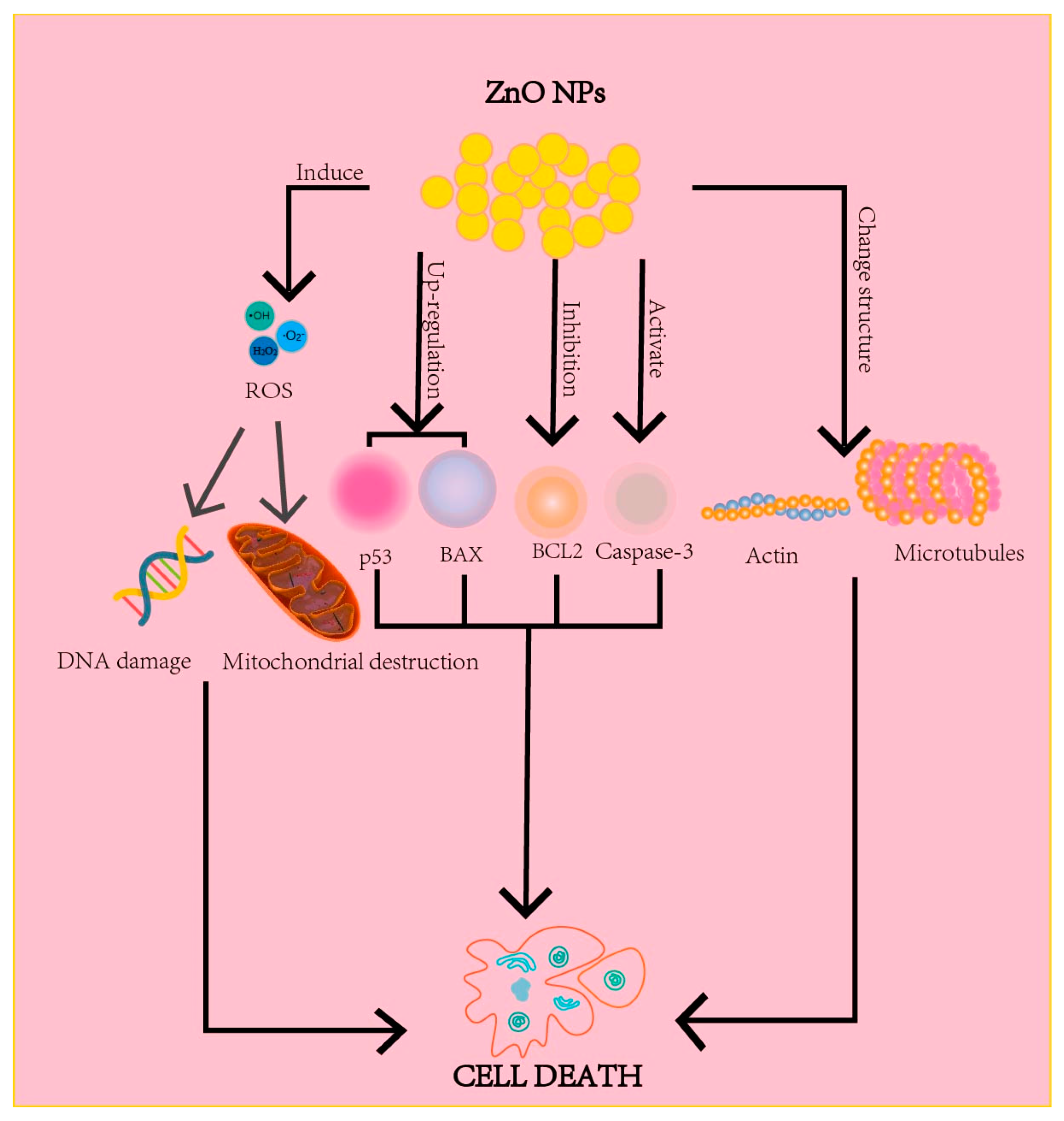
4.2.1. Anti-Cancer Mechanisms

The main mechanisms of ZnO nanomaterials as anti-cancer agents are: (1) Regulating the gene and protein expressions in tumor cells by the internalization of ZnO nanoparticles so as to induce tumor cell apoptosis [86,87,88]. (2) Generating high levels of ROS to induce cell destruction [86,88]. (3) Altering the structure of actin and microtubules to lead to cell necrosis and apoptosis [86] (Figure 4).

4.2.2. Anti-Cancer Applications

Here, we take the most popular tumor type in nano-ZnO anti-cancer studies, including liver, breast and lung cancer as paradigms, to reveal the promising potential of ZnO nanoparticles for anti-cancer applications.

Liver Cancer Treatment

As a highly prevalent malignant tumor, liver cancer threatens the life and health of people around the world. According to the statistics, the number of liver cancer cases in China in 2020 is as high as 410,000, which accounts for fifth place in the incidence of malignant tumors. Additionally, the number of deaths is 390,000, accounting for second place in cancer mortality. ZnO nanomaterials have been investigated in the treatment of liver cancer. Due to the lymphatic drainage obstruction and the insufficient vessel walls, ZnO nanomaterials are more likely to penetrate and remain in the cancerous tissue for a while [87]. Moreover, under physiological conditions, ZnO nanoparticles have a positive surface charge, while cancer cells have a negative membrane potential, and thus the electrostatic interactions drive the interaction of ZnO nanoparticles with cancer cells [88]. Yang et al. studied the effects of ZnO nanomaterials on Huh7 liver cancer cells in vitro. It found that ZnO nanomaterials could promote autophagy, upregulate the caspase3 and p53 expressions and trigger apoptosis, thus inhibiting the growth and proliferation of liver cancer cells [89]. Akhtar et al. found that ZnO nanoparticles promoted ROS production; upregulated the p53, bax and caspase3; downregulated the apoptosis-inhibiting bcl-2 gene; and induced DNA fragmentation, thus eventually causing HepG2 tumor cell apoptosis [90]. Rahimi Kalateh Shah Mohammad et al. investigated the effect of ZnO nanoparticles on Huh-7 and HepG2 cells via green synthesis of ZnO nanoparticles. The results showed that the IC50 values of ZnO nanoparticles for HepG2 cells and Huh-7 cells were 40 μg/mL at 72 h and 15 μg/mL at 48 h, respectively, which attributed to the enhancement of P53 and Bax gene expression profiles [91]. To further enhance the therapeutic efficacy of ZnO nanomaterials, researchers often integrate them with other nanoparticles that have similar properties. Alabyadh et al. synthesized ZnO/CeO2 nanocomposites via combustion method using metal-organic framework as precursor and explored the toxic effects on HepG2 liver cancer cells [92]. The results showed that the tumor growth inhibition increased with the increase in concentration of ZnO/CeO2 nanocomposites. More importantly, the anticancer effect of ZnO/CeO2 nanocomposites was better than that of ZnO or CeO2 alone.

Breast Cancer Treatment

The incidence of breast cancer in China ranks first place in female malignant tumors. The number of breast cancer cases in China increased from 315,000 in 2017 to 336,000 in 2021. As mentioned above, the long-term use of commonly therapeutic drugs such as docetaxel, paclitaxel, adriamycin and epi-amycin will induce tolerance in the body, and the increased dosage leads to more pronounced adverse effects. Therefore, the development of new drugs is necessary. Recently, ZnO nanomaterials presented promising potential for breast cancer treatment. Jasim Makkawi et al. prepared ZnO/CdS composites and investigated their cytotoxicity to breast cancer cells. They found that ZnO/CdS nanocomposites exhibited stronger anticancer activity against mammary gland (MCF-7) cells compared with ZnO nanoparticles alone [93]. This is attributed to the reduced bandgap energy after compositing ZnO nanoparticles with narrow bandgap semiconductor CdS, which increased the production of ROS. Abarna et al. synthesized ZnO/chitosan composites using a simple solid-state method [94]. They found that the number of MCF-7 breast cancer cells exposed to the composite was drastically reduced with an IC50 of 34.6% compared with a ZnO alone of 60%, which may be due to the synergistic anti-cancer effect of ZnO nanomaterials and chitosan. Sanad et al. produced structurally stable ZnO-5-FU nanocomposites using physical adsorption for breast cancer cell inhibition [95]. Both the 5-FU and ZnO nanomaterials have anticancer activity. Additionally, the increased ROS in cancer cells facilitate the high permeability, electrostatic interaction and selective cytotoxicity of ZnO. As a result, the incorporation of ZnO into the 5-FU surface significantly increased the killing rate of MCF-7 cells. Bisht et al. used trisodium citrate as a linker to conjugate ZnO nanoparticles and Fe3O4 to form ZnO-Fe3O4 magnetic composite nanomaterials (MCPs) [96]. The cytotoxicity of MCPs was investigated on a human breast cancer cell line (MDA-MB-231) and normal mouse fibroblasts (NIH-3T3). The MCPs can utilize both the selectivity of ZnO nanoparticles and the magnetic properties of Fe3O4 nanoparticles, and thus they had significant selectivity for MDA-MB-231 cells. Moreover, the Fe3O4 nanoparticles could convert the H2O2 produced by the interaction of ZnO nanoparticles with cancer cells into ROS, which enhanced the oxidative stress of cells. As a result, it showed that compared with naked ZnO nanoparticles, MCPs exhibited higher selectivity for cancer cells and lower toxicity for normal cells. Vimala et al. synthesized a folic-acid (FA)-modified PEG-ZnO nanosheet (FA-PEG-ZnO NS) and used it as a carrier to loaded anti-cancer drug adriamycin (DOX), which combined the targeted delivery of DOX and photothermal therapy for breast cancer treatment [8]. The DOX-FA-ZnO NS exhibited pH responsive, heat stimulative effects and sustained drug release properties. The experimental results confirmed that combined therapy based on DOX-FA-ZnO NS+NIR possesses a maximum death rate of MDA-MB-231 breast cancer cells compared with that of single chemotherapy or photothermal therapy.

Lung Cancer Treatment

Lung cancer is a malignant tumor originating from the bronchial mucosa or glands of the lungs. Its incidence and mortality are increasing extremely fast. WHO data show that China had 816,000 lung cancer cases and about 715,000 deaths in 2020. Lung cancer is currently divided into two categories: small cell lung cancer and non-small cell lung cancer. Additionally, non-small cell lung cancer accounts for approximately 80% of all lung cancers. In recent years, lung cancer therapy based on nano-drugs has developed rapidly, and many relative literature studies can be found. Compared with traditional drug therapy, nanocarrier delivery systems have obvious advantages [97], such as increasing the solubility of the drugs, avoiding drug degradation and reducing the interaction of drugs with non-target tissues so as to reduce the adverse reactions. In this subsection, we will introduce the application of ZnO nanomaterials in lung cancer therapy.
Cisplatin (Cp) and gemcitabine (Gem) are common broad-spectrum hydrophobic antitumor agents in clinic. They have significant efficacy but strong side effects. Based on this, Hu et al. used ZnO nanoparticles as carriers to load Cp and Gem to form ZnONPs(Cp/Gem) [98]. This nanosystem had properties as follows: (1). ZnONPs(Cp/Gem) could upregulate the expression of pro-apoptotic proteins caspase 3, caspase 9 and RARP, and thus promote the apoptosis of cancer cells. (2). ZnONPs(Cp/Gem) increase intracellular ROS, causing oxidative stress in cancer cells. (3). ZnONPs(Cp/Gem) decrease intracellular glutathione, accumulate harmful substances in cells and thus increase cytotoxicity. (4). ZnONPs(Cp/Gem) can damage the mitochondria of cancer cells and enhance cytotoxicity. As a result, the effect of ZnONPs(Cp/Gem) on human non-small cell lung cancer cells A549 was obviously higher than that of Cp, Gem and ZnO nanoparticles alone. Rupa et al. loaded indole-3-carbinol (I3C) on the ZnO nanoparticles under ultrasound to form DM-ZnO-I3C nanoemulsion (DM-ZnO-I3C-NE) [99]. DM-ZnO-I3C-NE can improve the solubility of anticancer drugs in the delivery system so as to improve the drugs’ efficacy. According to the result, DM-ZnO-I3C-NE presented more toxicity in non-small cell lung cancer cells than the free DM-ZnO nanoparticles or I3C; this may be due to the synergistic effect of I3C and Zn2+ ion. Zhao et al. synthesized ZnO@polymer nanoparticles, which can be self-assembled into capsule shells to load the hydrophobic drug isotretinoin (ISO), and obtained ZnO-ISO composite [100]. The ZnO@polymer delivery system had a tight structure and was less prone to drug leakage, and it had high pH sensitivity that could decompose under acidic conditions to facilitate drug release. From the results, the antitumor effect of ZnO-ISO composite is better than that of the existing Nintedanib and Crizotinib on the market within a certain drug concentration range. Cai et al. reported a PH-sensitive drug delivery platform based on NH2-ZnO quantum dots (QDs) [101]. This nanocarrier system was doped with some substances that have specific binding ability to lung cancer cells. First, the NH2-ZnO QDs surface was modified by PEG to improve its stability under physiological fluid. CD44 is a common marker of tumors, and hyaluronic acid (HA) can specifically bind to CD44. Thus, ZnO-PEG was combined with HA molecules to form HA-ZnO-PE.G., Subsequently, DOX drug was loaded onto the HA-ZnO-PE.G., After uptake of the QDs by cancer cells, the pH-sensitive ZnO QDs were dissolved to Zn2+ in acidic endosome/lysosome, which triggered the metal–drug complex dissociation and DOX controlled release, thus realizing a synergistic antitumor effect to A549 lung cancer cells by both Zn2+ and DOX.
Taken together, we list the ZnO nanomaterial-mediated anti-tumor applications in Table 2.

4.3. ZnO Nanomaterials for Anti-Inflammation

Inflammation is a defense response of living tissues to stimulation by various harmful external factors, which is dynamic in injury, anti-injury and recovery. In general, mild inflammation has positive effects on the body, such as limiting lesions and preventing the spread of pathogenic microorganisms to the whole body. Additionally, the fluid and leukocyte exudate can dilute toxins, remove necrotic tissues and repair and restore organ functions. However, severe inflammation has the potential to harm the organism. For example, inflammation can cause denaturation or necrosis of organs, inflammatory exudates can lead to fluid accumulation, mutual adhesions between organs, lesion swelling and compression of the surrounding tissues and organs. Long-term inflammation can cause asthma, rheumatoid arthritis, pancreatitis and other diseases [102]. Very severe acute inflammation may even be directly fatal. Therefore, anti-inflammatory applications are especially important in some sudden disease treatments. The usually used anti-inflammatory drugs are non-steroidal anti-inflammatory drugs (NSAIDs), including aspirin, ibuprofen, diclofenac, indomethacin and so on. However, when NSAIDs inhibit COX-2, they will inevitably restrain COX-1, where the COX-1 is beneficial to the gastrointestinal mucosa. As a result, this causes harmful gastrointestinal reactions, such as epigastric discomfort, nausea, vomiting and even gastric ulcers and bleeding in patients. These obvious adverse effects greatly limit the drug’s application. Moreover, in acute inflammation, the anti-inflammatory, antipyretic and analgesic efficacy of NSAIDs are delayed due to their slow absorption [102]. Therefore, the development of new drugs is particularly meaningful. Recently, many research studies indicated that ZnO nanomaterials exhibit potential in the treatment of inflammatory diseases. We thus discuss this part as follows.

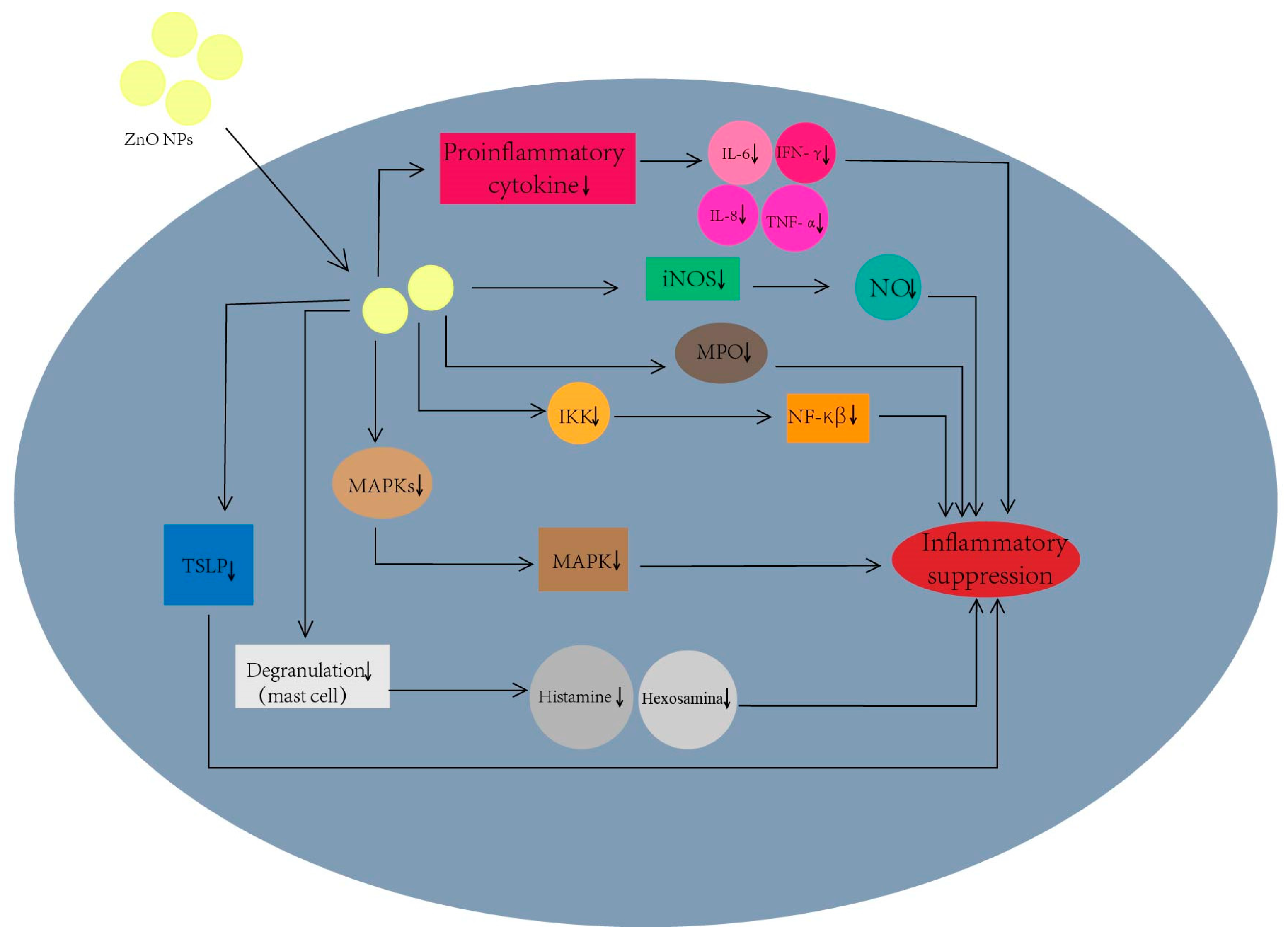
4.3.1. Anti-Inflammatory Mechanisms

As seen in Figure 5, the main mechanisms of anti-inflammatory effects of ZnO nanoparticles include: (1). Inhibition of inducible nitric oxide synthase (iNOS) protein expression [102]. Nitric oxide (NO) synthesis in the body is related to iNOS, which can participate in the rolling and migration of white blood cells, and can promote the genes’ transcription of pro-inflammatory factors. A high concentration of NO can cause the death of host immune cells. Thereby, the inhibition of iNOS can reduce NO synthesis so as to relieve inflammation in the body. (2). Inhibition of pro-inflammatory cytokine release [102,103,104]. Some cytokines, such as interleukin-6 (IL-6), interleukin-8 (IL-8), interleukin-17 (IL-17), interferon-γ (IFN-γ) and tumor necrosis factor-α (TNF-α), can regulate inflammation. Zn2+ can inhibit these cytokine releases to reduce inflammation. (3). Inhibition of myeloperoxidase (MPO) activity [102]. MPO is a heme-containing cationic enzyme, which can act as an inflammation mediator to alter neutrophil function and promote cytokine production. ZnO nanoparticles can effectively inhibit MPO activity and thus exert good anti-inflammatory effects. (4). Inhibition of mitogen-activated protein kinases (MAPKs) [102,103]. The MAPK family involved in the MAPK pathway is closely related to activating and regulating the inflammatory response. ZnO nanoparticles can inhibit the pathway to reduce inflammation. (5). Inhibition of the nuclear factor-κB (NF-κβ) pathway [102,103,105]. NF-κβ can mediate inflammation by promoting the production of pro-inflammatory cytokines and chemokines [102]. ZnO nanoparticles have exactly the IκB kinase (IKK) inhibiting effect to inhibit the NF-κβ pathway and thus exhibit anti-inflammatory activity. (6). Inhibition of mast cell degranulation [102]. In allergic inflammatory diseases such as allergic rhinitis, atopic dermatitis and asthma, mast cells are activated to release a series of inflammatory mediators (e.g., histamine and b-hexosaminidase). ZnO NPs can inhibit these inflammatory mediators to alleviate inflammation. (7). Inhibition of thymic stromal lymphopoietin (TSLP) generation [102,103]. TSLP activates mast cells and stimulates the release of Th2 cytokines to mediate the immune response. ZnO nanoparticles can inhibit the TSLP production to reduce inflammation.

4.3.2. Anti-Inflammatory Applications

The anti-inflammatory properties of ZnO nanoparticles are widely used in biomedical applications. For example, Mahadeva et al. prepared ZnO nanoparticles through a combustion method using lantana camara flowers as the fuel, and investigated their anti-inflammatory effect [106]. They showed that ZnO nanoparticles could obviously inhibit Phospholipase A2 (MIC value: 41 μg/mL) to reduce inflammation. This might be attributed to the binding of ZnO nanoparticles and Zn2+, which changes the enzyme conformation. Rajakumar et al. explored the anti-inflammatory activity of ZnO nanoparticles via evaluating the protein denaturation inhibition effect [107]. The ZnO nanoparticles presented significant anti-inflammatory potential with an inhibitory IC50 of 66.78 μg/mL, which was close to the reference (diclofenac, 62.55 μg/mL). Moreover, ZnO nanomaterials synthesized using kalanchoe pinnata [108], polygala tenuifolia root extract [109], Heritiera fomes and Sonneratia apetala extracts [110], pelargonium odoratissimum aqueous leaf extract [111], Trianthema portulacastrum Linn. [112] and acacia caesia bark extract [113] also have obvious anti-inflammatory activity. Xia et al. added ZnO nanoparticles to the diets of weaned piglets, which improved the intestinal flora to inhibit inflammation and thus reduced the incidence of diarrhea in piglets [104]. In ileum, the expression levels of TNF-α, IFN-γ, IL-1β and NF-κB were decreased. In addition, ZnO nanocomposites are also commonly used in anti-inflammatory applications. As a typical example, Yao et al. produced TNTs/ZnO composites via the electrodeposition of ZnO nanoparticles into TiO2 nanotubes (TNT) and investigated the anti-inflammatory effect of TNTs/ZnO based on a macrophages model [114]. It was found that TNTs/ZnO can effectively inhibit the macrophage proliferation and adhesion, and thus has the potential to control the inflammatory reaction and prevent chronic inflammation. In addition, ZnO nanocomposites can be indirectly used in anti-inflammatory therapy via acting as catalysts for the synthesis of anti-inflammatory drugs.
Overall, we sum up the ZnO nanomaterial-mediated anti-inflammatory applications in Table 3.

4.4. ZnO Nanomaterials for Skin Care

With the development of technology and the progress of industry, climate change is evident and the environmental pollution is serious, so skin problems are becoming more and more common among the general public. The need for skin care and makeup products in daily life has also become particularly urgent. Recently, nanomaterials have been widely used in the skin care industry, such as sunscreens, shampoos, creams, eye creams and color cosmetics [115,116,117,118]. Typically, ZnO nanomaterials have been used in various commercial skin care products, especially sunscreen products, because of their high stability, low irritancy, broad-spectrum absorption and other properties [119].

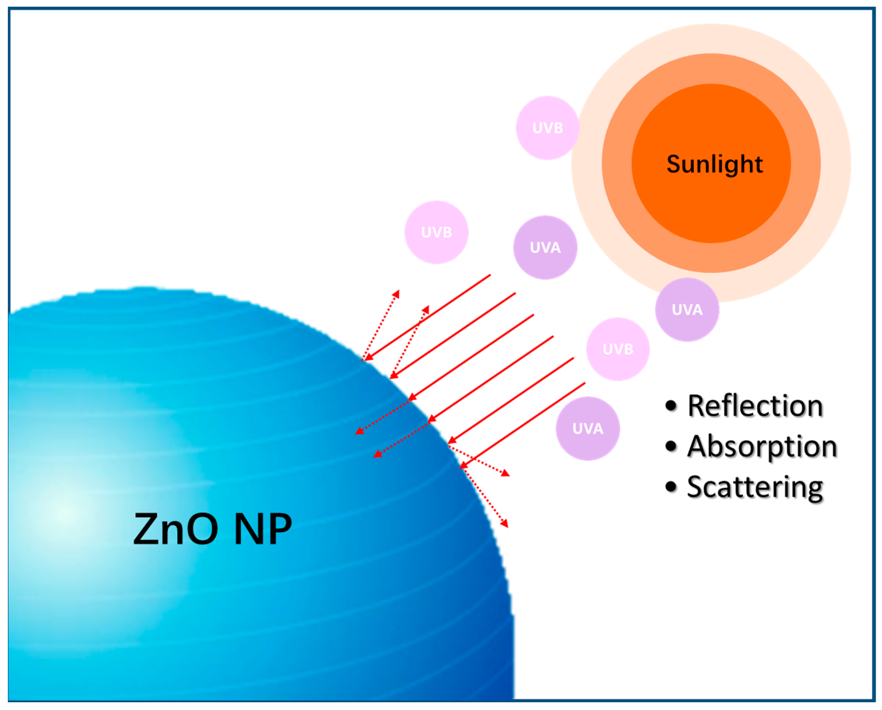
4.4.1. Sunscreen Mechanism

The usually so-called sunscreen mechanism is the protection of skin from damage caused by UV-rays in sunlight. As a kind of electromagnetic wave, UV radiation has a non-negligible damage to human skin. Additionally, it can be divided into four sections: (1). UV-A long-wave (320–400 nm); UV-A can penetrate the dermis, lead to melanin increase, damage lipids and collagen, cause skin aging and even induce skin cancer. (2). UV-B medium wave (280–320 nm); UV-B can cause skin sunburn, peeling and erythema, etc. (3). UV-C short wave (200–280 nm); (4) UV-D ultrashort wave (10-200 nm). Generally, UV-C does not invade human skin. Thus, the usual so-called UV injury is UV-A and UV-B injury. Appropriately, ZnO nanoparticles have the potential for absorption, reflection and scattering of UV light in sunlight [119,120]. For example, on the one hand, due to the band gap of ZnO nanoparticles, the electron can be excited and leap in the outer layer orbit by absorbing UV light to achieve the purpose of sun protection. On the other hand, nano-sized ZnO has the ability of UV light scattering and reflecting, and thus exhibits a physical shielding effect that decreases the touch of UV light to skin tissue (Figure 6).

4.4.2. Skin Care Applications

Just under the above-mentioned background, sunscreen products appear more and more frequently in daily life. Research has shown that the regular use of sunscreen can prevent actinic keratosis and squamous cell carcinoma [121]. At present, ZnO inorganic particles have become commonly used physical sunscreens in clinics, and their particle size has gone from traditional micrometer scale to nano-scale in order to improve their transparency and viscosity [122]. Meanwhile, the particle size of ZnO nanoparticles is one of the key factors that affects the sunscreen’s absorption function, where with the decreasing particle size of nanoparticles, the UV absorption performance may be improved [121]. There are many kinds of ZnO-containing sunscreens on the market and the content of ZnO nanoparticles in common commercial sunscreens is about 20% [123]. For example, sunscreens for pregnant women from the well-known Australian brand Kangaroo Mother added ZnO as a physical sunscreen ingredient, which has a theoretical sun protection time of 7.5 h. As another example, the sunscreen products of the well-known Australian brand Invisible Zinc also use ZnO as a main ingredient, which not only has a sun protection function but also has repairing and brightening abilities. Moreover, the sunscreen from American brand Aveeno Kids also contains ZnO, which can reflect UV-A and UV-B in sunlight without skin irritation. In addition, the sunscreen in French brand CeraVe’s moisturizing mineral also contains ZnO to form a protective barrier on the surface of the skin.
Although the sunscreen properties of nanoparticles are favored, it has recently been found that the hazards associated with their penetration into the body cannot be ignored, so we highlight safety studies here to inspire researchers to further optimize the properties of ZnO to improve its safety [124,125,126]. Studies have shown that the main factors affecting the permeation of Zn2+ are: (1). pH—Holmes et al. studied the effect of ZnO and Zn2+ permeation on the skin, and the results showed that the labile zinc within the viable epidermis increased with the decrease in pH, and the main zinc species are derived from the dissolution of ZnO that exist as Zn2+ [127]. (2). Skin barrier integrity—Studies have shown that an intact skin barrier can effectively block the penetration of Zn2+. When the skin stratum corneum is damaged, Zn2+ in the epidermis is significantly increased. For instance, after 24 h of material used on damaged skin, it is observed that the Zn2+ in vivo is up to 60–65 times higher than that used on normal skin. (3). The material retention time on the skin surface—Studies have shown that after 48 h staying on skin, the content of Zn2+ in the body is higher than that after 24 h staying. (4). Material size—the smaller the size of the nanoparticles, the greater the ratio of surface area to volume and the faster the dissolution of ZnO. Excessive material penetration will cause high levels of particles in vivo and thereby exhibit cytotoxicity. These factors provide important warnings for the right use of sunscreen containing ZnO nanoparticles. Special attention should also be paid to the fact that sprays cannot contain nanomaterials in order to avoid direct exposure of the lungs to ZnO nanoparticles [128]. Moreover, long-term exposure of ZnO nanoparticles to UV light can induce photocatalytic activity to produce toxic ROS and lose UV-blocking functions [128,129]. In order to synthesize nanoparticles with lower toxicity, Girigoswami et al. coated chitosan (CTS) and polyethylene glycol (PEG) on the surface of ZnO nanoparticles to produce ZnO-CTS and ZnO-PEG, respectively, and the experimental results showed that surface modified-ZnO nanoparticles have lower photocatalytic activity and higher UV-blocking efficiency than bare ZnO nanoparticles [129]. In addition to the preparation of the above two surface modifiers, the researchers also coated aluminum hydroxide, silica and vitamins on ZnO nanoparticles to reduce the toxicity of the materials [129,130].
In addition to sunscreens, ZnO nanomaterials are also used in other skin care products. The beauty industry giants currently using nano-products are mainly L’Oreal, Johnson & Johnson, Avon and Estee Lauder [131]. ZnO nanoparticles are one kind of nano-product used by these companies. The significant antibacterial and anti-inflammatory properties make ZnO nanoparticles used in moisturizers. The ability for cell proliferation and collagen formation make them of use in face and eye creams. Additionally, the appropriate concentration of zinc can facilitate skin wound healing and protects the body from UV-A and UV-B damage, which thereby are applied in body lotions. Additionally, the function of improving product stability makes ZnO nanoparticles doped in eyeliner [131]. Furthermore, different morphologies of ZnO nanomaterials have different applications. ZnO nanocrystals with a diameter less than 100 nm are applied in toothpaste to protect tooth enamel. ZnO nanocapsules can load active substances such as lipid mixtures and lipophilic substances to penetrate into the deep dermis, and thus are often used as emulsions or hydrogels in personal care products. ZnO nanoemulsions with uniformity and low viscosity are widely used in anti-aging products [123,130,131,132].

4.5. ZnO Nanomaterials for Bioimaging

Nowadays, semiconductor nanomaterials are increasingly used in the field of bioimaging, but the commonly used cadmium quantum dots (QDs) are limited due to their significant cytotoxicity [133]. In contrast, ZnO QDs have low toxicity and Zn is a trace element in the human body, which has a promising future in bioimaging [88,134,135,136,137,138]. Nowadays, many types of ZnO nanoparticles have been developed, such as ZnO QDs and ZnO nanoflowers [133,139,140,141]. The photoluminescence spectrum of ZnO includes UV emission and visible light emission, where the broad visible emission of ZnO nanomaterial makes it more suitable for bioimaging [140].

4.5.1. Imaging Mechanisms

The luminescent properties of ZnO nanomaterials are important for their use in imaging. The possible mechanisms of UV and visible emission of ZnO are described briefly below: (1). The recombination of photo-generated electrons with holes in the VB or in traps near the VB. (2). The recombination of electrons in shallow trap with a hole in a deep trap and the recombination of electrons in the singly ionized oxygen vacancies with the photo-generated holes in the VB [140,142]. As a result, the visible emission of ZnO QD is essentially due to the electrons leaping between energy levels [140] (Figure 7).

4.5.2. Imaging Applications

The surface defects, the band gap width and the physical properties of ZnO nanomaterials can affect their imaging applications. Herein, many researchers have modified the surface of ZnO nanoparticles, widened the band gap and developed new synthesis methods to improve their utilization value, and the ZnO nanoparticles can even be used as carriers to load colorant for assistant imaging [139,140,141,142,143,144,145,146,147,148,149]. Zang et al. synthesized ZnO nanoflowers using a biphasic reaction method. It had a band-edge absorption feature at λ = 360 nm (3.44 eV) that was greater than the bulk (~3.36 eV), which might be attributed to quantum-confinement [141]. In order to improve the water solubility of the materials that are good for bio-labeling, they exchanged the surface ligand with aminoethanol ethyl ester hydrochloride (AET), which can convert the hydrophobic surface of nanoparticles into a hydrophilic surface, but still can retain its photoluminescence performance. The biological cell labeling results showed that most Hela cells were successfully labeled by AET–ZnO nanoparticles and the corresponding blue emissions could be imaged under fluorescent microscopy. Wu et al. synthesized ZnO QDs using a soft chemical method and capped them with 3-aminopropyl trimethoxysilane (Am) to protect the core nanocrystals [150]. Subsequently, the ZnO QDs were capped with TiO2 via sol-gel method to synthesize ZnO-TiO2 QDs, where the nanoparticles prepared by the sol-gel method exhibit strong surface defects and thus can induce emission at longer wavelengths. They used mung bean seedlings as experimental objects and monitored the fluorescence emission intensity of seedling cross-sections to evaluate the bioimaging performance of ZnO nanomaterials. The results showed that the emission intensity of seedlings treated with TiO2-ZnO QDs was 18 times more than that of the untreated seedlings. Furthermore, it found that TiO2-ZnO QDs had a higher quantum yield than the standard fluorescein material. Senthilkumar et al. prepared a ZnO-ZnS core–shell structure where the nanostructure possessed a thin ZnS layer around ZnO nanoparticles [133]. The formation of the core–shell structure confined the charge carriers within the core band gap, which resulted in a significant increase in the photoluminescence intensity and thus was beneficial to imaging. Manaia et al. synthesized Mg-ZnO QD using a hydrolysis-condensation reaction and compared the photoluminescence effect between pure ZnO and Mg-ZnO QD [140]. They found that the luminescence performance of ZnO QD doped with Mg2+ was significantly improved. With the increase in Mg2+ proportion, both the emission and absorption spectra presented a blue shift phenomenon that increased the luminescence. When the doping concentration of Mg2+ in ZnO was 20 mol%, the luminescence exhibited a maximum with the quantum yield of 64%, which is significantly higher than that of pure ZnO suspension (10%).
With all of this research in mind, we list the ZnO nanomaterial-mediated bio-imaging applications in Table 4.

4.6. ZnO Nanomaterials for Food Packaging

With the frequent use of various food additives, antioxidants and antimicrobial agents in food, food-borne diseases also appear frequently, and people are paying more and more attention to food quality and food safety. Therefore, the development of new types of food additives with good biosafety is necessary. Zn2+ is a trace element required by the human body, and ZnO acting as a zinc supplement has been widely used in the food industry [151]. The FDA has classified ZnO as a safe material (GRAS) [152]. To avoid zinc deficiency, ZnO is often incorporated into food products as a food additive. In addition, the significant antimicrobial activity, high stability, low toxicity and large surface area of ZnO nanoparticles make them able to be used as active packaging [153,154,155,156,157,158,159]. This section will mainly describe the application of ZnO nanoparticles in active food packaging.

4.6.1. Material Performance Evaluation

Active packaging is food packaging that can give food a longer shelf-life and bring better economic efficiency to businesses. As a type of active packaging, antimicrobial packaging can extend the shelf-life of food by inhibiting and slowing the growth of microorganisms in the food [153]. Unlike traditional food preservatives that are directly added into food, antimicrobial packaging employs the antimicrobial agents to load onto the packaging material and gradually diffuse into the food matrix to provide antimicrobial effects that improve food safety [153]. The performance of packaging materials is usually evaluated in the following indicators [152,153,160]: (1). Mechanical properties—this includes the tensile strength (TS) and elongation at break (EAB) of the film. Generally speaking, the higher the TS and EAB, the stronger anti-damage ability and the better elasticity of the material. (2). Water solubility and swelling—this is the indicator of water resistance of bio-composite film. (3). Barrier properties—this includes water vapor permeability (WVP) and oxygen permeability (OP). WVP is the study of water transfer on the film; lower WVP values means better barrier performance of the packaging material and lower food moisture loss. Additionally, the smaller the OP, the better the oxygen separation performance of the packaging material. (4). Transparency—the lower the transparency, the better blocking performance of the material to UV-visible light. (5). Antibacterial activity. (6). Anti-oxidation properties.

4.6.2. Food Packaging Application

Chitosan (CS) is a biopolymer with significant antimicrobial activity, biocompatibility and low toxicity, which is now widely used as a direct coating for meat and fruit products [160]. Al-Naamani et al. once made CS-ZnO nanocomposites coat onto polyethylene (PE) film and investigated their antimicrobial efficacy compared with that of CS-coated PE [160]. The experimental results showed that compared with the control group, the counts of Escherichia coli, Salmonella and Staphylococcus aureus decreased only 10-fold after incubation with the CS-coated PE, while the CS-ZnO PE could completely inactivate as well as prevent the growth of food pathogens. Liu et al. used CS, ZnO nanoparticles and antioxidant bamboo leaves (AOB) to make active films and investigated their bioactivity and physical properties [161]. It was found that the adding of AOB enhanced the light-blocking effect of CS-ZnO film, particularly UV light blocking. Additionally, the light-blocking property improved with the increase in AOB concentration, which was probably due to the significant absorption of UV by the flavonoid compounds in AOB. The radical scavenging effects of material on 2,2-diphenyl-1-bitter hydrazide (DPPH) and 2,2-azido-bis-(3-ethylbenzothiazolidine-6-sulfonic acid) (ABTS) were also increased, which was mainly due to the antioxidant capacity of the flavonoid compounds in AOB. Meanwhile, the synergistic effect of AOB with ZnO greatly enhanced the antimicrobial effect against E. coli and S. aureus. Yadav et al. produced CS-ZnO@gal complex membranes using CS, ZnO nanoparticles and gallic acid (gal). It was found that the performance of CS-ZnO@gal membranes was greatly improved compared with that of pure CS membranes [152]. Firstly, the addition of ZnO@gal into CS improved the TS and EAB, which may be due to the fact that ZnO nanoparticles can fill the space of the CS matrix well and establish a strong interfacial interaction with the CS matrix. Secondly, the solubility and swelling of the CS-ZnO@gal membrane were reduced, which can be attributed to the denser and better sealing of the film. In addition, the antibacterial and antioxidant properties of the membranes were enhanced due to the doping of ZnO nanoparticles and gal, and the higher content of ZnO@gal, the higher the antioxidant properties of the membranes. Moreover, the WVP and OP values of CS-ZnO@gal membranes were lower compared with those of pure CS membranes, which were attributed to the ZnO@gal doping that made the network structure of the membrane tighter. Considering the environmental impact of packaging materials, Sarojini et al. used sesame oil-based polyurethane (PU) and CS as raw materials and incorporated different concentrations of ZnO nanoparticles to prepare biodegradable food packaging films [162]. The mechanical properties, water resistance and antimicrobial activity of the PC3-5Z film (containing 5% ZnO nanoparticles, a 3:1 weight ratio of PU and CS) were higher than those in pure CS film, which might be attributed to the denser structure of the film with the addition of PU and ZnO and the significant antimicrobial activity of ZnO. Additionally, the biodegradability of PC3-5Z was significantly enhanced, which possessed an 80% degradation rate within 28 days compared with plain PU films. This biodegradable food packaging film not only meets the expected requirements of packaging films, but is also very friendly to the environment. In addition, ZnO nanoparticles can also be combined with other natural-, organic- or biopolymers, such as acetylated cellulose nanocrystals [163], polylactic acid [157], starch-poly vinyl alcohol [164], mangosteen peel extract [165] and polycarbonate [166]. They can effectively enhance the mechanical properties, barrier properties, antibacterial and antioxidant properties, biodegradation properties and even pH sensing properties of the materials, which are promising escorts for food safety.
In all, we now list the ZnO nanomaterial-mediated food packaging applications in Table 5.

5. Biosafety of ZnO Nanomaterials

The assessment of the biosafety (or toxicity) of nanomaterials is a prerequisite for their practical application. Recently, diverse toxicity research results of ZnO nanoparticles have been reported in several species such as mammalian cells, animals, plant microbes and algae. It should be pointed out that we only discuss the toxicity of ZnO nanoparticles to mammalian cells and animals, but not to plants or other species, here because this paper primarily focuses on the biological applications of ZnO under human body exposure, which is more closely related to human health rather than eco-environmental influence. For instance, Sarkar et al. evaluated the bio-safety of mycosynthesized ZnO-NPs (75 ± 5 nm) using cytotoxicity and genotoxicity assays in human lymphocyte cells. The analysis of membrane integrity and mitochondrial dehydrogenase activity indicated that the cytotoxicity was significant at treatment doses of 500 µg/mL and above. Comet assay revealed that the obvious genotoxicity occurred at the highest concentration of 1000 µg/mL [167]. Of course, biosafety evaluation only at the cellular level is not enough; the in vivo data are more persuasive. Thus, researchers conduct more animal experiments in later studies. For example, Yu et al. investigated the oral acute toxicity of ZnO nanoparticles in rats. They showed that no obvious acute toxicity was observed after oral administration of ZnO nanoparticles (86.3 ± 23.8 nm, 100 mg/kg) for 14 days. Nevertheless, transcriptomic responses in the livers were affected, which showed that the ZnO nanoparticles up-regulated the metabolic process and metal biding [168]. Jung et al. studied the effect of the interaction between ZnO nanoparticles and food matrices on oral toxicity. The results indicated that after oral administration of ZnO nanoparticles (78 ± 26 nm, 100 mg/kg) interacting with glucose (5%) or albumin (5%) for 14 consecutive days, no acute toxicity was found in rats via the histopathological, hematological and serum biochemical analyses, revealing their low oral toxicity [169]. Kong et al. assessed the oral acute toxicity and oral long-term toxicity of unmodified ZnO nanoparticles (50 nm) in mice, respectively [170,171,172]. For the oral acute toxicity study, the experiments of clinical signs, body weights, organ weight, serum biochemistry, hematology, gross pathology, histopathology and mortality were measured, which demonstrated that the toxicity of 50 nm ZnO nanoparticles was greater than bulk ZnO, but similar to 20 nm ZnO nanoparticles, and the LD50 was 5177 mg/kg·bw [170]. For the oral long-term toxic effects study, it indicated that after a 90-day exposure, the unmodified 50 nm ZnO nanoparticles caused hepatic and renal function impairment, anemia and antioxidant system imbalance of mice. Remarkable toxic effects were observed under the doses > 80 mg/kg. Additionally, the lowest observed adverse effect level was 40 mg/kg·bw [171]. In addition, the unmodified 50 nm ZnO nanoparticles can cause lipid metabolism disorder, reproductive toxicity and hyperlipidemia to male mice through oxidative injury in long-term exposure [172].
As can be seen from the above case, bio-toxicity is influenced by many factors including dose, size, exposure time and so on. Thus, it can be predicted that more caution needs to be taken for ZnO nanoparticles, particularly with their use in food additives and skin care products that directly expose the human body, which need to strictly control the use parameters.

6. Conclusions and Outlook

The multifunctionality of ZnO nanomaterials provides a good opportunity for biological application. In this review, we systematically introduce the recent advances of ZnO nanomaterial-mediated biological applications and corresponding action mechanisms, which cover anti-bacterial, anti-tumor, anti-inflammatory, bioimaging, skin care and food packaging uses. Meanwhile, we also give a brief summary of the structure-dependent physicochemical properties, the common synthesis methods and the biosafety of ZnO nanoparticles. Note that although the fact that significant progress and achievements have been obtained in this field, challenges still exist that we have to face and tackle, such as the following:
(i).
Biosafety evaluation and improvement need further and deeper research. Although toxicity studies of ZnO nanoparticles have been extensively published, the reports about long-term effects are still not enough. It is not difficult to see that the acute toxicity of ZnO nanoparticles is low according to many reports. Thus, the chronic toxicity and long-term effects are more valuable to be concerned with. Meanwhile, the genetic toxicity evaluation of ZnO nanoparticles also needs to be further strengthened, which is closely related to the health of future generations. In addition, for the improvement of material biosafety, we can make material modifications such as alterations of the surface reactive property to improve its biocompatibility and minimize the adverse effects [173].
(ii).
Strengthening toxicity mechanisms study and establishing standardization. To date, the understanding of the toxicity mechanisms of ZnO nanoparticles is not deep enough, and the research results are diverse, even contradictory, in different models. The absence of key nanotoxicity information and the lack of consensus among researchers on the experimental evaluation index is unbeneficial to regulation control and enforcement in use. Therefore, the experimental protocols of toxicity mechanisms are demanded, and guidance and standardization for nanotoxicity evaluation that includes cytotoxicity, in vivo toxicity, genetic toxicity and so on should be established.
(iii).
The optimization of the synthesis. On the one hand, for existing mature synthesis methods, one can optimize the experimental parameters such as pH, temperature and ingredient proportion to rationally regulate the physicochemical properties of ZnO nanoparticles so as to obtain the particle with desired size, shape and specific surface area, etc. On the other hand, the yield demand of ZnO nanoparticles has increased dramatically due to their large-scale commercial use. Thus, simple, low-cost and eco-friendly new synthesis methods such as popular green synthesis are currently an urgent task.
(iv).
Proper surface functionalization of ZnO nanoparticles. The surface of a nanoparticle is closely related with its dispersibility, biocompatibility and biotoxicity in a physiological environment. Meanwhile, the modification of the surface with various functional biomolecules, such as aptamers and antibodies, is helpful to improve the selectivity and targeting ability to pathogenic tissue and so on. Thus, the proper surface functionalization of ZnO nanoparticles is significant to enhance its therapeutic efficacy for diseases such as tumors.
(v).
Deepening the research on the action mechanism. It is not difficult to see that the action mechanisms are more than one path of each ZnO nanomaterial-mediated application. For example, the ZnO-mediated antibacterial mechanisms cover not only ROS generation but also Zn2+ release, disturbance of the cell membrane and so on. So, which path is dominant? Or is there an inner link between these paths? Questions such as this need to be further explained.

Author Contributions

Conceptualization and methodology, J.X. and Z.G.; funding acquisition, J.X.; writing—original draft and investigation, J.X., H.L. and T.Z.; writing—review and editing, B.S., X.W. and Z.G. All authors have read and agreed to the published version of the manuscript.

Funding

The authors hereby acknowledge the financial support from the Natural Science Foundation of Sichuan province [2023NSFSC1452], the Talent Engineering Research Initiation Project of Chengdu University under Grant [2081920047], the Open Fund Project of the Key Laboratory of Chemistry in Ethnic Medicine Resource (Yunnan Minzu University), the State Ethnic Affairs Commission and the Ministry of Education [MZY2208].

Data Availability Statement

Not applicable.

Conflicts of Interest

The authors declare no conflict of interest.

References

  1. Kim, I.; Viswanathan, K.; Kasi, G.; Thanakkasaranee, S.; Sadeghi, K.; Seo, J. ZnO Nanostructures in Active Antibacterial Food Packaging: Preparation Methods, Antimicrobial Mechanisms, Safety Issues, Future Prospects, and Challenges. Food Rev. Int. 2020, 38, 537–565. [Google Scholar] [CrossRef]
  2. Zhong, L.; Yun, K. Graphene oxide-modified ZnO particles: Synthesis, characterization, and antibacterial properties. Int. J. Nanomed. 2015, 10, 79–92. [Google Scholar] [CrossRef]
  3. Lallo da Silva, B.; Caetano, B.L.; Chiari-Andreo, B.G.; Pietro, R.; Chiavacci, L.A. Increased antibacterial activity of ZnO nanoparticles: Influence of size and surface modification. Colloids Surf. B 2019, 177, 440–447. [Google Scholar] [CrossRef] [PubMed]
  4. Abebe, B.; Zereffa, E.A.; Tadesse, A.; Murthy, H.C.A. A Review on Enhancing the Antibacterial Activity of ZnO: Mechanisms and Microscopic Investigation. Nanoscale Res. Lett. 2020, 15, 190. [Google Scholar] [CrossRef]
  5. Gad, S.S.; Fayez, A.M.; Abdelaziz, M.; Abou El-Ezz, D. Amelioration of autoimmunity and inflammation by zinc oxide nanoparticles in experimental rheumatoid arthritis. N-S Arch. Pharmacol. 2021, 394, 1975–1981. [Google Scholar] [CrossRef]
  6. Feltis, B.N.; Elbaz, A.; Wright, P.F.; Mackay, G.A.; Turney, T.W.; Lopata, A.L. Characterizing the inhibitory action of zinc oxide nanoparticles on allergic-type mast cell activation. Mol. Immunol. 2015, 66, 139–146. [Google Scholar] [CrossRef]
  7. de Lucas-Gil, E.; Del Campo, A.; Pascual, L.; Monte-Serrano, M.; Menendez, J.; Fernandez, J.F.; Rubio-Marcos, F. The fight against multidrug-resistant organisms: The role of ZnO crystalline defects. Mater. Sci. Eng. C 2019, 99, 575–581. [Google Scholar] [CrossRef]
  8. Vimala, K.; Shanthi, K.; Sundarraj, S.; Kannan, S. Synergistic effect of chemo-photothermal for breast cancer therapy using folic acid (FA) modified zinc oxide nanosheet. J. Colloid Interface Sci. 2017, 488, 92–108. [Google Scholar] [CrossRef]
  9. Yin, X.; Fan, X.; Zhou, Z.; Li, Q. Encapsulation of berberine decorated ZnO nano-colloids into injectable hydrogel using for diabetic wound healing. Front. Chem. 2022, 10, 964662. [Google Scholar] [CrossRef]
  10. Sarkar, S.; Debnath, S.K.; Srivastava, R.; Kulkarni, A.R. Continuous flow scale-up of biofunctionalized defective ZnO quantum dots: A safer inorganic ingredient for skin UV protection. Acta Biomater. 2022, 147, 377–390. [Google Scholar] [CrossRef]
  11. Du, T.; Zhao, C.; ur Rehman, F.; Lai, L.; Li, X.; Sun, Y.; Luo, S.; Jiang, H.; Selke, M.; Wang, X. Rapid and multimodal in vivo bioimaging of cancer cells through in situ biosynthesis of Zn&Fe nanoclusters. Nano Res. 2017, 10, 2626–2632. [Google Scholar] [CrossRef]
  12. Janani, N.; Zare, E.N.; Salimi, F.; Makvandi, P. Antibacterial tragacanth gum-based nanocomposite films carrying ascorbic acid antioxidant for bioactive food packaging. Carbohydr. Polym. 2020, 247, 116678. [Google Scholar] [CrossRef] [PubMed]
  13. Secu, C.E.; Sima, M. Photoluminescence and thermoluminescence of ZnO nano-needle arrays and films. Opt. Mater. 2009, 31, 876–880. [Google Scholar] [CrossRef]
  14. Sharma, P.; Jang, N.Y.; Lee, J.W.; Park, B.C.; Kim, Y.K.; Cho, N.H. Application of ZnO-Based Nanocomposites for Vaccines and Cancer Immunotherapy. Pharmaceutics 2019, 11, 493. [Google Scholar] [CrossRef]
  15. Xie, J.; Gong, L.; Zhu, S.; Yong, Y.; Gu, Z.; Zhao, Y. Emerging Strategies of Nanomaterial-Mediated Tumor Radiosensitization. Adv. Mater. 2019, 31, e1802244. [Google Scholar] [CrossRef]
  16. Chen, C.; Liu, P.; Lu, C. Synthesis and characterization of nano-sized ZnO powders by direct precipitation method. Chem. Eng. J. 2008, 144, 509–513. [Google Scholar] [CrossRef]
  17. Dhanya, R.; Suganthi, K.S.; Rajan, K.S. Studies on Scale-up of Synthesis of ZnO Nanoparticles. Asian J. Chem. 2014, 26, 4273–4276. [Google Scholar] [CrossRef]
  18. Maroufi, S.; Nekouei, R.K.; Assefi, M.; Sahajwalla, V. Waste-cleaning waste: Synthesis of ZnO porous nano-sheets from batteries for dye degradation. Environ. Sci. Pollut. Res. 2018, 25, 28594–28600. [Google Scholar] [CrossRef]
  19. Raj, C.J.; Joshi, R.K.; Varma, K.B.R. Synthesis from zinc oxalate, growth mechanism and optical properties of ZnO nano/micro structures. Cryst. Res. Technol. 2011, 46, 1181–1188. [Google Scholar] [CrossRef]
  20. Wang, H.; Li, C.; Zhao, H.; Liu, J. Preparation of nano-sized flower-like ZnO bunches by a direct precipitation method. Adv. Powder Technol. 2013, 24, 599–604. [Google Scholar] [CrossRef]
  21. Yildiz, Ö. A Novel Synthesis Process of ZnO Nano Powders in Granule Form. Trans. Ind. Ceram. Soc. 2015, 74, 41–48. [Google Scholar] [CrossRef]
  22. Ulyankina, A.; Molodtsova, T.; Gorshenkov, M.; Leontyev, I.; Zhigunov, D.; Konstantinova, E.; Lastovina, T.; Tolasz, J.; Henych, J.; Licciardello, N.; et al. Photocatalytic degradation of ciprofloxacin in water at nano-ZnO prepared by pulse alternating current electrochemical synthesis. J. Water Process. Eng. 2021, 40, 101809. [Google Scholar] [CrossRef]
  23. Bai, X.; Yi, L.; Liu, D.; Nie, E.; Sun, C.; Feng, H.; Jin, Y.; Jiao, Z.; Sun, X. Electrodeposition from ZnO nano-rods to nano-sheets with only zinc nitrate electrolyte and its photoluminescence. Appl. Surf. Sci. 2011, 257, 10317–10321. [Google Scholar] [CrossRef]
  24. Zhang, S.; Yan, C.; Zhang, H.; Lu, G. Effects of bath temperature on the morphology of ZnO nano-rods and its optical properties. Mater. Lett. 2015, 148, 1–4. [Google Scholar] [CrossRef]
  25. An, D.; Tong, X.; Liu, J.; Wang, Q.; Zhou, Q.; Dong, J.; Li, Y. Template-free hydrothermal synthesis of ZnO micro/nano-materials and their application in acetone sensing properties. Superlattices Microstruct. 2015, 77, 1–11. [Google Scholar] [CrossRef]
  26. Ben Saad, L.; Soltane, L.; Sediri, F. Nano-ZnO Sand Flowers and Rods: Hydrothermal Synthesis and Optical Properties. Russ. J. Phys. Chem. A 2019, 93, 2269–2274. [Google Scholar] [CrossRef]
  27. Du, Y.; Yang, Y.; Wang, X.; Li, X.; Zhou, Q. Hydrothermal synthesis and optical properties of CTAB modified nano-ZnO. Integr. Ferroelectr. 2019, 200, 161–167. [Google Scholar] [CrossRef]
  28. Liu, W.; Wang, S.; Wang, J.; Zhang, B.; Liu, L.; Liu, H.; Yang, J. Supercritical hydrothermal synthesis of nano-zinc oxide: Process and mechanism. Ceram. Int. 2022, 48, 22629–22646. [Google Scholar] [CrossRef]
  29. Wen, M.; Yang, B.; Yan, H.; Fu, Z.; Cai, C.; Liu, K.; Chen, Y.; Xu, J.; Fu, S.; Zhang, S. Morphology-controlled synthesis of flowerlike ZnO nano/microstructures and their photocatalytic property. J. Nanosci. Nanotechnol. 2009, 9, 2038–2044. [Google Scholar] [CrossRef]
  30. Xiang-Qi, L.I.; Qing-Fei, F.A.N.; Guang-Li, L.I.; Yao-Han, H.; Zhao, G.A.O.; Xi-Mei, F.A.N.; Chao-Liang, Z.; Zuo-Wan, Z. Syntheses of ZnO Nano-Arrays and Spike-Shaped CuO/ZnO Heterostructure. Acta Phys.-Chim. Sin. 2015, 31, 783–792. [Google Scholar] [CrossRef]
  31. Zhang, L.; Zhao, J.; Zheng, J.; Li, L.; Zhu, Z. Shuttle-like ZnO nano/microrods: Facile synthesis, optical characterization and high formaldehyde sensing properties. Appl. Surf. Sci. 2011, 258, 711–718. [Google Scholar] [CrossRef]
  32. Zhou, Q.; Zhang, F.; Zhang, S.; Wang, W.; Wang, X.; Yang, Y. Study on hydrothermal synthesis and photoelectric properties of nano-ZnO. Ferroelectrics 2019, 549, 212–219. [Google Scholar] [CrossRef]
  33. Kumar, D.; Singh Mehata, M. Synthesis of diamagnetic ZnO nano-crystallites via sol-gel method and their photocatalytic activity. Indian J. Eng. Mater. Sci. 2022, 29, 437–444. [Google Scholar] [CrossRef]
  34. Fu, D.; Han, G.; Meng, C. Size-controlled synthesis and photocatalytic degradation properties of nano-sized ZnO nanorods. Mater. Lett. 2012, 72, 53–56. [Google Scholar] [CrossRef]
  35. John, R.; Shanmugaraj, S.; Rajaram, R.; Endo, T. Synthesis and characterization of nano ZnO and CdO. J. Ceram. Soc. JPN 2010, 118, 329–332. [Google Scholar] [CrossRef]
  36. Noroozi, A.H.; Sadeghi, M.; Tehrani, A.A.; Sarabadani, P.; Rajabaifar, S.; Mirzaee, M. Synthesis and Characterization of Zinc Oxide Nano-particles by Solid State Chemical Reaction Method. J. Clust. Sci. 2013, 24, 757–770. [Google Scholar] [CrossRef]
  37. Ali, M.; Friedenberger, N.; Spasova, M.; Winterer, M. A Novel Approach for Chemical Vapor Synthesis of ZnO Nanocrystals: Optimization of Yield, Crystallinity. Chem. Vap. Depos. 2009, 15, 192–198. [Google Scholar] [CrossRef]
  38. Riahi-Noori, N.; Sarraf-Mamoory, R.; Alizadeh, P.; Mehdikhanic, A. Synthesis of ZnO nano powder by a gel combustion method. J. Ceram. Process. Res. 2008, 9, 246–249. [Google Scholar]
  39. Teng, H.; Xu, S.; Meng, W. Controllable Synthesis of Different Dimensions Nano-ZnO by Microemulsion and Photocatalytic Activity. J. Inorg. Mater. 2010, 25, 1034–1040. [Google Scholar] [CrossRef]
  40. Luković Golić, D.; Ćirković, J.; Šćepanović, M.; Srećković, T.; Longo, E.; Varela, J.A.; Daneu, N.; Stamenković, V.; Branković, G.; Branković, Z. The modification of structural and optical properties of nano- and submicron ZnO powders by variation of solvothermal syntheses conditions. J. Nanopart. Res. 2014, 16, 2670. [Google Scholar] [CrossRef]
  41. Moulahi, A.; Sediri, F. Controlled synthesis of nano-ZnO via hydro/solvothermal process and study of their optical properties. Optik 2016, 127, 7586–7593. [Google Scholar] [CrossRef]
  42. Song, Y.L.; Zhang, T.J.; Jie Du, H.; Ji, P.F.; Li, Y.; Zhou, F.Q. Synthesis, structures and temperature-dependent photoluminescence from ZnO nano/micro-rods on Zn foil. Mater. Lett. 2016, 176, 139–142. [Google Scholar] [CrossRef]
  43. Agouram, S.; Bushiri, M.J.; Montenegro, D.N.; Reig, C.; Martinez-Tomas, M.C.; Munoz-Sanjose, V. Synthesis and characterization of ZnO nano and micro structures grown by low temperature spray pyrolysis and vapor transport. J. Nanosci. Nanotechnol. 2012, 12, 6792–6799. [Google Scholar] [CrossRef] [PubMed]
  44. Li, X.; Zhai, F.; Liu, Y.; Cao, M.; Wang, F.; Zhang, X. Synthesis and photoluminescence study on ZnO nano-particles. Chin. Phys. 2007, 16, 2769–2772. [Google Scholar]
  45. Seo, S.H.; Kang, H.C. Self-assembled ZnO hexagonal nano-disks grown by radio-frequency magnetron sputtering. Mater. Lett. 2013, 94, 34–37. [Google Scholar] [CrossRef]
  46. Sun, J.H.; Kang, H.C. Synthesis and characterization of self-assembled ZnO nano-dots grown on SiNx/Si(001) substrates by radio frequency magnetron sputtering. Thin Solid Films 2010, 518, 6522–6525. [Google Scholar] [CrossRef]
  47. Khanna, P.K.; Kate, K.; Dhanabalan, K.; Banerjee, S.; Reji, N.; Shinde, S.D.; Jain, G.H. Sono-chemical synthesis of ZnO nano-particles and their application in hydrogen sulphide gas sensing. J. Nanosci. Nanotechnol. 2012, 12, 2791–2796. [Google Scholar] [CrossRef]
  48. Shi, L.-E.; Fang, X.-J.; Zhang, Z.-L.; Zhou, T.; Jiang, D.; Wu, H.-H.; Tang, Z.-X. Preparation of nano-ZnO using sonication method and its antibacterial characteristics. Int. J. Food Sci. Technol. 2012, 47, 1866–1871. [Google Scholar] [CrossRef]
  49. Song, H.; Zhu, K.; Liu, Y.; Zhai, X. Microwave-assisted synthesis of ZnO and its photocatalytic activity in degradation of CTAB. Russ. J. Phys. Chem. A 2017, 91, 59–62. [Google Scholar] [CrossRef]
  50. Lv, H.; Sang, D.; Li, H.; Du, X.; Li, D.; Zou, G. Thermal Evaporation Synthesis and Properties of ZnO Nano/Microstructures Using Carbon Group Elements as the Reducing Agents. Nanoscale Res. Lett. 2010, 5, 620–624. [Google Scholar] [CrossRef]
  51. Park, N.-K.; Han, G.B.; Lee, J.D.; Ryu, S.O.; Lee, T.J.; Chang, W.C.; Chang, C.H. The growth of ZnO nano-wire by a thermal evaporation method with very small amount of oxygen. Curr. Appl. Phys. 2006, 6, e176–e181. [Google Scholar] [CrossRef]
  52. Zhang, J.; Yang, Y.; Xu, B.; Jiang, F.; Li, J. Shape-controlled synthesis of ZnO nano- and micro-structures. J. Cryst. Growth 2005, 280, 509–515. [Google Scholar] [CrossRef]
  53. Chen, C.; Yu, B.; Liu, P.; Liu, J.; Wang, L. Investigation of nano-sized ZnO particles fabricated by various synthesis routes. J. Ceram. Process. Res. 2011, 12, 420–425. [Google Scholar]
  54. Gu, Z.; Paranthaman, M.P.; Xu, J.; Pan, Z.W. Aligned ZnO nanorod arrays grown directly on zinc foils and zinc spheres by a low-temperature oxidization method. ACS Nano 2009, 3, 273–278. [Google Scholar] [CrossRef]
  55. Prasad, A.R.; Williams, L.; Garvasis, J.; Shamsheera, K.O.; Basheer, S.M.; Kuruvilla, M.; Joseph, A. Applications of phytogenic ZnO nanoparticles: A review on recent advancements. J. Mol. Liq. 2021, 331, 115805. [Google Scholar] [CrossRef]
  56. Essawy, A.A.; Alsohaimi, I.H.; Alhumaimess, M.S.; Hassan, H.M.A.; Kamel, M.M. Green synthesis of spongy Nano-ZnO productive of hydroxyl radicals for unconventional solar-driven photocatalytic remediation of antibiotic enriched wastewater. J. Environ. Manag. 2020, 271, 110961. [Google Scholar] [CrossRef]
  57. Jeevanandam, J.; Kiew, S.F.; Boakye-Ansah, S.; Lau, S.Y.; Barhoum, A.; Danquah, M.K.; Rodrigues, J. Green approaches for the synthesis of metal and metal oxide nanoparticles using microbial and plant extracts. Nanoscale 2022, 14, 2534–2571. [Google Scholar] [CrossRef]
  58. Svetlichnyi, V.; Shabalina, A.; Lapin, I.; Goncharova, D.; Nemoykina, A. ZnO nanoparticles obtained by pulsed laser ablation and their composite with cotton fabric: Preparation and study of antibacterial activity. Appl. Surf. Sci. 2016, 372, 20–29. [Google Scholar] [CrossRef]
  59. Sharma, S.C. ZnO nano-flowers from Carica papaya milk: Degradation of Alizarin Red-S dye and antibacterial activity against Pseudomonas aeruginosa and Staphylococcus aureus. Optik 2016, 127, 6498–6512. [Google Scholar] [CrossRef]
  60. Happy, A.; Soumya, M.; Venkat Kumar, S.; Rajeshkumar, S. Mechanistic study on antibacterial action of zinc oxide nanoparticles synthesized using green route. Chem.-Biol. Interact. 2018, 286, 60–70. [Google Scholar] [CrossRef]
  61. Iqbal, G.; Faisal, S.; Khan, S.; Shams, D.F.; Nadhman, A. Photo-inactivation and efflux pump inhibition of methicillin resistant Staphylococcus aureus using thiolated cobalt doped ZnO nanoparticles. J. Photochem. Photobiol. B 2019, 192, 141–146. [Google Scholar] [CrossRef] [PubMed]
  62. Verma, S.K.; Jha, E.; Panda, P.K.; Das, J.K.; Thirumurugan, A.; Suar, M.; Parashar, S. Molecular aspects of core-shell intrinsic defect induced enhanced antibacterial activity of ZnO nanocrystals. Nanomedicine 2018, 13, 43–68. [Google Scholar] [CrossRef] [PubMed]
  63. Saha, R.; Subramani, K.; Petchi Muthu Raju, S.A.K.; Rangaraj, S.; Venkatachalam, R. Psidium guajava leaf extract-mediated synthesis of ZnO nanoparticles under different processing parameters for hydrophobic and antibacterial finishing over cotton fabrics. Prog. Org. Coat. 2018, 124, 80–91. [Google Scholar] [CrossRef]
  64. Babitha, N.; Priya, L.S.; Christy, S.R.; Manikandan, A.; Dinesh, A.; Durka, M.; Arunadevi, S. Enhanced Antibacterial Activity and Photo-Catalytic Properties of ZnO Nanoparticles: Pedalium Murex Plant Extract-Assisted Synthesis. J. Nanosci. Nanotechnol. 2019, 19, 2888–2894. [Google Scholar] [CrossRef]
  65. Alavi, M.; Karimi, N.; Salimikia, I. Phytosynthesis of zinc oxide nanoparticles and its antibacterial, antiquorum sensing, antimotility, and antioxidant capacities against multidrug resistant bacteria. J. Ind. Eng. Chem. 2019, 72, 457–473. [Google Scholar] [CrossRef]
  66. Datta, A.; Patra, C.; Bharadwaj, H.; Kaur, S.; Dimri, N.; Khajuria, R. Green Synthesis of Zinc Oxide Nanoparticles Using Parthenium hysterophorus Leaf Extract and Evaluation of their Antibacterial Properties. J. Biotechnol. Biomater. 2017, 7, 1000271. [Google Scholar] [CrossRef]
  67. Karthikeyan, M.; Jafar Ahamed, A.; Karthikeyan, C.; Vijaya Kumar, P. Enhancement of antibacterial and anticancer properties of pure and REM doped ZnO nanoparticles synthesized using Gymnema sylvestre leaves extract. SN Appl. Sci. 2019, 1, 355. [Google Scholar] [CrossRef]
  68. Mahendiran, D.; Subash, G.; Arumai Selvan, D.; Rehana, D.; Senthil Kumar, R.; Kalilur Rahiman, A. Biosynthesis of Zinc Oxide Nanoparticles Using Plant Extracts of Aloe vera and Hibiscus sabdariffa: Phytochemical, Antibacterial, Antioxidant and Anti-proliferative Studies. BioNanoScience 2017, 7, 530–545. [Google Scholar] [CrossRef]
  69. Nguyen, V.; Vu, V.; Nguyen, T.; Nguyen, T.; Tran, V.; Nguyen-Tri, P. Antibacterial Activity of TiO2- and ZnO-Decorated with Silver Nanoparticles. J. Compos. Sci. 2019, 3, 61. [Google Scholar] [CrossRef]
  70. Yusof, N.A.A.; Zain, N.M.; Pauzi, N. Synthesis of ZnO nanoparticles with chitosan as stabilizing agent and their antibacterial properties against Gram-positive and Gram-negative bacteria. Int. J. Biol. Macromol. 2019, 124, 1132–1136. [Google Scholar] [CrossRef]
  71. Afzal, G.; Jamal, A.; Kiran, S.; Mustafa, G.; Ahmad, F.; Saeed, S.; Ahmad, H.I.; Dawood, S. Aerva javanica Mediated Synthesis, Characterization and Antimicrobial Evaluation of Zinc Oxide Nanoparticles. J. Anim. Plant Sci. 2021, 32, 547–553. [Google Scholar] [CrossRef]
  72. Ahmed, J.; Ali, M.; Sheikh, H.M.; Al-Kattan, M.O.; Farhana; Haroon, U.; Safaeishakib, M.; Akbar, M.; Kamal, A.; Zubair, M.S.; et al. Biocontrol of Fruit Rot of Litchi chinensis Using Zinc Oxide Nanoparticles Synthesized in Azadirachta indica. Micromachines 2022, 13, 1461. [Google Scholar] [CrossRef] [PubMed]
  73. Gondal, M.A.; Alzahrani, A.J.; Randhawa, M.A.; Siddiqui, M.N. Morphology and antifungal effect of nano-ZnO and nano-Pd-doped nano-ZnO against Aspergillus and Candida. J. Environ. Sci. Health A 2012, 47, 1413–1418. [Google Scholar] [CrossRef] [PubMed]
  74. Haghighi, N.; Abdi, Y.; Haghighi, F. Light-induced antifungal activity of TiO2 nanoparticles/ZnO nanowires. Appl. Surf. Sci. 2011, 257, 10096–10100. [Google Scholar] [CrossRef]
  75. Khan, M.F.; Ansari, A.H.; Hameedullah, M.; Ahmad, E.; Husain, F.M.; Zia, Q.; Baig, U.; Zaheer, M.R.; Alam, M.M.; Khan, A.M.; et al. Sol-gel synthesis of thorn-like ZnO nanoparticles endorsing mechanical stirring effect and their antimicrobial activities: Potential role as nano-antibiotics. Sci. Rep. 2016, 6, 27689. [Google Scholar] [CrossRef]
  76. Krishnamoorthy, N.; Sivasankarapillai, V.S.; Natarajan, V.K.; Eldesoky, G.E.; Wabaidur, S.M.; Eswaran, M.; Dhanusuraman, R. Biocidal activity of ZnO NPs against pathogens and antioxidant activity—A greener approach by Citrus hystrix leaf extract as bio-reductant. Biochem. Eng. J. 2023, 192, 108818. [Google Scholar] [CrossRef]
  77. Liu, H.; Qiao, Z.; Jang, Y.O.; Kim, M.G.; Zou, Q.; Lee, H.J.; Koo, B.; Kim, S.H.; Yun, K.; Kim, H.S.; et al. Diatomaceous earth/zinc oxide micro-composite assisted antibiotics in fungal therapy. Nano Converg. 2021, 8, 32. [Google Scholar] [CrossRef]
  78. Munnawar, I.; Iqbal, S.S.; Anwar, M.N.; Batool, M.; Tariq, S.; Faitma, N.; Khan, A.L.; Khan, A.U.; Nazar, U.; Jamil, T.; et al. Synergistic effect of Chitosan-Zinc Oxide Hybrid Nanoparticles on antibiofouling and water disinfection of mixed matrix polyethersulfone nanocomposite membranes. Carbohydr. Polym. 2017, 175, 661–670. [Google Scholar] [CrossRef]
  79. Roy, A.; Joshi, M.; Butola, B.S. Preparation and antimicrobial assessment of zinc-montmorillonite intercalates based HDPE nanocomposites: A cost-effective and safe bioactive plastic. J. Clean. Prod. 2019, 212, 1518–1525. [Google Scholar] [CrossRef]
  80. Sharma, R.K.; Ghose, R. Synthesis of zinc oxide nanoparticles by homogeneous precipitation method and its application in antifungal activity against Candida albicans. Ceram. Int. 2015, 41, 967–975. [Google Scholar] [CrossRef]
  81. Sivasankarapillai, V.S.; Krishnamoorthy, N.; Eldesoky, G.E.; Wabaidur, S.M.; Islam, M.A.; Dhanusuraman, R.; Ponnusamy, V.K. One-pot green synthesis of ZnO nanoparticles using Scoparia Dulcis plant extract for antimicrobial and antioxidant activities. Appl. Nanosci. 2022, 13, 1–11. [Google Scholar] [CrossRef] [PubMed]
  82. Tryfon, P.; Kamou, N.N.; Ntalli, N.; Mourdikoudis, S.; Karamanoli, K.; Karfaridis, D.; Menkissoglu-Spiroudi, U.; Dendrinou-Samara, C. Coated Cu-doped ZnO and Cu nanoparticles as control agents against plant pathogenic fungi and nematodes. NanoImpact 2022, 28, 100430. [Google Scholar] [CrossRef] [PubMed]
  83. Anjum, S.; Hashim, M.; Malik, S.A.; Khan, M.; Lorenzo, J.M.; Abbasi, B.H.; Hano, C. Recent Advances in Zinc Oxide Nanoparticles (ZnO NPs) for Cancer Diagnosis, Target Drug Delivery, and Treatment. Cancers 2021, 13, 4570. [Google Scholar] [CrossRef] [PubMed]
  84. Ali, A.; Phull, A.-R.; Zia, M. Elemental zinc to zinc nanoparticles: Is ZnO NPs crucial for life? Synthesis, toxicological, and environmental concerns. Nanotechnol. Rev. 2018, 7, 413–441. [Google Scholar] [CrossRef]
  85. Anitha, R.; Ramesh, K.V.; Ravishankar, T.N.; Sudheer Kumar, K.H.; Ramakrishnappa, T. Cytotoxicity, antibacterial and antifungal activities of ZnO nanoparticles prepared by the Artocarpus gomezianus fruit mediated facile green combustion method. J. Sci. Adv. Mater. Devices 2018, 3, 440–451. [Google Scholar] [CrossRef]
  86. Fu, W.; Zhou, W.; Chu, P.K.; Yu, X.F. Inherent Chemotherapeutic Anti-Cancer Effects of Low-Dimensional Nanomaterials. Chemistry 2019, 25, 10995–11006. [Google Scholar] [CrossRef]
  87. Puisney, C.; Baeza-Squiban, A.; Boland, S. Mechanisms of uptake and translocation of nanomaterials in the lung. Adv. Exp. Med. Biol. 2018, 1048, 21–36. [Google Scholar] [CrossRef]
  88. Mishra, P.K.; Mishra, H.; Ekielski, A.; Talegaonkar, S.; Vaidya, B. Zinc oxide nanoparticles: A promising nanomaterial for biomedical applications. Drug Discov. Today 2017, 22, 1825–1834. [Google Scholar] [CrossRef]
  89. Yang, R.; Wu, R.; Mei, J.; Hu, F.-R.; Lei, C.-J. Zinc oxide nanoparticles promotes liver cancer cell apoptosis through inducing autophagy and promoting p53. Eur. Rev. Med. Pharmacol. Sci. 2021, 25, 1557–1563. [Google Scholar]
  90. Akhtar, M.J.; Ahamed, M.; Kumar, S.; Khan, M.M.; Ahmad, J.; Alrokayan, S.A. Zinc oxide nanoparticles selectively induce apoptosis in human cancer cells through reactive oxygen species. Int. J. Nanomed. 2012, 7, 845–857. [Google Scholar] [CrossRef]
  91. Rahimi Kalateh Shah Mohammad, G.; Seyedi, S.M.R.; Karimi, E.; Homayouni-Tabrizi, M. The cytotoxic properties of zinc oxide nanoparticles on the rat liver and spleen, and its anticancer impacts on human liver cancer cell lines. J. Biochem. Mol. Toxicol. 2019, 33, e22324. [Google Scholar] [CrossRef] [PubMed]
  92. Alabyadh, T.; Albadri, R.; Es-Haghi, A.; Yazdi, M.E.T.; Ajalli, N.; Rahdar, A.; Thakur, V.K. ZnO/CeO2 Nanocomposites: Metal-Organic Framework-Mediated Synthesis, Characterization, and Estimation of Cellular Toxicity toward Liver Cancer Cells. J. Funct. Biomater. 2022, 13, 139. [Google Scholar] [CrossRef] [PubMed]
  93. Jasim Makkawi, A.J.; Aysa, N.H.; Gassim, F.-A.G. Anticancer Activity of Zinc Oxide and Zinc Oxide/Cadmium Sulfide Nanocomposites. Asian J. Pharm. Clin. Res. 2019, 12, 535–539. [Google Scholar] [CrossRef]
  94. Abarna, B.; Preethi, T.; Rajarajeswari, G.R. Single-pot solid-state synthesis of ZnO/chitosan composite for photocatalytic and antitumour applications. J. Mater. Sci. Mater. Electron. 2019, 30, 21355–21368. [Google Scholar] [CrossRef]
  95. Sanad, F.; Nabih, S.; Goda, M.A. A Lot of Promise for ZnO-5FU Nanoparticles Cytotoxicity against Breast Cancer Cell Lines. J. Nanomed. Nanotechnol. 2018, 9, 1000486. [Google Scholar] [CrossRef]
  96. Bisht, G.; Rayamajhi, S.; Kc, B.; Paudel, S.N.; Karna, D.; Shrestha, B.G. Synthesis, Characterization, and Study of In Vitro Cytotoxicity of ZnO-Fe3O4 Magnetic Composite Nanoparticles in Human Breast Cancer Cell Line (MDA-MB-231) and Mouse Fibroblast (NIH 3T3). Nanoscale Res. Lett. 2016, 11, 537. [Google Scholar] [CrossRef]
  97. Martinez-Carmona, M.; Gun’ko, Y.; Vallet-Regi, M. ZnO Nanostructures for Drug Delivery and Theranostic Applications. Nanomaterials 2018, 8, 268. [Google Scholar] [CrossRef]
  98. Hu, C.; Du, W. Zinc oxide nanoparticles (ZnO NPs combined with cisplatin and gemcitabine inhibits tumor activity of NSCLC cells. Aging 2020, 12, 25767–25777. [Google Scholar] [CrossRef]
  99. Rupa, E.J.; Arunkumar, L.; Han, Y.; Kang, J.P.; Ahn, J.C.; Jung, S.K.; Kim, M.; Kim, J.Y.; Yang, D.C.; Lee, G.J. Dendropanax morbifera Extract-Mediated ZnO Nanoparticles Loaded with Indole-3-Carbinol for Enhancement of Anticancer Efficacy in the A549 Human Lung Carcinoma Cell Line. Materials 2020, 13, 3197. [Google Scholar] [CrossRef]
  100. Zhao, W.; Wei, J.S.; Zhang, P.; Chen, J.; Kong, J.L.; Sun, L.H.; Xiong, H.M.; Mohwald, H. Self-Assembled ZnO Nanoparticle Capsules for Carrying and Delivering Isotretinoin to Cancer Cells. ACS Appl. Mater. Interfaces 2017, 9, 18474–18481. [Google Scholar] [CrossRef]
  101. Cai, X.; Luo, Y.; Zhang, W.; Du, D.; Lin, Y. pH-Sensitive ZnO Quantum Dots-Doxorubicin Nanoparticles for Lung Cancer Targeted Drug Delivery. ACS Appl. Mater. Interfaces 2016, 8, 22442–22450. [Google Scholar] [CrossRef] [PubMed]
  102. Agarwal, H.; Shanmugam, V. A review on anti-inflammatory activity of green synthesized zinc oxide nanoparticle: Mechanism-based approach. Bioorg. Chem. 2020, 94, 103423. [Google Scholar] [CrossRef] [PubMed]
  103. Mobarez, E.; Azoz, H.; Alkalamawy, N.; Nada, A.-F. Evaluation the Effectiveness of Green Zinc Oxide Nanoparticles on the Anti-Inflammatory Effect of Dexamethasone and Its Side Effects in Rats. SVU-Int. J. Vet. Sci. 2018, 1, 25–54. [Google Scholar] [CrossRef]
  104. Xia, T.; Lai, W.; Han, M.; Han, M.; Ma, X.; Zhang, L. Dietary ZnO nanoparticles alters intestinal microbiota and inflammation response in weaned piglets. Oncotarget 2017, 8, 64878–64891. [Google Scholar] [CrossRef]
  105. Abd El-Baset, S.A.; Mazen, N.F.; Abdul-Maksoud, R.S.; Kattaia, A.A.A. The therapeutic prospect of zinc oxide nanoparticles in experimentally induced diabetic nephropathy. Tissue Barriers 2022, 11, 2069966. [Google Scholar] [CrossRef]
  106. Surendra, B.S.; Mallikarjunaswamy, C.; Pramila, S.; Rekha, N.D. Bio-mediated synthesis of ZnO nanoparticles using Lantana camara flower extract: Its characterizations, photocatalytic, electrochemical and anti-inflammatory applications. Environ. Nanotechnol. Monit. Manag. 2021, 15, 100442. [Google Scholar] [CrossRef]
  107. Rajakumar, G.; Thiruvengadam, M.; Mydhili, G.; Gomathi, T.; Chung, I.M. Green approach for synthesis of zinc oxide nanoparticles from Andrographis paniculata leaf extract and evaluation of their antioxidant, anti-diabetic, and anti-inflammatory activities. Bioprocess Biosyst. Eng. 2018, 41, 21–30. [Google Scholar] [CrossRef]
  108. Agarwal, H.; Shanmugam, V.K. Synthesis and optimization of zinc oxide nanoparticles using Kalanchoe pinnata towards the evaluation of its anti-inflammatory activity. J. Drug Deliv. Sci. Technol. 2019, 54, 101291. [Google Scholar] [CrossRef]
  109. Nagajyothi, P.C.; Cha, S.J.; Yang, I.J.; Sreekanth, T.V.; Kim, K.J.; Shin, H.M. Antioxidant and anti-inflammatory activities of zinc oxide nanoparticles synthesized using Polygala tenuifolia root extract. J. Photochem. Photobiol. B 2015, 146, 10–17. [Google Scholar] [CrossRef]
  110. Thatoi, P.; Kerry, R.G.; Gouda, S.; Das, G.; Pramanik, K.; Thatoi, H.; Patra, J.K. Photo-mediated green synthesis of silver and zinc oxide nanoparticles using aqueous extracts of two mangrove plant species, Heritiera fomes and Sonneratia apetala and investigation of their biomedical applications. J. Photochem. Photobiol. B 2016, 163, 311–318. [Google Scholar] [CrossRef]
  111. Abdelbaky, A.S.; Abd El-Mageed, T.A.; Babalghith, A.O.; Selim, S.; Mohamed, A. Green Synthesis and Characterization of ZnO Nanoparticles Using Pelargonium odoratissimum (L.) Aqueous Leaf Extract and Their Antioxidant, Antibacterial and Anti-inflammatory Activities. Antioxidants 2022, 11, 1444. [Google Scholar] [CrossRef] [PubMed]
  112. Yadav, E.; Singh, D.; Yadav, P.; Verma, A. Ameliorative effect of biofabricated ZnO nanoparticles of Trianthema portulacastrum Linn. on dermal wounds via removal of oxidative stress and inflammation. RSC Adv. 2018, 8, 21621–21635. [Google Scholar] [CrossRef] [PubMed]
  113. Ashwini, J.; Aswathy, T.R.; Rahul, A.B.; Thara, G.M.; Nair, A.S. Synthesis and Characterization of Zinc Oxide Nanoparticles Using Acacia caesia Bark Extract and Its Photocatalytic and Antimicrobial Activities. Catalysts 2021, 11, 1507. [Google Scholar] [CrossRef]
  114. Yao, S.; Feng, X.; Lu, J.; Zheng, Y.; Wang, X.; Volinsky, A.A.; Wang, L.N. Antibacterial activity and inflammation inhibition of ZnO nanoparticles embedded TiO(2) nanotubes. Nanotechnology 2018, 29, 244003. [Google Scholar] [CrossRef] [PubMed]
  115. Séby, F. Metal and metal oxide nanoparticles in cosmetics and skin care products. Compr. Anal. Chem. 2021, 93, 381–427. [Google Scholar]
  116. Osmond-McLeod, M.J.; Oytam, Y.; Kirby, J.K.; Gomez-Fernandez, L.; Baxter, B.; McCall, M.J. Dermal absorption and short-term biological impact in hairless mice from sunscreens containing zinc oxide nano- or larger particles. Nanotoxicology 2014, 8 (Suppl. S1), 72–84. [Google Scholar] [CrossRef]
  117. Miljković, S.; Tomić, M.; Hut, I.; Pelemis, S. Nanomaterials for Skin Care. In Commercialization of Nanotechnologies—A Case Study Approach; Springer: Cham, Switzerland, 2018; pp. 205–226. [Google Scholar]
  118. Gubitosa, J.; Rizzi, V.; Fini, P.; Cosma, P. Nanomaterials in sun-care products. In Nanocosmetics; Elsevier: Amsterdam, The Netherlands, 2020; pp. 349–373. [Google Scholar]
  119. Al-Hilli, S.M.; Willander, M. Optical Properties of Zinc Oxide Nano-particles Embedded in Dielectric Medium for UV region: Numerical Simulation. J. Nanopart. Res. 2006, 8, 79–97. [Google Scholar] [CrossRef]
  120. Egambaram, O.P.; Kesavan Pillai, S.; Ray, S.S. Materials Science Challenges in Skin UV Protection: A Review. Photochem. Photobiol. 2020, 96, 779–797. [Google Scholar] [CrossRef]
  121. Filipe, P.; Silva, J.N.; Silva, R.; Cirne de Castro, J.L.; Marques Gomes, M.; Alves, L.C.; Santus, R.; Pinheiro, T. Stratum corneum is an effective barrier to TiO2 and ZnO nanoparticle percutaneous absorption. Skin Pharmacol. Physiol. 2009, 22, 266–275. [Google Scholar] [CrossRef]
  122. Kim, S.-H.; Heo, Y.; Choi, S.-J.; Kim, Y.-J.; Kim, M.-S.; Kim, H.; Jo, E.; Song, C.-W.; Lee, K. Safety evaluation of zinc oxide nanoparticles in terms of acute dermal toxicity, dermal irritation and corrosion, and skin sensitization. Mol. Cell Toxicol. 2016, 12, 93–99. [Google Scholar] [CrossRef]
  123. Holmes, A.M.; Kempson, I.; Turnbull, T.; Paterson, D.; Roberts, M.S. Penetration of Zinc into Human Skin after Topical Application of Nano Zinc Oxide Used in Commercial Sunscreen Formulations. ACS Appl. Bio Mater. 2020, 3, 3640–3647. [Google Scholar] [CrossRef] [PubMed]
  124. Ezealisiji, K.M.; Siwe-Noundou, X.; Maduelosi, B.; Nwachukwu, N.; Krause, R.W.M. Green synthesis of zinc oxide nanoparticles using Solanum torvum (L.) leaf extract and evaluation of the toxicological profile of the ZnO nanoparticles–hydrogel composite in Wistar albino rats. Int. Nano Lett. 2019, 9, 99–107. [Google Scholar] [CrossRef]
  125. Leite-Silva, V.R.; Sanchez, W.Y.; Studier, H.; Liu, D.C.; Mohammed, Y.H.; Holmes, A.M.; Ryan, E.M.; Haridass, I.N.; Chandrasekaran, N.C.; Becker, W.; et al. Human skin penetration and local effects of topical nano zinc oxide after occlusion and barrier impairment. Eur. J. Pharm. Biopharm. 2016, 104, 140–147. [Google Scholar] [CrossRef] [PubMed]
  126. Lee, C.-C.; Lin, Y.-H.; Hou, W.-C.; Li, M.-H.; Chang, J.-W. Exposure to ZnO/TiO2 Nanoparticles Affects Health Outcomes in Cosmetics Salesclerks. Int. J. Environ. Res. Public Health 2020, 17, 6088. [Google Scholar] [CrossRef]
  127. Holmes, A.M.; Song, Z.; Moghimi, H.R.; Roberts, M.S. Relative Penetration of Zinc Oxide and Zinc Ions into Human Skin after Application of Different Zinc Oxide Formulations. ACS Nano 2016, 10, 1810–1819. [Google Scholar] [CrossRef] [PubMed]
  128. Sogne, V.; Meier, F.; Klein, T.; Contado, C. Investigation of zinc oxide particles in cosmetic products by means of centrifugal and asymmetrical flow field-flow fractionation. J. Chromatogr. A 2017, 1515, 196–208. [Google Scholar] [CrossRef]
  129. Girigoswami, K.; Viswanathan, M.; Murugesan, R.; Girigoswami, A. Studies on polymer-coated zinc oxide nanoparticles: UV-blocking efficacy and in vivo toxicity. Mater. Sci. Eng. C-Mater. 2015, 56, 501–510. [Google Scholar] [CrossRef]
  130. Hameed, A.; Fatima, G.R.; Malik, K. Scope of Nanotechnology in Cosmetics: Dermatology and Skin Care Products. J. Med. Chem. Sci. 2019, 2, 9–16. [Google Scholar] [CrossRef]
  131. Niska, K.; Zielinska, E.; Radomski, M.W.; Inkielewicz-Stepniak, I. Metal nanoparticles in dermatology and cosmetology: Interactions with human skin cells. Chem.-Biol. Interact. 2018, 295, 38–51. [Google Scholar] [CrossRef]
  132. Gupta, S.; Bansal, R.; Gupta, S.; Jindal, N.; Jindal, A. Nanocarriers and nanoparticles for skin care and dermatological treatments. Indian Dermatol. Online J. 2013, 4, 267–272. [Google Scholar] [CrossRef]
  133. Senthilkumar, K.; Senthilkumar, O.; Yamauchi, K.; Sato, M.; Morito, S.; Ohba, T.; Nakamura, M.; Fujita, Y. Preparation of ZnO nanoparticles for bio-imaging applications. Phys. Status Solidi B 2009, 246, 885–888. [Google Scholar] [CrossRef]
  134. Neogi, A. Zno Nanoparticles in Hydrogel Polymer Network for Bio-Imaging. Glob. J. Nanomed. 2017, 1, 555572. [Google Scholar] [CrossRef]
  135. Yan, N.; Tang, B.Z.; Wang, W.X. In Vivo Bioimaging of Silver Nanoparticle Dissolution in the Gut Environment of Zooplankton. ACS Nano 2018, 12, 12212–12223. [Google Scholar] [CrossRef] [PubMed]
  136. Sureshkumar, S.; Jothimani, B.; Sridhar, T.M.; Santhosh, A.; Venkatachalapathy, B. Synthesis of Hexagonal ZnO-PQ7 Nano Disks Conjugated with Folic Acid to Image MCF–7 Cancer Cells. J. Fluoresc. 2017, 27, 21–29. [Google Scholar] [CrossRef]
  137. Yao, J.; Li, P.; Li, L.; Yang, M. Biochemistry and biomedicine of quantum dots: From biodetection to bioimaging, drug discovery, diagnostics, and therapy. Acta Biomater. 2018, 74, 36–55. [Google Scholar] [CrossRef]
  138. Li, Y.; Wang, R.; Zheng, W.; Li, Y. Silica-Coated Ga(III)-Doped ZnO: Yb(3+), Tm(3+) Upconversion Nanoparticles for High-Resolution in vivo Bioimaging using Near-Infrared to Near-Infrared Upconversion Emission. Inorg. Chem. 2019, 58, 8230–8236. [Google Scholar] [CrossRef]
  139. Matsuyama, K.; Ihsan, N.; Irie, K.; Mishima, K.; Okuyama, T.; Muto, H. Bioimaging application of highly luminescent silica-coated ZnO-nanoparticle quantum dots with biotin. J. Colloid Interface Sci. 2013, 399, 19–25. [Google Scholar] [CrossRef]
  140. Manaia, E.B.; Kiatkoski Kaminski, R.C.; Caetano, B.L.; Briois, V.; Chiavacci, L.A.; Bourgaux, C. Surface modified Mg-doped ZnO QDs for biological imaging. Eur. J. Nanomed. 2015, 7, 109–120. [Google Scholar] [CrossRef]
  141. Zang, Z.; Tang, X. Enhanced fluorescence imaging performance of hydrophobic colloidal ZnO nanoparticles by a facile method. J. Alloys Compd. 2015, 619, 98–101. [Google Scholar] [CrossRef]
  142. Gulia; Rita Kakkar, S. ZnO Quantum Dots for Biomedical Applications. Adv. Mater. Lett. 2013, 4, 876–887. [Google Scholar] [CrossRef]
  143. Xiong, H.M.; Xu, Y.; Ren, Q.G.; Xia, Y.Y. Stable aqueous ZnO@polymer core-shell nanoparticles with tunable photoluminescence and their application in cell imaging. J. Am. Chem. Soc. 2008, 130, 7522–7523. [Google Scholar] [CrossRef] [PubMed]
  144. Venkatesha, N.; Qurishi, Y.; Atreya, H.S.; Srivastava, C. ZnO coated CoFe2O4 nanoparticles for multimodal bio-imaging. RSC Adv. 2016, 6, 18843–18851. [Google Scholar] [CrossRef]
  145. Lei, G.; Yang, S.; Cao, R.; Zhou, P.; Peng, H.; Peng, R.; Zhang, X.; Yang, Y.; Li, Y.; Wang, M.; et al. In Situ Preparation of Amphibious ZnO Quantum Dots with Blue Fluorescence Based on Hyperbranched Polymers and their Application in Bio-Imaging. Polymers 2020, 12, 144. [Google Scholar] [CrossRef] [PubMed]
  146. Prasanna, A.P.S.; Venkataprasanna, K.S.; Pannerselvam, B.; Asokan, V.; Jeniffer, R.S.; Venkatasubbu, G.D. Multifunctional ZnO/SiO(2) Core/Shell Nanoparticles for Bioimaging and Drug Delivery Application. J. Fluoresc. 2020, 30, 1075–1083. [Google Scholar] [CrossRef]
  147. Kaur, P.; Choudhury, D. Functionality of receptor targeted zinc-insulin quantum clusters in skin tissue augmentation and bioimaging. J. Drug Target. 2021, 29, 541–550. [Google Scholar] [CrossRef]
  148. Giannousi, K.; Karageorgou, M.E.; Oikonomou, I.M.; Komninou, P.; Dendrinou-Samara, C. ZnO NPs immobilized by Alizarin as in vitro predictive and imaging biomarkers for protein amyloidosis. J. Inorg. Biochem. 2022, 236, 111971. [Google Scholar] [CrossRef]
  149. Wanas, W.; Abd El-Kaream, S.A.; Ebrahim, S.; Soliman, M.; Karim, M. Cancer bioimaging using dual mode luminescence of graphene/FA-ZnO nanocomposite based on novel green technique. Sci. Rep. 2023, 13, 27. [Google Scholar] [CrossRef]
  150. Wu, Y.L.; Lim, C.S.; Fu, S.; Tok, A.I.Y.; Lau, H.M.; Boey, F.Y.C.; Zeng, X.T. Surface modifications of ZnO quantum dots for bio-imaging. Nanotechnology 2007, 18, 215604. [Google Scholar] [CrossRef]
  151. Youn, S.M.; Choi, S.J. Food Additive Zinc Oxide Nanoparticles: Dissolution, Interaction, Fate, Cytotoxicity, and Oral Toxicity. Int. J. Mol. Sci. 2022, 23, 6074. [Google Scholar] [CrossRef]
  152. Yadav, S.; Mehrotra, G.K.; Dutta, P.K. Chitosan based ZnO nanoparticles loaded gallic-acid films for active food packaging. Food Chem. 2021, 334, 127605. [Google Scholar] [CrossRef]
  153. Espitia, P.J.P.; Otoni, C.G.; Soares, N.F.F. Zinc Oxide Nanoparticles for Food Packaging Applications. In Antimicrobial Food Packaging; Academic Press: San Diego, CA, USA, 2016; pp. 425–431. [Google Scholar]
  154. Priyadarshi, R.; Negi, Y.S. Effect of Varying Filler Concentration on Zinc Oxide Nanoparticle Embedded Chitosan Films as Potential Food Packaging Material. J. Polym. Environ. 2016, 25, 1087–1098. [Google Scholar] [CrossRef]
  155. Zahiri Oghani, F.; Tahvildari, K.; Nozari, M. Novel Antibacterial Food Packaging Based on Chitosan Loaded ZnO Nano Particles Prepared by Green Synthesis from Nettle Leaf Extract. J. Inorg. Organomet. Polym. Mater. 2020, 31, 43–54. [Google Scholar] [CrossRef]
  156. Sun, J.; Jiang, H.; Wu, H.; Tong, C.; Pang, J.; Wu, C. Multifunctional bionanocomposite films based on konjac glucomannan/chitosan with nano-ZnO and mulberry anthocyanin extract for active food packaging. Food Hydrocoll. 2020, 107, 105942. [Google Scholar] [CrossRef]
  157. Marra, A.; Silvestre, C.; Duraccio, D.; Cimmino, S. Polylactic acid/zinc oxide biocomposite films for food packaging application. Int. J. Biol. Macromol. 2016, 88, 254–262. [Google Scholar] [CrossRef]
  158. Amjadi, S.; Emaminia, S.; Nazari, M.; Davudian, S.H.; Roufegarinejad, L.; Hamishehkar, H. Application of Reinforced ZnO Nanoparticle-Incorporated Gelatin Bionanocomposite Film with Chitosan Nanofiber for Packaging of Chicken Fillet and Cheese as Food Models. Food Bioprocess Technol. 2019, 12, 1205–1219. [Google Scholar] [CrossRef]
  159. Esmailzadeh, H.; Sangpour, P.; Shahraz, F.; Hejazi, J.; Khaksar, R. Effect of nanocomposite packaging containing ZnO on growth of Bacillus subtilis and Enterobacter aerogenes. Mater. Sci. Eng. C 2016, 58, 1058–1063. [Google Scholar] [CrossRef]
  160. Al-Naamani, L.; Dobretsov, S.; Dutta, J. Chitosan-zinc oxide nanoparticle composite coating for active food packaging applications. Innov. Food Sci. Emerg. Technol. 2016, 38, 231–237. [Google Scholar] [CrossRef]
  161. Liu, J.; Huang, J.; Hu, Z.; Li, G.; Hu, L.; Chen, X.; Hu, Y. Chitosan-based films with antioxidant of bamboo leaves and ZnO nanoparticles for application in active food packaging. Int. J. Biol. Macromol. 2021, 189, 363–369. [Google Scholar] [CrossRef]
  162. Sarojini, K.S.; Indumathi, M.P.; Rajarajeswari, G.R. Mahua oil-based polyurethane/chitosan/nano ZnO composite films for biodegradable food packaging applications. Int. J. Biol. Macromol. 2019, 124, 163–174. [Google Scholar] [CrossRef]
  163. Yu, F.; Fei, X.; He, Y.; Li, H. Poly(lactic acid)-based composite film reinforced with acetylated cellulose nanocrystals and ZnO nanoparticles for active food packaging. Int. J. Biol. Macromol. 2021, 186, 770–779. [Google Scholar] [CrossRef]
  164. Jayakumar, A.; Heera, K.V.; Sumi, T.S.; Joseph, M.; Mathew, S.; Praveen, G.; Nair, I.C.; Radhakrishnan, E.K. Starch-PVA composite films with zinc-oxide nanoparticles and phytochemicals as intelligent pH sensing wraps for food packaging application. Int. J. Biol. Macromol. 2019, 136, 395–403. [Google Scholar] [CrossRef] [PubMed]
  165. Huang, X.; Zhou, X.; Dai, Q.; Qin, Z. Antibacterial, Antioxidation, UV-Blocking, and Biodegradable Soy Protein Isolate Food Packaging Film with Mangosteen Peel Extract and ZnO Nanoparticles. Nanomaterials 2021, 11, 3337. [Google Scholar] [CrossRef] [PubMed]
  166. Dhapte, V.; Gaikwad, N.; More, P.V.; Banerjee, S.; Dhapte, V.V.; Kadam, S.; Khanna, P.K. Transparent ZnO/polycarbonate nanocomposite for food packaging application. Nanocomposites 2015, 1, 106–112. [Google Scholar] [CrossRef]
  167. Sarkar, J.; Ghosh, M.; Mukherjee, A.; Chattopadhyay, D.; Acharya, K. Biosynthesis and safety evaluation of ZnO nanoparticles. Bioprocess Biosyst. Eng. 2014, 37, 165–171. [Google Scholar] [CrossRef]
  168. Yu, J.; Choi, S.J. Particle Size and Biological Fate of ZnO Do Not Cause Acute Toxicity, but Affect Toxicokinetics and Gene Expression Profiles in the Rat Livers after Oral Administration. Int. J. Mol. Sci. 2021, 22, 1698. [Google Scholar] [CrossRef]
  169. Jung, E.B.; Yu, J.; Choi, S.J. Interaction between ZnO Nanoparticles and Albumin and Its Effect on Cytotoxicity, Cellular Uptake, Intestinal Transport, Toxicokinetics, and Acute Oral Toxicity. Nanomaterials 2021, 11, 2922. [Google Scholar] [CrossRef]
  170. Kong, T.; Zhang, S.H.; Zhang, J.L.; Hao, X.Q.; Yang, F.; Zhang, C.; Yang, Z.J.; Zhang, M.Y.; Wang, J. Acute and Cumulative Effects of Unmodified 50-nm Nano-ZnO on Mice. Biol. Trace Elem. Res. 2018, 185, 124–134. [Google Scholar] [CrossRef]
  171. Kong, T.; Zhang, S.H.; Zhang, C.; Zhang, J.L.; Yang, F.; Wang, G.Y.; Yang, Z.J.; Bai, D.Y.; Zhang, M.Y.; Wang, J.; et al. Long-Term Effects of Unmodified 50 nm ZnO in Mice. Biol. Trace Elem. Res. 2019, 189, 478–489. [Google Scholar] [CrossRef]
  172. Kong, T.; Zhang, S.H.; Zhang, C.; Zhang, J.L.; Yang, F.; Wang, G.Y.; Yang, Z.J.; Bai, D.Y.; Shi, Y.Y.; Liu, T.Q.; et al. The Effects of 50 nm Unmodified Nano-ZnO on Lipid Metabolism and Semen Quality in Male Mice. Biol. Trace Elem. Res. 2020, 194, 432–442. [Google Scholar] [CrossRef]
  173. Srivastav, A.K.; Dhiman, N.; Tiwari, R.; Arjaria, N.; Prakash, J.; Jagdale, P.; Ayanur, A.; Singh, D.; Patnaik, S.; Kumar, M. Sub-acute oral exposure of zinc oxide nanoparticles causes alteration in iron homeostasis through acute phase response: A protective effect by surface modification. J. Trace Elem. Med. Biol. 2019, 52, 270–287. [Google Scholar] [CrossRef]
Figure 1. The biological applications of ZnO nanomaterials. Anti-bacteria: Adapted with permission [7]. Copyright 2019, Elsevier B.V. Anti-tumor: Adapted with permission [8]. Copyright 2017, Elsevier B.V. Anti-inflammation: Adapted with permission [9]. Copyright 2022, Frontiers Media S.A. Skin protection: Adapted with permission [10]. Copyright 2022, Elsevier B.V. Bio-imaging: Adapted with permission [11]. Copyright 2017, Springer Nature Switzerland AG. Food packaging: Adapted with permission [12]. Copyright 2020, Elsevier B.V.
Nanomaterials 13 01500 g001
Figure 2. The structure-dependent properties of ZnO nanoparticles.
Nanomaterials 13 01500 g002
Figure 3. The anti-bacterial mechanism of ZnO nanomaterials.
Nanomaterials 13 01500 g003
Figure 4. The anti-cancer mechanisms of ZnO nanomaterials.
Nanomaterials 13 01500 g004
Figure 5. The anti-inflammatory mechanisms of ZnO nanomaterials.
Nanomaterials 13 01500 g005
Figure 6. The sunscreen mechanism of ZnO nanomaterials.
Nanomaterials 13 01500 g006
Figure 7. The bio-imaging mechanisms of ZnO nanomaterials.
Nanomaterials 13 01500 g007
Table 1. A list of representative nano-ZnO-mediated anti-bacterial and anti-fungal applications.
NanomaterialsSynthetic MethodParticle SizeType of MicrobeAnti-Microbial OutcomesRef.
GPTMS-ZnO nanoparticleSol-gelVarying sizes from 5.3 to 38.2 nmStaphylococcus aureusAntibacterial activity increased with decreased particle size [3]
ZnO nanocrystallineHEBMVarying sizes from 20 to 250 nmStaphylococcus, Streptococcus, Micrococcus, Escherichia Coli, Enterobacter and PseudomonasAntibacterial activity increased with decreased particle size [62]
ZnO nanoparticleBio-mediated solution combustion11–26 nm of crystallite sizePseudomonas aeruginosa and Staphylococcus aureusZnO nano-flower exhibited excellent photocatalytic activity over the ZnO with other morphology[59]
ZnO nanoparticleBio-mediated method using Aloe vera and Hibiscus sabdariffa plant extracts9–18 nmEscherichia coli, Klebsiella pneumoniae, Pseudomonas aeruginosa and Staphylococcus aureusMore efficient bactericidal activity was found in nanoparticles via biosynthesis than those from chemical synthesis[68]
Ag-decorated ZnO nanoparticleChemical reduction30–50 nmStaphylococcus aureus and Escherichia coliThe hybrid of nanoparticles improved the antibacterial performance[69]
ZnO/graphene oxide compositesCovalent bonding170 nm of ZnOEscherichia coli, Salmonella typhimurium, Bacillus subtilis and Enterococcus faecalis The composite action of materials improved antibacterial properties[2]
Chitosan-stabilized ZnO nanoparticlesMicrowave heating50–70 nmStaphylococcus aureus and Escherichia coliChitosan-stabilized ZnO nanoparticles exhibited excellent antibacterial
activity
[70]
ZnO nanoparticlesSol-gelLess than 50 nmCandida albicansEffective antifungal effect was observed[75]
ZnO nanoparticlesHomogeneous precipitation method~30 nmCandida albicansThe antifungal activity was positively correlated with the ZnO concentration[80]
ZnO nanoparticlesBio-mediated method using Citrus hystrix leaf extract26−69 ± 0.5 nmCandida albicans and Aspergillus nigerExhibition of good antifungal activity at a higher concentration of 5 μg/mL[76]
ZnO nanoparticlesBio-mediated method using Azadirachta indica leaf extract29.024 nmAspergillus nigerMaximum suppression of lychee fruit rot was obtained at the dose of 1.0 mg/mL[72]
HDPE/ZnO-MMT nanocompositesA combination of simple ion exchange and reduction reactions~20 nmAspergillus nigerSignificant antifungal properties were found at 5 wt% of loading[79]
5%Pd-doped nano-ZnOThermal decomposition method35 nmAspergilus niger and Candida albicansPd-doped nano-ZnO increased the antifungal activity over pure nano-ZnO[73]
TiO2/ZnO nanocompositesChemical vapor deposition50−100 nmCandida albicans biofilmsTiO2/ZnO nanocomposites enhanced antifungal activity more than single component[74]
Table 2. A list of the representative nano-ZnO-mediated anti-tumor applications.
NanomaterialsSynthetic MethodParticle SizeType of Cancer CellsAnti-Cancer OutcomesRef.
ZnO nanoparticles/14.13 ± 0.92 nmHuh7 liver cancer cellsEffectively inhibited liver cancer cells’ growth and proliferation[89]
ZnO nanoparticlesCo-precipitation21.59 ± 4.89 nmHepG2 liver cancer cellsThe HepG2 cell viability was significantly decreased to 39% at dose of 15 μg/mL[90]
ZnO nanoparticlesBio-mediated method using H. officinalis plant extracts10−100 nmHepG2 and Huh7 liver cancer cellsIC50 values for HepG2 and Huh-7 cells were 40 μg/mL at 72 h and 15 μg/mL at 48 h, respectively[91]
ZnO/CeO2 nanocompositesCombustion31.9 nmHepG2 liver cancer cellsThe anticancer effect of ZnO/CeO2 nanocomposites was better than that of single component[92]
ZnO/CdS nanocompositesChemical synthesis/MCF-7 breast cancer cellsThe anticancer effect of ZnO/CdS nanocomposites was better than that of ZnO alone[93]
ZnO/chitosan nanocompositeSolid-state synthesis25−31.67 nmMCF-7 breast cancer cellsZnO/chitosan nanocomposites exhibited excellent anti-tumor activity compared with ZnO alone[94]
ZnO-Fe3O4 magnetic nanocompositeEx situ conjugation44.05 ± 1.2 nmMDA-MB-231 breast cancer cellsZnO-Fe3O4 showed preferential toxicity to breast cancer cells but low ctoxicity towards noncancerous cells[96]
DOX-FA-ZnO NSEDC/NHS coupling and physical absorption160 nmMDA-MB-231 breast cancer cellsThe combined therapy based on DOX-FA-ZnO NS possessed maximum death rate of breast cancer cells compared with single therapy[8]
ZnONPs(Cp/Gem)Mixing ~20 nmA549 lung cancer cellsThe effect of ZnONPs(Cp/Gem) on lung cancer cells was higher than that of single component[98]
DM-ZnO-I3C nanoemulsionUltrasonication239.6 ± 6.13 nmA549 lung cancer cellsDM-ZnO-I3C-NE presented more toxicity in lung cancer cells than free DM-ZnO or I3C[99]
ZnO−ISO nanocompositeSelf-assembly ~100 nmA549 lung cancer cellsThe antitumor effect of ZnO-ISO composite was better than the existing drugs of Nintedanib and Crizotinib on the market[100]
HA-ZnO-DOXEDC/NHS coupling~3 nm of ZnOA549 lung cancer cellsHA-ZnO-DOX exhibited a good synergistic antitumor effect to lung cancer cells[101]
Table 3. A list of ZnO nanomaterial-mediated anti-inflammatory applications.
NanomaterialsSynthetic MethodParticle SizeInflammatory IndicatorsAnti-Inflammatory OutcomesRef.
ZnO nanoparticlesCombustion method21.4–27.2 nmPhospholipase A2Presenting high inhibition for Phospholipase A2[106]
ZnO nanoparticlesBio-mediated method using Andrographis paniculata leaf extractThe sizes of 96–115 and 57 ± 0.3 nm with spherical and hexagonal shapes, respectivelyInhibiting protein denaturationPresenting significant inhibitory effects with IC50 of 66.78 μg/mL[107]
ZnO nanoparticles/23.0 nmIFN-γ, IL-1β, TNF-α and NF-κBInhibiting the expression of the inflammatory factor[104]
ZnO nanoparticlesBio-mediated method using kalanchoe pinnata leaf extract24 nmTNF-α, IL-1β, IL-6 and COX-2Blocking the production and release of inflammatory mediators[108]
ZnO nanoparticlesBio-mediated method using Polygala tenuifolia root leaf extract33.03−73.48 nmiNOS, COX-2, IL-1β, IL-6 and TNF-α.Suppressing both mRNA and protein expressions of iNOS, COX-2, IL-1β, IL-6 and TNF-α.[109]
ZnO nanoparticlesBio-mediated method using Heritiera fomes and Sonneratia apetala extracts40–50 nmInhibiting protein denaturation Presenting significant inhibitory effects with IC50 of 63.25 μg/mL[110]
ZnO nanoparticlesBio-mediated method using pelargonium odoratissimum aqueous leaf extract21.6 nmHuman red blood cells’ membrane stabilizationPromoting the stabilization of the red blood cells’ membrane[111]
ZnO nanoparticlesBio-mediated method using Trianthema portulacastrum Linn.10–20 nmHuman red blood cells’ membrane stabilization, protein denaturation and proteinase inhibitor activity Good membrane stabilization efficiency against human red blood cell membrane, effectively preventing albumin denaturation and obvious proteinase inhibitory activity[112]
ZnO nanoparticlesBio-mediated method using acacia caesia bark extract32.32 nmCOXInhibiting the expression of the COX[113]
TNTs/ZnOElectrodepositionInner diameter 50 nmMacrophageTNTs/ZnO can effectively inhibit the macrophage proliferation and adhesion[114]
Table 4. A list of representative nano-ZnO-mediated bio-imaging applications.
NanomaterialsSynthetic MethodParticle SizeLabeling ModelBio-Imaging OutcomesRef.
AET–ZnO nanoparticlesDouble-phase reaction30 nmHela cellsHela cells were successfully labeled and imaged via blue emission [141]
ZnO-TiO2 QDsSol-gel method ~50 nmMung bean seedling plant cellsGood bio-imaging capability on plant cells[150]
ZnO@Polymer core–shell nanoparticlesSol-gel method 3–4 nmHuman hepatoma cells (QGY 7763)The ZnO QDs penetrated into the living cells and exhibited bright fluorescence imaging[143]
CoFe2O4-ZnO core–shell nanoparticlesWet chemical synthesis11.6 ± 1.8 nmMCF-7 (human breast cancer cells)The cells containing the core–shell nanoparticles were visible in blue, green and red emission[144]
ZnO/HPEI nanocompositesChemical synthesis3 nm of ZnOCOS-7 cellsThe water-soluble ZnO/HPEI nanocomposites could easily be endocytosed by the COS-7 cells without transfection reagent and exhibited excellent biological imaging behavior[145]
ZnO/SiO2 core–shell nanoparticlesWet chemical synthesis60 nm of ZnO core and 7–10 nm of the width shellNIH 3 t3 fibroblast cellsThis nanoparticle showed high visible florescence even at lower concentrations[146]
ZnO@alizarin nanoparticlesSolvothermal synthesis47.4 nm of ZnOAmyloid oligomers and plaquesEffective imaging properties[148]
Graphene/FA-ZnO nanocompositeCo-precipitation and conjugation method22.4–35.4 nm of diameter and 84.1–154 nm of length.Swiss albino mice implanted with Ehrlich TumorThe position of fluorescence in the tumor confirmed that the nanocomposite was prepared for in vivo tumor targeting.[149]
Table 5. A list of representative nano-ZnO-mediated food packaging applications.
NanomaterialsSynthetic MethodParticle SizeEvaluation IndicatorsOutcomesRef.
CS-ZnO PEStirring ~55 nm of ZnOAntimicrobial efficacy Completely inactivated and prevented the food pathogens’ growth[160]
CS-ZnOStirring50 ± 10 nm of ZnOLight-blocking effect, radical scavenging effects, antimicrobial effectUV light blocking was the most obvious, DPPH and ABTS radical scavenging effects was obvious and antimicrobial effect against E. coli and S. aureus was powerful[161]
CS-ZnO@gal complexSolution casting19.2 nm of ZnO@galTS, EAB, solubility, swelling, antibacterial and antioxidant effect, WVP and OPImproved the TS and EAB, reduced the solubility and swelling, enhanced the antibacterial and antioxidant effect and decreased WVP and OP [152]
PC3-5Z filmSolvent casting~55 nm of ZnOMechanical properties, water resistance, antimicrobial activity, biodegradability All these properties of PC3-5Z film were better than those of plain PU films[162]
PLA/ACNC/ZnO filmsSolution casting50 nm of ZnOUV blocking, mechanical strength, oxygen and water vapor barrier, antibacterial activityImproved the UV barrier with a slight decrease in transparency, mechanical and barrier properties. The tensile strength, oxygen barrier and water vapor barrier increased compared with those of pure PLA film[163]
PLA/ZnOMelt mixing100−500 nm of ZnOMechanical and barrier properties, antimicrobial propertyGood mechanical properties, a slight increase in water vapor and excellent antimicrobial property against E. Coli[157]
PSNZJSolvent casting/Water barrier, UV barrier, mechanical and antimicrobial propertiesThis film enhanced the water barrier, UV barrier, mechanical and antimicrobial properties.[164]
SPI/MPE/ZnO composite filmSolution casting30 ± 10 nm of ZnOMechanical strength, water vapor transmission, UV-blocking, antioxidant and antibacterial propertiesThe composite film exhibited excellent UV-blocking, antioxidant and antibacterial properties against Escherichia coli and Staphylococcus aureus[165]
ZnO/polycarbonate
nanocomposite
Blade coating15–20 nm of ZnOAntibacterial properties, UV-blocking properties and hydrophobicityGood UV-blocking capability, enhanced bacteriostatic action against S. aureus and E. coli and enhanced hydrophobic character.[166]
Disclaimer/Publisher’s Note: The statements, opinions and data contained in all publications are solely those of the individual author(s) and contributor(s) and not of MDPI and/or the editor(s). MDPI and/or the editor(s) disclaim responsibility for any injury to people or property resulting from any ideas, methods, instructions or products referred to in the content.

