Article

Microwave-Assisted Synthesis of MoS2/BiVO4 Heterojunction for Photocatalytic Degradation of Tetracycline Hydrochloride

1
Guangxi Colleges and Universities Key Laboratory of Environmental-Friendly Materials and New Technology for Carbon Neutralization, Guangxi Key Laboratory of Advanced Structural Materials and Carbon Neutralization, School of Materials and Environment, Guangxi Minzu University, Nanning 530105, China
2
Guangxi Research Institute of Chemical Industry Co., Ltd., Nanning 530006, China
3
Faculty of Material Science and Engineering, Kunming University of Science and Technology, Kunming 650093, China
4
School of Chemistry and Chemical Engineering, Guangxi Minzu University, Nanning 530006, China
5
Key Laboratory of Advanced Materials of Yunnan Province, Kunming 650093, China
6
Institute for Advanced Study, Chengdu University, Chengdu 610106, China
*
Authors to whom correspondence should be addressed.
Nanomaterials 2023, 13(9), 1522; https://doi.org/10.3390/nano13091522
Submission received: 7 April 2023 / Revised: 25 April 2023 / Accepted: 26 April 2023 / Published: 30 April 2023

Abstract

:
Compared with traditional hydrothermal synthesis, microwave-assisted synthesis has the advantages of being faster and more energy efficient. In this work, the MoS2/BiVO4 heterojunction photocatalyst was synthesized by the microwave-assisted hydrothermal method within 30 min. The morphology, structure and chemical composition were characterized by X-ray diffraction (XRD), Raman, X-ray photoelectron spectroscopy (XPS), scanning electron microscope (SEM), and high-resolution transmission electron microscopy (HRTEM). The results of characterizations demonstrated that the synthesized MoS2/BiVO4 heterojunction was a spherical structure with dimensions in the nanorange. In addition, the photocatalytic activity of the samples was investigated by degrading tetracycline hydrochloride (TC) under visible light irradiation. Results indicated that the MoS2/BiVO4 heterojunction significantly improved the photocatalytic performance compared with BiVO4 and MoS2, in which the degradation rate of TC (5 mg L−1) by compound where the mass ratio of MoS2/BiVO4 was 5 wt% (MB5) was 93.7% in 90 min, which was 2.36 times of BiVO4. The active species capture experiments indicated that •OH, •O2 and h+ active species play a major role in the degradation of TC. The degradation mechanism and pathway of the photocatalysts were proposed through the analysis of the band structure and element valence state. Therefore, microwave technology provided a quick and efficient way to prepare MoS2/BiVO4 heterojunction photocatalytic efficiently.

1. Introduction

The 2022 Global Environmental Performance Index (EPI) issued in June 2022 pointed out that two billion people in the world could not get clean drinking water due to the shortage of water resources and water pollution [1]. Wastewater treatment is a major global problem. Antibiotics from livestock raising [2], sewage treatment plants [3] and other channels are rampant in the water environment [4]. Tetracycline hydrochloride (TC) is a major broad-spectrum antibiotic, cannot be removed through spontaneous degradation because of its high structural stability, and its existence can bring potential risks to plants and human health [5]. Many methods have been used to remove TC from water, including photocatalysis [6], adsorption [7], biodegradation [8], and efficient activation of peroxydisulfate (PS) [9]. In comparison, photocatalytic technology is one of the practical techniques for the degradation of contaminants in water, due to the advantage of its high-efficiency and no secondary pollution [10].
Since BiVO4 was applied as a photocatalyst [11], it has attracted significant attention in the field of photocatalysis. BiVO4 as an n-type semiconductor, is considered to be a promising visible-light-driven photocatalyst with a low band gap (Eg = 2.4 eV), sensitive visible light response, strong chemical stability, and nontoxicity [12]. However, the photocatalytic performance of BiVO4 has limited by the high photogenerated carriers recombination rate and weak surface adsorption ability [13]. As previous reports [14,15] suggest, building heterojunctions is an effective way to solve the above problems of BiVO4.
Creating heterostructures by coupling more than two semiconductors with matched band positions is a way to significantly improve the photocatalytic capability of single photocatalysts. For instance, Guo et al. [16] prepared the BiVO4/Bi0.6Y0.4VO4 heterojunction by an in-situ pressure method following by hydrothermal, which significantly enhanced the overall photocatalytic water splitting activity. Jin et al. [17] used a self-assembly method to construct a La-BiVO4/CN step-scheme heterojunction photocatalyst and applied the photocatalyst to degrade TC, which showed enhancing photocatalytic activity. Hu et al. [18] fabricated a g-C3N4/BiVO4 heterojunction photocatalyst by hydrothermal synthesis, where the degradation rate of benzyl-paraben by g-C3N4/BiVO4 was 1.42 times higher than that of BiVO4 under direct natural sunlight. Therefore, the construction of heterostructures between BiVO4 and semiconductors with a narrow band gap could effectively improve the photocatalytic ability of BiVO4.
As a 2D-layered and p-type semiconductor, molybdenum disulfide (MoS2) has a high surface area and excellent adsorptive properties [19]. MoS2 also has a narrow and adjustable band gap between 1.2–1.9 eV benefited by different layer structures [20], thus having a nice visible light absorption capacity. To date, the MoS2/BiVO4 heterojunction has been developed for the photocatalytic degradation of methylene blue [21], tetracycline [22], bisphenol A [23] and other pollutants [24,25] with excellent degradation results. According to some previous reports, the techniques used for constructing MoS2/BiVO4 heterojunction materials such as hydrothermal synthesis [21], electrospinning [24], and the ultrasonic agitation [26] have the disadvantages of being time consuming and technically complex. Microwave synthesis is a promising synthesis method. Microwave radiation can improve the reaction yields by penetrating materials and coupling directly with ions [27]. In addition, microwave synthesis has the advantages of a rapid temperature rise, uniform heat transfer, high controllability, and quick reaction rate [28,29]. The microwave synthesis has been used for the preparation of heterojunction materials, such as BiOI/BiF3 [30], CdS/BiOBr [31], La(OH)3/BiOCl [32], BiVO4/WO3 [33], and so on. Sriram et al. [34] used a hydrothermal-microwave synthesis to prepare a MoS2/BiVO4 modified electrode, and then measure 3-nitro-L-tyrosine in a biological media with this electrode without any pretreatment. So far, we have not seen any reports on the degradation of environmental pollutants by microwave synthesis of MoS2/BiVO4 heterojunction. We used a microwave-assisted hydrothermal method which was similar to Sriram to prepare MoS2/BiVO4 photocatalyst for photocatalytic degradation of pollutants in water.
The preparation of MoS2/BiVO4 heterojunction by the hydrothermal method usually takes several hours. In this paper, a MoS2/BiVO4 heterojunction was efficiently constructed via a microwave-assisted synthesis within 30 min. Additionally, the MoS2/BiVO4 photocatalysts were applied to the degradation of TC under visible light. The morphology, structure and element composition of the samples were determined by a series of characterizations. In addition, the free radical quenching experiments were carried out to investigate the reaction mechanism. The MoS2/BiVO4 heterojunction photocatalyst showed superior adsorption and visible light photocatalytic capability for TC.

2. Experimental

2.1. Materials

Thiourea (CH4N2S), sodium molybdate (Na2MoO4), bismuth nitrate hydrate (Bi(NO3)3·5H2O), ethylene glycol ((CH2OH)2), ammonium metavanadate (NH4VO3), tetracycline hydrochloride (TC, C22H25ClN2O8), tert-butyl alcohol (TBA, C4H10O), ammonium oxalate (AO, (NH4)2C2O4), and p-benzoquinone (PBQ, C6H4O2) were obtained from Macklin Biochemical Co., Ltd., (Shanghai, China). The reagents used were all analytically pure and all water used for experiments was ultrapure water.

2.2. Synthesis of MoS2 Nanosphere

The hydrothermal treatment was used to manufacture the MoS2 nanoflower. Na2MoO4 (0.6096 g) and CH4N2S (0.9104 g) were dissolved in 60 mL ultrapure water. Subsequently, the mixing solution was transferred into a 100 mL Teflon-lined autoclave and heated under 200 °C for 24 h. The product was cooled to room temperature and washed with ethanol and ultrapure water. The obtained black compound was dried at 60 °C for 12 h.

2.3. Preparation of MoS2/BiVO4 Heterojunction

Bi(NO3)3·5H2O (3.1920 g) and NH4VO3 (0.9453 g) were added to 80 mL (CH2OH)2, then stirred until fully dissolution. Then, a certain mass of MoS2 (0.0099, 0.0168, 0.0241 g) was added to the aforementioned solution and ultrasound dispersion. The above mixing solution was transferred into a reaction bottle and reacted at 300 W and 120 °C reacted for 30 min in a microwave reactor (CEM-Discover SP, CEM, Matthews, NC, USA). The product was cooled to room temperature and washed with ethanol and ultrapure water several times. Obtained yellow compound was dried at 60 °C for 12 h. Composites with different loading amounts of MoS2 were prepared by adding different quality of MoS2, which were denoted as MB3, MB5 and MB7, respectively (“3” meant the mass ratio of MoS2/BiVO4 was 3 wt%).

2.4. Characterization and Analysis Methods

The surface morphology of the prepared materials was characterized by a scanning electron microscope (SEM, SUPRA 55 Sapphire, Zeiss, Oberkochen, Germany). X-ray diffractometer (XRD, D8 advance, Bruker, Karlsruhe, Germany) was employed to test physical phase and crystalline size of the materials at the 2θ range of 10–80° at a step size of 0.02° and a scan rate of 6°/min (Cu Ka radiation, λ = 0.15814 nm). Raman spectroscopy (Raman, inVia Reflex, Renishaw, Gloucestershire, UK) was used to investigate molecular structure of the samples with an emission wavelength of 532 nm, 10% laser power, and the scanning range was 200–1000 cm−1. Using FT-IR spectrometer (IRAffinity-1s, Shimadzu, kyoto, Japan) with the scan range from 2000 to 400 cm−1 and the resolution of 4 cm−1 to test the chemical bond type and structure of materials. A UV-vis spectroscopy (UV-2700, Shimadzu, Japan) with a range of 200–800 nm was used to examine UV-visible absorption spectra of catalysts. The specific surface area of the samples was studied by Brunner-Emmet-Teller (BET) measurements (ASAP2460, Micromeritics, Norcross, GA, USA). The elemental composition and changes of the sample surface were investigated by X-ray photoelectron spectroscopy (XPS, Axis Ultra DLD Kratos AXIS SUPRA, Shimadzu, Japan).

2.5. Photocatalytic Activity Tests

The visible-light source was generated by using a Xenon lamp (300 W, China Education AU-Light, Beijing, China) with a 420 nm cut-off filter to remove light of λ < 420 nm. The photocatalytic degradation of TC was used to test photocatalytic performance of the prepared samples. A double-layer beaker with a water circulation cooling system was used as the container. Specifically, 50 mg of photocatalyst was added to 100 mL of TC solution (5 mg L−1) was stirred in dark for 30 min to achieve the adsorption-desorption equilibrium. And then turned on the light illuminated 90 min for photocatalytic reaction. The absorption at 356 nm was used to analyze the TC concentration [35]. The active species capture experiments were performed to explore the major active species in the MB5 photocatalyst and the mechanism of photocatalytic degradation of TC. The active species capture agents were TBA (1.8 mL), AO (0.3 mmol), and PBQ (0.3 mmol), which were used to capture •OH, h+ and •O2, respectively.

2.6. PEC Spectra Measurements

Electrochemical tests were performed in a standard three-electrode system using a CHI 760E electrochemical workstation (CH Instruments Ins., Shanghai, China). A Pt electrode was used as a counter electrode, and an Ag/AgCl electrode was used as a reference electrode. A total of 50 mg of photocatalyst was dispersed in 2 mL of ultrapure water by ultrasound, then dropped 100 μL of the mixed solution on to FTO glass, where the resultant electrode used as a working electrode [21]. In this study, Na2SO4 (0.1 mol L−1) solution was employed as the electrolyte. A Xenon lamp light source (300 W) with a 420 nm cut-off filter to remove light of λ < 420 nm was used as the visible-light source. Nyquist plots tested by scanning the frequency range were 100 mHz–10 kHz with a voltage amplitude of 5 mV. The applied voltage is 1 V vs. Ag/AgCl during current versus time curve (i-t) for testing photocurrent density.

3. Results and Discussion

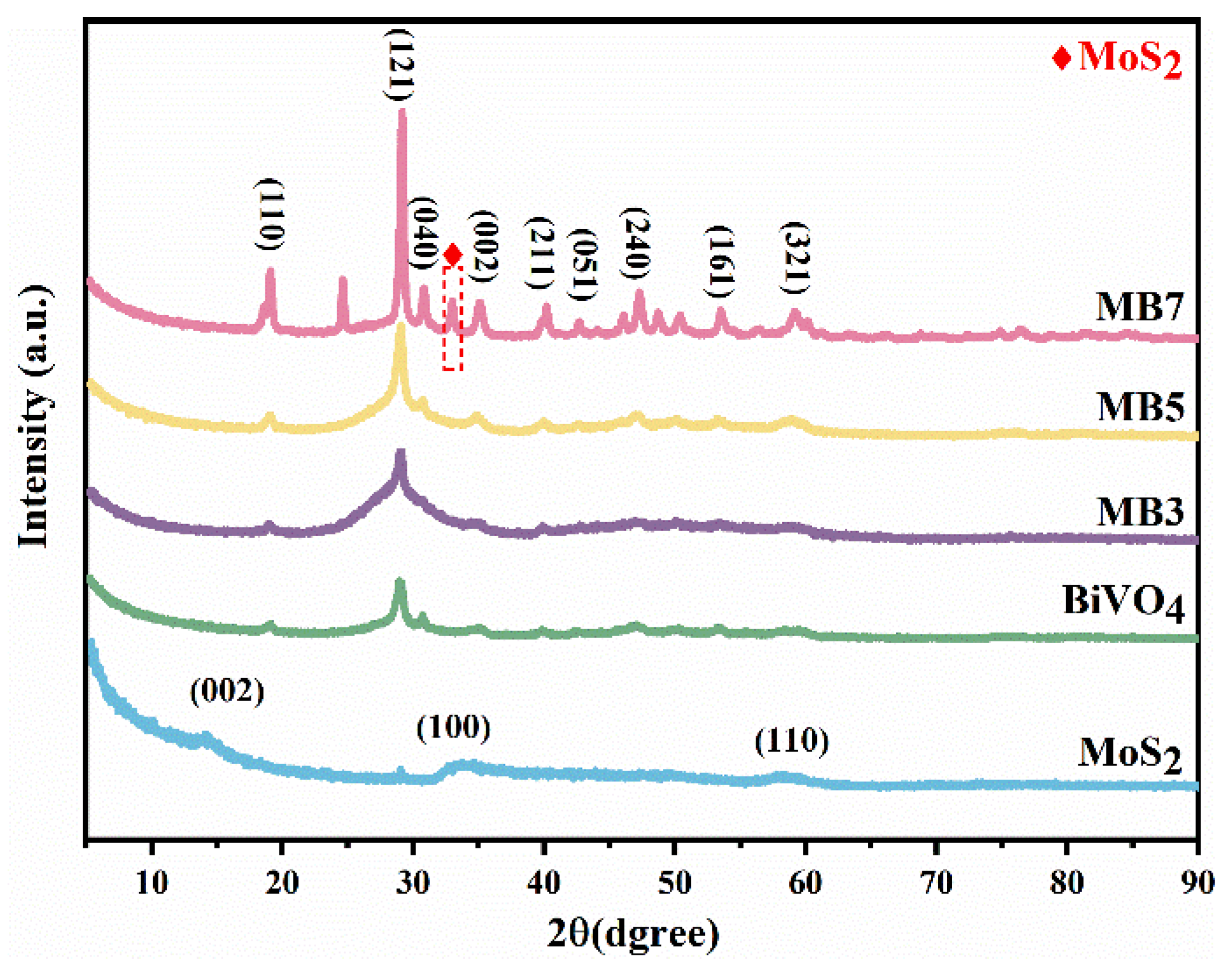
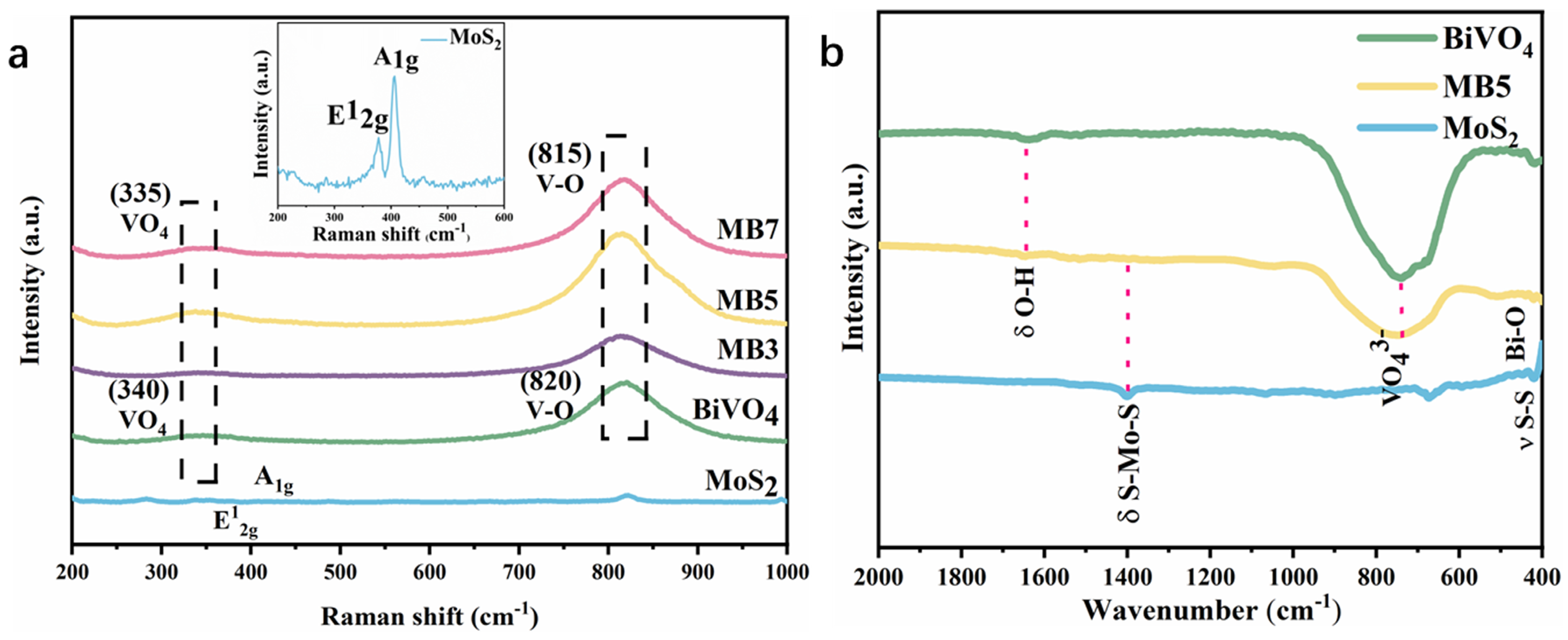
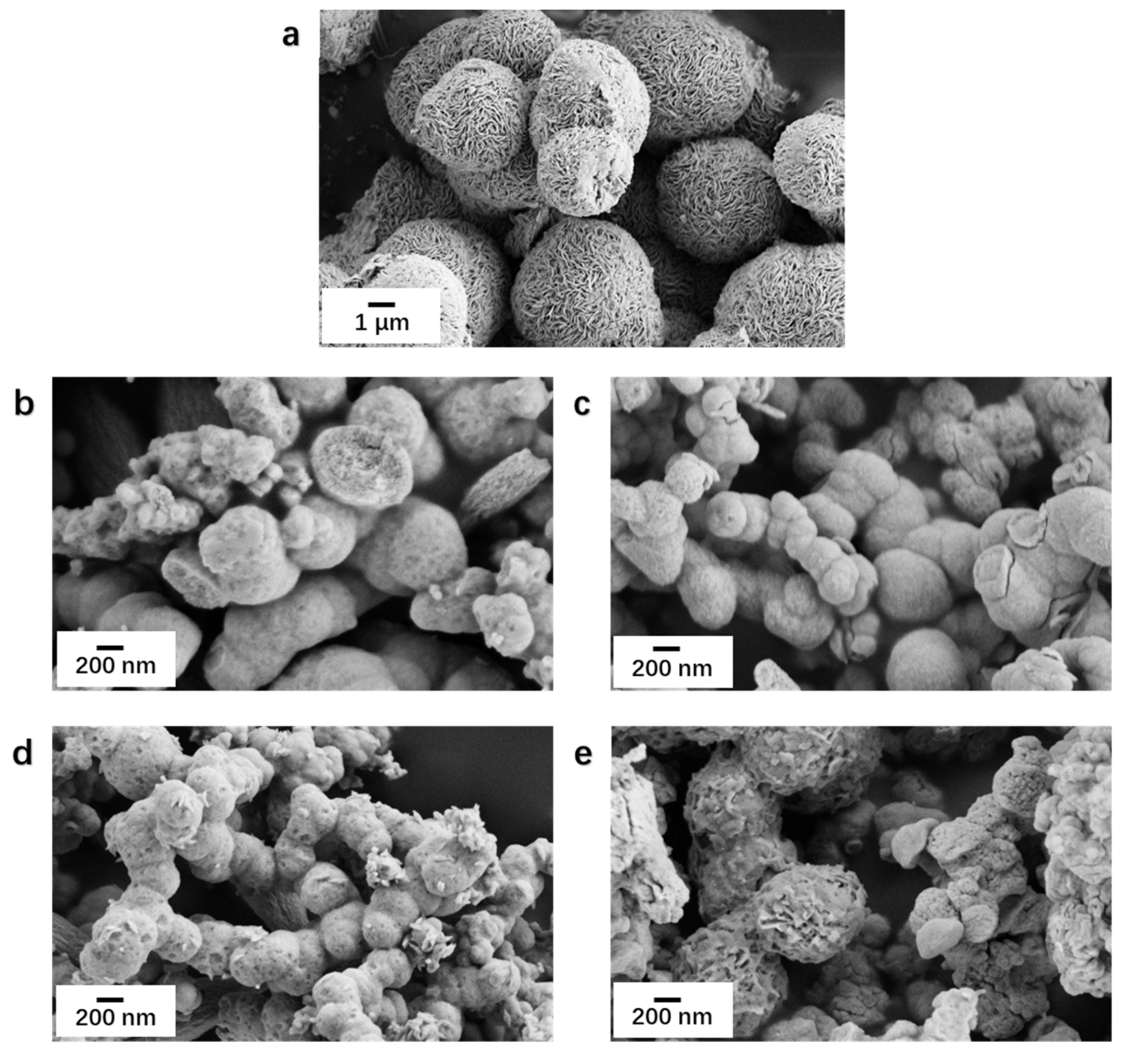
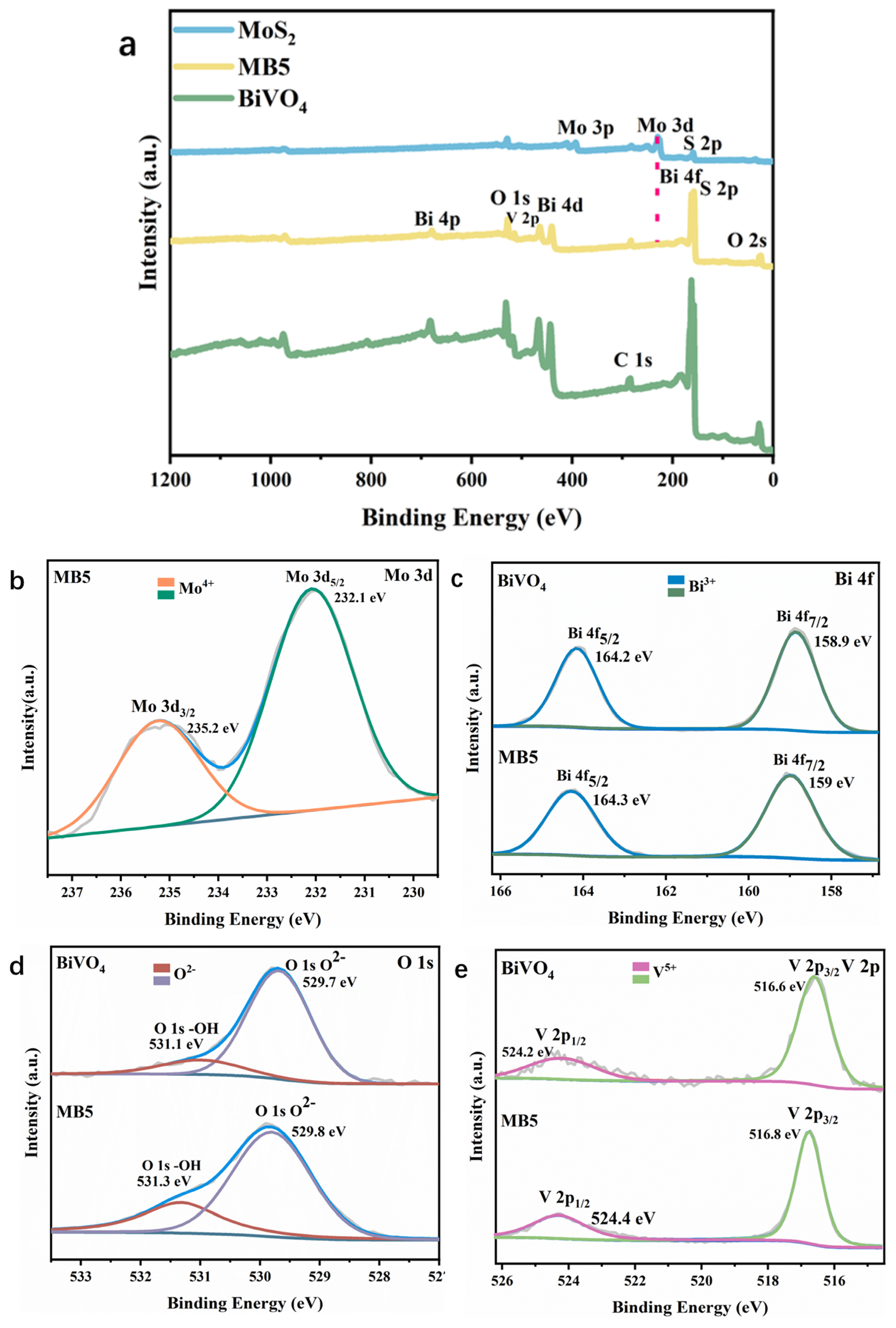
3.1. Characterization

The crystal phase of the samples was characterized by XRD. As shown in Figure 1, the diffraction peaks of BiVO4, and all MoS2/BiVO4 heterojunctions had a good corresponding relationship with the monoclinic scheelite BiVO4 (JCPDS No. 14-0688) [36]. The diffraction peaks at 2θ = 14.0°, 33.0° and 58.3° of MoS2 are derived from the contributions of the (002), (100) and (110) crystal planes of hexagonal MoS2 (JCPDS No.37-1492) [37]. Remarkably, when the loading amount of MoS2 was low, the diffraction peaks of MoS2 were absent in the XRD spectra of MB3 and MB5. As the amount of MoS2 continued to increase, the diffraction peak located at 33.0° corresponds to the (100) crystal plane of MoS2 which appeared in MB7 composites. Moreover, it proved the successful preparation of MoS2/BiVO4 [38]. The enhancement of the diffraction peak intensity of the (121) crystal plane in MB5 and MB7 was significantly greater than the other diffraction peaks, and this probably attributed to the addition of MoS2 affected the crystal structure and the growth direction of BiVO4 [39,40] The grain size can be calculated by the Debye-Scherrer equation [41] (Equation (1)),
D = K λ Bcos θ
where D is the average grain size, K = 0.89, λ = 1.5406 nm, B is the full width at half maximum of the diffraction peaks, θ is the Bragg’s angle, which in this study is the diffraction angle corresponding to the (121) crystal plane. The average crystallite sizes of BiVO4, MB3, MB5 and MB7 were calculated as 6.9, 2.9, 5.5, 17.2 nm, respectively. The addition of MoS2 influenced the particle size of MoS2/BiVO4 composites, where the smallest size was MB3.
Raman spectroscopy analysis (Figure 2a) and FT-IR spectra (Figure 2b) could be helpful to further validate the structure of samples. As shown in Figure 2a, BiVO4 and MoS2/BiVO4 composites have a strong peak at 820 cm−1 which is caused by V-O bond tensile vibration, and the peak at 340 cm−1 is the asymmetric and symmetric bending vibration of VO43− [42]. Further, the characteristic peaks of MoS2 at 378 and 406 cm−1 correspond to the E12g and A1g vibrational modes [43]. In Raman spectra, red shifts in the MoS2/BiVO4 spectral band can be clearly seen, the peak at 815 cm−1 has an increase of the intensity, and along with the stretching vibration of the V-O bond and the bending vibration of VO43− become irregular, which can be attributed to the interaction between MoS2 and BiVO4. According to the available literature [44,45], such a red shift and increase in peak intensity of MB5 were due to successful preparation of heterojunction. FT-IR spectra of MoS2, BiVO4 and MB5 were shown in Figure 2b. For BiVO4, the peak at 416 cm−1 corresponds to the chemical stretching of Bi-O. The absorption band observed at 745 cm−1 was attributed to VO43− symmetric and tensile vibration peak [46]. For MoS2, the peaks at 420, and 1401 cm−1 were allocated to S-S and S-Mo-S bond vibration and tensile vibration [47]. For MoS2/BiVO4 composites, all single-phase vibrations corresponded well.
SEM was employed to observe the micro morphology of the photocatalyst (Figure 3). Figure 3a indicated that MoS2 is a nanosphere structure with an average diameter of about 4 μm. Figure 3b showed BiVO4 as a nanosphere structure with the size of about 0.5 um. The plots of MoS2/BiVO4 with different ratios were placed in Figure 3c–e, when MoS2 nanospheres were added to the microwave reactor, they were separated into nano-microspheres of smaller size by microwave radiation. It can be observed that the surface morphology of the MoS2/BiVO4 heterojunction with a different mass ratio of MoS2 and BiVO4 is different. First, MB3 was similar to BiVO4 nanospheres, and the surface of MB3 that MoS2 was completely covered by BiVO4 (Figure 3c). In the second place, some irregular depressions could be observed on the surface of MB5 (Figure 3d). Finally, with MB7 it could be seen that there were many MoS2 exposed on the surface, which was due to the fact that as the added MoS2 increases MoS2, it was not completely covered by BiVO4 (Figure 3e). The results showed that the growth process and the structure are similar to MoS2/BiVO4 heterojunction prepared by Peng et al. [25]. During the reaction, with MoS2 as a substrate, BiVO4 would nucleate and continue to grow on the surface of MoS2 nano-microspheres, eventually covered the entire surface of MoS2, and grew into a nanosphere structure.
Further investigation of the microstructure of MoS2/BiVO4 heterostructures was conducted through HRTEM (Figure 4). The interplanar spacing of 0.31 nm and 0.27 nm were clearly found in Figure 4b, which were correspond to the (121) plane of BiVO4 [48] and the (100) plane of MoS2 [49], respectively. In addition, the chemical composition and elemental distribution of MB5 were analyzed by SEM-EDS. As shown in Figure 4c–g, the MB5 composites photocatalyst was composed of five elements (Bi, V, O, Mo, and S). The above images could be clearly observed that BiVO4 and MoS2 formed an interface in close contact further provided evidence for the successful preparation of MoS2/BiVO4 heterostructures.
With the use of XPS, the surface molecular structure and chemical states of MoS2, BiVO4, and MB5 were identified. The presence of Bi, V, O, Mo, and S element in the MB5 were revealed by the full scan XPS spectrum (Figure 5a). Figure 5b shows the Mo 3d high resolution XPS spectra. Mo 3d5/2 and Mo 3d3/2 of MB5 were matched with two peaks at 232.1 eV and 235.2 eV. The energy gap of these two peaks is approximately 3.1 eV, indicating that molybdenum ions are Mo4+ in a lower oxidation state [25]. There is no characteristic peak of Mo3d in BiVO4, while it is present in MB5, which proves the presence of MoS2 in the MoS2/BiVO4 heterojunction. In Figure 5c, two significant peaks that occurred around 164.3 eV and 159.0 eV were identified to the Bi 4f5/2 and Bi 4f7/2; the energy gap of 5.3 eV confirmed that the bismuth species occurred as Bi3+ in MB5. Vanadium ions in MB5 existed in the shape of V5+. Two significant peaks near 524.2 eV and 516.6 eV were attributed to the V 2p split signals (V 2p1/2 and V 2p3/2) clearly seen in Figure 5e [24]. Likewise, the main peak of O 1s in the high resolution XPS spectra could be divided into two bands, implying two different types of oxygen existed on the surface of MB5 [23]. The XPS results indicate a strong interaction between MoS2 and BiVO4 and the successful preparation of a heterostructure.
Specific surface area is an important factor affecting the catalytic efficiency of photocatalysts. Figure 6 is the N2 adsorption-desorption curve and the pore size distribution map of MoS2, BiVO4 and MB5 measured by the Brunauer-Emmett-Teller (BET) and Barret-Joyner-Halenda (BJH), respectively. As shown in Figure 6a, all photocatalysts showed type-IV isotherms with H3 hysteresis loops [50]. The specific surface areas of 9.79 m2 g−1, 5.07 m2 g−1, and 11.02 m2 g−1 were for MoS2, BiVO4, and MB5, respectively. The findings indicated that MB5 had the largest specific surface area. This was because in the synthesis process of MB5, where MoS2 were broken down into smaller-sized MoS2 by microwave energy. Through the microwave reaction, the increased surface area of the MoS2 led to an overall increase in the specific surface area of MB5 [51]. The large specific surface area provides more active sites and higher adsorption capacity. In addition, Figure 6b showed the presence of a mesoporous structure, which could serve as a fast transfer path for photogenerated electrons [52].

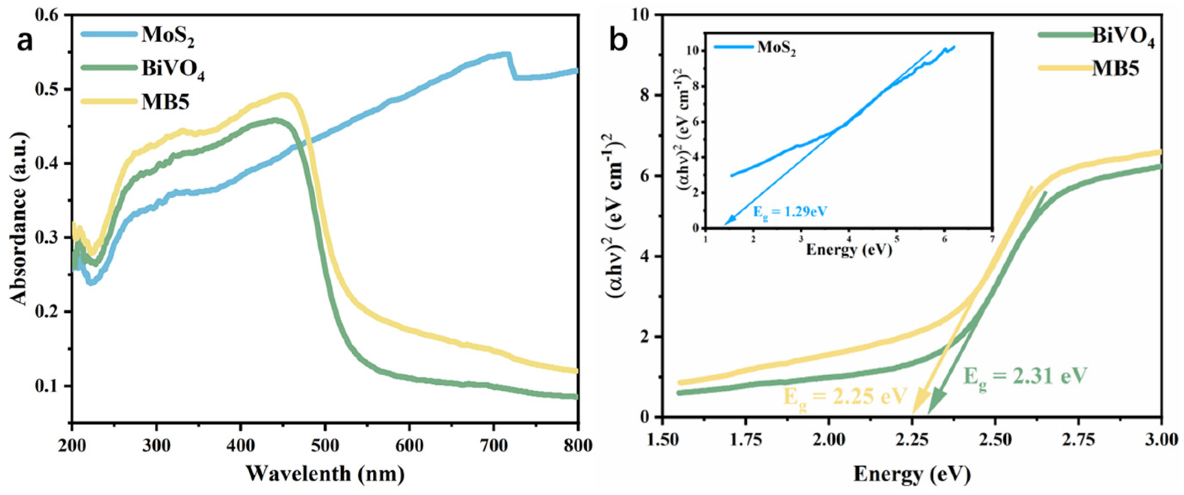
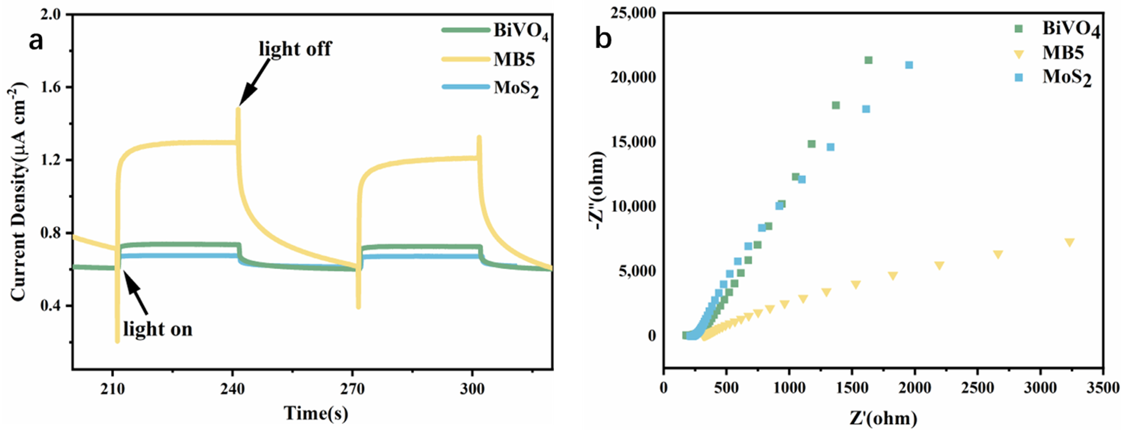
3.2. Light Absorption and Charge Transfer Performance

The optical features of the photocatalysts could be assessed by using UV-visible diffuse reflectance spectroscopy (UV-vis DRS). In Figure 7a, BiVO4 had an absorption band at 500 nm. MoS2 showed a response to ultraviolet and visible light region, indicating its wide absorption rage [53]. MB5 had an increased absorption capacity in both the ultraviolet and visible light region compared with BiVO4, contributing to the higher utilization of solar light. Tauc plot (Figure 7b) showed the band gaps of 1.29 eV, 2.31 eV and 2.25 eV for MoS2, BiVO4 and MB5, respectively. Compared to BiVO4, MB5 had a reduced band gap. According to the above analysis, compared with MoS2 and BiVO4, electrons in the MB5 were more easily excited from the valence band to the conduction band due to the stronger optical absorption capability and the reduced band gap width. In order to reveal the photogenerated charge divorce process of the heterojunction in the photocatalysis, the conduction band (CB) and valence band (VB) of BiVO4 and MoS2 were calculated by the following formula [54] (Equations (2) and (3)):
E C B = X E e 0.5 E g
E V B = E C B + E g
where, Ee is the energy of free electrons at hydrogen standard potential, 4.5 eV. Eg is band gap. X is the absolute electronegativity of the semiconductor. The X values of BiVO4 and MoS2 are 6.16 eV and 5.32 eV, which are calculated by the following formula (Equation (4)):
X = [ x ( A ) a x ( B ) b x ( C ) c ] 1 / ( a + b + c )
in which x(Bi) = 4.69 eV, x(V) = 3.60 eV, x(O) = 7.54 eV, x(Mo) = 3.90 eV, x(S) = 6.22 eV. a, b, and c are the number of atoms in compound [55].
The calculated CB values of BiVO4, MoS2 are 0.505 eV and 0.175 eV, respectively. The calculated VB values of BiVO4 is 2.815 eV, and MoS2 is 1.465 eV. Apparently, MoS2 and BiVO4 could form a heterojunction because they have a staggered band position.
To further investigate the charge separation of MoS2/BiVO4 heterojunction, the photocurrent response and electrochemical impedance spectroscopy (EIS) of MoS2, BiVO4 and MB5 were measured (Figure 8). Figure 8a showed that, MB5 displayed the highest photocurrent response (1.3 μA cm−2) under visible light (λ > 420 nm) illumination, which was 3.25 times that of MoS2 and 1.85 times that of BiVO4. A higher photocurrent response of the MB5 suggests the increased charges separation efficiency, which increased the lifetime of the photogenerated charge and thus improved the photocatalytic rate [56,57]. In the EIS curve, when compared with MoS2 and BiVO4, it was evident that MB5 had the smallest arc radius (Figure 8b), which was consistent with the results of the photocurrent density test. The minimum arc radius implies that MB5 has the lowest resistance and highest charge transfer efficiency. The results suggested that the separation efficiency and transport performance of photogenerated carriers in heterojunction were hugely enhanced.

3.3. Catalytic Capacity of MoS2/BiVO4

To assess the visible light photocatalytic activity of BiVO4, MB3, MB5, and MB7, TC degradation experiments were carried out. Figure 9a indicated that 39.7% of TC could be degraded in 90 min by BiVO4. In the same conditions, 93.7% of TC could be degraded by MB5, it is 2.36 times greater than BiVO4. Moreover, with the increase of MoS2 compounded in MoS2/BiVO4, the degradation rate of TC by MoS2/BiVO4 photocatalyst showed a trend of increasing and then decreasing, and MB5 showed the best enhancement of photocatalytic activity. Compared with BiVO4, there may be three reasons for the enhanced photocatalytic performance of MB5: firstly, the larger specific surface area signifies an increased adsorption capacity, secondly, the improved utilization of visible light and the narrower band gap boosts the excitation of electrons, and thirdly, the generation of transport channels between MoS2 and BiVO4 are beneficial to the rapid transfer and separation of photogenerated electron-hole pairs. Figure 9b showed the result of the TC in three cyclic degradation experiments. Due to the loss of photocatalytic materials in the recycling process, the degradation rate decreases with the number of uses. After the catalyst was reused three times, the degradation efficiency remained above 80%. By a pseudo first-order model (lnC0/C = kt) [58], the degradation processes were fitted. Figure 9c showed that all fitted curves are nearly linear. As shown in Figure 9d, the kinetic constants k for the synthesized BiVO4, MB3, MB5 and MB7 are 0.0015, 0.0031, 0.0215 and 0.0012 min−1, respectively. The kinetic constants k of MB5 is much higher than other photocatalysts, which was 14.3 times that of BiVO4. The above results illustrated the effectiveness and stability of MoS2/BiVO4 heterojunction as a photocatalyst.

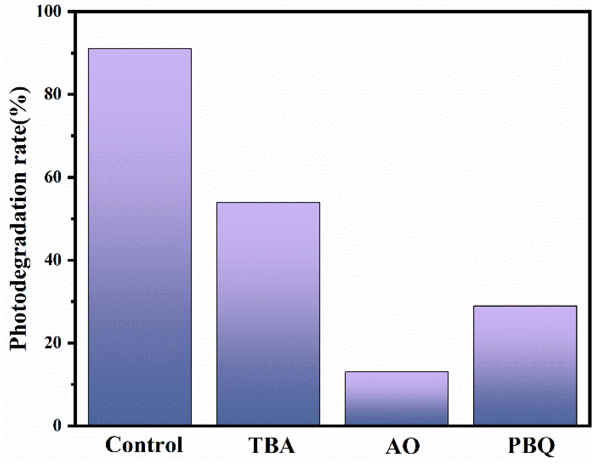
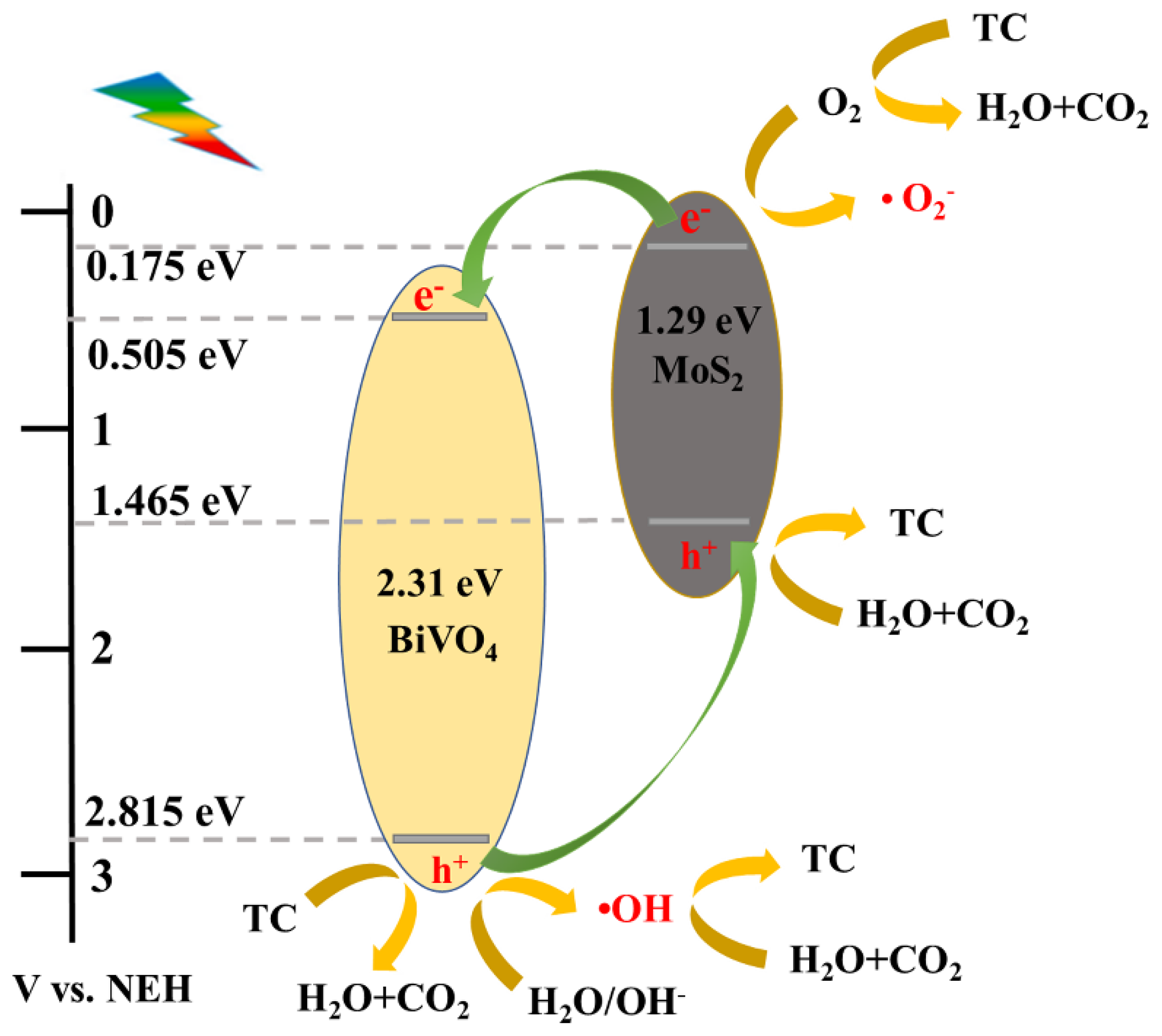
3.4. Photocatalytic Mechanism Study

A radical capture test was used to explore the photocatalytic mechanism. Under the same experimental conditions, TBA, AO and PBQ were added as radical trapping agents for •OH, h+ and •O2, respectively [59,60]. The outcomes in Figure 10 revealed that after the addition of the three catchers TBA, AO, and PBQ, respectively, the degradation efficiency reduced to 54%, 13% and 29%. It could be concluded from the test results that the efficient degradation of TC benefited from the synergistic effect of •OH, h+, and •O2. Among them, h+ had two working modes, one was the direct oxidative degradation of TC, and the other was reaction with the water adsorbed on the surface of the material to form •OH, which then oxidizes and degrades TC. Owing to the addition of AO had the most pronounced inhibitory effect on the degradation of TC, which indicated that the primary active species for the oxidative degradation TC was h+ in the photocatalytic reaction for MB5.
Hence, based on the relative positions of energy bands, and the above radical catch test results, possible photodegradation mechanisms of MoS2/BiVO4 photocatalysis is posited (Figure 11). The p-type MoS2 and the n-type BiVO4 form a p-n heterojunction, and MoS2 had higher CB and VB than BiVO4 based on the energy band structure. Firstly, because the adsorption property of MB5 was enhanced by the addition of MoS2, the large amount of TC was adsorbed on the surface of the photocatalytic materials. Under visible light irradiation, MoS2 and BiVO4 were excited simultaneously. The photogenerated holes of BiVO4 and MoS2 degraded TC as h+ radicals. And some of the photogenerated holes in the VB of BiVO4 migrated to the VB of MoS2 and then formed •OH with water or hydroxyl groups which was adsorbed on the surface of the material. MoS2 and BiVO4 originally had different Fermi levels, and the Fermi levels became the same after the heterojunction was formed, which moved the CB of MoS2 up to a higher level [61]. Therefore, in the heterojunction, the e in the CB of MoS2 had a stronger redox ability to react with dissolved oxygen to form •O2. The matching energy band position between MoS2 and BiVO4 allows the generation of fast separation channels for photogenerated carriers at the heterojunction interface, which contributes to the generation of •OH, h+ and •O2. Moreover, h+ played the most dominant role in the degradation of TC. Finally, the three active substances synergize with each other to oxidize and degrade the pollutants which are adsorbed on the surface of the photocatalytic materials into H2O and CO2.

4. Conclusions

Herein, a p-n heterojunction photocatalyst MoS2/BiVO4 was obtained by microwave-assisted synthesis of BiVO4 crystals that were grown on the surface of MoS2 nano-microspheres. Compared with BiVO4, the heterojunction structure formed by MoS2/BiVO4 has a stronger adsorption capacity, greater visible light utilization, smaller band gap and higher separation of photogenerated carriers, and these advantages synergistically lead to the enhanced photocatalytic activity of the MoS2/BiVO4. For the above reasons, MB5 showed better photocatalytic activity under visible light, and the degradation rate of TC reached 93.7% within 90 min, and the first-order kinetic constant of MB5 was 0.0215 min−1, which was 14.3 times higher over BiVO4. Cyclic degradation experiments demonstrated the reusability and stability of the MoS2/BiVO4 photocatalyst. This study provides an energy-saving and an efficient, feasible method for the preparation of the MoS2/BiVO4 heterojunction photocatalyst with excellent photocatalytic performance, and the MoS2/BiVO4 heterojunction photocatalyst is expected to realize broader applications in the wastewater treatment.

Author Contributions

Conceptualization, Q.S. and W.Z.; methodology, C.C.; formal analysis, Y.Z., W.S. and Z.L.; investigation, Q.S., W.Z. and C.C.; resources, J.Y.; writing—original draft preparation, C.C.; writing—review and editing, Q.S., W.Z. and K.C.; visualization, C.C.; supervision, J.Y., K.C., Q.W. and J.L.; funding acquisition, Q.S., W.Z., J.Y. and Q.W. All authors have read and agreed to the published version of the manuscript.

Funding

This work has been financially supported by China Postdoctoral Science Foundation (No 2022MD713733), Basic Ability Improvement Project for Young and Middle-Aged Teachers in Guangxi Province, China (No 2019KY0187), Scientific Research Foundation for introduced Talents of Guangxi Minzu University (No 2018KJQD07), Yunnan Ten Thousand Talents Plan Young & Elite Talents Project (YNWR-QNBJ-2019-015, KKRD202051006), the Natural Science Foundation of Yunnan Province (202101AT070104).

Data Availability Statement

The data that support the findings of this study are available from the corresponding author upon reasonable request.

Conflicts of Interest

The authors declare no conflict of interest.

References

  1. Wolf, M.J.; Emerson, J.W.; Esty, D.C.; de Sherbinin, A.; Wendling, Z.A. 2020 Environmental Performance Index; Yale Cener for Environmental Law & Policy: New Haven, CT, USA, 2020. [Google Scholar]
  2. Danner, M.C.; Robertson, A.; Behrends, V.; Reiss, J. Antibiotic pollution in surface fresh waters: Occurrence and effects. Sci. Total Environ. 2019, 664C, 793–804. [Google Scholar] [CrossRef] [PubMed]
  3. Wang, R.; Ji, M.; Zhai, H.; Guo, Y.; Liu, Y. Occurrence of antibiotics and antibiotic resistance genes in WWTP effluent-receiving water bodies and reclaimed wastewater treatment plants. Sci. Total Environ. 2021, 796, 148919. [Google Scholar] [CrossRef] [PubMed]
  4. Li, J.; Li, W.; Liu, K.; Guo, Y.; Ding, C.; Han, J.; Li, P. Global review of macrolide antibiotics in the aquatic environment: Sources, occurrence, fate, ecotoxicity, and risk assessment. J. Hazard. Mater. 2022, 439, 129628. [Google Scholar] [CrossRef] [PubMed]
  5. Larsson, D.G.J.; Flach, C.F. Antibiotic resistance in the environment. Nat. Rev. Microbiol. 2022, 20, 257–269. [Google Scholar] [CrossRef] [PubMed]
  6. Guo, J.; Liu, T.; Peng, H.; Zheng, X. Efficient Adsorption-Photocatalytic Removal of Tetracycline Hydrochloride over Octahedral MnS. Int. J. Mol. Sci. 2022, 23, 9343. [Google Scholar] [CrossRef]
  7. Yang, M.; Cui, C.; Liu, L.; Dai, L.; Bai, W.; Zhai, J.; Jiang, S.; Wang, W.; Ren, E.; Cheng, C.; et al. Porous activated carbons derived from bamboo pulp black liquor for effective adsorption removal of tetracycline hydrochloride and malachite green from water. Water Sci. Technol. 2022, 86, 244–260. [Google Scholar] [CrossRef]
  8. Zhou, Y.; You, S.; Zhang, J.; Wu, M.; Yan, X.; Zhang, C.; Liu, Y.; Qi, W.; Su, R.; He, Z. Copper ions binding regulation for the high-efficiency biodegradation of ciprofloxacin and tetracycline-HCl by low-cost permeabilized-cells. Bioresour. Technol. 2022, 344, 126297. [Google Scholar] [CrossRef]
  9. Tian, T.; Zhu, X.; Song, Z.; Li, X.; Zhang, W.; Mao, Y.; Chen, S.; Wu, J.; Ouyang, G. The potential of a natural iron ore residue application in the efficient removal of tetracycline hydrochloride from an aqueous solution: Insight into the degradation mechanism. Environ. Sci. Pollut. Res. Int. 2022, 29, 76782–76792. [Google Scholar] [CrossRef]
  10. Wang, H.; Li, X.; Zhao, X.; Li, C.; Song, X.; Zhang, P.; Huo, P.; Li, X. A review on heterogeneous photocatalysis for environmental remediation: From semiconductors to modification strategies. Chin. J. Catal. 2022, 43, 178–214. [Google Scholar] [CrossRef]
  11. Kudo, A.; Ueda, K.; Kato, H.; Mikami, I. Photocatalytic O2 evolution under visible light irradiation on BiVO4 in aqueous AgNO3 solution. Catal. Lett. 1998, 53, 229–230. [Google Scholar] [CrossRef]
  12. Shi, H.; Guo, H.; Wang, S.; Zhang, G.; Hu, Y.; Jiang, W.; Liu, G. Visible Light Photoanode Material for Photoelectrochemical Water Splitting: A Review of Bismuth Vanadate. Energy Fuels 2022, 36, 11404–11427. [Google Scholar] [CrossRef]
  13. Wang, L.; Shi, X.; Jia, Y.; Cheng, H.; Wang, L.; Wang, Q. Recent advances in bismuth vanadate-based photocatalysts for photoelectrochemical water splitting. Chin. Chem. Lett. 2021, 32, 1869–1878. [Google Scholar] [CrossRef]
  14. Zhong, X.; Li, Y.; Wu, H.; Xie, R. Recent progress in BiVO4-based heterojunction nanomaterials for photocatalytic applications. Mater. Sci. Eng. B 2023, 289, 116278. [Google Scholar] [CrossRef]
  15. Xu, X.; Zhang, J.; Tao, F.; Dong, Y.; Wang, L.; Hong, T. Facile construction of Z-scheme g-C3N4/BiVO4 heterojunctions for boosting visible-light photocatalytic activity. Mater. Sci. Eng. B 2022, 279, 115676. [Google Scholar] [CrossRef]
  16. Guo, W.; Luo, H.; Jiang, Z.; Shangguan, W. In-situ pressure-induced BiVO4/Bi0.6Y0.4VO4 S-scheme heterojunction for enhanced photocatalytic overall water splitting activity. Chin. J. Catal. 2022, 43, 316–328. [Google Scholar] [CrossRef]
  17. Jin, Z.; Zhang, Y.; Liu, D.; Ding, H.; Mamba, B.B.; Kuvarega, A.T.; Gui, J. Fabrication of a La-doped BiVO4@CN step-scheme heterojunction for effective tetracycline degradation with dual-enhanced molecular oxygen activation. Sep. Purif. Technol. 2021, 277, 119224. [Google Scholar] [CrossRef]
  18. Hu, C.; Tian, M.; Wu, L.; Chen, L. Enhanced photocatalytic degradation of paraben preservative over designed g-C3N4/BiVO4 S-scheme system and toxicity assessment. Ecotoxicol. Environ. Saf. 2022, 231, 113175. [Google Scholar] [CrossRef] [PubMed]
  19. Li, M.; Liu, B.; Guo, H.; Wang, H.; Shi, Q.; Xu, M.; Yang, M.; Luo, X.; Wang, L. Reclaimable MoS2 Sponge Absorbent for Drinking Water Purification Driven by Solar Energy. Environ. Sci. Technol. 2022, 56, 11718–11728. [Google Scholar] [CrossRef]
  20. Liu, H.; Wu, R.; Zhang, H.; Ma, M. Microwave Hydrothermal Synthesis of 1T@2H−MoS2 as an Excellent Photocatalyst. Chem-catchem 2019, 12, 893–902. [Google Scholar] [CrossRef]
  21. Li, H.; Yu, K.; Lei, X.; Guo, B.; Fu, H.; Zhu, Z. Hydrothermal Synthesis of Novel MoS2/BiVO4 Hetero-Nanoflowers with Enhanced Photocatalytic Activity and a Mechanism Investigation. J. Phys. Chem. C 2015, 119, 22681–22689. [Google Scholar] [CrossRef]
  22. Koutavarapu, R.; Jang, W.Y.; Rao, M.C.; Arumugam, M.; Shim, J. Novel BiVO4-nanosheet-supported MoS2-nanoflake-heterostructure with synergistic enhanced photocatalytic removal of tetracycline under visible light irradiation. Chemosphere 2022, 305, 135465. [Google Scholar] [CrossRef] [PubMed]
  23. Zheng, Z.; Ng, Y.H.; Tang, Y.; Li, Y.; Chen, W.; Wang, J.; Li, X.; Li, L. Visible-light-driven photoelectrocatalytic activation of chloride by nanoporous MoS2@BiVO4 photoanode for enhanced degradation of bisphenol A. Chemosphere 2021, 263, 128279. [Google Scholar] [CrossRef] [PubMed]
  24. Xu, A.; Tu, W.; Shen, S.; Lin, Z.; Gao, N.; Zhong, W. BiVO4@MoS2 core-shell heterojunction with improved photocatalytic activity for discoloration of Rhodamine B. Appl. Surf. Sci. 2020, 528, 146949. [Google Scholar] [CrossRef]
  25. Ju, P.; Hao, L.; Zhang, Y.; Sun, J.; Dou, K.; Lu, Z.; Liao, D.; Zhai, X.; Sun, C. Facile fabrication of a novel spindlelike MoS2/BiVO4 Z-scheme heterostructure with superior visible-light-driven photocatalytic disinfection performance. Sep. Purif. Technol. 2022, 299, 121706. [Google Scholar] [CrossRef]
  26. Liu, X.; Xu, L.; Zhou, G.; Liu, Q.; Song, M.; Han, S.; Esakkimuthu, S.; Vinh, J.; Barati, B.; Lu, Z. Greatly improved photocatalytic performance of BiVO4/MoS2 heterojunction with enhanced hole transfer and attack capability by ultrasonic agitation and in-situ hydrothermal method. J. Taiwan Chem. Eng. 2020, 117, 48–55. [Google Scholar] [CrossRef]
  27. Acevedo, L.; Usón, S.; Uche, J. Exergy transfer analysis of microwave heating systems. Energy 2014, 68, 349–363. [Google Scholar] [CrossRef]
  28. Selvam, S.M.; Paramasivan, B. Microwave assisted carbonization and activation of biochar for energy-environment nexus: A review. Chemosphere 2022, 286, 131631. [Google Scholar] [CrossRef]
  29. Acevedo, L.; Ferreira, G.; López-Sabirón, A.M. Exergy transfer principles of microwavable materials under electromagnetic effects. Mater. Today Commun. 2021, 27, 102313. [Google Scholar] [CrossRef]
  30. Lu, H.; Ju, T.; She, H.; Wang, L.; Wang, Q. Microwave-assisted synthesis and characterization of BiOI/BiF3 p–n heterojunctions and its enhanced photocatalytic properties. J. Mater. Sci. Mater. Electron. 2020, 31, 13787–13795. [Google Scholar] [CrossRef]
  31. Hu, H.; Wang, T.; Peng, L.; Ling, X.; He, Y.; Sun, M.; Yang, M.; Deng, C. Microwave-assisted synthesis of Z-scheme CdS/BiOBr heterojunction for improved visible-light photocatalytic degradation of organic dyes. Appl. Phys. A 2022, 128, 452. [Google Scholar] [CrossRef]
  32. Zhou, Z.; Xu, H.; Li, D.; Zou, Z.; Xia, D. Microwave-assisted synthesis of La(OH)3/BiOCl n-n heterojunctions with high oxygen vacancies and its enhanced photocatalytic properties. Chem. Phys. Lett. 2019, 736, 136805. [Google Scholar] [CrossRef]
  33. Claudino, C.H.; Kuznetsova, M.; Rodrigues, B.S.; Chen, C.; Wang, Z.; Sardela, M.; Souza, J.S. Facile one-pot microwave-assisted synthesis of tungsten-doped BiVO4/WO3 heterojunctions with enhanced photocatalytic activity. Mater. Res. Bull. 2020, 125, 110783. [Google Scholar] [CrossRef]
  34. Sriram, B.; Baby, J.N.; Hsu, Y.F.; Wang, S.F.; George, M. In Situ Synthesis of a Bismuth Vanadate/Molybdenum Disulfide Composite: An Electrochemical Tool for 3-Nitro-l-Tyrosine Analysis. Inorg. Chem. 2022, 61, 14046–14057. [Google Scholar] [CrossRef] [PubMed]
  35. Gao, Y.; Cong, S.; He, Y.; Zou, D.; Liu, Y.; Yao, B.; Sun, W. Study on the mechanism of degradation of tetracycline hydrochloride by microwave-activated sodium persulfate. Water Sci. Technol. 2020, 82, 1961–1970. [Google Scholar] [CrossRef]
  36. Fardush Tanha, J.; Farhad, S.F.U.; Honey, U.; Tanvir, N.I.; Hasan, T.; Shahriyar Nishat, S.; Kabir, A.; Ahmed, S.; Hakim, M.; Khan, M.N.I.; et al. A DFT+U look into experimentally synthesized monoclinic scheelite BiVO4. J. Appl. Phys. 2021, 130, 235107. [Google Scholar] [CrossRef]
  37. Li, Q.; Huang, L.; Dai, W.; Zhang, Z. Controlling 1T/2H heterophase junctions in the MoS2 microsphere for the highly efficient photocatalytic hydrogen evolution. Catal. Sci. Technol. 2021, 11, 7914–7921. [Google Scholar] [CrossRef]
  38. Zhao, W.; Liu, Y.; Wei, Z.; Yang, S.; He, H.; Sun, C. Fabrication of a novel p–n heterojunction photocatalyst n-BiVO4@p-MoS2 with core–shell structure and its excellent visible-light photocatalytic reduction and oxidation activities. Appl. Catal. B 2016, 185, 242–252. [Google Scholar] [CrossRef]
  39. Liu, Y.; Xu, X.; Ma, C.; Zhao, F.; Chen, K. Morphology Effect of Bismuth Vanadate on Electrochemical Sensing for the Detection of Paracetamol. Nanomaterials 2022, 12, 1173. [Google Scholar] [CrossRef]
  40. Lutsko, J.F. How crystals form: A theory of nucleation pathways. Sci. Adv. 2019, 5, 7399. [Google Scholar] [CrossRef]
  41. Verma, A.; Bisen, D.P.; Brahme, N.; Sahu, I.P.; Singh, A.K. Yttrium aluminum garnet based novel and advanced phosphor synthesized by combustion route activated by Dy, Eu, and Tb rare earth metals. J. Mater. Sci. Electron. 2023, 34, 644. [Google Scholar] [CrossRef]
  42. Ullah, S.; Fayeza; Khan, A.A.; Jan, A.; Aain, S.Q.; Neto, E.P.; Serge-Correales, Y.E.; Parveen, R.; Wender, H.; Rodrigues-Filho, U.P.; et al. Enhanced photoactivity of BiVO4/Ag/Ag2O Z-scheme photocatalyst for efficient environmental remediation under natural sunlight and low-cost LED illumination. Colloids Surf. A Physicochem. Eng. Asp. 2020, 600, 124946. [Google Scholar] [CrossRef]
  43. van Nguyen, T.; Tekalgne, M.; Nguyen, T.P.; Wang, W.; Hong, S.H.; Cho, J.H.; van Le, Q.; Jang, H.W.; Ahn, S.H.; Kim, S.Y. Control of the morphologies of molybdenum disulfide for hydrogen evolution reaction. Int. J. Energy Res. 2022, 46, 11479–11491. [Google Scholar] [CrossRef]
  44. Liu, J.J.; Jiang, Z.W.; Hsu, S.W. Investigation of the Performance of Heterogeneous MOF-Silver Nanocube Nanocomposites as CO2 Reduction Photocatalysts by In Situ Raman Spectroscopy. ACS Appl. Mater. Interfaces 2023, 15, 6716–6725. [Google Scholar] [CrossRef] [PubMed]
  45. Li, M.; Wei, Y.; Fan, X.; Li, G.; Hao, Q.; Qiu, T. Mixed-dimensional van der Waals heterojunction-enhanced Raman scattering. Nano Res. 2021, 15, 637–643. [Google Scholar] [CrossRef]
  46. Chawla, H.; Garg, S.; Rohilla, J.; Szamosvölgyi, Á.; Efremova, A.; Szenti, I.; Ingole, P.P.; Sápi, A.; Kónya, Z.; Chandra, A. Visible LED-light driven photocatalytic degradation of organochlorine pesticides (2,4-D & 2,4-DP) by Curcuma longa mediated bismuth vanadate. J. Clean. Prod. 2022, 367, 132923. [Google Scholar] [CrossRef]
  47. Kite, S.V.; Kadam, A.N.; Sathe, D.J.; Patil, S.; Mali, S.S.; Hong, C.K.; Lee, S.W.; Garadkar, K.M. Nanostructured TiO2 Sensitized with MoS2 Nanoflowers for Enhanced Photodegradation Efficiency toward Methyl Orange. ACS Omega 2021, 6, 17071–17085. [Google Scholar] [CrossRef]
  48. Wang, J.; Jin, J.; Wang, X.; Yang, S.; Zhao, Y.; Wu, Y.; Dong, S.; Sun, J.; Sun, J. Facile fabrication of novel BiVO4/Bi2S3/MoS2 n-p heterojunction with enhanced photocatalytic activities towards pollutant degradation under natural sunlight. J. Colloid Interface Sci. 2017, 505, 805–815. [Google Scholar] [CrossRef]
  49. Zhang, Z.; Dong, Y.; Sun, H.; Liu, G.; Liu, S.; Yang, X. Defect-rich 2D reticulated MoS2 monolayers: Facile hydrothermal preparation and marvellous photoelectric properties. J. Taiwan Chem. Eng. 2019, 101, 221–230. [Google Scholar] [CrossRef]
  50. Dong, P.; Xi, X.; Zhang, X.; Hou, G.; Guan, R. Template-Free Synthesis of Monoclinic BiVO4 with Porous Structure and Its High Photocatalytic Activity. Materials 2016, 9, 685. [Google Scholar] [CrossRef]
  51. Zhong, Y.; Yu, L.; Chen, Z.F.; He, H.; Ye, F.; Cheng, G.; Zhang, Q. Microwave-Assisted Synthesis of Fe3O4 Nanocrystals with Predominantly Exposed Facets and Their Heterogeneous UVA/Fenton Catalytic Activity. ACS Appl. Mater. Interfaces 2017, 9, 29203–29212. [Google Scholar] [CrossRef]
  52. Chen, S.; Huang, D.; Du, L.; Lei, L.; Chen, Y.; Wang, G.; Wang, Z.; Zhou, W.; Tao, J.; Li, R.; et al. Peroxymonosulfate activation by surface-modified bismuth vanadate for ciprofloxacin abatement under visible light: Insights into the generation of singlet oxygen. Chem. Eng. J. 2022, 444, 136373. [Google Scholar] [CrossRef]
  53. Zhang, X.; Suo, H.; Zhang, R.; Niu, S.; Zhao, X.q.; Zheng, J.; Guo, C. Photocatalytic activity of 3D flower-like MoS2 hemispheres. Mater. Res. Bull. 2018, 100, 249–253. [Google Scholar] [CrossRef]
  54. Geosciences, D.O. The absolute energy positions of conduction and valence bands of selected semiconducting minerals. Am. Mineral. 2000, 85, 543–556. [Google Scholar]
  55. Pearson, R.G. Absolute Electronegativity and Hardness: Applications to Organic. J. Org. Chem. 1989, 54, 1423–1430. [Google Scholar] [CrossRef]
  56. Li, J.; Jin, B.; Jiao, Z. Rationally embedded zinc oxide nanospheres serving as electron transport channels in bismuth vanadate/zinc oxide heterostructures for improved photoelectrochemical efficiency. J. Colloid Interface Sci. 2021, 592, 127–134. [Google Scholar] [CrossRef]
  57. Li, J.; Li, L.; Ma, X.; Han, X.; Xing, C.; Qi, X.; He, R.; Arbiol, J.; Pan, H.; Zhao, J.; et al. Selective Ethylene Glycol Oxidation to Formate on Nickel Selenide with Simultaneous Evolution of Hydrogen. Adv. Sci. 2023, 10, 2300841. [Google Scholar] [CrossRef]
  58. Hu, C.; Xiong, C.; Lin, Y.L.; Zhu, Y.; Wang, Q.; Xu, L.; Huang, D. Degradation of 2-phenylbenzimidazole 5-sulfonic acid by UV/chlorine advanced oxidation technology: Kinetic model, degradation byproducts and reaction pathways. J. Hazard. Mater. 2022, 431, 128574. [Google Scholar] [CrossRef]
  59. Yang, W.; Ding, K.; Chen, G.; Wang, H.; Deng, X. Synergistic Multisystem Photocatalytic Degradation of Anionic and Cationic Dyes Using Graphitic Phase Carbon Nitride. Molecules 2023, 28, 2796. [Google Scholar] [CrossRef]
  60. Xia, L.; Liang, W.; Chen, G.; Li, W.; Gao, M. Catalytic Ozonation of Quinoline Utilizing Manganese-Based Catalyst with Abundant Oxygen Vacancies. Catal. Lett. 2021, 152, 1669–1677. [Google Scholar] [CrossRef]
  61. Masoumi, Z.; Tayebi, M.; Kolaei, M.; Lee, B.-K. Unified surface modification by double heterojunction of MoS2 nanosheets and BiVO4 nanoparticles to enhance the photoelectrochemical water splitting of hematite photoanode. J. Alloys Compd. 2022, 890, 161802. [Google Scholar] [CrossRef]
Figure 1. XRD patterns of MoS2, BiVO4, MB3, MB5 and MB7 ( mark shows the presence of MoS2).
Nanomaterials 13 01522 g001
Figure 2. (a) Raman spectrum of MoS2, BiVO4, MB3, MB5 and MB7 and (b) FT−IR spectra of MoS2, BiVO4 and MB5.
Nanomaterials 13 01522 g002
Figure 3. SEM image of (a) MoS2, (b) BiVO4, (c) MB3, (d) MB5, and (e) MB7.
Nanomaterials 13 01522 g003
Figure 4. (a,b) HRTEM image and (cg) SEM-EDS elemental mapping of MB5.
Nanomaterials 13 01522 g004
Figure 5. XPS spectra: (a) overall spectra of MoS2, BiVO4 and MB5, (b) Mo 3d spectra, (c) Bi 4f spectra, (d) O 1s spectra, and (e) V 2p spectra of BiVO4 and MB5.
Nanomaterials 13 01522 g005
Figure 6. (a) N2 adsorption-desorption curves and (b) pore size distribution of MoS2, BiVO4 and MB5.
Nanomaterials 13 01522 g006
Figure 7. (a) UV–vis absorption spectra and (b) the plot of (αhν)2 vs. energy hν and band gap energy of t of MoS2, BiVO4 and MB5.
Nanomaterials 13 01522 g007
Figure 8. (a) Photocurrent response (1 V vs. Ag/AgCl) under visible light irradiation (λ > 420 nm) and (b) EIS spectra of MoS2, BiVO4 and MB5.
Nanomaterials 13 01522 g008
Figure 9. (a) Degradation efficiency of TC in visible light, (b) Recycle degradation of TC, (c) quasi first-order kinetics of degradation of TC, and (d) the kinetic constants k of degradation of TC (MB5 = 50 mg, TC = 100 mL, 5 mg L−1, time = 90 min).
Nanomaterials 13 01522 g009
Figure 10. Active species capture tests of MB5 in photocatalytic degradation (TBA = 1.8 mL, AO = 0.3 mmol, PBQ = 0.3 mmol).
Nanomaterials 13 01522 g010
Figure 11. Mechanism of TC degradation by MoS2/BiVO4 heterojunction photocatalyst.
Nanomaterials 13 01522 g011
Disclaimer/Publisher’s Note: The statements, opinions and data contained in all publications are solely those of the individual author(s) and contributor(s) and not of MDPI and/or the editor(s). MDPI and/or the editor(s) disclaim responsibility for any injury to people or property resulting from any ideas, methods, instructions or products referred to in the content.

