Article

Synthesis, Structural Characterization, and Infrared Analysis of Double Perovskites Pr2NiMnO6, Gd2NiMnO6, and Er2NiMnO6 Functional Nano-Ceramics

by
Mebark Elhamel
1,
Zoulikha Hebboul
1,*,
Djamal Benbertal
1,
Pablo Botella
2 and
Daniel Errandonea
2,*
1
Laboratoire Physico-Chimie des Matériaux, Université Amar Telidji de Laghouat, BP 37G, Route de Ghardaia, Laghouat 03000, Algeria
2
Departamento de Física Aplicada—ICMUV, MALTA Consolider Team, Universidad de Valencia, Edificio de Investigación, Carrer del Doctor Moliner 50, 46100 Burjassot, Valencia, Spain
*
Authors to whom correspondence should be addressed.
Nanomaterials 2024, 14(11), 960; https://doi.org/10.3390/nano14110960
Submission received: 16 May 2024 / Revised: 24 May 2024 / Accepted: 29 May 2024 / Published: 30 May 2024
(This article belongs to the Section Nanocomposite Materials)

Abstract

:
We synthesized Pr2NiMnO6, Gd2NiMnO6, and Er2NiMnO6 double perovskites in a nano-ceramic form by a sol–gel method. By means of room-temperature X-ray powder diffraction measurements, we determined the crystal structure of the three compounds, which is monoclinic, corresponding to a double perovskite structure, described by space group P21/n structure. From the determined structures, the bulk moduli were estimated to be 173–179 GPa. The average size particle of nanoparticles was determined from X-ray diffraction by the Langford method plot and by the Scherrer formula. The morphology and homogeneity of nanoparticles were analyzed by scanning electron microscopy. We found that they form compact agglomerations of approximately 200 nm in diameter. Fourier transform infrared spectroscopy measurements were performed, determining the absorption spectrum. The assignment of the measured infrared absorption bands is discussed.

1. Introduction

Double perovskites expand the chemical and structural space of the perovskite family and have emerged as promising alternatives with improved catalytic performance and many other interesting properties for technological applications [1,2]. Double perovskite R2NiMnO6 compounds, where R is a lanthanide element, are materials that stand out for electronic interaction which give rise to a ferromagnetic alignment of d electron spins, due to the super-exchange interaction between Mn4+ and Ni2+ ions according to the Goodenough–Kanamori rules [3]. These materials have recently received increased interest because of their unique optical, electronic, and magnetic properties [4,5]. They have also attracted much attention due to their potential use in magnetic cooling, environmentally friendly technologies based on the magnetocaloric effect [6,7,8,9]. It has been also proposed that, thanks to their complex physical properties, R2NiMnO6 compounds may be used to design a variety of innovative devices, including multiple-state memory elements [10]. The discovery of the existence of a magnetodielectric effect around the ferromagnetic Curie temperature of compounds like La2NiMnO6 represented a step forward for these developments [11]. R2NiMnO6 compounds are also ferromagnetic semiconductors, being therefore promising materials for spintronic devices for energy-efficient data storage and energy harvesting [12]. These materials also exhibit magnetocapacitance and magnetoresistance near room temperature [13] and have been used for improving the performance of solid oxide fuel cells [14], which adds more value to R2NiMnO6 compounds as multifunctional materials. Another interesting fact in lanthanide double perovskites is related to the lanthanide contraction in ionic radius and gadolinium break in ionic electronegativity. These facts have been found to regulate the properties in the perovskite structure, including the catalytic performance [15]. Thus, R2NiMnO6 compounds have direct interest from both fundamental and technological points of view. More information on the multiple recent developments in the study of R2NiMnO6 double perovskite can be found in a recently published review [16].
The prospective of technological utilization of the multifunctional properties of R2NiMnO6 compounds could be boosted by the synthesis of these materials as nanoparticles [17]. Nanomaterials have currently a considerable impact in practically all domains of science and technology because, thanks to size effects, the properties of materials, such as reactivity, strength, and electrical and optical characteristics, can be tuned for different applications [18]. Polycrystalline R2NiMnO6 double perovskite compounds are usually synthesized by conventional solid-state methods [6], by low-temperature auto-ignition methods [19], or by annealing a compact mixture of R2O3, NiO, and MnO2 under high pressure using a belt-type apparatus [20]. They have been also prepared from citrate precursors obtained by a soft-chemistry procedure [17].
In this work, we present a simple, cheap, and environmentally friendly soft-chemistry method to prepare nanoparticles of Pr2NiMnO6, Gd2NiMnO6, and Er2NiMnO6. This is the first time nanoparticles of the studied compounds are reported. To the best of the authors knowledge, the used method has never been used before to prepare R2NiMnO6 compounds. The morphology, crystal structure, and infrared transmittance of the samples were characterized via scanning electron microscopy (SEM), powder X-ray diffraction (XRD), and Fourier transform infrared (FTIR) spectroscopy. The bulk modulus and important elastic modulus have been also estimated. The size of the obtained particles ranges from 29–36 nm to 40–49 nm in the different compounds according to the two methods used for determining the particle size.

2. Materials and Methods

All the reagents used in the synthesis were purchased from Sigma–Aldrich (St. Louis, MO, USA) with 99.99% of purity. They include nickel nitrate hexahydrate (Ni(NO3)2·6H2O), praseodymium(III) nitrate hexahydrate (Pr(NO3)3·6H2O), erbium(III) oxide (Er2O3), gadolinium nitrate hexahydrate (Gd(NO3)3·6H2O), and manganese chloride tetrahydrate (MnCl2·4H2O). The samples R2NiMnO6, R = Pr, Gd, and Er, as nanoparticles, were prepared by sol–gel synthesis. The weights of all reagents and reaction conditions are summarized in Table 1. For each sol preparation, MnCl2·4H2O and Ni(NO3)·6H2O were mixed in methanol for 10 min using a magnetic mixer; then, Pr(NO3)3·6H2O, Er2O3, and Gd(NO3)3·6H2O were added separately to the independent solutions and mixed for another 20, 10, and 10 min, respectively. After that, a solution of citric acid (C6H8O7), adjusting the solution pH to 6.5, was added to the mixture and heating was kept at 70 °C until gel formation at 22, 48, and 32 min, respectively. The three gels were dried at 80 °C in a cabinet drier for 24 h to obtain precursor powders. Then, an agate mortar and pestle were used to grind the mixture prepared for each sample. Subsequently pellets were prepared under a pressure of ~45 bars and were subsequently annealed at 1100 °C for 3 h under air atmosphere.
Sample morphology, homogeneity, and chemical composition of the obtained products were analyzed by scanning SEM and energy-dispersive X-ray analysis (EDX) using a Tescan Vega (Brno, Czech Republic) scanning electron microscope connected to a Peltier cooled XFlashTM Bruker (Billerica, MA, USA) silicon drift detector and using an accelerating voltage of 30 kV. Crystal structures were characterized by powder XRD using a Panalytical EMPYREAN diffractometer (Malvern, UK) operating in a Bragg–Brentano geometry (Cu Kα1, 40 mA, 30 kV) with a 0.01° step size and an acquisition time of 6 s/step in the 10−90° 2θ range. FTIR spectra were recorded in transmission mode using a Jasco FT/IR-4200 (Tokyo Japan) set up using ultrapure KBr pellets.

3. Results and Discussion

3.1. Morphology

SEM images of the synthesized samples illustrating their microstructure are shown in Figure 1. The micrographs notably show that the three samples have a similar homogeneous morphology, which consisted of quasi-spherical nanoparticles having uniform grain sizes and distributions throughout the three samples. Figure 1 also shows that R2NiMnO6 nanoparticles are arranged sequentially as chains, forming strongly bound agglomerations and leading to submicron-sized entities. The average diameter of agglomerations is between 100 and 200 nm in diameter. In the case of Pr2NiMnO6 and Gd2NiMnO6, the agglomerations bear a resemblance to zeolite-type structures. In the case of Er2NiMnO6, the agglomerations look like stacked coral reefs. The differences on the shape of agglomerations might be related to the different concentration of impurities of the samples (see discussion of XRD). As a consequence of the strong agglomeration between nanoparticles, we could not determine the particle of nanoparticles from SEM measurements. The particle size was determined from XRD. The mean chemical compositions of the synthesized samples were analyzed by EDX analyses. We found within experimental uncertainties that the composition of the samples, by both weight percent and atomic percent of Pr/Gd/Er, Ni, and Mn, were consistent with their corresponding nominal compositions of 2:1:1. The readers should note that, in this manuscript, we do not characterize and analyze the materials’ lattice oxygen and oxygen vacancies, which might be important for oxides [21]. Future studies using O 1s XPS or chemical titration are needed to obtain such information, but they are beyond the scope of the present study.

3.2. XRD Analysis

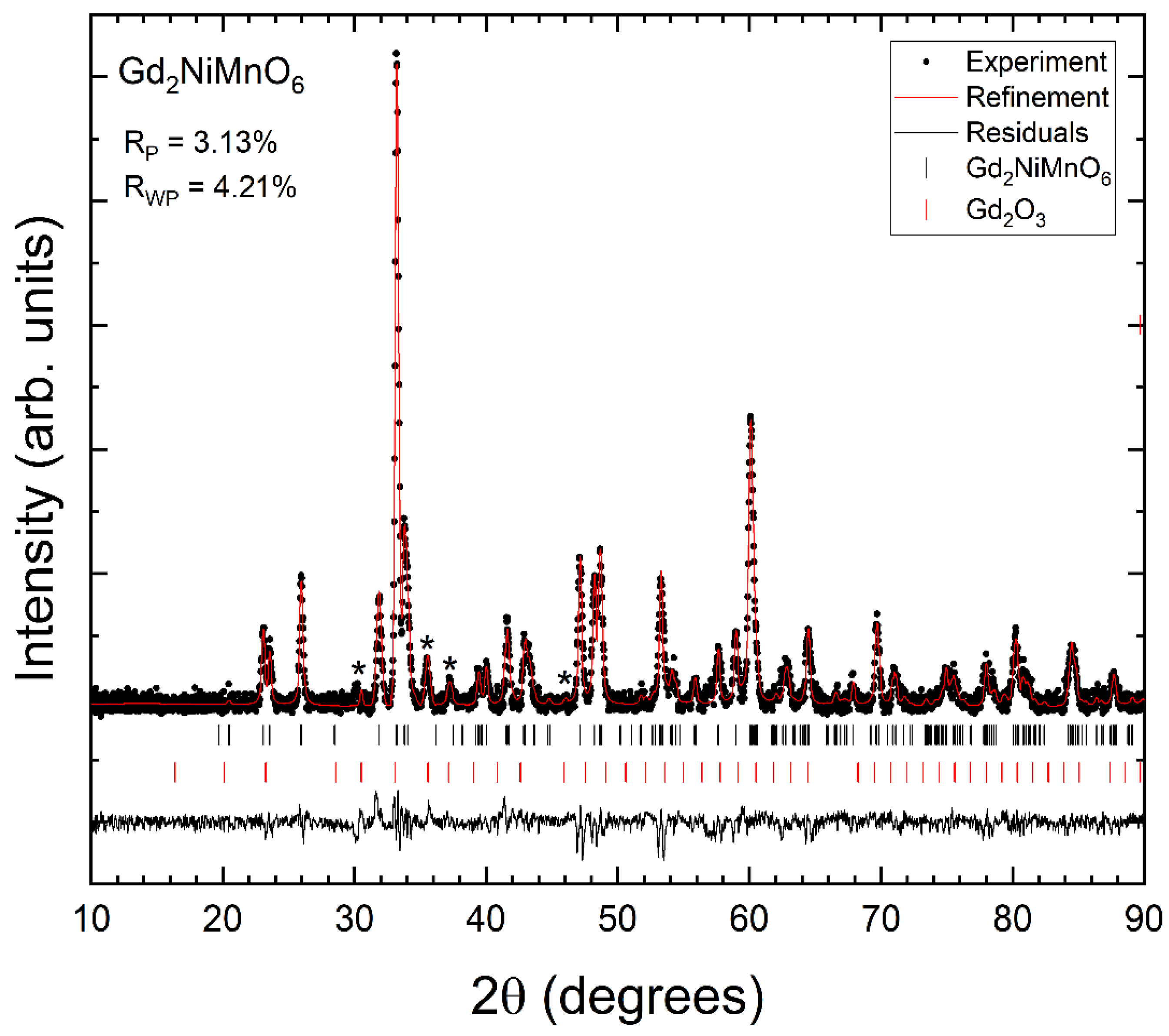
Powder XRD patterns measured in the three synthesized samples at room temperature are shown in Figure 2, Figure 3 and Figure 4. We found that XRD patterns can be well explained by the known double perovskite structure of R2NiMnO6 compounds, which is usually described by the non-standard monoclinic space group P21/n [17]. In addition to the reflection peaks assigned to the studied compounds, we observed extra weak reflections (indicated by asterisks in Figure 2, Figure 3 and Figure 4) that can be assigned to the known bixbyite-type structure of Pr2O3 (in Pr2NiMnO6 see Figure 2), of Gd2O3 (in Gd2NiMnO6 see Figure 3), and of Er2O3 (in Er2NiMnO6 see Figure 4), which is described by space group Ia 3 ¯ [22,23]. The amount of impurities has been estimated from Rietveld refinements [24] and they are indicated in Table 1. The presence of impurities does not preclude the accurate structural determination of the crystal structure of the studied R2NiMnO6 compounds. The Rietveld refinements are shown in Figure 2, Figure 3 and Figure 4, including R-values. The refinements show that the assumed structural model for the three studied compounds matches well with the measured XRD patterns. In the refinements, the background was fitted with a Chebyshev polynomial function of the first kind with six coefficients [25]. The shapes of peaks were modelled using a pseudo-Voigt function and a Caglioti model [26]. The unit-cell parameters obtained from the refinements are summarized in Table 2. They agree with the literature [16,17,27]. The atomic positions are summarized in Table 3. The oxygen positions were refined with precision, in spite of the fact that the compounds contain heavy atoms, thanks to the fact that the positions of Ni and Mn are fixed by symmetry.
In Table 2, by comparing the three compounds, it can be seen that there is a monotonic decrease of the unit-cell volume as the size of the rare-earth ion decreases from Pr to Er, which is a consequence of the lanthanide contraction. Table 4 contains some selected bond distances and angles. There it can be seen than in the three compounds the MnO6 octahedron is smaller and more regular than the NiO6 octahedron. Regarding the coordination polyhedron of the lanthanide atom, in contrast to conventional double perovskites, where the A-site coordination number is 12, in the specific type of R2NiMnO6, the coordination number of the lanthanide atom is 8, as previously highlighted by Maneesha et al. [16]. Notice that the eight R-O bonds are shorter than 2.7 Å. In the three compounds, the ninth bond is at a distance larger than 3.15 Å. An additional fact to highlight about the coordination of R atoms is that, in the three compounds, there are four distances shorter 2.35 Å and four distances larger than 2.49 Å, which makes the effective coordination number to be close to six. We would like to add here that, according to the calculated Ni-O-Mn angles, the tilting angle φ = 180 < N i O M n > 2 is close to 17.5° for the three compounds, which is consistent with the fact that the three compounds have a similar β angle.
From the XRD patterns, we also obtained information on the average particle size using the X’Pert high-score plus v5.2 software and applied both a Langford (L) analysis [28] and the equation proposed by Scherrer applied to the strongest peak [29]. The average crystallite size we determined for each sample is summarized in Table 1. Both methods provide consistent particle sizes, with the Langford method providing slightly smaller sizes.
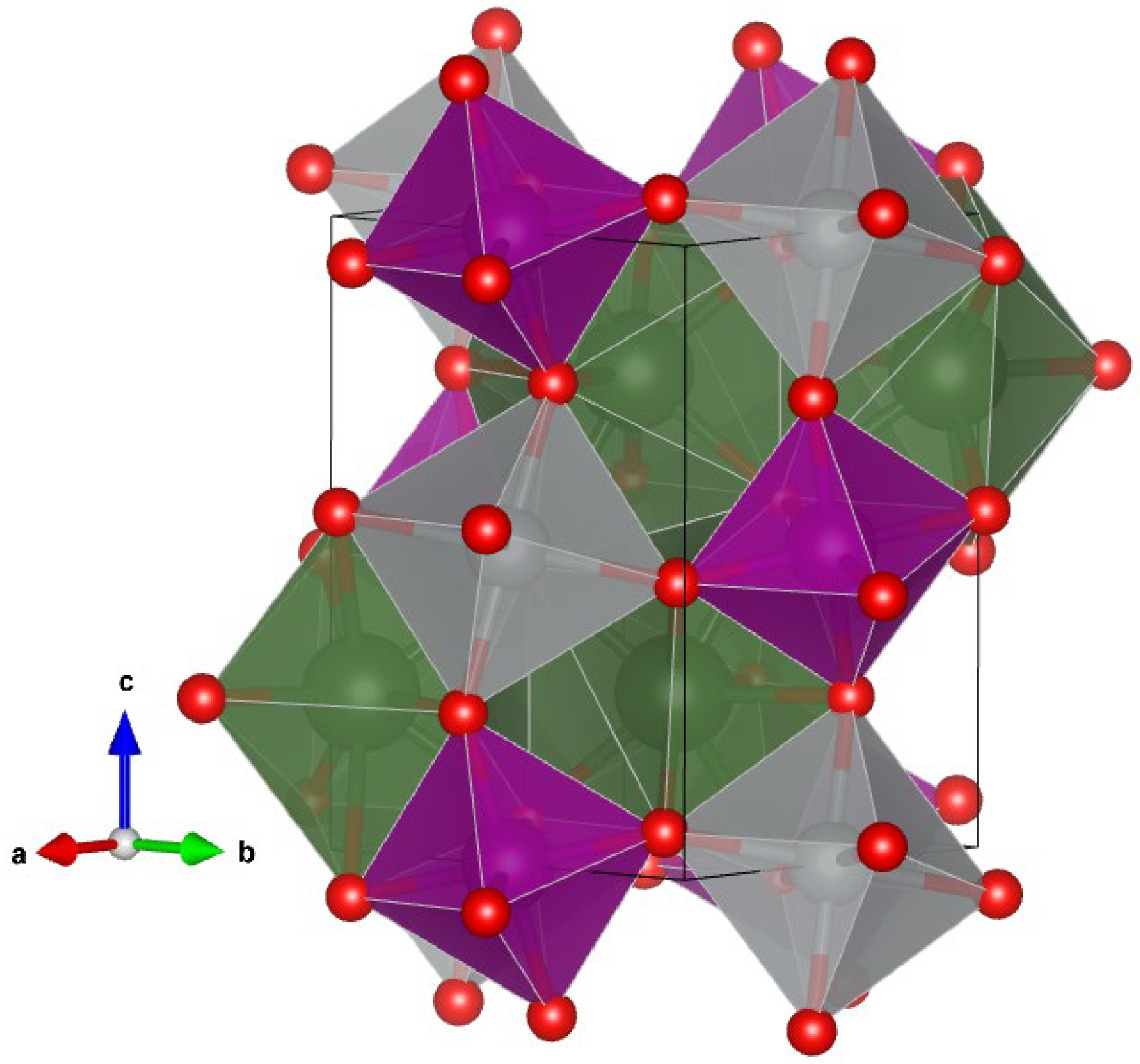
A schematic representation of the crystal structure of the three R2NiMnO6 compounds here studied is shown in Figure 5. The structure can be described as a linear chain of corner-sharing NiO6 and MnO6 octahedral units running parallel to the c-axis, which are connected by edge-sharing RO8 dodecahedra. The NiO6 and MnO6 units show a significant octahedral tilting. Since NiO6 and MnO6 units can be considered as uncompressible units in comparison with RO8 dodecahedra, the compressibility (and consequently the bulk modulus) of the three studied compounds can be assumed as determined basically by the RO8 dodecahedra [30]. Under this hypothesis, and using the empirical formula proposed by Errandonea and Manjon [31], we estimated the bulk modulus (K) of Pr2NiMnO6, Gd2NiMnO6, and Er2NiMnO6 to be 173(5), 176(5), and 179(5) GPa, respectively. The bulk modulus is the measure of how resistant a material is to compression and is very relevant for technological applications, since small changes in the unit-cell volume (and consequently of bond distances) could strongly affect physical properties like the band-gap energy or the magnetic moment. The values here reported for the bulk moduli are very similar to the bulk modulus determined experimentally for La2NiMnO6, 179(8)–188(28) [32]. It is also comparable to the bulk modulus of perovskite CeScO3, 165(7) GPa [33], and the bulk modulus of Ho2O3, 178(5) GPa [34], two compounds where the bulk modulus is defined by the coordination polyhedron of the lanthanide atom. Both facts support the soundness of our estimations of the bulk modulus, confirming that the coordination polyhedra of the rare-earth atom determines the bulk modulus of the R2NiMnO6 double perovskite compounds.

3.3. FTIR Measurements

Fourier transform infrared spectra were measured in near-normal incidence mode at room temperature in the three studied samples. The FTIR spectra for the samples are shown in Figure 6. They show two strong and well-defined absorption bands. The strongest one has the maximum of the absorption (minimum of transmission) centered at approximately 590–600 cm−1 and the other band has it centered at 440–470 cm−1. The frequency of the strongest absorption is in the same range that the IR absorption reported for Eu2NiMnO6 [35] and La2NiMnO6 [36]. A frequency outside the 590–600 cm−1 range was only reported for Sm2NiMnO6 [37], 575 cm−1; probably, this frequency has been underestimated. The frequency of the second absorption is similar to that of the second absorption band reported for La2NiMnO6 [36]. The frequencies determined from this work are summarized in Table 5. We named the highest and lowest frequency modes as ν1 and ν2, respectively.
Having the primitive cell of R2NiMnO6 compounds two formula units, sixty lattice vibrations are expected. Their mechanical representation is Γ = 12Ag + 18Au + 12Bg + 18Bu, where one Au mode plus two Bu modes are the acoustic modes. Therefore, at the center of the Brillouin zone there are 24 Raman-active modes (12Ag + 12Bg) and 33 infrared-active modes (17Au + 16Bu). In previous studies, only two Raman modes and two infrared modes were reported. In La2NiMnO6, the reported Raman modes are at 640 and 530 cm−1 [37] and the observed infrared modes are redshifted to lower frequencies, 600 and 430 cm−1 [38]. The same redshift between Raman and infrared modes is observed when comparing the Raman modes of Pr2NiMnO6, which are at 657 and 511 cm−1 [39], and the infrared modes here reported, which are at 590 and 440 cm−1.
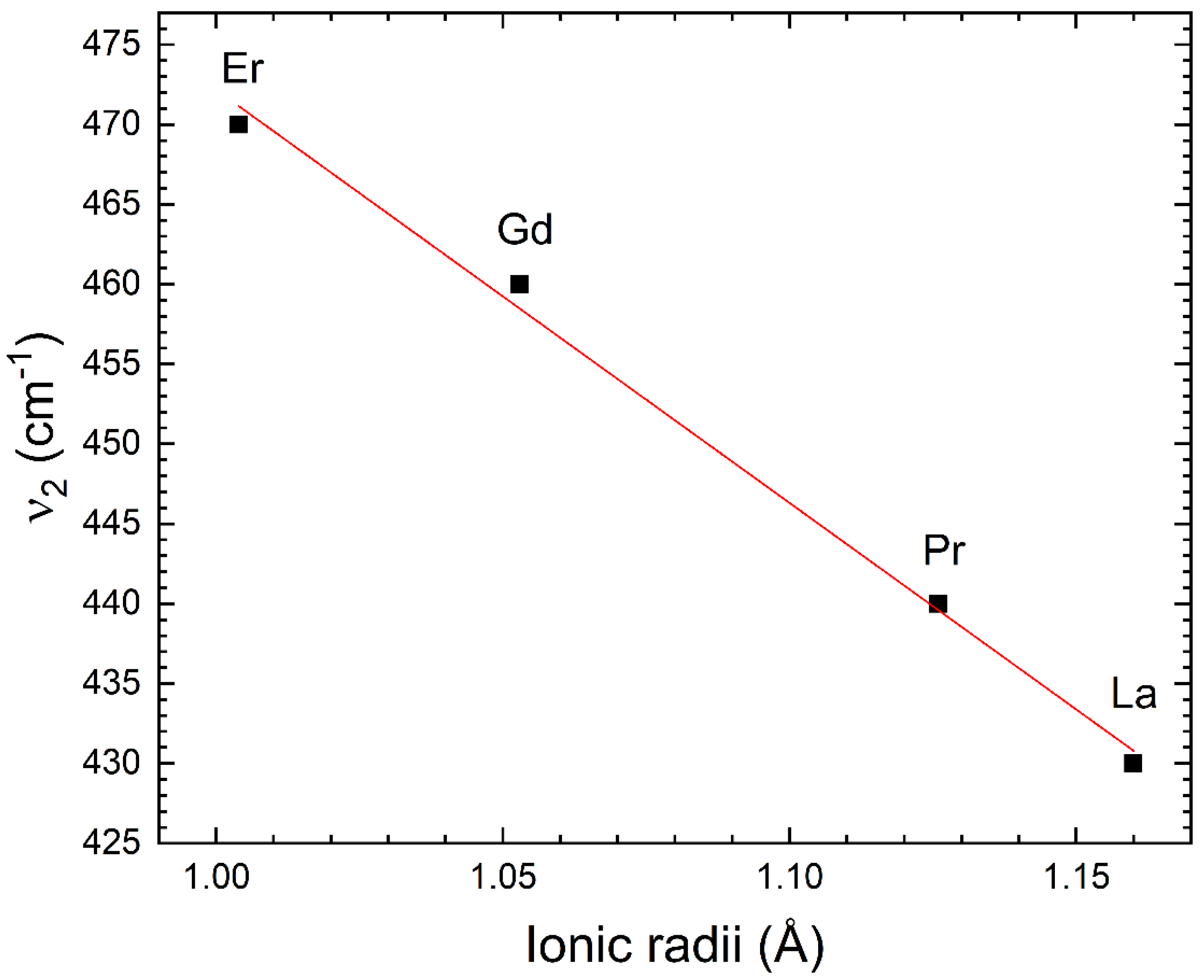
The highest frequency Raman and infrared modes have been assigned in the literature to stretching–breathing internal vibrations of the MnO6 octahedron [40]. This interpretation is consistent with the fact that the frequency of the ν1 mode is basically not affected by the change of the rare-earth atom, as can be seen in Table 5. On the other hand, the frequency of the ν2 mode increases as the atomic number increases, showing that this mode is a more complex mode, involving movements of the rare-earth atoms [41]. The increase in the phonon frequency as the mass the lanthanide atom is opposite to the behavior of the Raman modes, where the frequency decreases as the mass of the lanthanide mode increases [42]. Indeed, this is what we would naively expect according to the harmonic approximation, where the frequency is inversely proportional to the square root of the reduced mass of the atoms involved in the vibration [43]. The increase in the frequency of the infrared mode when moving from La to Er along the lanthanide series might be related with the known lanthanide contraction, which makes the ionic radii decrease as the atomic number (and the mass) increases [44]. As a consequence of it, the bond distance within the RO8 polyhedron decreases as the atomic mass decreases (see Table 4). Then, if the mode ν2 is related to vibrations involving the R-O bonds, an observation of an increase in the frequency of it would be expected, as observed. Note that the reduction in the radius of the lanthanide produces a reduction in the R-O distance and that the force constant of the phonons, and consequently the frequency, which as a first approximation, would follow an inverse relationship with the average R–O bond distance and the ionic radii of the lanthanide atom [43,45] as shown in Table 6 and Figure 7. This figure shows that there is a linear relationship between the frequency of the mode ν2 and the ionic radii r of the lanthanide element, being ν 2 = 730 13 258 12 × r . In Table 6, comprehensive estimations of the vibrational frequency ( ν 2 ) are presented, specifically focusing on the entirety of the lanthanide family in the context of the remaining R2NiMnO6 double perovskites, using the equation provided above.

4. Conclusions

Double perovskites Pr2NiMnO6, Gd2NiMnO6, and Er2NiMnO6 were synthesized as nanoparticles by a sol–gel method, and a monoclinic crystal structure described by space group P21/n was identified for them using powder X-ray diffraction and the Rietveld method. Bond distances and angles were determined. The morphology of the samples was studied by electron scanning microscopy, showing that spherical nanoparticles form 100–200 μm agglomerations. The composition was confirmed by energy-dispersive X-ray spectroscopy. The infrared transmittance was measured and the frequencies of the two main phonons determined. Results from infrared spectroscopy were discussed in comparison with previous Raman and infrared studies in related compounds, being the mode assignment discussed. A systematic for the phonon frequencies was discussed and predictions for other R2NiMnO6 were presented. The bulk modulus of the three compounds was estimated, providing this important parameter for the response of the studied materials to external compression. The synthesis of Pr2NiMnO6, Gd2NiMnO6, and Er2NiMnO6 could open the avenue for additional applications for these materials. The reported results are relevant for energy storage and magnetocaloric applications, and, consequently, to the developing of green technologies.

Author Contributions

M.E.: formal analysis, methodology, writing—original draft, writing—review and editing; Z.H.: formal analysis, methodology, writing—review and editing; D.B.: formal analysis; P.B.: writing—review and editing; D.E.: conceptualization, formal analysis, writing—original draft, writing—review and editing. All authors have read and agreed to the published version of the manuscript.

Funding

Authors from Université Amar Telidji were supported by the Algerian Ministry of Higher Education and Scientific Research through Projects de Recherche Formation-Universitaire PRFU under grant B00L01UN030120220002. They gratefully acknowledge the support of the Technical Platform of Physico-Chemical Analysis-Laghouat-Algeria. Authors from Univ. de Valencia thank the financial support from the Spanish Research Agency (AEI) and Spanish Ministry of Science and Investigation (MCIN) under grants PID2022-138076NB-C41 and RED2022-134388-T (DOI:10.13039/501100011033). D.E. thanks the support of Generalitat Valenciana (GVA) under grants PROMETEO CIPROM/2021/075 and MFA/2022/007. This study forms part of the Advanced Materials program and is supported by MCIN with funding from the European Union Next-Generation EU (PRTR-C17.I1) and by GVA.

Data Availability Statement

Data will be made available on request.

Conflicts of Interest

The authors declare that they have no known competing financial interests or personal relationships that could have appeared to influence the work reported in this paper.

References

  1. Sun, H.; Chen, X.X.G.; Zhou, Y.; Lin, H.-J.; Chen, C.-T.; Ran, R.; Zhou, W.; Shao, Z. Smart Control of Composition for Double Perovskite Electrocatalysts toward Enhanced Oxygen Evolution Reaction. ChemSusChem 2019, 12, 5111–5116. [Google Scholar] [CrossRef] [PubMed]
  2. Xu, X.; Zhong, Y.; Shao, Z. Double Perovskites in Catalysis, Electrocatalysis, and Photo(electro)catalysis. Trends Chem. 2019, 1, 410–424. [Google Scholar] [CrossRef]
  3. Oleś, M.; Horsch, P.; Feiner, L.F.; Khaliullin, G. Spin-Orbital Entanglement and Violation of the Goodenough-Kanamori Rules. Phys. Rev. Lett. 2006, 96, 147205. [Google Scholar] [CrossRef] [PubMed]
  4. Abass, S.; Bagri, A.; Sultan, K. Modifications induced in structural, electronic, and dielectric properties of Nd2NiMnO6 double perovskite by Sr doping. J. Alloys Compd. 2023, 930, 167463. [Google Scholar] [CrossRef]
  5. Booth, R.J.; Fillman, R.; Whitaker, H.; Nag, A.; Tiwari, R.M.; Ramanujachary, K.V.; Gopalakrishnan, J.; Lofland, S.E. An investigation of structural, magnetic, and dielectric properties of R2NiMnO6 (R = rare earth, Y). Mater. Res. Bull. 2009, 44, 1559–1564. [Google Scholar] [CrossRef]
  6. Jia, Y.; Cheng, Y.; Wang, H.; Zhang, Z.; Li, L. Magnetocaloric properties and critical behavior in double perovskite RE2CrMnO6 (RE = La, Pr, and Nd) compounds. Ceram. Int. 2020, 46, 25043–25049. [Google Scholar] [CrossRef]
  7. Shinde, K.P.; Lee, E.J.; Manawan, M.; Lee, A.; Park, S.Y.; Jo, Y.; Ku, K.; Kim, J.M.; Park, J.S. Structural; magnetic, and magnetocaloric properties of R2NiMnO6 (R = Eu, Gd, Tb). Sci. Rep. 2021, 11, 20206. [Google Scholar] [CrossRef]
  8. Kumar, N.; Kaushik, S.D.; Rao, K.S.; Babu, P.D.; Deshpande, S.K.; Achary, S.N.; Errandonea, D. Temperature Dependent Crystal Structure of Nd2CuTiO6: An In Situ Low Temperature Powder Neutron Diffraction Study. Crystals 2023, 13, 503. [Google Scholar] [CrossRef]
  9. Bessimou, M.; Masrour, R. Study of Optical, Magnetic, and Magnetocaloric Properties of Double Perovskites Dy2NiMnO6: First Principles Approach and Monte Carlo Simulations. J. Inorg. Organomet. Polym. Mater. 2024, 1–8. [Google Scholar] [CrossRef]
  10. Eerenstein, W.; Mathur, N.D.; Scott, J.F. Multiferroic and magnetoelectric materials. Nature 2006, 442, 759. [Google Scholar] [CrossRef]
  11. Singh, M.P.; Truong, K.D.; Jandl, S.; Fournier, P. Magnetic properties and phonon behavior of Pr2NiMnO6 thin films. Appl. Phys. Lett. 2011, 98, 162506. [Google Scholar] [CrossRef]
  12. Hashisaka, M.; Kan, D.; Masuno, A.; Takano, M.; Shimakawa, Y.; Terashima, T.; Mibu, K. Epitaxial growth of ferromagnetic La2NiMnO6 with ordered double-perovskite structure. Appl. Phys. Lett. 2006, 89, 032504. [Google Scholar] [CrossRef]
  13. Rogado, N.S.; Li, J.; Sleight, A.W.; Subramanian, M.A. Magnetocapacitance and magnetoresistance near room temperature in a ferromagnetic semiconductor: La2NiMnO6. Adv. Mater. 2005, 17, 2225–2227. [Google Scholar] [CrossRef]
  14. Li, H.; Sun, L.P.; Feng, Q.; Huo, L.H.; Zhao, H.; Bassat, J.M.; Rougier, A.; Fourcade, S.; Grenier, J.C. Investigation of Pr2NiMnO6-Ce0.9Gd0.1O1.95 composite cathode for intermediate-temperature solid oxide fuel cells. J. Solid State Electrochem. 2017, 21, 273–280. [Google Scholar] [CrossRef]
  15. Guan, D.; Zhou, J.; Huang, Y.C.; Dong, C.-L.; Wang, J.-Q.; Zhou, W.; Shao, Z. Screening highly active perovskites for hydrogen-evolving reaction via unifying ionic electronegativity descriptor. Nat. Commun. 2019, 10, 3755. [Google Scholar] [CrossRef] [PubMed]
  16. Maneesha, P.; Baral, S.C.; Rini, E.G.; Sen, S. An overview of the recent developments in the structural correlation of magnetic and electrical properties of Pr2NiMnO6 double perovskite. Prog. Solid. State Chem. 2023, 70, 100402. [Google Scholar] [CrossRef]
  17. Retuerto, M.; Muñoz, A.; Martínez-Lope, M.J.; Alonso, J.A.; Mompeán, F.J.; Fernández-Díaz, M.T.; Sánchez-Benítez, J. Magnetic Interactions in the Double Perovskites R2NiMnO6 (R = Tb, Ho, Er, Tm) Investigated by Neutron Diffraction. Inorg. Chem. 2015, 54, 10890–10900. [Google Scholar] [CrossRef] [PubMed]
  18. Miguel, A.S. Nanomaterials under high-pressure. Chem. Soc. Rev. 2006, 35, 876–889. [Google Scholar] [CrossRef] [PubMed]
  19. Anirban, S.; Dutta, A. Understanding the structure and charge transport mechanism of Sm2NiMnO6 double perovskite prepared via low temperature auto-ignition method. Phys. Lett. A 2021, 397, 127256. [Google Scholar] [CrossRef]
  20. Yi, W.; Liang, Q.; Matsushita, Y.; Tanaka, M.; Belik, A.A. High-Pressure Synthesis, Crystal Structure, and Properties of In2NiMnO6 with Antiferromagnetic Order and Field-Induced Phase Transition. Inorg. Chem. 2013, 52, 14108–14115. [Google Scholar] [CrossRef]
  21. Zou, D.; Yi, Y.; Song, Y.; Guan, D.; Xu, M.; Ran, R.; Wang, W.; Zhou, W.; Shao, Z. The BaCe0.16Y0.04Fe0.8O3−δ nanocomposite: A new high-performance cobalt-free triple-conducting cathode for protonic ceramic fuel cells operating at reduced temperatures. J. Mater. Chem. A 2022, 10, 5381–5390. [Google Scholar] [CrossRef]
  22. Saiki, A.; Ishizawa, N.; Mizutani, N.; Kato, M. Structural Change of C-Rare Earth Sesquioxides Yb2O3 and Er2O3 as a Function of Temperature. J. Ceram. Soc. Jpn. 1985, 93, 649–654. [Google Scholar] [CrossRef]
  23. Rudenko, V.S.; Boganov, A.G. Stoichiometry and phase transitions in rare earth oxides. Inorg. Mater. 1970, 6, 1893–1898. [Google Scholar]
  24. Rietveld, H.M. A profile refinement method for nuclear and magnetic structures. J. Appl. Crystallogr. 1969, 2, 65–71. [Google Scholar] [CrossRef]
  25. Kaduk, J.A. A Rietveld tutorial-Mullite. Powder Diffr. 2009, 24, 351–361. [Google Scholar] [CrossRef]
  26. Scardi, P. Diffraction Line Profiles in the Rietveld Method. Cryst. Growth Des. 2020, 20, 6903–6916. [Google Scholar] [CrossRef]
  27. Mohapatra, S.R.; Sahu, B.; Raut, S.; Kaushik, S.D.; Singh, A.K. Investigation on structural, optical and magnetic properties of double perovskite Gd2NiMnO6. AIP Conf. Proc. 2015, 1665, 140032. [Google Scholar] [CrossRef]
  28. Langford, J.I.; Wilson, A.J.C. Scherrer after sixty years: A survey and some new results in the determination of crystallite size. J. Appl. Cryst. 1978, 11, 102–113. [Google Scholar] [CrossRef]
  29. Patterson, A. The Scherrer Formula for X-Ray Particle Size Determination. Phys. Rev. 1939, 56, 978–982. [Google Scholar] [CrossRef]
  30. Errandonea, D.; Garg, A.B. Recent progress on the characterization of the high-pressure behaviour of AVO4 orthovanadates. Prog. Mater. Sci. 2018, 97, 123–169. [Google Scholar] [CrossRef]
  31. Errandonea, D.; Manjon, F.J. Pressure effects on the structural and electronic properties of ABX4 scintillating crystals. Prog. Mater. Sci. 2008, 53, 711. [Google Scholar] [CrossRef]
  32. Ridley, C.J.; Daisenberger, D.; Wilson, C.W.; Stenning, G.B.G.; Sankar, G.; Knight, K.S.; Tucker, M.G.; Smith, R.I.; Bull, C.L. High-Pressure Study of the Elpasolite Perovskite La2NiMnO6. Inorg. Chem. 2019, 58, 9016–9027. [Google Scholar] [CrossRef] [PubMed]
  33. Errandonea, D.; Santamaria-Perez, D.; Martinez-Garcia, D.; Gomis, O.; Shukla, R.; Achary, S.N.; Tyagi, A.K.; Popescu, C. Pressure Impact on the Stability and Distortion of the Crystal Structure of CeScO3. Inorg. Chem. 2017, 56, 8363–8371. [Google Scholar] [CrossRef]
  34. Garg, A.B.; Muñoz, A.; Anzellini, S.; Sánchez-Martín, J.; Turnbull, R.; Díaz-Anichtchenko, D.; Popescu, C.; Errandonea, D. Role of GdO addition in the structural stability of cubic Gd2O3 at high pressures: Determination of the equation of states of GdO and Gd2O3. Materialia 2024, 34, 102064. [Google Scholar]
  35. Elhamel, M.; Hebboul, Z.; Naidjate, M.E.; Draoui, A.; Benghia, A.; Fadla, M.A.; Kanoun, M.B.; Goumri-Said, S. Experimental synthesis of double perovskite functional nano-ceramic Eu2NiMnO6: Combining optical characterization and DFT calculations. J. Solid State Chem. 2023, 323, 124022. [Google Scholar] [CrossRef]
  36. Ahmad, J.; Siddique, M.; Khan, J.A.; Bukhari, S.H.; Sultan, T. Impact of rare earth substitution on structural and optical properties of multiferroic La2−xGdxNiMnO6. Mater. Res. Express 2019, 6, 126311. [Google Scholar] [CrossRef]
  37. Mukherjee, R.; Sheikh, M.S.; Sinha, T.P. Sintering Temperature Dependent Optical and Vibrational Properties of Sm2NiMnO6 Nanoparticle. J. Nano-Electron. Phys. 2019, 11, 06010. [Google Scholar] [CrossRef] [PubMed]
  38. Yang, D.; Lampronti, G.I.; Haines, C.R.S.; Carpenter, M.A. Magnetoelastic coupling behavior at the ferromagnetic transition in the partially disordered double perovskite La2NiMnO6. Phys. Rev. B 2019, 100, 014304. [Google Scholar] [CrossRef]
  39. Truong, K.D.; Singh, M.P.; Jandl, S.; Fournier, P. Investigation of phonon behavior in Pr2NiMnO6 by micro-Raman spectroscopy. J. Phys. Condens. Matter 2011, 23, 052202. [Google Scholar] [CrossRef]
  40. Iliev, M.N.; Guo, H.; Gupta, A. Raman spectroscopy evidence of strong spin-phonon coupling in epitaxial thin films of the double perovskite La2NiMnO6. Appl. Phys. Lett. 2007, 90, 151914. [Google Scholar] [CrossRef]
  41. Ruiz-Fuertes, J.; Errandonea, D.; López-Moreno, S.; González, J.; Gomis, O.; Vilaplana, R.; Manjón, F.J.; Muñoz, A.; Rodríguez-Hernández, P.; Friedrich, A.; et al. High-pressure Raman spectroscopy and lattice-dynamics calculations on scintillating MgWO4: Comparison with isomorphic compounds. Phys. Rev. B 2011, 83, 214112. [Google Scholar] [CrossRef]
  42. Nasir, M.; Kumar, S.; Patra, N.; Bhattacharya, D.; Jha, S.N.; Basaula, D.R.; Bhatt, S.; Khan, M.; Liu, S.W.; Biring, S.; et al. Role of Antisite Disorder, Rare-Earth Size, and Superexchange Angle on Band Gap, Curie Temperature, and Magnetization of R2NiMnO6 Double Perovskites. ACS Appl. Electron. Mater. 2019, 1, 141–153. [Google Scholar] [CrossRef]
  43. Bucknum, M.J. Chemical Physics of Phonons and Superconductivity: A Heuristic Approach. Nat. Preced. 2008, 1586, 2. [Google Scholar] [CrossRef]
  44. Errandonea, D.; Boehler, R.; Ross, M. Melting of the Rare Earth Metals and f-Electron Delocalization. Phys. Rev. Lett. 2000, 85, 3444. [Google Scholar] [CrossRef]
  45. Liang, A.; Rahman, S.; Rodriguez-Hernandez, P.; Muñoz, A.; Manjón, F.J.; Nenert, G.; Errandonea, D. High-Pressure Raman Study of Fe(IO3)3: Soft-Mode Behavior Driven by Coordination Changes of Iodine Atoms. J. Phys. Chem. C 2020, 124, 21329–21337. [Google Scholar] [CrossRef]
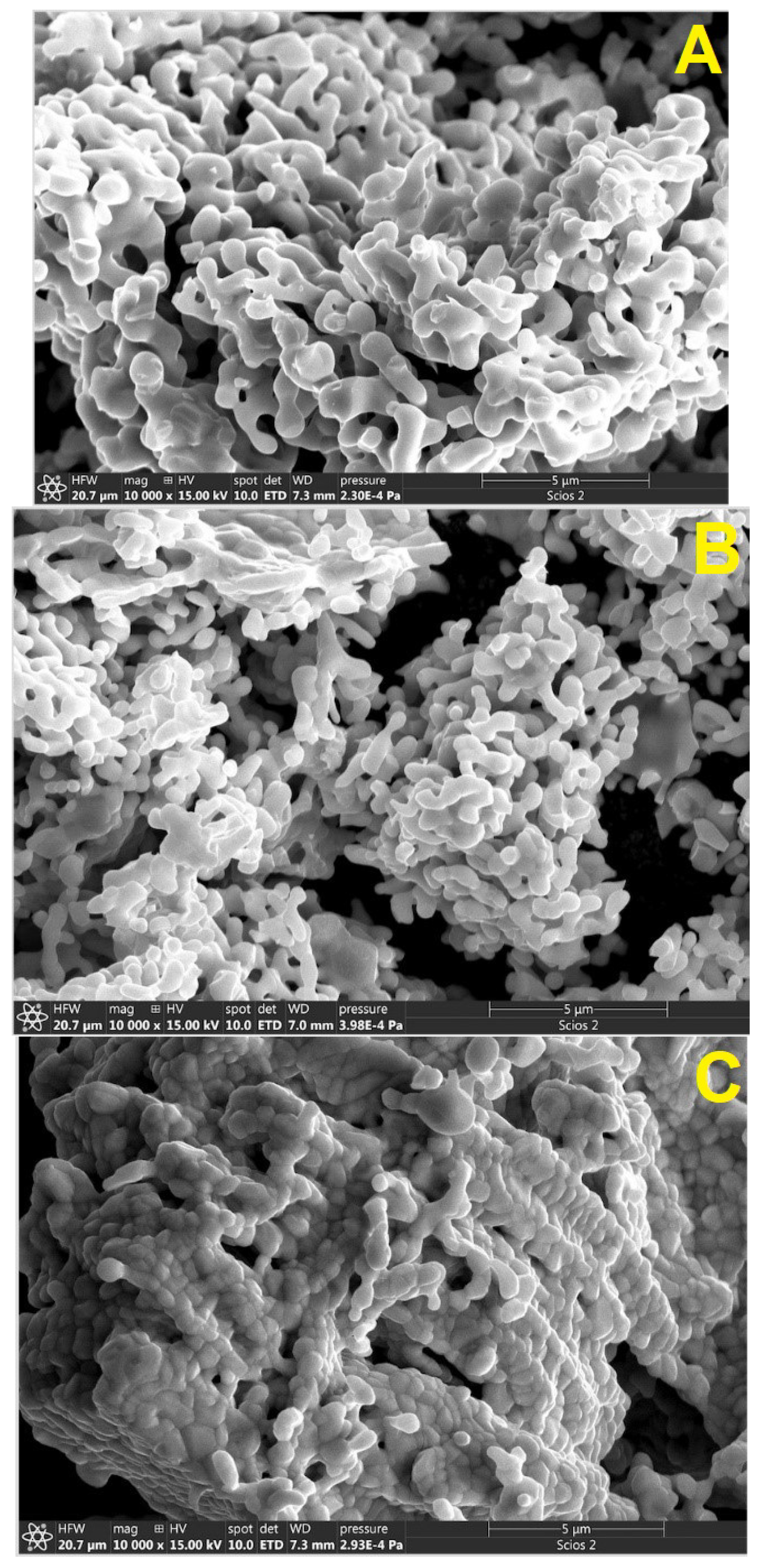
Figure 1. SEM images of agglomerations of synthesized nanoparticles. (A) Pr2NiMnO6, (B) Gd2NiMnO6, and (C) Er2NiMnO6.
Nanomaterials 14 00960 g001
Figure 2. XRD pattern measured in Pr2NiMnO6 (black symbols), Rietveld refinement (red line), and residuals (black line). The ticks show the position of the calculated peaks. R-values are provided in the figure. Asterisks are the most intense peaks of the minority phase.
Nanomaterials 14 00960 g002
Figure 3. XRD pattern measured in Gd2NiMnO6 (black symbols), Rietveld refinement (red line), and residuals (black line). The ticks show the position of the calculated peaks. R-values are provided in the figure. Asterisks are the most intense peaks of the minority phase.
Nanomaterials 14 00960 g003
Figure 4. XRD pattern measured in Er2NiMnO6 (black symbols), Rietveld refinement (red line), and residuals (black line). The ticks show the position of the calculated peaks. R-values are provided in the figure. Asterisks are the most intense peaks of the minority phase.
Nanomaterials 14 00960 g004
Figure 5. Crystal structure of R2NiMnO6 compounds. NiO6 and MnO6 octahedral units are shown in grey and violet, respectively. The RO8 dodecahedra are shown in green and the oxygen atoms in red.
Nanomaterials 14 00960 g005
Figure 6. FTIR transmittance spectra measured in the three R2NiMnO6 double perovskites studied.
Nanomaterials 14 00960 g006
Figure 7. Frequency of the mode ν2 versus ionic radii of the lanthanide atom.
Nanomaterials 14 00960 g007
Table 1. Reagent weights used in reactions, products of synthesis, and particle sizes determined using the Langford (L) and Scherrer (S) methods.
Ni(NO3)2·6H2OMnCl2·4H2OProductParticle Size (nm)
LS
Pr(NO3)3·6H2O
0.435 g
0.182 g0.197 gPr2NiMnO6 98%
Pr2O3 2%
37(4)45(4)
Er2O3
0.382 g
Er2NiMnO6 94%
Er2O3 6%
40(4)49(5)
Gd(NO3)3·6H2O
0.451 g
Gd2NiMnO6 98%
Gd2O3 4%
29(3)36(3)
Table 2. Unit-cell parameters and volume determined for the three synthesized compounds. The estimated bulk modulus (K) is also included.
Pr2NiMnO6Gd2NiMnO6Er2NiMnO6
a (Å)5.4432(5)5.2978(5)5.2302(5)
b (Å)5.5095(5)5.6119(5)5.6392(5)
c (Å)7.7101(7)7.5406(7)7.4533(5)
β (°)90.17(3)90.21(3)90.21(3)
V (Å3)231.2(1) Å3224.2(1) Å3219.8(1) Å3
K (GPa)173(5)176(5)179(5)
Table 3. Atomic positions obtained from Rietveld refinements for the studied materials.
Pr2NiMnO6
sitexyz
Pr4e0.9824(5)0.0705(5)0.2504(5)
Ni2d0.500
Mn2c0.500.5
O14e0.1074(12)0.4627(12)0.2423(12)
O24e0.7009(12)0.3123(12)0.0505(12)
O34e0.1783(12)0.2057(12)0.9446(12)
Gd2NiMnO6
sitexyz
Gdsite0.9832(5)0.0713(5)0.2501(5)
Ni4e0.500
Mn2d0.500.5
O12c0.1082(12)0.4621(12)0.2435(12)
O24e0.7016(12)0.3118(12)0.0515(12)
O34e0.1771(12)0.2059(12)0.9441(12)
Er2NiMnO6
sitexyz
Ersite0.9821(5)0.0701(5)0.2512(5)
Ni4e0.500
Mn2d0.500.5
O12c0.1077(12)0.4330(12)0.2428(12)
O24e0.7013(12)0.3133(12)0.0499(12)
O34e0.1788(12)0.2051(12)0.9448(12)
Table 4. Selected bond distances and angles.
LanthanidePrGdEr
NiO6 octahedron
Ni-O1 (x2)2.083(10) Å2.031(9) Å2.030(9) Å
Ni-O2 (x2)2.075(7) Å2.086(7) Å2.091(7) Å
Ni-O3 (x2)2.128(7) Å2.106(7) Å2.079(7) Å
MnO6 octahedron
Mn-O1 (x2)1.967(10) Å1.933(9) Å1.936(9) Å
Mn-O2 (x2)1.969(7) Å1.942(7) Å1.924(7) Å
Mn-O3 (x2)1.938(7) Å1.945(7) Å1.952(7) Å
Ni-O-Mn angles
Ni-O1-Mn144.3(3)144.5(3)144.6(3)
Ni-O2-Mn146.5(3)146.4(3)146.6(3)
Ni-O3-Mn144.3(3)144.2(3)144.1(3)
RO8 polyhedron
R-O12.266(8) Å2.251(7) Å2.150(8) Å
R-O12.311(8) Å2.292(8) Å2.281(7) Å
R-O22.320(6) Å2.308(6) Å2.283(6) Å
R-O22.546(7) Å2.504(7) Å2.493(7) Å
R-O22.677(5) Å2.630(5) Å2.603(5) Å
R-O32.309(6) Å2.297(7) Å2.284(7) Å
R-O32.553(7) Å2.520(7) Å2.499(7) Å
R-O32.696(5) Å2.640(5) Å2.614(5) Å
Table 5. Frequencies of modes ν1 and ν2, atomic number of the lanthanide atom, and ionic radii. The frequencies for Eu2NiMnO6, La2NiMnO6, and Sm2NiMnO6 were taken from the literature [35,36,37].
CompoundZIonic Radii (Å)ν1 (cm−1)ν2 (cm−1)
La2NiMnO6571.160600430
Pr2NiMnO6591.126590440
Sm2NiMnO6621.079575
Eu2NiMnO6631.066602
Gd2NiMnO6641.053595460
Er2NiMnO6681.004602470
Table 6. Estimations of the frequencies of ν 2 for the lanthanide family hosted in R2NiMnO6 double perovskites using ν 2 = 730 13 258 12 × r .
ElementShannon Ionic Radii. Coord. 8 * Frequency (cm−1)
La1.160431(19)
Ce1.143435(19)
Pr1.126439(19)
Nd1.109444(19)
Pm1.093448(19)
Sm1.079452(19)
Eu1.066455(18)
Gd1.053458(18)
Tb1.040462(18)
Dy1.027465(18)
Ho1.015468(18)
Er1.004471(18)
Tm0.994474(18)
Yb0.985476(18)
Lu0.977478(18)
* Shannon ionic radii from the database of the Atomistic Simulation Group in the Materials Department of Imperial College.
Disclaimer/Publisher’s Note: The statements, opinions and data contained in all publications are solely those of the individual author(s) and contributor(s) and not of MDPI and/or the editor(s). MDPI and/or the editor(s) disclaim responsibility for any injury to people or property resulting from any ideas, methods, instructions or products referred to in the content.