Article

A Label-Free Photoelectrochemical Biosensor Based on ZnO/Cs3MnBr5 Heterogeneous Films for Alpha-Fetoprotein Detection

by
Long Shao
1,2,*,
Biyu Zhang
2,
Wei Wu
2,
Gengyan Cui
3 and
Mao Liu
1,*
1
School of Metallurgy, Northeastern University, Shenyang 110819, China
2
School of Materials Science and Engineering, Changchun University of Science and Technology, Changchun 130012, China
3
School of Mechanical Engineering, Henan Polytechnic Institute, Nanyang 473000, China
*
Authors to whom correspondence should be addressed.
Nanomaterials 2024, 14(13), 1127; https://doi.org/10.3390/nano14131127
Submission received: 18 May 2024 / Revised: 19 June 2024 / Accepted: 27 June 2024 / Published: 29 June 2024
(This article belongs to the Special Issue Nano-Enabled Sensors for High Performance in Detection and Monitoring)

Abstract

:
Highly sensitive and specific biomarker detection is of outstanding importance for the diagnosis and treatment of cancers. Herein, we developed robust photoelectrochemical (PEC) biosensors with low background noise and high sensitivity based on a heterojunction, which can improve semiconductor photoelectric properties by limiting the recombination of photogenerated electron–hole pairs and successfully widening the range of light absorption. Alpha-fetoprotein (AFP) was used as a target model to examine the analytical performances of the designed PEC biosensors. ZnO/Cs3MnBr5 heterogeneous film with a uniform porous structure and large surface area enhanced electron transfer and biomolecule immobilization, and significantly increased the photocurrent response. Under the optimal conditions, the designed PEC biosensor exhibited a linear detection range of 0.01–500 ng/mL and a detection limit of 12 pg/mL. In addition, this PEC biosensor performed well when testing human serum samples and exhibited good repeatability, stability over time, and specificity, showing enormous potential for the detection of cancer markers in future biological and clinical research.

1. Introduction

Hepatocellular carcinoma (HCC) is a serious disease that poses a global threat to human life and health [1,2,3,4,5,6]. As one of the most common cancers, HCC has a 60 percent mortality rate, which is fairly significant; therefore, patients with HCC need early diagnosis and treatment [7,8,9,10]. One of the most common blood proteins in HCC is alpha-fetoprotein (AFP), a distinct tumor marker that is commonly used in HCC liver transplantation as well as HCC screening, diagnosis, treatment, and prognosis [11,12,13]. Normal human serum AFP concentrations are less than 10 ng/mL, and high AFP levels in adult serum have been associated with the development of liver cell carcinoma [9]. Hence, several techniques for detecting AFP have been developed, including electrochemical approaches [13], enzyme-linked immunosorbent assays [14,15], fluorescence immunoassays [16,17], surface plasmon resonance (SPR)-based immunoassays [18,19,20,21], and electro-chemiluminescent assays [22,23]. Despite their high sensitivity, these approaches usually require complex procedures, high costs, and lengthy processing periods. Therefore, there is a need for a novel, rapid, and highly sensitive AFP detection technology to efficiently diagnose HCC and reduce its mortality rate.
Photoelectrochemical (PEC) biosensors are widely recognized as a new and powerful technology for biological analysis, owing to their excellent affinity to biomolecules and superior inherent sensitivity compared to conventional methods [24,25]. For example, Li et al. successfully designed a novel label-free PEC biosensor based on Au/CsxWO3 heterogeneous films for AFP detection; the as-prepared PEC biosensor had good specificity, repeatability, and long-term stability, and showed satisfactory results in human serum sample analysis [26]. This detection technique is based on the photoelectric conversion capabilities of photoactive compounds, which are used to determine the concentration of targeted biological markers [24]. In photoelectrochemical detection, appropriate light wavelengths are used to excite photosensitive substances, and electrical signals are detected. PEC detection technology is, therefore, highly sensitive and shows great promise for identifying small biomolecules and chemicals. The photocurrent, which is the output signal obtained from the photoelectric conversion of the photosensitive material in PEC biosensors, is required for the prompt detection of biomarkers. Depending on the nature of the photosensitive material, PEC biosensors can be classified into inorganic [25], organic [27,28], or composite photosensitive materials [29,30]. ZnO [31,32], TiO2 [33,34,35], and reduced graphene oxide [36,37] are common inorganic photosensitive materials. ZnO is a popular photocatalyst, owing to its outstanding electrical conductivity communication properties, nontoxicity, and chemical stability. However, the wide band gap of ZnO restricts its absorption to the UV region (~380 nm), limiting its application across the solar spectrum [31,38]. Furthermore, UV light has a high oxidative power that may destroy biomolecules [39]. The key challenge faced by ZnO electrodes is, therefore, the effective utilization of visible light, and coupling two materials to form a heterostructure is an effective method for enhancing the photoelectric response and overcoming this challenge [32,37,40,41].
Heterojunctions can improve semiconductor photoelectric properties by limiting the recombination of photogenerated electron–hole pairs and successfully widening the range of light absorption. To date, many studies have reported improvements in PEC activity by modifying the surface of ZnO with narrow-bandgap semiconductors, including CdX (X = S, Se, or Te) and PbS [42]. However, the heavy metal toxicity of the above-mentioned materials limits their further commercial applications. Cs3MnBr5 is a nontoxic material that has garnered research interest owing to its high visible-light absorption coefficient and optical stability for photovoltaic applications. In addition, Cs3MnBr5 is an inorganic semiconductor with outstanding chemical stability and excellent biocompatibility, which render it a good candidate for PEC biosensing of substrate [43].
In this study, we developed a novel ZnO/Cs3MnBr5 heterogeneous film-based label-free PEC biosensor for AFP detection (Scheme 1). The ZnO layer adopts a three-dimensional inverse opal (IO) photonic crystal structure that improves photochemical processes and solar cell photovoltaic response by increasing the effective optical path length [44]. Furthermore, the ZnO IO has a uniform porous structure and large surface area, which reduce the distance between the targeted substance and the fluorine-doped tin oxide (FTO) substrate, thereby enhancing electron transfer and biomolecule immobilization [45]. The matched effective energy level between ZnO IO and Cs3MnBr5 nanocrystals (NCs) further enables the ZnO/Cs3MnBr5 heterostructure’s full use of the visible-light spectrum, thus, facilitating charge transfer, significantly improving photocurrent responsiveness, and preventing UV damage to proteins. While the application of ZnO/Cs3MnBr5 heterostructures in PEC biosensor fabrication has not been explored to date, our experimental results indicate their immense potential for disease diagnosis.

2. Materials and Methods

2.1. Materials

Zinc nitrate (Zn (NO3)2·6H2O), methyl methacrylate (MMA), and glutaraldehyde (GLD) 50% were purchased from Tianjin Chemical Plant Co., Ltd. (Tianjin, China). Citric acid, ethanol, hydrogen peroxide (H2O2), and glucose were purchased from Beijing Chemical Plant Co. (Beijing, China). Tetraethoxysilane (TEOS) and sodium borohydride (NaBH4) were purchased from Sinopharm Chemical Reagent Co. (Beijing, China). Glucose oxidase (GOD) was purchased from Amresco Co., Ltd. (Shanghai, China). Chitosan (CS) was purchased from Solarbio Co., Ltd. (Beijing, China). Alpha-fetoprotein (AFP), carcinoembryonic antigen (CEA) prostate-specific antigen (PSA), and anti-AFP antibody (Ab, polyclonal antibody) were purchased from Beijing Boisynthese Biotechnology Co., Ltd. (Beijing, China). Bovine serum albumin (BSA, 96–99%) was purchased from Beijing DingGuo Biotechnology Company (Beijing, China). 1-ethyl-3-(3-dimethylaminopropyl) carbodiimide (EDC) was purchased from Sigma-Aldrich (Steinheim, Germany). L-cysteine was purchased from Alfa Aesar Co., Ltd. (Beijing, China). Human serum samples were purchased from a local hospital. Phosphate-buffered saline (PBS) was prepared in a conventional way to configuring the CS, Ab, BSA, and AFP to pH = 7.2 (0.01 M) and the test solution to pH = 7.4 (0.1 M), respectively. All reagents were of analytical purity and were used without further purification. All the aqueous solutions were prepared with deionized water (DI water, 18.25 MΏ/cm) that was obtained from a water purification system. Cs2CO3 (99.9%), octadecene (ODE, 90%), oleic acid (OA, 90%), oleylamine (OAm, 70%), manganese (II) acetate tetrahydrate (MnC4H6O4·4H2O, 99.999%), C18H37Br (OAmBr, 99%), toluene (ACS grade), and ODE were purchased from Macklin; others were purchased from Sigma-Aldrich, and all were used directly.

2.2. Apparatus

UV–visible (UV–VIS) transmittance spectra were measured using a UV-3101PC scanning spectrophotometer (Shimadzu Instruments Co., Ltd., Shanghai, China) ranging from 200 to 1100 nm. The morphology of the samples was characterized using a JSM-7500F field-emission scanning electron microscope (SEM) (JEOL Instruments Co., Ltd., Shanghai, China). Fourier transform infrared (FTIR) absorption spectra were measured using a Shimadzu DT-40 model 883 infrared spectrophotometer (Shimadzu Instruments Co., Ltd., Shanghai, China). X-ray diffractometry (XRD) analysis was conducted using a RigakuD/max 2550 X-ray diffractometer (Tiger Instruments Co., Ltd., Wuhan, China). Electrochemical impedance spectroscopies (EISs) were calculated using a model CHI660D electrochemical analyzer (ChenHua Instruments Co., Ltd., Shanghai, China). Electrochemical tests were performed with a model CHI630D electrochemical analyzer (ChenHua Instruments Co., Ltd., Shanghai, China), using a conventional three-electrode system comprising a platinum wire as a counter electrode, a saturated calomel electrode (SCE) as a reference electrode, and a modified electrode with a geometrical area of 1.0 ± 0.1 cm2 as a working electrode. The cyclic voltammetry curves (CVs) were recorded at a scan rate of 100 mV/s. All the photocurrent measurements were conducted under a 500 W xenon irradiation lamp at a constant potential of 0.6 V (relative to a saturated Ag/AgCl electrode) in PBS (pH = 7.4, 0.1 M).

2.3. Preparation of ZnO IO

Evenly monodispersed polymethyl methacrylate (PMMA) balls of a predetermined size were synthesized using a previously reported method [45]. The PMMA template was generated using a vertical-deposition self-assembly process. The obtained PMMA colloidal solution was immersed in the hydrophilic fluorine-doped tin oxide (FTO)-coated glass substrate, which was subsequently baked for 24 h at 35 °C. The surface tension generated by liquid evaporation prompted gradual self-assembly of the PMMA colloidal spheres into a highly ordered array on the FTO substrate. Subsequently, the PMMA template was sintered for 40 min at 120 °C to improve its physical properties. Figure S1 displays the morphology of the PMMA spheres.
The Zn ion precursor solution was prepared by dissolving Zn (NO3)2·6H2O in ethanol. Appropriate amounts of tetraethyl orthosilicate (TEOS) and citric acid were added as chelating agents, and then the solution was stirred until it became colorless and transparent. Subsequently, the precursor solution was extensively infiltrated at room temperature, and capillary forces were applied to the interstices of the PMMA templates. To obtain the ZnO IO structure, the samples were heated to 500 °C for 3 h, which burned the original PMMA template. The phase purity of the ZnO IO was determined by using X-ray powder diffraction (XRD) (Tiger Instruments Co., Ltd., Wuhan, China) (Figure S2).

2.4. Preparation and Modification of Cs3MnBr5 NCs

2.4.1. Synthesis of Cs-Oleates

The synthesis of Cs-oleates was performed under a N2 atmosphere. A 25 mL three-neck round-bottom flask was filled with 0.4 g of Cs2CO3, 13 mL of octadecene (ODE), and 1 mL of oleic acid (OA). The mixture was heated at 130 °C for 1 h, or until the powder was entirely dissolved.

2.4.2. Synthesis of Cs3MnBr5 NCs

The synthesis of Cs3MnBr5 NCs was performed under a N2 atmosphere. In a 50 mL three-neck bottle, 0.245 g MnCl4H6O4·4H2O and appropriate amounts of oleylamine, OA, oleylammonium bromide, and ODE were mixed, stirred, and the bottle filled with N2. The solution was heated to 140 °C for 2 h and further heated to 260 °C. Subsequently, 0.8 mL Cs-oleate was rapidly injected into the bottle and ice water was used to cool the solution, thereby producing Cs3MnBr5 NCs. Following acetone precipitation and centrifugation, the Cs3MnBr5 NCs were dissolved in toluene. Figure S3 displays the experimental flowchart. The best crystalline nanomaterials were created by optimizing the synthesis conditions (Figures S4–S6).

2.5. Preparation of FTO/ZnO/Cs3MnBr5 Electrode

The FTO/ZnO electrode was spin-coated with 50 μL of a 5 mL toluene solution with scattered Cs3MnBr5 NCs. The electrode was annealed at 70 °C for 1 h, dried at room temperature, and washed with phosphate-buffered solution (pH = 7.2).

2.6. PEC Measurements

The surface of the FTO working electrode was modified with the ZnO/Cs3MnBr5 heterogeneous layer, followed by application of 10 μL 0.05 wt% chitosan (CS) and drying at ambient temperature for 30 min. The FTO/ZnO/Cs3MnBr5 electrode was then thoroughly cleaned using NaOH solution and deionized water. After the electrode had dried for 30 min at room temperature, 20 μL of an aqueous solution containing 5% glutaric dialdehyde was added to activate the CS. Thereafter, deionized water was used to rinse the electrodes. The surface of the working electrode was subsequently altered using 25 μL of 10 μg/mL of the antibody (Ab) and kept at 4 °C for over 12 h of incubation. After removing any uncombined antibody with phosphate-buffered saline (PBS) (pH = 7.2), a 20 μL solution of 1% bovine serum albumin (BSA) was applied to the electrode surface for 30 min at 37 °C to block nonspecific active sites. After rinsing thoroughly with PBS (pH = 7.2), 25 μL solutions of the AFP antigen at various concentrations were added to the generated electrode. The electrode was incubated at 37 °C for 1 h and then thoroughly cleaned with PBS (pH = 7.2) to produce the PEC biosensor used in the subsequent experiments.
A three-electrode setup was employed for the electrochemical and PEC tests. Saturated calomel and Pt wire electrodes were used as the reference and counter electrode, respectively. The working electrode was a modified FTO electrode with an area of 1.0 ± 0.1 cm2. The photocurrent was measured using a constant potential of 0.6 V, supplied by a 500 W xenon lamp in a PBS solution containing 0.07 M AA (pH = 7.4) [46]. The photoelectrochemical measurement process is shown in Scheme S1.

3. Results and Discussion

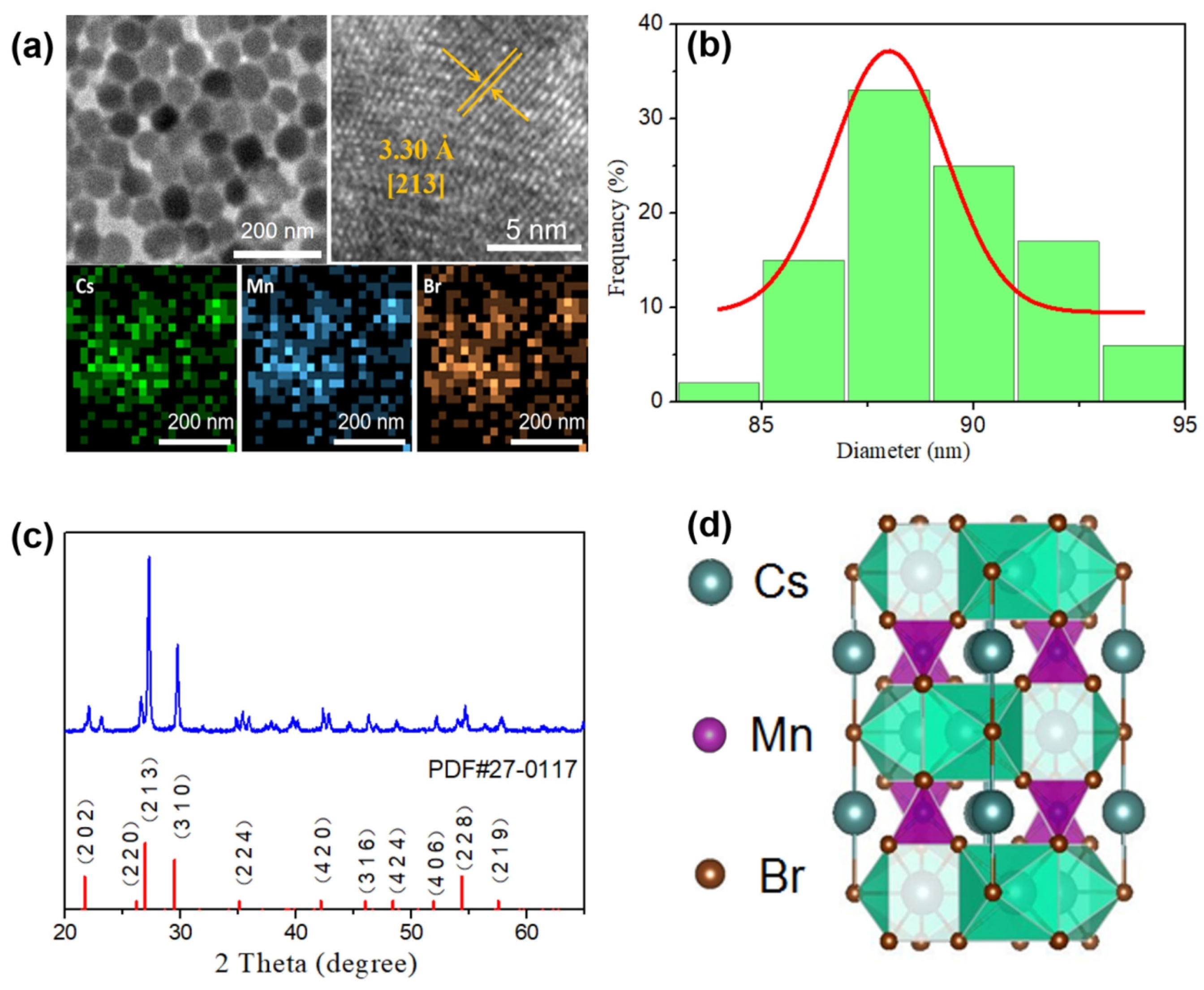
3.1. Characterization

Transmission electron microscopy (TEM) was used to confirm the successful synthesis of Cs3MnBr5 NCs. The TEM image of the as-prepared Cs3MnBr5 NCs (Figure 1a) reveals that they form a regular arrangement on the TEM grid while having excellent uniformity and a particle size of approximately 87 nm. The high-resolution transmission electron microscopy (HRTEM) image reveals the lattice fringes of the [213] planes, with a distance of 3.30 Å. In addition, the elemental mapping images verify the existence of Cs, Mn, and Br in the NCs. The NC size conformed to the mathematical statistical law of Gaussian distribution, and the size was mainly distributed in the narrow range of 82–94 nm, further indicating that the NCs were monodispersed (Figure 1b). XRD measurements were utilized to determine the phase purity of the as-prepared Cs3MnBr5 NCs. TOPAS 4.2 software was used to fit the Cs3MnBr5 diffractogram line profile using the Rietveld method (Figure 1c). All peaks were indexed to a tetragonal unit cell, and their values correspond to those of the standard Cs3MnBr5 pattern (PDF#27-0117). The XRD pattern did not contain any other diffraction peaks, and the sharp signals indicate that the NCs had good crystallinity. The Rietveld refinement confirmed that the Cs3MnBr5 NCs were isostructural with Cs3CoCl5 and belonged to the tetragonal phase system with the I4/mcm space group (Figure 1d).
The field-emission scanning electron microscopy image of the ZnO IO thin film is displayed in Figure 2a, exhibiting a pore size of approximately 700 nm and a well-organized 3D pore structure. Furthermore, a considerable number of as-prepared Cs3MnBr5 NCs adhered to the exterior and interior of the ZnO IO, as shown in the SEM image of the ZnO IO thin film modified with the Cs3MnBr5 NCs (Figure 2b). The large specific surface area of the enhanced electrode significantly enhances biological protein adsorption, hence improving the detection capacity of the sensor.
The FTO/ZnO/Cs3MnBr5 heterogeneous film outperformed the FTO/ZnO film in terms of photocurrent responsiveness (Figure 3a). However, as the number of spin-coated Cs3MnBr5 NC layers increased to above six, the photocurrent of the composite electrode decreased. This is because the excessive deposition of Cs3MnBr5 NCs on the electrode surface increases its diffusion resistance and generates more recombination centers.
Furthermore, we investigated the spectral response capability of the ZnO/Cs3MnBr5 heterogeneous film. As shown in Figure 3b, the ZnO IO mainly absorbs in the ultraviolet range, whereas the Cs3MnBr5 NCs absorb in the visible range. Consequently, the ZnO/Cs3MnBr5 heterogeneous film absorbed in both the ultraviolet and visible-light ranges. In addition, there was more absorption in the visible-light range compared to the ZnO IO, highlighting the material’s improved photoelectric conversion efficiency.

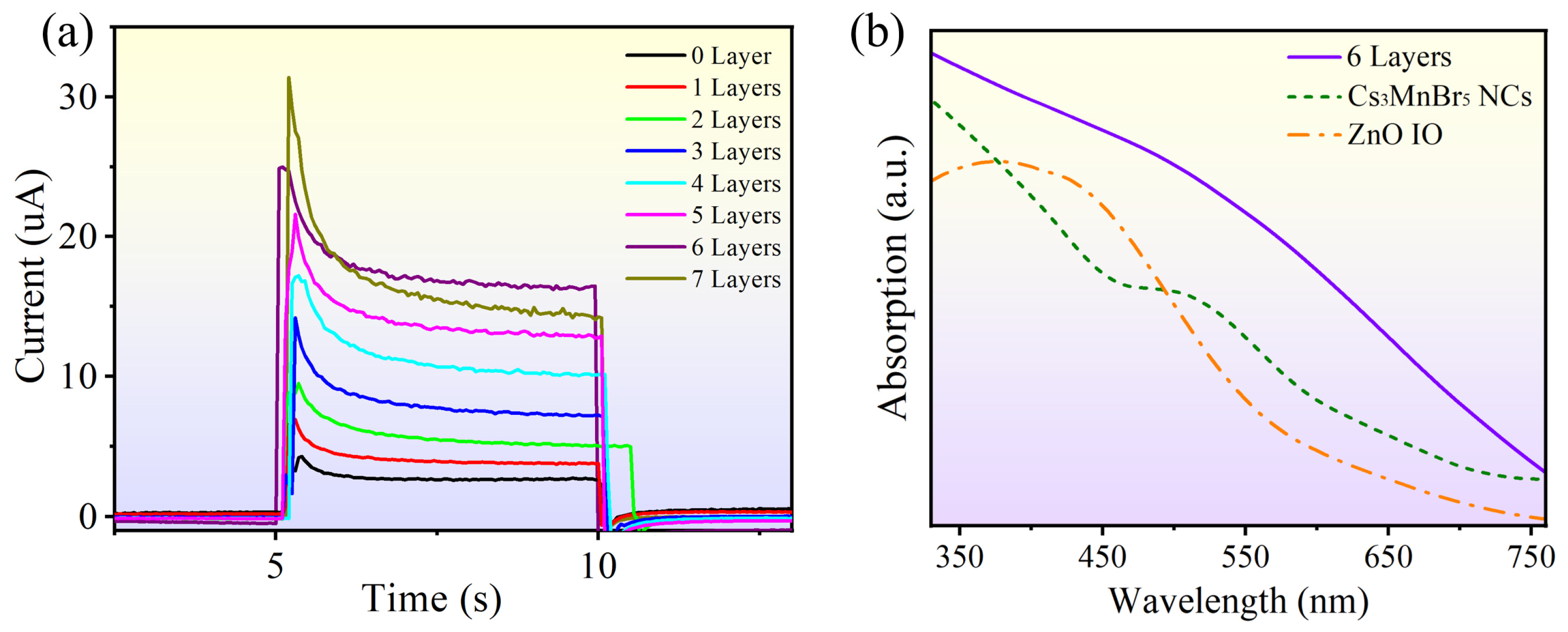
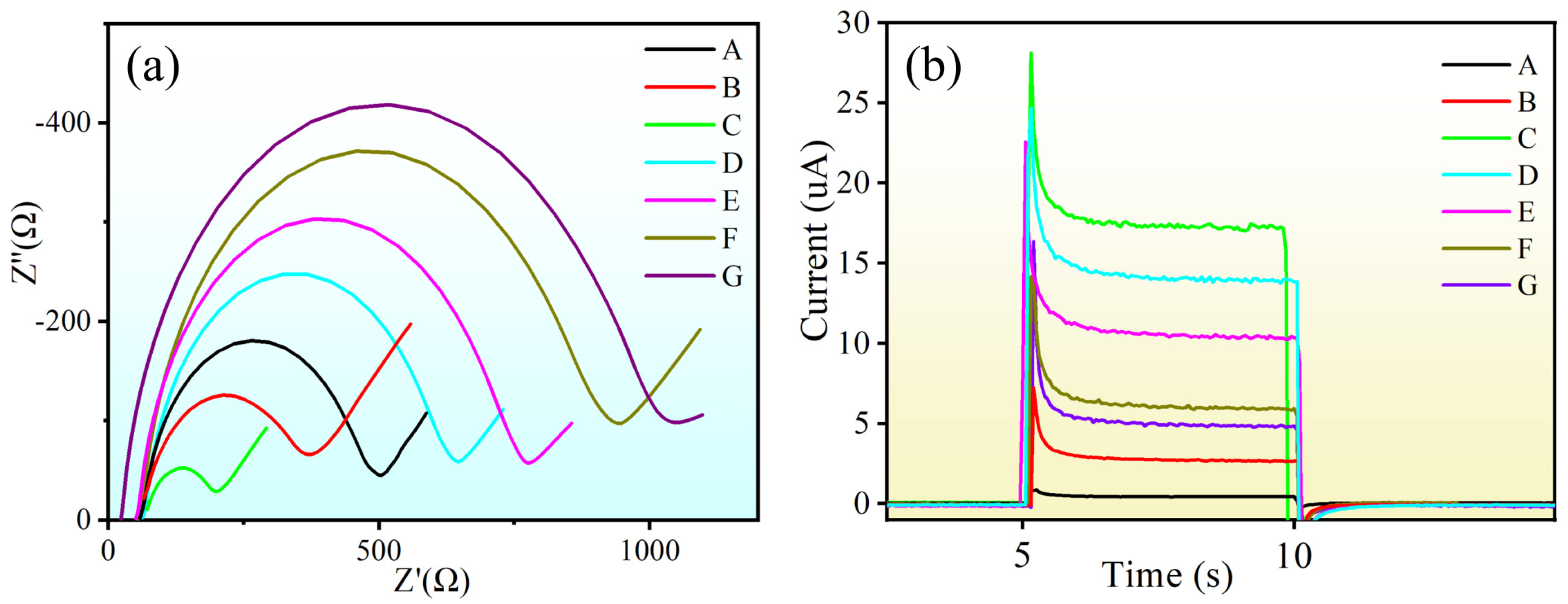
3.2. Characterization of Photoelectrochemical Biosensor Construction Process

The fabrication of the biosensor electrode was monitored using electrochemical impedance spectroscopy (EIS) and photocurrent response measurements (Figure 4). The values of electron transfer resistance (Ret) were obtained by electrochemical impedance spectroscopy (EIS), which is normally used to analyze the construction process of biosensor electrodes. The Ret spectra were measured in a solution mixture that contained 0.1 M KCl and 0.05 M K3[Fe(CN)6]/K4[Fe(CN)6] (1:1) in 0.01 M PBS (pH = 7.4) buffer solution, with an open circuit voltage of 0.6 V and a frequency of 0.01–100 Hz. The Ret is equal to the diameter of the semicircle. The ZnO IO exhibited superconductivity, as shown in Figure 4a, resulting in a notable drop in electrode resistance. It was also observed that the FTO/ZnO/Cs3MnBr5 electrode showed greater Ret than the FTO/ZnO electrode. Modifying the electrode with CS caused a significant increase in the resistance because CS has an insulating property [16]. The resistance of the electrode increased slightly after further modification with Ab (25 μL, 10 μg/mL) and BSA (20 μL, 1%) because these organic molecules inhibited electron transport [45]. The impedance spectrum showed notable changes in the electrode’s surface with the use of 25 μL 10 ng/mL AFP antigen. Thus, a ZnO/Cs3MnBr5 heterogeneous film-based label-free PEC biosensor was successfully produced. The PEC biosensor was then measured using a constant potential of 0.6 V supplied by a 500 W xenon lamp in a PBS solution containing 0.07M AA (pH = 7.4).
Figure 4b shows the variations in the photocurrent response of the electrode for each processing step, and the observed photocurrent shift further validates the successful synthesis of the PEC biosensor. The results reveal that the photocurrent of the FTO/ZnO electrode increased compared to the bare FTO, confirming both the high conductivity and photoelectric conversion capabilities of the Cs3MnBr5 NCs and the successful modification of ZnO IO. The photocurrent response of the FTO substrate modified with the ZnO/Cs3MnBr5 heterogeneous film was approximately enhanced by a factor of 7, demonstrating that the heterogeneous film greatly increased the photoelectric conversion efficiency of the photoelectrochemical biosensor. The primary explanation for this enhancement is the comparable energy levels of ZnO IO and Cs3MnBr5 NCs, which enable a more efficient injection of the excited-state electrons into the ZnO IO layer, thereby facilitating charge transfer, decreasing carrier recombination, and significantly enhancing photocurrent. Furthermore, the slow-light effect of the ZnO IO can lengthen the photovoltaic response and effective optical path. These advantages have the potential to greatly improve light utilization and photoelectric conversion efficiency. The current gradually decreased after adding CS, Ab, BSA, and AFP antigens to the electrode surface due to the insulating properties of these organic compounds, which obstructed electron transfer and reduced current responsiveness [46]. The observed change in photocurrent at each stage of the modification process indicates the effective development of a label-free PEC biosensor based on the ZnO/Cs3MnBr5 heterogeneous film.

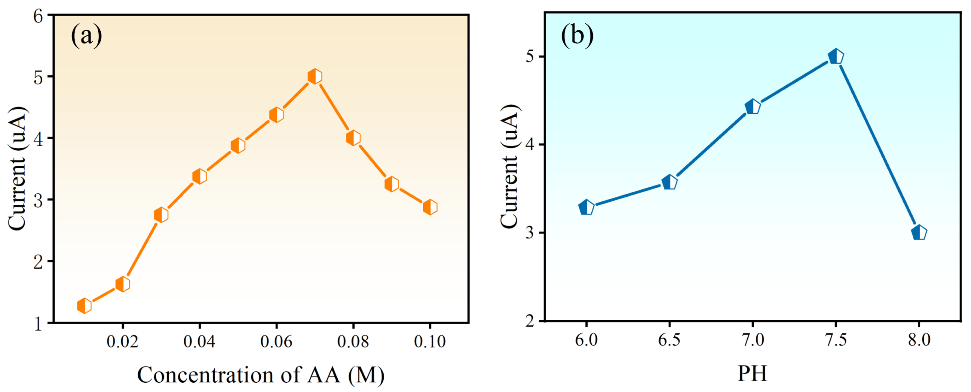
3.3. Optimization of the Detection Conditions

To build a functional and optimized PEC biosensor, an experiment that involved adjusting the pH and AA levels was performed. In the detection solution, AA functioned as an electron donor to create a consistent and adequate photocurrent signal while also minimizing electron–hole recombination [47]. Therefore, the AA concentration is a crucial parameter that was studied. As shown in Figure 5a, the photocurrent intensity rises with an AA concentration of 0.02–0.07 M, whereas it decreases with an AA concentration of 0.07–0.10 M. This suggests that an AA concentration of 0.07 M provides a sufficient number of available electrons for photoinduced holes and saturated electron donors. Furthermore, the photoelectric response of the electrode is modified by the pH of the solution, which alters the biomolecular activity on the electrode [45]. A pH of 7.4 was selected as the ideal pH for identification because it produced the highest photocurrent (Figure 5b).

3.4. Photoelectrochemical Detection of AFP

Under optimal conditions, variations in the AFP concentrations (25 μL) were recognized by measuring photocurrent fluctuation in the synthesized PEC biosensor. Figure 6a shows the photocurrent response calibration curve for various AFP concentrations. Notably, the photocurrent is a decreasing logarithmic function of the AFP concentration in the range of 0.01–500 ng/mL. With a determination coefficient of 0.997, the regression equation is ΔI = 5.476 − 0.572logCAFP, where ΔI is the photocurrent of the FTO/ZnO/Cs3MnBr5/CS/Ab/BSA heterogeneous film electrode incubated with 25 μL solutions of varying AFP concentrations. As the concentration of AFP increased, the electronic specialization of AA towards the PEC sensor was blocked, resulting in a gradual decrease in the photocurrent intensity. By calculating the signal-to-noise ratio (S/N = 3) [48], the detection limit of the PEC biosensor was found to 12 pg/mL. Our as-prepared PEC biosensor provides a larger linear range for AFP detection and a lower detection limit than heterostructure photoelectrochemical biosensors based on other materials. Table S1 shows the good performance of the PEC biosensor based on the heterogeneous ZnO/Cs3MnBr5 film.
AFP standard solutions of various concentrations were added to a standard human serum sample to determine the relative standard deviation (RSD). Table S2 shows that the analytical recovery range is 95–104%, with RSD values smaller than 5.3%. These findings demonstrate that the ZnO/Cs3MnBr5 heterogeneous film-based label-free PEC biosensor is very sensitive and accurate for AFP detection in real samples.

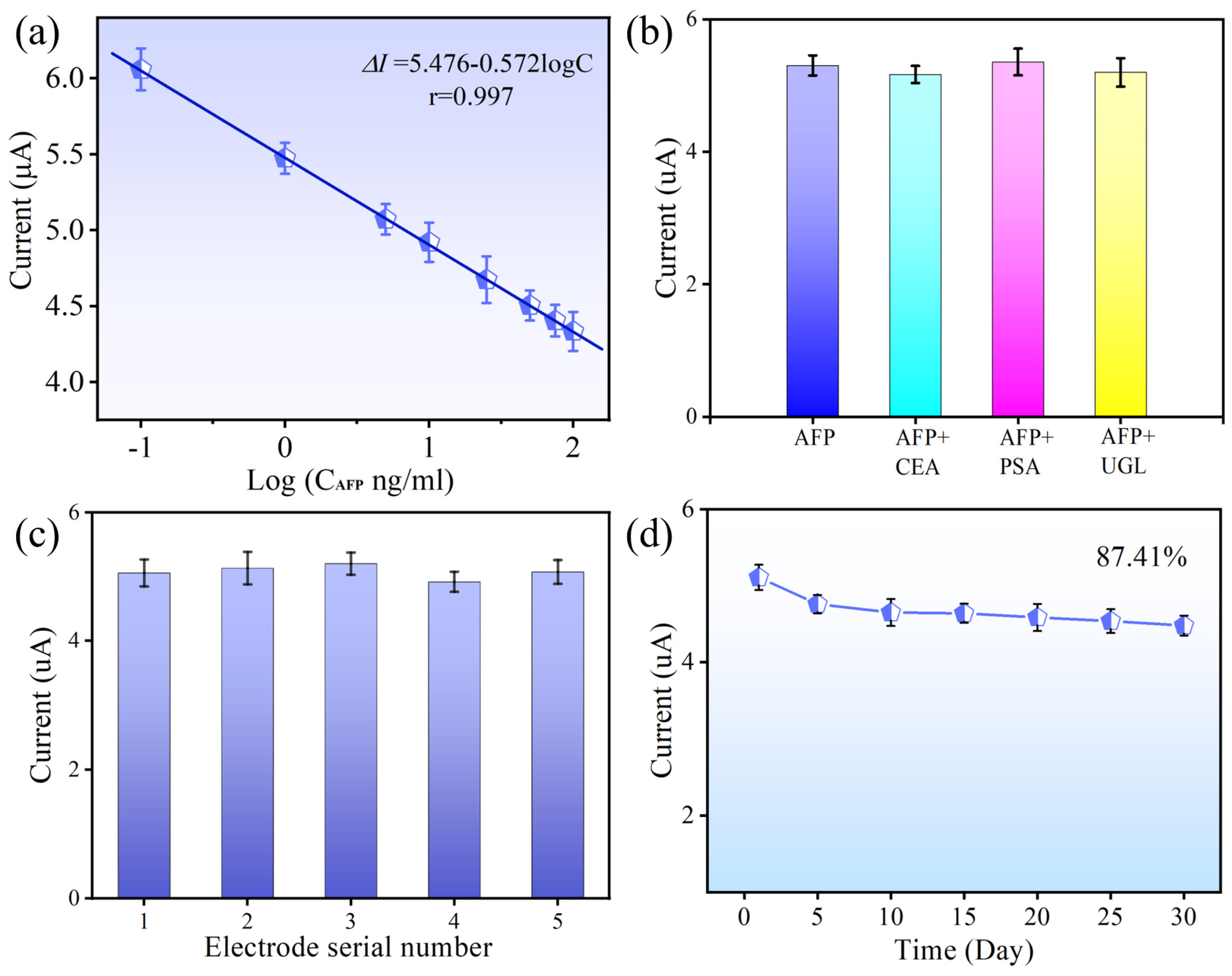
3.5. Specificity, Reproducibility, and Stability of the PEC Biosensor

The selectivity of the proposed AFP biosensor was evaluated by adding other interfering species, including carcinoembryonic antigen, prostate-specific antigen, and uteroglobin. The three electrodes treated with solutions containing 100 ng/mL interfering protein and 10 ng/mL AFP produced photocurrent responses due to the specific binding (Figure 6b). The photocurrent did not vary significantly, indicating that nonspecific adsorption had no effect on the PEC biosensor. As a result, the ZnO/Cs3MnBr5 heterogeneous film-based label-free PEC biosensor exhibited excellent specificity.
Five PEC biosensors were created under identical experimental conditions to assess their photoelectric response at 10 ng/mL of AFP. As shown in Figure 6c, there was little variance in the photocurrent intensity generated by the five PEC biosensors, demonstrating the reproducibility of the ZnO/Cs3MnBr5 heterogeneous film-based label-free PEC biosensor’s performance. The long-term stability of a ZnO/Cs3MnBr5 heterogeneous film electrode that was kept in a refrigerator at 4 °C is displayed in Figure 6d. Every five days, the photoelectric response of this sensor was tested, using 10 ng/mL of AFP. After 30 d, the electrode retained 87.41% of its initial performance, and the photocurrent slightly decreased with storage time, demonstrating the strong long-term durability of the ZnO/Cs3MnBr5 heterogeneous film-based label-free PEC biosensor.

4. Conclusions

In this study, a ZnO/Cs3MnBr5 heterogeneous film was synthesized to effectively create a novel label-free PEC biosensor for AFP detection. The delayed light effect in the ZnO IO improved the photovoltaic performance of the electrode in solar cells and photochemical processes by increasing the effective optical path. The ZnO/Cs3MnBr5 heterogeneous film leverages the homogeneous porosity structures of the ZnO IO due to its large surface area for the immobilization of biomolecules and electron transport. The addition of Cs3MnBr5 NCs increases the photoelectric response of the ZnO IO, allowing it to fully utilize visible light, stimulating charge transfer, and significantly increasing the photocurrent response. We also demonstrated the use of a ZnO/Cs3MnBr5 heterogeneous film in a photoelectrochemical bioassay for the first time. Under ideal conditions, a linear detection range of 0.01–500 ng/mL with a detection limit of 12 pg/mL was noted. Additionally, the as-prepared PEC biosensor performed well while testing human serum samples and exhibited good repeatability, stability over time, and specificity. Therefore, our ZnO/Cs3MnBr5 heterogeneous film-based label-free photoelectrochemical biosensor has enormous potential for the identification of cancer markers in biological and clinical research.

Supplementary Materials

The following supporting information can be downloaded at: https://www.mdpi.com/article/10.3390/nano14131127/s1. Figure S1: SEM image of PMMA spheres; Figure S2: XRD image of ZnO IO; Figure S3: Schematic diagram of Cs3MnBr5 NCs synthesis; Figure S4: (a) TEM images, (b) XRD, and (c) Luminescence of synthesized products at different reaction temperatures; Figure S5: (a) TEM images and (b) Luminescence spectrum of the synthesized product with varying amounts of OA; Figure S6: (a) TEM images and (b) Luminescence spectrum of the synthesized product with varying amounts of OAm; Scheme S1: The photoelectrochemical measurement process; Table S1: Performance comparison of heterostructure photoelectrochemical biosensors based on other materials for AFP detection; and Table S2: Detection of AFP in human standard serum. Reference [49] are cited in the Supplementary Materials.

Author Contributions

Conceptualization, L.S. and M.L.; methodology, L.S. and M.L.; validation, B.Z. and W.W.; investigation, G.C. and M.L.; resources, M.L.; data curation, L.S., M.L., W.W. and G.C.; writing—original draft preparation, L.S.; writing—review and editing L.S. and M.L.; supervision, M.L. All authors have read and agreed to the published version of the manuscript.

Funding

This work was supported by the National Natural Science Foundation of China [Grant Nos. 62305045] from L.S. and the Nanyang City Basic and Frontier Technology Research Special Project [Grant Nos. 23JCQY2021] from G.C.

Data Availability Statement

The data presented in this study are available on request from the corresponding author.

Conflicts of Interest

The authors declare that they have no conflicts of interest.

References

  1. Ganesan, P.; Kulik, L.M. Hepatocellular Carcinoma New Developments. Clin. Liver Dis. 2023, 27, 85–102. [Google Scholar] [CrossRef] [PubMed]
  2. Greten, T.F.; Lai, C.W.; Li, G.; Staveley-O’Carroll, K.F. Targeted and Immune-Based Therapies for Hepatocellular Carcinoma. Gastroenterology 2019, 156, 510–524. [Google Scholar] [CrossRef] [PubMed]
  3. Jiang, Y.; Sun, A.; Zhao, Y.; Ying, W.; Sun, H.; Yang, X.; Xing, B.; Sun, W.; Ren, L.; Hu, B.; et al. Proteomics identifies new therapeutic targets of early-stage hepatocellular carcinoma. Nature 2019, 567, 257–261. [Google Scholar] [CrossRef] [PubMed]
  4. Tan, D.J.H.; Ng, C.H.; Lin, S.Y.; Pan, X.H.; Tay, P.; Lim, W.H.; Teng, M.; Syn, N.; Lim, G.; Yong, J.N.; et al. Clinical characteristics, surveillance, treatment allocation, and outcomes of non-alcoholic fatty liver disease-related hepatocellular carcinoma: A systematic review and meta-analysis. Lancet Oncol. 2022, 23, 521–530. [Google Scholar] [CrossRef] [PubMed]
  5. Torimura, T.; Iwamoto, H. Treatment and the prognosis of hepatocellular carcinoma in Asia. Liver Int. 2022, 42, 2042–2054. [Google Scholar] [CrossRef] [PubMed]
  6. Vogel, A.; Meyer, T.; Sapisochin, G.; Salem, R.; Saborowski, A. Hepatocellular carcinoma. Lancet 2022, 400, 1345–1362. [Google Scholar] [CrossRef]
  7. Beaufrere, A.; Calderaro, J.; Paradis, V. Combined hepatocellular-cholangiocarcinoma: An update. J. Hepatol. 2021, 74, 1212–1224. [Google Scholar] [CrossRef]
  8. El-Mahdy, H.A.; Sallam, A.-A.M.; Ismail, A.; Elkhawaga, S.Y.; Elrebehy, M.A.; Doghish, A.S. miRNAs inspirations in hepatocellular carcinoma: Detrimental and favorable aspects of key performers. Pathol. Res. Pract. 2022, 233, 153886. [Google Scholar] [CrossRef]
  9. Hanif, H.; Ali, M.J.; Susheela, A.T.; Khan, I.W.; Luna-Cuadros, M.A.; Khan, M.M.; Lau, D.T.-Y. Update on the applications and limitations of alpha-fetoprotein for hepatocellular carcinoma. World J. Gastroenterol. 2022, 28, 216–229. [Google Scholar] [CrossRef]
  10. Llovet, J.M.; Pinyol, R.; Kelley, R.K.; El-Khoueiry, A.; Reeves, H.L.; Wang, X.W.; Gores, G.J.; Villanueva, A. Molecular pathogenesis and systemic therapies for hepatocellular carcinoma. Nat. Cancer 2022, 3, 386–401. [Google Scholar] [CrossRef]
  11. Hu, X.; Chen, R.; Wei, Q.; Xu, X. The Landscape of Alpha Fetoprotein in Hepatocellular Carcinoma: Where Are We? Int. J. Biol. Sci. 2022, 18, 536–551. [Google Scholar] [CrossRef] [PubMed]
  12. Johnson, P.; Zhou, Q.; Dao, D.Y.; Lo, Y.M.D. Circulating biomarkers in the diagnosis and management of hepatocellular carcinoma. Nat. Rev. Gastroenterol. Hepatol. 2022, 19, 670–681. [Google Scholar] [CrossRef] [PubMed]
  13. Pilvenyte, G.; Ratautaite, V.; Boguzaite, R.; Ramanavicius, A.; Viter, R.; Ramanavicius, S. Molecularly Imprinted Polymers for the Determination of Cancer Biomarkers. Int. J. Mol. Sci. 2023, 24, 4105. [Google Scholar] [CrossRef] [PubMed]
  14. Cimbalo, A.; Alonso-Garrido, M.; Font, G.; Manyes, L. Toxicity of mycotoxins in vivo on vertebrate organisms: A review. Food Chem. Toxicol. 2020, 137, 111161. [Google Scholar] [CrossRef] [PubMed]
  15. Tan, G.; Wang, S.; Yu, J.; Chen, J.; Liao, D.; Liu, M.; Nezamzadeh-Ejhieh, A.; Pan, Y.; Liu, J. Detection mechanism and the outlook of metal-organic frameworks for the detection of hazardous substances in milk. Food Chem. 2024, 430, 136934. [Google Scholar] [CrossRef] [PubMed]
  16. Haupt, K.; Rangel, P.X.M.; Bui, B.T.S. Molecularly Imprinted Polymers: Antibody Mimics for Bioimaging and Therapy. Chem. Rev. 2020, 120, 9554–9582. [Google Scholar] [CrossRef]
  17. Hlavacek, A.; Farka, Z.; Mickert, M.J.; Kostiv, U.; Brandmeier, J.C.; Horak, D.; Skladal, P.; Foret, F.; Gorris, H.H. Bioconjugates of photon-upconversion nanoparticles for cancer biomarker detection and imaging. Nat. Protoc. 2022, 17, 1028–1072. [Google Scholar] [CrossRef]
  18. Khursheed, R.; Dua, K.; Vishwas, S.; Gulati, M.; Jha, N.K.; Aldhafeeri, G.M.; Alanazi, F.G.; Goh, B.H.; Gupta, G.; Paudel, K.R.; et al. Biomedical applications of metallic nanoparticles in cancer: Current status and future perspectives. Biomed. Pharmacother. 2022, 150, 112951. [Google Scholar] [CrossRef] [PubMed]
  19. Nejabat, M.; Samie, A.; Ramezani, M.; Alibolandi, M.; Abnous, K.; Taghdisi, S.M. An Overview on Gold Nanorods as Versatile Nanoparticles in Cancer Therapy. J. Control. Release 2023, 354, 221–242. [Google Scholar] [CrossRef]
  20. Pan, C.; Mao, Z.; Yuan, X.; Zhang, H.; Mei, L.; Ji, X. Heterojunction Nanomedicine. Adv. Sci. 2022, 9, 2105747. [Google Scholar] [CrossRef]
  21. Singh, S.K.; Mazumder, S.; Vincy, A.; Hiremath, N.; Kumar, R.; Banerjee, I.; Vankayala, R. Review of Photoresponsive Plasmonic Nanoparticles That Produce Reactive Chemical Species for Photodynamic Therapy of Cancer and Bacterial Infections. ACS Appl. Nano Mater. 2023, 6, 1508–1521. [Google Scholar] [CrossRef]
  22. Chaudhary, V.; Khanna, V.; Awan, H.T.A.; Singh, K.; Khalid, M.; Mishra, Y.K.; Bhansali, S.; Li, C.-Z.; Kaushik, A. Towards hospital-on-chip supported by 2D MXenes-based 5th generation intelligent biosensors. Biosens. Bioelectron. 2023, 220. [Google Scholar] [CrossRef] [PubMed]
  23. Iravani, S.; Varma, R.S. Green synthesis, biomedical and biotechnological applications of carbon and graphene quantum dots. A review. Environ. Chem. Lett. 2020, 18, 703–727. [Google Scholar] [CrossRef] [PubMed]
  24. Jafari, S.; Mahyad, B.; Hashemzadeh, H.; Janfaza, S.; Gholikhani, T.; Tayebi, L. Biomedical Applications of TiO2 Nanostructures: Recent Advances. Int. J. Nanomed. 2020, 15, 3447–3470. [Google Scholar] [CrossRef] [PubMed]
  25. Zhou, Q.; Tang, D. Recent advances in photoelectrochemical biosensors for analysis of mycotoxins in food. Trac-Trends Anal. Chem. 2020, 124, 115814. [Google Scholar] [CrossRef]
  26. Li, J.; Liu, D.; Zhou, D.; Shao, L.; Chen, X.; Song, H. Label-free photoelectrochemical biosensor for alpha-fetoprotein detection based on Au/CsxWO3 heterogeneous films. Talanta 2021, 225, 122074. [Google Scholar] [CrossRef]
  27. Li, N.; Wang, C.; Chen, L.; Ye, C.; Peng, Y. Ultrathin Covalent Organic Framework Nanosheets/Ti3C2Tx-Based Photoelectrochemical Biosensor for Efficient Detection of Prostate-Specific Antigen. Molecules 2022, 27, 6732. [Google Scholar] [CrossRef] [PubMed]
  28. Xu, L.; Zhang, X.; Zhong, S.; Gao, Y.; Cui, X. Aggregation-induced emission property of pectin from orange peel and its multiple applications. Colloids Surf. A-Physicochem. Eng. Asp. 2022, 647, 129087. [Google Scholar] [CrossRef]
  29. Xing, Y.; Chen, X.; Jin, B.; Chen, P.; Huang, C.; Jin, Z. Photoelectrochemical Aptasensors Constructed with Photosensitive PbS Quantum Dots/TiO2 Nanoparticles for Detection of Kanamycin. Langmuir 2021, 37, 3612–3619. [Google Scholar] [CrossRef] [PubMed]
  30. Zhao, Y.; Li, X.; Xiang, M.-H.; Gao, F.; Qu, F.; Li, M.; Lu, L. Enzyme-free nucleic acid dual-amplification strategy combined with mimic enzyme catalytic precipitation reaction for the photoelectrochemical detection of microRNA-21. Microchim. Acta 2022, 189, 249. [Google Scholar] [CrossRef]
  31. Qiu, Z.; Lei, Y.; Lin, X.; Zhu, J.; Zeng, R.; Sa, R.; Tang, D.; Chen, Q.; Chen, Y. A laser-induced zinc oxide/graphene photoelectrode for a photocurrent-polarity-switching photoelectrochemical biosensor with bipedal DNA walker amplification. J. Mater. Chem. B 2024, 12, 984–990. [Google Scholar] [CrossRef]
  32. Tseng, K.-W.; Hsiao, Y.-P.; Jen, C.-P.; Chang, T.-S.; Wang, H.-C. Cu2O/PEDOT:PSS/ZnO Nanocomposite Material Biosensor for Esophageal Cancer Detection. Sensors 2020, 20, 2455. [Google Scholar] [CrossRef]
  33. Ge, L.; Hou, R.; Cao, Y.; Tu, J.; Wu, Q. Photoelectrochemical enzymatic sensor for glucose based on Au@C/TiO2 nanorod arrays. Rsc Adv. 2020, 10, 44225–44231. [Google Scholar] [CrossRef]
  34. Yang, J.; Fu, S.; Luo, F.; Guo, L.; Qiu, B.; Lin, Z. Homogeneous photoelectrochemical biosensor for microRNA based on target-responsive hydrogel coupled with exonuclease III and nicking endonuclease Nb.BbvCI assistant cascaded amplification strategy. Microchim. Acta 2021, 188, 267. [Google Scholar] [CrossRef]
  35. Yu, Q.; Fu, Y.; Xiao, K.; Zhang, X.; Du, C.; Chen, J. A label-free photoelectrochemical biosensor with ultra-low-background noise for lead ion assay based on the Cu2O-CuO-TiO2 heterojunction. Anal. Chim. Acta 2022, 1195, 339456. [Google Scholar] [CrossRef]
  36. Zhao, C.; Jing, T.; Tian, J.; Guo, J.; Wu, M.; Shi, D.; Zhao, Z.; Guo, Z. Visible light-driven photoelectrochemical enzyme biosensor based on reduced graphene oxide/titania for detection of glucose. J. Nanostruct. Chem. 2022, 12, 193–205. [Google Scholar] [CrossRef]
  37. Zhao, K.; Yan, X.; Gu, Y.; Kang, Z.; Bai, Z.; Cao, S.; Liu, Y.; Zhang, X.; Zhang, Y. Self-Powered Photoelectrochemical Biosensor Based on CdS/RGO/ZnO Nanowire Array Heterostructure. Small 2016, 12, 245–251. [Google Scholar] [CrossRef]
  38. Xiao, M.; Zhu, M.; Yuan, R.; Yuan, Y. Dual-sensitized heterojunction PDA/ZnO@MoS2 QDs combined with multilocus domino-like DNA cascade reaction for ultrasensitive photoelectrochemical biosensor. Biosens. Bioelectron. 2023, 227, 115151. [Google Scholar] [CrossRef]
  39. Weng, Q.; Zheng, X.; Zhang, S.; Zhu, L.; Huang, Q.; Liu, P.; Li, X.; Kang, J.; Han, Z. A photoelectrochemical immunosensor based on natural pigment sensitized ZnO for alpha-fetoprotein detection. J. Photochem. Photobiol. A-Chem. 2020, 388, 112200. [Google Scholar] [CrossRef]
  40. Huang, J.; Cui, K.; Li, L.; Li, X.; Wang, F.; Wang, Y.; Zhang, Y.; Ge, S.; Yu, J. Paper-Supported Photoelectrochemical Biosensor for Dual-Mode miRNA-106a Assay: Integration of Luminescence-Confined Upconversion-Actuated Fluorescent Resonance Energy Transfer and CRISPR/Cas13a-Powered Cascade DNA Circuits. Langmuir 2023, 39, 16048–16059. [Google Scholar] [CrossRef]
  41. Zheng, D.; Yang, J.; Zheng, Z.; Peng, M.; Ng, K.-M.; Chen, Y.; Huang, L.; Gao, W. Sensitive photoelectrochemical detection of colitoxin DNA based on NCDs@CuO/ZnO heterostructured nanocomposites with efficient separation capacity of photo-induced carriers. Microchim. Acta 2022, 189, 166. [Google Scholar] [CrossRef]
  42. Zhao, Y.; Gong, J.; Zhang, X.; Kong, R.; Qu, F. Enhanced biosensing platform constructed using urchin-like ZnO-Au@CdS microspheres based on the combination of photoelectrochemical and bioetching strategies. Sens. Actuators B-Chem. 2018, 255, 1753–1761. [Google Scholar] [CrossRef]
  43. Shao, L.; Zhou, D.; Ding, N.; Sun, R.; Xu, W.; Wang, N.; Xu, S.; Liu, D.; Song, H. Broadband Ultraviolet Photodetectors Based on Cerium Doped Lead-Free Cs3MnBr5 Metal Halide Nanocrystals. Acs Sustain. Chem. Eng. 2021, 9, 4980–4987. [Google Scholar] [CrossRef]
  44. You, X.; Pikul, J.H.; King, W.P.; Pak, J.J. Zinc oxide inverse opal enzymatic biosensor. Appl. Phys. Lett. 2013, 102, 253103. [Google Scholar] [CrossRef]
  45. Jiang, Y.; Liu, D.; Yang, Y.; Xu, R.; Zhang, T.; Sheng, K.; Song, H. Photoelectrochemical detection of alpha-fetoprotein based on ZnO inverse opals structure electrodes modified by Ag2S nanoparticles. Sci. Rep. 2016, 6, 38400. [Google Scholar] [CrossRef]
  46. Xu, R.; Jiang, Y.; Xia, L.; Zhang, T.; Xu, L.; Zhang, S.; Liu, D.; Song, H. A sensitive photoelectrochemical biosensor for AFP detection based on ZnO inverse opal electrodes with signal amplification of CdS-QDs. Biosens. Bioelectron. 2015, 74, 411–417. [Google Scholar] [CrossRef]
  47. Xia, L.; Song, J.; Xu, R.; Liu, D.; Dong, B.; Xu, L.; Song, H. Zinc oxide inverse opal electrodes modified by glucose oxidase for electrochemical and photoelectrochemical biosensor. Biosens. Bioelectron. 2014, 59, 350–357. [Google Scholar] [CrossRef]
  48. Rahmanian, R.; Mozaffari, S.A. Electrochemical fabrication of ZnO-polyvinyl alcohol nanostructured hybrid film for application to urea biosensor. Sens. Actuators B-Chem. 2015, 207, 772–781. [Google Scholar] [CrossRef]
  49. Chen, X.; Xu, W.; Jiang, Y.; Pan, G.; Zhou, D.; Zhu, J.; Wang, H.; Chen, C.; Li, D.; Song, H. A novel upconversion luminescence derived photoelectrochemical immunoassay: Ultrasensitive detection to alpha-fetoprotein. Nanoscale 2017, 9, 16357–16364. [Google Scholar] [CrossRef]
Scheme 1. (a) Fabrication process of the ZnO/Cs3MnBr5 photoelectrochemical immunosensor and (b) its operational mechanism. VB and CB denote the valence and conduction bands, respectively.
Nanomaterials 14 01127 sch001
Figure 1. (a) TEM (left), HRTEM (right), and elemental mapping (bottom) images of the Cs3MnBr5 NCs. (b) Particle size statistics, (c) XRD pattern, and (d) schematic of the crystal structure of the Cs3MnBr5 NCs.
Nanomaterials 14 01127 g001
Figure 2. (a) SEM image of the FTO/ZnO Cs3MnBr5 thin films and (b) SEM image of FTO/ZnO/Cs3MnBr5 thin films.
Nanomaterials 14 01127 g002
Figure 3. (a) Photocurrent of ZnO/Cs3MnBr5 composite electrodes with different number of Cs3MnBr5 NC spin-coated layers. (b) Absorption spectra of the ZnO IO, Cs3MnBr5 NCs, and composite electrodes coated with six layers of Cs3MnBr5 NCs.
Nanomaterials 14 01127 g003
Figure 4. (a) EIS profile and (b) photocurrent response at each step of electrode modification: (A) FTO, (B) FTO/ZnO, (C) FTO/ZnO/Cs3MnBr5, (D) FTO/ZnO/Cs3MnBr5/CS, (E) FTO/ZnO/Cs3MnBr5/CS/Ab, (F) FTO/ZnO/Cs3MnBr5/CS/AB/BSA, and (G) FTO/ZnO/Cs3MnBr5/CS/AB/BSA/AFP.
Nanomaterials 14 01127 g004
Figure 5. Current (a) with different AA concentrations and (b) at various pH levels (6.0–8.0). The concentrations of Ab and AFP are 10 μg/mL and 10 ng/mL, respectively.
Nanomaterials 14 01127 g005
Figure 6. (a) Calibration curves of the photocurrent response of the ZnO/Cs3MnBr5 sensor for different concentrations of AFP. (b) Photocurrent of the sensor in the presence of different interfering substances. (c) Repeatability of the photocurrent response of the sensor. (d) Stability of the sensor over 30 days.
Nanomaterials 14 01127 g006
Disclaimer/Publisher’s Note: The statements, opinions and data contained in all publications are solely those of the individual author(s) and contributor(s) and not of MDPI and/or the editor(s). MDPI and/or the editor(s) disclaim responsibility for any injury to people or property resulting from any ideas, methods, instructions or products referred to in the content.

