Article

Quantum Prism—Nano Source of Light with Dispersive Spectrum and Optical Upconversion

1
Insitute of Technical Physics, Riga Technical University, Paula Valdena 3/7, 1048 Riga, Latvia
2
Institute of Photonics and Nanotechnology, Faculty of Physics, Vilnius University, Saulėtekio Ave. 3, 10257 Vilnius, Lithuania
3
Research Institute of Electronics, Shizuoka University, 3-5-1 Johoku, Naka-ku, Hamamatsu 432-8561, Japan
*
Authors to whom correspondence should be addressed.
Nanomaterials 2024, 14(15), 1277; https://doi.org/10.3390/nano14151277
Submission received: 26 June 2024 / Revised: 19 July 2024 / Accepted: 28 July 2024 / Published: 30 July 2024
(This article belongs to the Special Issue Innovations in Nano-Based Optoelectronic Devices)

Abstract

:
A quantum prism, a new structure, consisting of many quantum wires with a diameter that gradually decreases from the base to the top, is the focus of our research. This distribution of quantum wires leads to a dispersive emitted spectrum. The red edge of the spectrum is determined by the band gap width of the bulk semiconductor, and the blue edge is determined by the quantum size of the excitons at the top of the prism. The PL spectrum of the silicon prismatic sample was excited by weak and strong light absorption. At weak absorption (hνex = 1.2 eV), the PL spectrum is located in the visible part of the spectrum, from 1.4 eV to 1.9 eV, with an energy higher than the band gap of the Si crystal. Such a “blue shift” of PL spectra by 0.7 eV is characteristic of the quantum confinement effect. It is a rainbow spectrum with an optical upconversion. The quantum prism is a new type of nano light source, as it replaces two elements in a conventional spectrometer: a light source and a dispersive element. These features enable to create a nano-spectrometer for measuring the absorption spectrum of individual molecules or viruses.

1. Introduction

Nanostructures (NSs) are the most studied objects in solid-state physics, especially the quantum size effect in systems of quantum dots—0D, quantum wires—1D, and quantum wells—2D. Thanks to these conditions, it is possible to create new electronic and optical devices. On the other hand, the tendency to reduce the size of artificial light sources has made it possible to use such sources in microelectronics, medicine, and biology to study micro-objects. At the same time, the requirements for improving the quality of such sources, increasing the intensity, expanding the radiation spectrum, and increasing the uniformity and controllability of its parameters have increased. The importance of this direction is confirmed by the appearance of international conferences and monographs in this direction. Spectroscopic studies of microscopic objects, such as microbes, molecules, and blood cells, require nontraditional research methods, such as an optical probe [1], since the size of such objects is smaller than the wavelength of visible light; therefore, the micro-object does not absorb and scatter it effectively. To study a micro-object, a light source much smaller than the object itself is required, such as quantum dots—0D. Hence, the awarding of the 2023 Nobel Prize in Chemistry to Mungo J. Bawenda, Louis E. Bruce, and Oleksiy I. Yekimov “for the discovery and synthesis of quantum dots” confirms that the quantum confinement effect is a significant focus in solid-state physics today [2]. Therefore, one of the main tasks of scientists is the development of new optical and electronic devices and technologies based on the effect of quantum confinement: quantum dots [3,4] or quantum wires.
We have shown the possibility of the formation of quantum cones [5,6,7]. The quantum cone consists of many points, and the diameter gradually increases from the top to the base of the cone. This distribution of quantum dots leads to a dispersion spectrum. In addition, a change in the diameter of the dots leads to a change in the diameter and energy of excitons [8] and, therefore, their lifetime. This light source is characterized by radiation at a solid angle of 380° and low intensity due to the whole solid angle and the small size of the quantum dot.
The aim of this study is to elaborate a quantum prism as a nano source of light with a dispersive spectrum and optical upconversion. We propose a new light source, a quantum nanoprism, and investigate its photoluminescence properties for applications in nano-spectrometers.

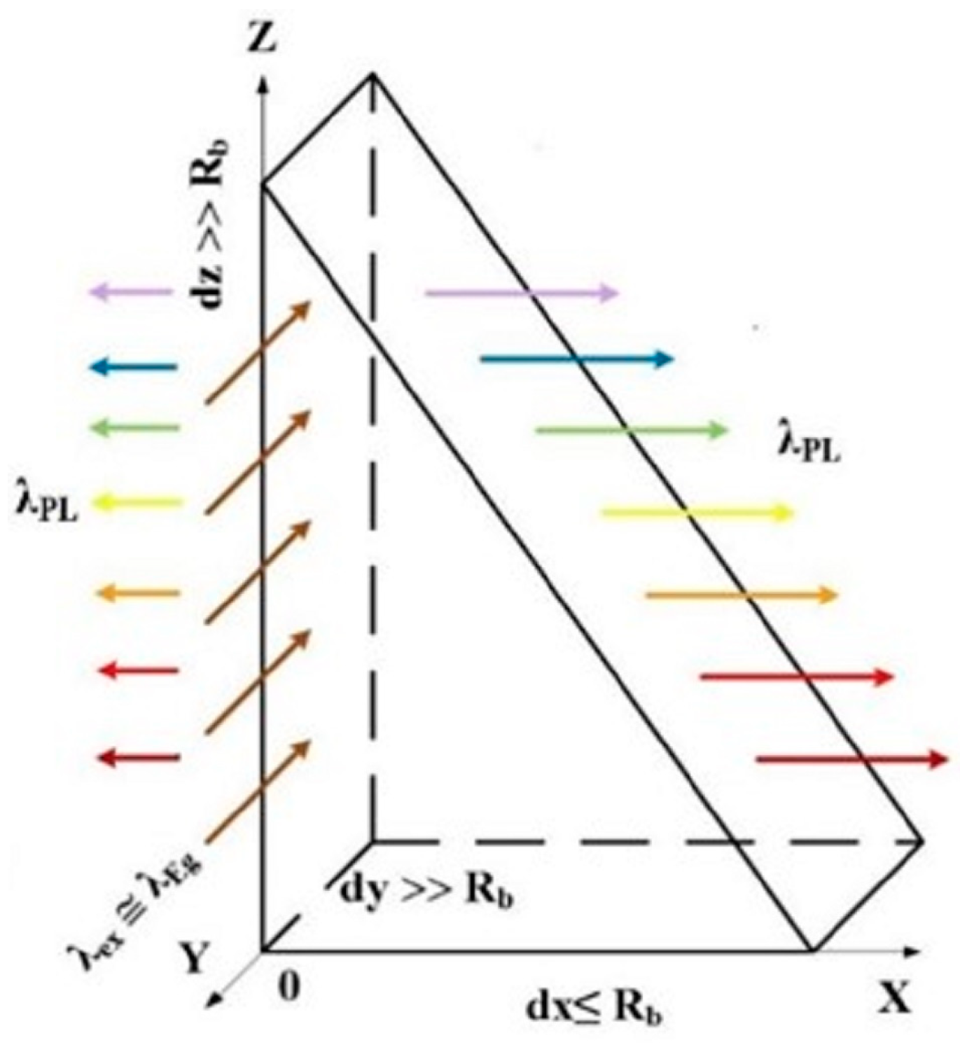
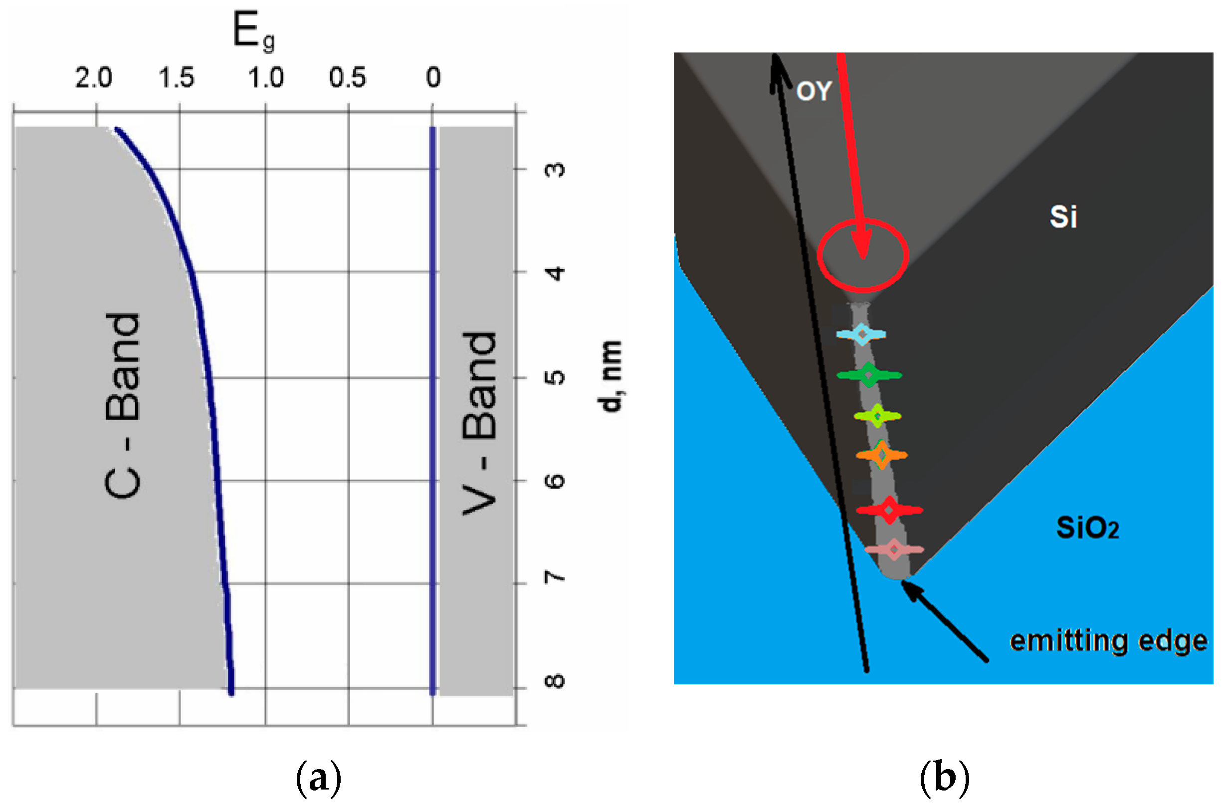
2. Theory and Materials

The quantum nanoprism is schematically shown in Figure 1. The radiation intensity of the quantum prism is higher than that of the quantum cone [5,6,7] due to the limit dy ≤ RB (RB is the Bohr’s exciton radius) and without limitations in the OY direction. This is the proportional relationship between the length of the quantum prism in the OY direction and the diameter of the quantum dot at the same point. Also, the radiation of a quantum prism occurs at a full angle of 180°. In addition, irradiation of the prism in the OY direction by light with low absorption λex ≤ λEg leads to the formation of excitons with gradually decreasing energy from the top of the prism to its base. Since the bandgap Eg in the OZ direction gradually decreases from the top of the prism to its base, according to the modified formula for Si, the ∆Eg function on the diameter of the nanowire is calculated [9].
∆Eg = (2Ћ2ζ2)/(m*d(OZ)2)
where 1/(m*) = 1/(me*) + 1/(mh*), me* and mh* are electron and hole effective masses, respectively, and d is the nanowire diameter. For QWs, ζ = 2.4048. In our case, the diameter of the nanowires is a function of the height d(OZ). Thus, it is a semiconductor structure with a graded band gap. First of all, we have a light source with a dispersion spectrum in the OX direction from hνmax = Eg max to hνmin = Ego, where Ego is the band gap of the bulk semiconductor. Secondly, since the energy of the exciting light quantum is less than Ego, anti-Stokes photoluminescence (ASPL), the so-called optical upconversion should be observed [10,11]. The upconversion phenomenon benefits many applications, such as solar cells, to improve efficiency [12], condensed-phase optical cooling [13], etc.
The experiments were carried out on micro-cutting of an SOI (silicon on insulator) (110) structure with the following parameters: top Si thickness 200 nm, box 400 nm, resistivity 20–30 Ω·cm, doped by boron. After cutting the structure, we chose a prism with an angle at the top of the prism less than 60°. An SEM microscope image of the SOI sample with nanoprism is shown in Figure 2. All experiments were carried out at room temperature.
The photoluminescence (PL) spectra were measured using a Pharos Light Conversion laser coupled with an optical parametric amplifier via an Orpheus module. The width of the laser pulse was approximately 160 fs, and the intensity was up to 1 GW/cm2. Light from the sample was collected via an Acton monochromator and directed to the Hamamatsu streak camera. Excitation of PL in the SOI sample with strong hνex2 = 2.1eV and weak hνex1 = 1.2 eV light absorptions for silicon was carried out in the OY direction, and the PL spectra were measured in the OX direction, as shown in Figure 1.

3. Results and Discussion

The measured PL spectra of the nanoprism are shown in Figure 3. The PL spectrum for strong absorption by hνex2 = 2.1 eV is atypical for a Si crystal because it is situated at an energy higher than the band gap of Si; a “blue shift” to 1.5 eV takes place. It is an optical downconversion.
The PL spectrum with weak absorption light at hνex1 = 1.2 eV is broader than strong absorption with four maxima and shoulders. Higher PL intensity and broad spectrum, compared to strong absorption, are due to the volume absorption of light by the prism [14]. More maxima and shoulders in the PL spectra are explained by the inhomogeneous prism or the presence of the addition of tiny prisms, as can be seen in Figure 2. Moreover, the PL spectrum is continuous from red to green color. It is an optical upconversion with a rainbow-like spectrum. This means that there is the possibility of upconverting excitation light with controllable energy of light quanta. It depends on the excitation place of the prism; for example, if the excitation takes place at the top of the prism, the energy light quanta will be maximal, and when it goes down to the base of the prism (in the OY direction), the energy of light quanta will decrease. This infrared absorption is important, as prism excitation could be achieved inside organism tissues due to their transparency to infrared.
The study of PL kinetics (PL(t)) showed that the decay time is ~20 ps. The lifetime values were determined by deconvolution of the decays using the system response function IRF = const × exp(−t2/2σ2), σ = (20 ± 2) ps (Figure 3b) and fitting the function PL(t)~exp[(σ2 − 2tτ)/(2τ2)] × [1 + Erf{(tτ − σ2)/(21/2στ)}] [15], where τ is the decay lifetime. The determined τ value of (21 ± 2) ps was fitted for both high and low excitation quanta. The decay time did not depend on the emission wavelength. In the experiment, the SiO2 layer is also irradiated. Therefore, it is necessary to consider this possibility as well. The PL spectrum of the SiO2 layer is located in the visible part of the spectra at hν = 1.9 eV [16,17], but it is having usually one band, and the PL decay time is in the range of several milliseconds. The supposed mechanism of upconversion is in two steps: the first creates a cold population of free electrons and holes using a light tuned to the Si bandgap, and the second subsequent phonon coupling results in the carrier with higher energy than the incident light, a so-called Pringsheim mechanism [18]. Evidence of this idea is investigated, for example, by upconversion in shell–core quantum structures [19].
The calculation of the change in Eg as a function of height in the OZ direction using the spectrum in Figure 3 (“blue edge” position) and Formula 1 and silicon parameters (the range for d variation was determined from the red and blue edges in Figure 3a) is shown in Figure 4a. It can be seen that this prism is a graded bandgap with a gradual decrease in Eg from the top of the prism to its base only due to the quantum size effect in quantum wires. The most-emitting places of the prism are localized on the sharp prism top. The prism top dimensions according the curve in Figure 4a and shortest emitted quanta in Figure 3 are in the range of 2.5–4 nm, and are determined by micromachining precision affecting the prism sharpness. If we take a section of a nanoprism with 60° triangles with a base of 8 nm, this is the diameter of the lowest nanowire in the nanoprism in which there is the weakest QCE, as taken from Figure 4a. The indirect bandgap of the Si crystal is Ego = 1.1 eV, and the height of the active part of the nanoprism will be only 7 nm. Eight Gaussian peaks were fitted with different peak emission wavelengths to describe the PL spectra in Figure 3a. They correspond to approximately six dominant nanowires located on the prim top along its edge, and their shortest emission wavelength is shown in Figure 4b. The nanowires of shorter emission wavelength are located closer to the sample surface and are the shortest ones as the prism top radius increases towards its interface with SiO2. This means that the quantum prism is a nano source of light with a dispersion spectrum [6].
Such a nano source of light is usable for the study of small medical and biological objects, such as organelles, microbes, and viruses.

4. Conclusions

This study shows the possibility of constructing a nano source of light, a quantum prism with a dispersive spectrum distributed along the prism height. The range of the dispersive spectrum depends on the material of the quantum prism and its parameters. The red edge of the spectrum is determined by the band gap of the bulk semiconductor, and the blue edge by the quantum confinement of electron–hole pairs/excitons on top of the quantum prism.
The possibility of upconverting light with a controllable energy of light quanta is shown. This quantum prism is a new type of nano source of light because it substitutes for two elements in a conventional spectrometer: a source of light and a dispersive element.

Author Contributions

Conceptualization, A.M.; methodology, P.Š. and S.M.; investigation, A.M., P.Š. and S.M.; data curation, A.M., P.Š. and S.M.; writing—original draft preparation, A.M., P.Š. and S.M.; project administration, K.H. All authors have read and agreed to the published version of the manuscript.

Funding

We acknowledge the support by the COST action: “European Network for Innovative and Advanced Epitaxy” (CA20116, P-COST- 21- 6, 2021–2025).

Data Availability Statement

The data that support the findings of this study are available upon reasonable request from the authors.

Conflicts of Interest

The authors declare no conflicts of interest.

References

  1. Motz, J.T.; Hunter, M.; Galindo, L.H.; Gardecki, J.A.; Kramer, J.R.; Dasari, R.R.; Feld, M.S. Optical fiber probe for biomedical Raman spectroscopy. Appl. Opt. 2004, 43, 542–554. [Google Scholar] [CrossRef] [PubMed]
  2. Brus, L.E. Electron–electron and electron-hole interactions in small semiconductor crystallites: The size dependence of the lowest excited electronic state. J. Chem. Phys. 1984, 80, 4403–4444. [Google Scholar] [CrossRef]
  3. Medvid’, A.; Fukuda, Y.; Michko, A.; Onufrievs, P.; Anma, Y. 2D lattice formation by YAG:Nd laser on the surface of Ge single crystal. Appl. Surf. Sci. 2005, 244, 120–123. [Google Scholar] [CrossRef]
  4. Medvid’, A.; Mychko, A.; Gnatyuk, V.; Levytskyi, S.; Naseka, Y. Mechanism of nano-cone formation on Cd0.9Zn0.1Te crystal by laser radiation. Opt. Mater. 2010, 32, 836–839. [Google Scholar] [CrossRef]
  5. Medvid’, A.; Dmytruk, I.; Onufrijevs, P.; Pundyk, I. Quantum confinement effect in nanohills formed on a surface of Ge by laser radiation. Phys. Status Solidi C 2007, 4, 3066–3069. [Google Scholar] [CrossRef]
  6. Medvid’, A.; Dmitruk, I.; Onufrijevs, P.; Pundyk, I. Properties of nanostructure formed on SiO2/Si interface by laser radiation. Solid State Phenom. 2008, 131–133, 559–562. [Google Scholar]
  7. Medvid’, A.; Onufrijevs, P. Properties of nanocones formed on a surface of semiconductors by laser radiation: QC effect of electrons, phonons, and excitons. Nanoscale Res. Lett. 2011, 6, 582. [Google Scholar] [CrossRef] [PubMed]
  8. Takahahara, T.; Takeda, K. Theory of the quantum confinement effect on excitons in quantum dots of indirect-gap materials. Phys. Rev. B 1992, 46, 15578–15581. [Google Scholar] [CrossRef]
  9. Li, J.; Wang, L. Comparison between Quantum Confinement Effect of Quantum Wires and Dots. Chem. Mater. 2004, 16, 4012–4015. [Google Scholar] [CrossRef]
  10. Morozov, Y.V.; Draguta, S. Defect-Mediated CdS Nanobelt Photoluminescence Up-Conversion. J. Phys. Chem. C 2017, 121, 16607–16616. [Google Scholar] [CrossRef]
  11. Sellers, D.G.; Doty, M.F. Design, synthesis and photophysical properties of InP/CdS/CdSe and CdTe/CdS/CdSe (core/shelVshell) quantum dots for photon upconversion. In Proceedings of the 2015 IEEE 42nd Photovoltaic Specialist Conference (PVSC), New Orleans, LA, USA, 14–19 June 2015. [Google Scholar] [CrossRef]
  12. Goldschmidt, J.C.; Fischer, S. Upconversion for Photovoltaics—A Review of Materials, Devices and Concepts for Performance Enhancement. Adv. Opt. Mater. 2015, 3, 510–535. [Google Scholar] [CrossRef]
  13. Sheik-Bahae, M.; Epstein, R.I. Optical Refrigeration. Nat. Photonics 2007, 1, 693–699. [Google Scholar] [CrossRef]
  14. Kang, H. Crystalline Silicon vs. Amorphous Silicon: The Significance of Structural Differences in Photovoltaic Applications. IOP Conf. Ser. Earth Environ. Sci. 2021, 726, 012001. [Google Scholar] [CrossRef]
  15. Farr, E.P.; Quintana, J.C.; Reynoso, V.; Ruberry, J.D.; Shin, W.R.; Swartz, K.R. Introduction to Time-Resolved Spectroscopy: Nanosecond Transient Absorption and Time-Resolved Fluorescence of Eosin B. J. Chem. Educ. 2018, 95, 864–871. [Google Scholar] [CrossRef]
  16. Chen, E.Y.; Li, Z.; Milleville, C.C.; Lennon, K.R.; Zide, J.M.O.; Doty, M.F. CdSe(Te)/CdS/CdSe Rods Versus CdTe/CdS/CdSe Spheres: Morphology-Dependent Carrier Dynamics for Photon Upconversion. IEEE J. Photovolt. 2018, 8, 746–751. [Google Scholar] [CrossRef]
  17. Skuja, L.; Ollier, N.; Kajihara, K.; Smits, K. Creation of glass-characteristic point defects in crystalline SiO2 by 2.5 MeV electrons and by fast neutrons. J. Non-Cryst. Solids 2019, 505, 252–259. [Google Scholar] [CrossRef]
  18. Skuja, L.; Ollier, N.; Kajihara, K. Luminescence of non-bridging oxygen hole centers as a marker of particle irradiation of α-quartz. Radiat. Meas. 2020, 135, 106373–106377. [Google Scholar] [CrossRef]
  19. Zwei, P.P. Bemerkungen uber Denunterschied von Lumineszenz- Und Temperaturatutstrahlung. Z. Fur Phys. 1929, 57, 739–746. [Google Scholar]
Figure 1. Geometric scheme of a quantum prism with quantum confinement in the OX direction and without it in the OY and OZ directions. Irradiation of the prism is in OY and photoluminescence in the OX directions, respectively.
Nanomaterials 14 01277 g001
Figure 2. SEM microscope image of SOI sample with nanoprism. The arrow shows the excitation spot.
Nanomaterials 14 01277 g002
Figure 3. (a) PL spectra of quantum prism formed on SOI and excited with strong silicon absorption at hνex2 = 2.1 eV (red curve), and weak silicon absorption hνex1 = 1.2 eV (black curve). (b) The corresponding PL decays and system instrumental response function (dashed line with Gauss fit of 40 ps FWHM).
Nanomaterials 14 01277 g003
Figure 4. (a) Calculation of Eg along the height of the prism in the direction of OZ, using PL spectrum red and blue edge peak energies (for black curve in Figure 3) and Formula (1). (b) Schematic prism top roundness dependence on its thickness and its correlation with nanowire shortest emission wavelength, shown by different color. The red circle shows the excitation spot. The laser beam excites the prism in the OY direction.
Nanomaterials 14 01277 g004
Disclaimer/Publisher’s Note: The statements, opinions and data contained in all publications are solely those of the individual author(s) and contributor(s) and not of MDPI and/or the editor(s). MDPI and/or the editor(s) disclaim responsibility for any injury to people or property resulting from any ideas, methods, instructions or products referred to in the content.

