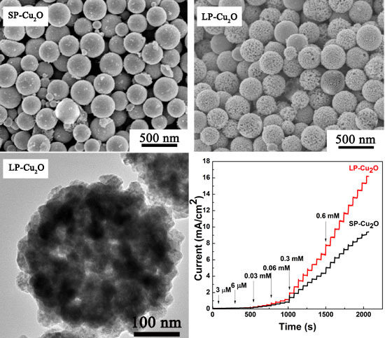
Article

Preparation of Cuprous Oxide Mesoporous Spheres with Different Pore Sizes for Non-Enzymatic Glucose Detection

Lab of Advanced Nano-Structures & Transfer Processes, Department of Chemical Engineering, Tianjin University, Tianjin 300354, China
*
Authors to whom correspondence should be addressed.
Nanomaterials 2018, 8(2), 73; https://doi.org/10.3390/nano8020073
Submission received: 9 January 2018 / Revised: 26 January 2018 / Accepted: 26 January 2018 / Published: 29 January 2018
(This article belongs to the Special Issue Nanomaterials for Electrocatalytic Applications in Energy and Sensing)

Abstract

:
Mass transfer plays a significant role in a sensor’s performance, because the substrate can be detected only when it contacts with the active catalytic surface. In this work, cuprous oxide mesoporous nanospheres (Cu2O MPNS) with different pore size distributions are fabricated and applied as electrocatalysts for glucose detection. The small pore Cu2O (SP-Cu2O, mean pore size of 5.3 nm) and large pore Cu2O (LP-Cu2O, mean pore size of 16.4 nm) spheres are prepared by the template method and an etching treatment. The obtained two kinds of Cu2O MPNS exhibit high porosity with a similar specific surface area of 61.2 and 63.4 (m2·g−1), respectively. The prepared Cu2O MPNS are used to construct an electrochemical non-enzymatic glucose sensor. The results show that the LP-Cu2O exhibits better performance than SP-Cu2O, which illustrates that the internal diffusion takes a great impact on the performance of the sensor. The LP-Cu2O modified electrode possesses a high and reproducible sensitivity of 2116.9 μA mM−1·cm−2 at the applied potential of 0.6 V with a wide detection range of 0.003–7.8 mM and a low detection limit of 0.42 μM.

Graphical Abstract

1. Introduction

Glucose biosensors, which play an important role in the monitoring of blood glucose, have become a reliable tool for the treatment and control of diabetes [1,2,3]. The majority of amperometric biosensors for glucose detection focus on two major types: enzymatic and non-enzymatic sensors [4]. Due to the high sensitivity and good selectivity, most glucose biosensors focus on glucose oxidase. However, they suffer from poor reproducibility, a complicated immobilization process, and thermal and chemical instability because of the intrinsic properties of the enzymes. To solve this problem, plentiful efforts have been made to the research of non-enzymatic glucose biosensors. As a member of glucose sensors, non-enzymatic glucose sensors based on the direct electrocatalysis of glucose have recently drawn great attention due to their efficient glucose detection sensitivity and stability [2,5,6,7]. Owing to the high electrocatalytic ability, materials based on noble metals, such as Pt [8], Pd [9], and Ag [10], transition metals, such as Cu [11], Ni [12,13], Mn [14], and Co [15], and their oxides [16], or polymers, such as polyaniline [17], have been found to be active electrocatalytic materials for glucose biosensors. Among them, cuprous oxide (Cu2O), an important semiconductor with a band gap of 2.2 eV [18], has exhibited promising applications in the field of sensing, which is attributed to its minimal cost, stability, and significant catalytic activity [19,20,21,22]. Recently, in order to introduce larger surface areas and more catalytically active sites for the electrocatalytic reactions, porous Cu2O nanomaterials have been prepared and used to build biosensors with improved performances [23,24,25,26].
However, the efficiency of electrochemical sensors is significantly influenced by the substrate adsorption and charge transfer on the electrode surface, because the glucose molecules can be oxidized only when they contact with the active sites. As a consequence, the performances of these electrocatalytic sensors may be subject to mass transfer limitations, especially for the porous systems. In principle, the mass transfer resistance comes from both external and internal mass transfer [27]. Similar to the reactions with normal porous catalysts, the mass transfer of the substrate first occurs from the bulk fluid to the external surface of the catalyst, then diffuses from the external surface into and through the pores within the pellet [28,29]. The external diffusion can be neglected with vigorous stirring, hence the internal diffusion will play a significant role in the detection ability of the biosensor [30]. Therefore, promoting the internal diffusion rate may facilitate the sensitivity of the biosensors. Altering the pore size of the materials can adjust the internal diffusion rate due to the fact that the internal diffusion rate has a proportional relationship with the pore size. However, there is little research about the influence of different pore sizes on glucose sensors.
Herein, we report the fabrication of mesoporous Cu2O nanospheres with different pore sizes and their performances on the glucose sensors (Scheme 1). Comparative studies reveal that a Cu2O sphere with a relatively larger pore size shows higher activity, a wider detection range, and a lower detection limitation for glucose detection, demonstrating the important role of internal diffusion on their performances for glucose detection.

2. Materials and Methods

The porous Cu2O nanospheres with a small pore size (SP-Cu2O) were prepared according to the template synthetic process with some modification. Firstly, 0.5 g triblock copolymer pluronic P123 (MW 5800) was dissolved in 40 mL deionized water and stirred for at least 5 h. Then, 3 mL of 0.2 M Cu(NH3)42+ solution (10:1, NH3:Cu2+) was transferred into the P123 solution under constant stirring. After 40 min, 6 mL of 0.6 M ascorbic acid solution was added dropwise into the above mixture. After stirring for 40 min, the resulting precipitate was collected by centrifugation, washed with ethanol at 333 K overnight to remove the P123, and finally dried in vacuum at 333 K for 4 h.
The porous Cu2O nanospheres with a large pore size (LP-Cu2O) were prepared using the etching method. Typically, 5 mg of the as-prepared SP-Cu2O was added into 10 mL of the ethanol solution containing 1.2 mg·mL−1 of L-proline under constant stirring. After 8 h, the precipitate was collected by centrifugation, washed several times with distilled water and ethanol, and finally dried in vacuum at 333 K for 4 h.
An X-ray power diffraction (XRD) analysis was carried out using a Bruker advance-D8 power diffractometer (Karlsruhe, Germany) with Cu Kα radiation (λ = 0.154178 nm). Field-emission scanning electron microscopy (FESEM) images were obtained on a Hitachi S-4800 microscope (Tokyo, Japan) operating at 5 kV. X−ray photoelectron spectrum (XPS) was conducted on a PHI 1600 spectrometer (PerkinElmer, Shelton, CT, USA) to analyze the surface composition. For transmission electron microscopy (TEM) and high-resolution transmission electron microscopy (HRTEM), the structures were imaged with a JEM-2011F microscope (JEOL, Tokyo, Japan) with an accelerating voltage of 200 kV and a JEOL JEM-3010 microscope (JEOL, Tokyo, Japan) operated at 200 kV, respectively. The nitrogen adsorption-desorption isotherms were obtained with ASAP2010 apparatus (Micrometritics, Atlanta, GA, USA) at 77 K. The surface areas of the obtained samples were calculated by the Brunauer–Emmett–Teller (BET) method, and the pore size distribution curve was derived from the adsorption using the Barrett–Joyner–Halenda (BJH) theory.
Electrochemical measurements were conducted using an electrochemical workstation (CHI 660E, Chenhua, Shanghai, China) connected to the computer for glucose detection. All of the electrochemical measurements were carried out using a conventional three-electrode system with a magnetic stirrer. Platinum wire was used as the counter electrode, saturated calomel electrode was used as the reference electrode, and a Cu2O mesoporous nanospheres (MPNS)-modified glassy carbon electrode (GCE) was used as the working electrode. The GCE was polished with alumina slurries and cleaned by sonication in the deionized water and ethanol, respectively. Four milligrams of the prepared Cu2O sample was added in 2 mL of 5 wt % Nafion solution and dispersed in an ultrasonic bath for 30 min. Then, a 10 μL dispersion (2 mg·mL−1) was dropped onto the GCE’s surface and dried in air overnight to afford a Cu2O-modified GCE.

3. Results and Discussion

The morphologies of the samples were characterized by field emission scanning electron microscopy (FESEM) and transmission electron microscopy (TEM). Figure 1a shows the FESEM image of SP-Cu2O. Figure 1b shows FESEM images of LP-Cu2O. It can be found that both the SP-Cu2O and LP-Cu2O spheres are uniformly dispersed and with an average diameter of 320 nm. Moreover, the distinct widespread pores could be observed on the SEM image of LP-Cu2O, which demonstrates its large porous property. The magnified SEM image of a typical sphere (Figure 1c) illustrates its mesoporous structure feature. As shown in Figure S1, the mesoporous structure is destroyed by extending the etching time to 10 h. Figure 1d,e and Figure S2 are the corresponding TEM images of the LP-Cu2O and SP-Cu2O, which illustrate that the Cu2O spheres have a porous structure. The observed lattice fringes in the HRTEM image (Figure 1f) show a clear lattice spacing of 0.24 nm, corresponding to the distance of the (111) planes of Cu2O.
Figure 2 shows the X-ray diffraction (XRD) patterns of SP-Cu2O and LP-Cu2O, in which can be seen four sharp peaks at 2θ = 30, 37, 42.6, and 62.4°, corresponding to the (110), (111), (200), and (220) planes of Cu2O (JCDPS No. 05-0667) [31]. There is no obvious peak showing the formation of any phase of copper (Cu) or copper oxide (CuO), which indicates the high purity of the prepared samples. Additionally, XPS is employed to determine the surface state. Figure S3 displays the XPS spectrum of Cu 2p. The peaks at 952.1 and 932.3 eV are assigned to Cu 2p1/2 and Cu 2p3/2 of Cu2O, respectively. No shake-up satellite peaks are found at 940–945 eV, indicating that Cu (I) is dominant in the LP-Cu2O.
The N2 adsorption-desorption isotherms are measured to testify to the porosity of the as-prepared Cu2O spheres. Figure 3a shows the N2 adsorption-desorption isotherms of SP-Cu2O and LP-Cu2O, which indicate that the two isotherms belong to type IV hysteresis loops, providing the evidence for the mesoporous structure. The BET specific surface area of the SP-Cu2O is 61.2 m2·g−1, which is almost identical with that of the LP-Cu2O (63.4 m2·g−1), indicating that the surface area is slightly increased after the etching treatment. This may be due to the crystal size of LP-Cu2O, which as calculated from XRD, is slightly decreased after etching [32]. Figure 3b displays the pore size distribution curves. The mean pore size of LP-Cu2O is 16.4 nm, which is much larger than that of SP-Cu2O (5.3 nm). Nevertheless, a weak peak appears at about 5 nm from the BJH curve of the LP-Cu2O in Figure 3b, which may be ascribed to a small part of unetched pores.
Generally, the diffusion of liquid in the pore of the porous catalyst is in accordance with Fick-diffusion according to mass transfer theory [33]. The effective diffusion coefficient (DABP) can be defined as follows:
D ABP = ε   D AB τ
The pore size and the void fraction (ε) have a proportional relationship, while it has an inverse relationship with the twist factor (τ). DAB is constant in a certain solution system. On the basis of Equation (1), the DABP value of LP-Cu2O is larger than that of SP-Cu2O. Therefore, the liquid diffuses faster in the pores of LP-Cu2O than in those of SP-Cu2O.
The non-enzymatic glucose sensor is created by direct deposition of SP-Cu2O or LP-Cu2O on the surface of a glassy carbon electrode (GCE). Figure 4 depicts the cycle voltammetry curves (CVs) of the SP-Cu2O and LP-Cu2O in 0.1 M KOH solution with and without the addition of glucose at a scan rate of 50 mV s‒1. Comparing the CVs in Figure 4a, a relatively larger current signal can be observed in Figure 4b, which illustrates that the LP-Cu2O exhibits a higher activity towards glucose electro-oxidation. The enhanced electrocatalytic activity may be attributed to the kinetic effect by the improved internal diffusion rate of the LP-Cu2O, because with the improved internal diffusion ability there will be more catalytically active sites, which could be attained by the glucose molecules.
The amperometric response of the Cu2O MPNS modified electrodes towards glucose oxidation is conducted at 0.6 V for the successive addition of various concentrations of glucose solution with violent stirring. Upon the injection of glucose solution (Figure 5), the modified electrodes exhibit an obvious increase of current density to the change of glucose concentration. Both catalysts can acquire 97% of steady-state current within 5 s, indicating the sensitive and rapid response behavior of SP-Cu2O and LP-Cu2O towards a glucose electro-oxidation reaction. Moreover, the current response of the LP-Cu2O for an equal concentration of glucose solution is obviously larger than that of the SP-Cu2O.
Figure 6 illustrates the linear relationship between the current response and the glucose concentration of SP-Cu2O- and LP-Cu2O-modified electrodes, which shows that both catalysts exhibit a good linear relationship towards the concentration of the glucose. The linear regression equations acquired from the amperometric response of SP-Cu2O and LP-Cu2O are J(y) = 0.6775 + 1.5314C(x) and J(y) = 0.7405 + 2.1169C(x), respectively. Hence, the factor J refers to the current response density and C is the concentration of the glucose. The sensitivity of the LP-Cu2O (2116.9 μA mM−1 cm−2), which is calculated from the calibration curves, is about 1.38-fold greater than that of the SP-Cu2O (1531.4 μA mM−1 cm−2). The detection limit of LP-Cu2O (0.42 μM) is lower than that of the SP-Cu2O (0.63 μM), and the linear range of LP-Cu2O (up to 7.234 mM) is much wider than that of the SP-Cu2O (up to 3.861 mM). The above results indicate that in comparison with SP-Cu2O, the LP-Cu2O exhibits higher electrocatalytic activity towards glucose electro-oxidation with higher sensitivity and a lower detection limit. The catalytic activity is proportional to the effective catalytic surface area where the glucose can contact. We use the same amount of SP-Cu2O and LP-Cu2O to modify the electrodes. The total surface area is considered equal on the basis of the almost same BET specific surface area of the two samples. Therefore, supposing that all of the surface is used as the active surface, the sensor effect should be same. The effect of the external diffusion can be neglected, which is owing to the fact that the experiments are conducted with under a sufficient stirring condition. Therefore, the internal diffusion plays a vital role on the sensors’ performance. Compared with SP-Cu2O, LP-Cu2O has the same surface area but an improved sensor performance due to the higher internal diffusion rate of the LP-Cu2O. The results prove that the fabrication of a large-pore electrocatalyst can improve the efficiency of mass transfer so as to improve the electrocatalytic activity. Additionally, the comparison of the LP-Cu2O and other reported Cu-based glucose sensors is summarized in Table S1. Obviously, the sensor in this work exhibits good analytical properties, which are comparable to or even higher than those of other reported Cu-based electrochemical sensors for glucose.
Another important analytical factor for an electrochemical sensor is its ability to discriminate the interfering species having similar electroactivities on the electrocatalyst. So, the selectivity of the as-prepared biosensor is evaluated against some interfering compounds, including dopamine (DA), ascorbic acid (AA), uric acid (UA), fructose, and sucrose, which usually co-exist with glucose in human blood serum [4,34]. The blood glucose level of a normal human body is between 4 and 7 mM, which is approximately 30 to 50 times higher than that of the interferences. The interfering species with only 5–10% of glucose concentration are applied in the selectivity tests in the previous studies. In this work, a higher concentration of DA, AA, UA, fructose, and sucrose are used to test the selectivity of the catalyst. Figure 7a shows the amperometric response of the LP-Cu2O-modified electrode toward the successive addition of 0.3 mM glucose, 0.1 mM ascorbic acid (AA), 0.1 mM uric acid (UA), 0.1 mM dopamine (DA), 0.1 mM NaCl, 0.1 mM fructose, 0.1 mM sucrose, and 0.3 mM glucose in a stirred 0.1 M KOH solution at the applied potential of 0.6 V. It can be seen that the current responses to interfering species are rather weak compared to that from glucose oxidation, suggesting the excellent selectivity of the LP-Cu2O-modified electrode. Therefore, the LP-Cu2O-modified electrode is a promising candidate in practical application for glucose detection.
The long-term storage stability of the electrode is characterized by storing under ambient conditions and studying the response intermittently. As shown in Figure 7b, the activity of the LP-Cu2O-modified electrode is nearly unchanged within 10 days, and retains 97.2% of its original sensitivity after 30 days. The reproducibility of the sensor is also evaluated by preparing three electrodes through the same method. A relative standard deviation (RSD) of 3.88% is acquired, which suggests the good reproducibility of the LP-Cu2O-modified electrode.

4. Conclusions

In summary, different pore-size Cu2O nanospheres with a high specific surface area and a uniformly porous spherical morphology are successfully synthesized by the template method with an etching treatment. In the electrochemical measurements for glucose detection, the LP-Cu2O modified electrode exhibits better electrocatalytic activity compared with the SP-Cu2O modified electrode. The results show that internal diffusion rate plays a vital role in the performance of the sensor. The obtained LP-Cu2O biosensor exhibits a low detection limit, good sensitivity, excellent selectivity, good stability, reproducibility, and a wide detection range (0.003 to 7.8 mM) due to the improved internal diffusion and electron transfer ability.

Supplementary Materials

The following are available online at https://www.mdpi.com/2079-4991/8/2/73/s1. Figure S1: SEM image of LP-Cu2O with longer etching time; Figure S2: TEM image of SP-Cu2O; Figure S3: XPS spectrum of Cu 2p for LP-Cu2O; Table S1: Comparison of similar non-enzymatic glucose sensors.

Acknowledgments

This study was supported by the Specialized Research Funds for the National Natural Science Foundation of China (No. 20156157), the Doctoral Program of Higher Education of China (No. 20130032120017), and the Program of Introducing Talents of Discipline to Universities (No. B06006).

Author Contributions

Jingwen Ma, Fengbao Zhang, Xiaobin Fan, and Yang Li conceived and designed the experiments; Jingwen Ma and Jun Wang performed the experiments; Jingwen Ma, Jun Wang, Min Wang, Wenchao Peng, and Guoliang Zhang analyzed the data; and Jingwen Ma wrote the paper.

Conflicts of Interest

The authors declare no conflict of interest.

References

  1. Galant, A.L.; Kaufman, R.C.; Wilson, J.D. Glucose: Detection and analysis. Food Chem. 2015, 188, 149–160. [Google Scholar] [CrossRef] [PubMed]
  2. Zhu, H.; Li, L.; Zhou, W.; Shao, Z.; Chen, X. Advances in non-enzymatic glucose sensors based on metal oxides. J. Mater. Chem. B 2016, 4, 7333–7349. [Google Scholar] [CrossRef]
  3. Chen, A.; Ding, Y.; Yang, Z.; Yang, S. Constructing heterostructure on highly roughened caterpillar-like gold nanotubes with cuprous oxide grains for ultrasensitive and stable nonenzymatic glucose sensor. Biosens. Bioelectron. 2015, 74, 967–973. [Google Scholar] [CrossRef] [PubMed]
  4. Chung, R.J.; Wang, A.N.; Liao, Q.L.; Chuang, K.Y. Non-enzymatic glucose sensor composed of carbon-coated nano-zinc oxide. Nanomaterials 2017, 7, 36. [Google Scholar] [CrossRef] [PubMed]
  5. Yuan, R.-M.; Li, H.-J.; Yin, X.-M.; Lu, J.-H.; Zhang, L.-L. 3D CuO nanosheet wrapped nanofilm grown on Cu foil for high-performance non-enzymatic glucose biosensor electrode. Talanta 2017, 174, 514–520. [Google Scholar] [CrossRef] [PubMed]
  6. Zhang, E.; Xie, Y.; Ci, S.; Jia, J.; Wen, Z. Porous Co3O4 hollow nanododecahedra for nonenzymatic glucose biosensor and biofuel cell. Biosens. Bioelectron. 2016, 81, 46–53. [Google Scholar] [CrossRef] [PubMed]
  7. Ahmad, R.; Tripathy, N.; Ahn, M.S.; Bhat, K.S.; Mahmoudi, T.; Wang, Y.; Yoo, J.Y.; Kwon, D.W.; Yang, H.Y.; Hahn, Y.B. Highly Efficient Non-Enzymatic Glucose Sensor Based on CuO Modified Vertically-Grown ZnO Nanorods on Electrode. Sci. Rep. 2017, 7, 5715. [Google Scholar] [CrossRef] [PubMed]
  8. Li, Z.; Gao, F.; Gu, Z. Vertically aligned pt nanowire array/au nanoparticle hybrid structure as highly sensitive amperometric biosensors. Sens. Actuators B Chem. 2017, 243, 1092–1101. [Google Scholar] [CrossRef]
  9. Wen, D.; Herrmann, A.-K.; Borchardt, L.; Simon, F.; Liu, W.; Kaskel, S.; Eychmüller, A. Controlling the growth of palladium aerogels with high-performance toward bioelectrocatalytic oxidation of glucose. J. Am. Chem. Soc. 2014, 136, 2727–2730. [Google Scholar] [CrossRef] [PubMed]
  10. Chen, S.; Hai, X.; Chen, X.-W.; Wang, J.-H. In situ growth of silver nanoparticles on graphene quantum dots for ultrasensitive colorimetric detection of H2O2 and glucose. Anal. Chem. 2014, 86, 6689–6694. [Google Scholar] [CrossRef] [PubMed]
  11. Wang, B.; Wu, Y.; Chen, Y.; Weng, B.; Li, C. Flexible paper sensor fabricated via in situ growth of cu nanoflower on rgo sheets towards amperometrically non-enzymatic detection of glucose. Sens. Actuators B Chem. 2017, 238, 802–808. [Google Scholar] [CrossRef]
  12. Wu, H.; Yu, Y.; Gao, W.; Gao, A.; Qasim, A.M.; Zhang, F.; Wang, J.; Ding, K.; Wu, G.; Chu, P.K. Nickel plasma modification of graphene for high-performance non-enzymatic glucose sensing. Sens. Actuators B Chem. 2017, 251, 842–850. [Google Scholar] [CrossRef]
  13. Shu, Y.; Yan, Y.; Chen, J.; Xu, Q.; Pang, H.; Hu, X. Ni and nio nanoparticles decorated metal–organic framework nanosheets: Facile synthesis and high-performance nonenzymatic glucose detection in human serum. ACS Appl. Mater. Interfaces 2017, 9, 22342–22349. [Google Scholar] [CrossRef] [PubMed]
  14. Han, L.; Shi, J.; Liu, A. Novel biotemplated MnO2 1D nanozyme with controllable peroxidase-like activity and unique catalytic mechanism and its application for glucose sensing. Sens. Actuators B Chem. 2017, 252, 919–926. [Google Scholar] [CrossRef]
  15. Kannan, P.; Maiyalagan, T.; Marsili, E.; Ghosh, S.; Guo, L.; Huang, Y.; Rather, J.A.; Thiruppathi, D.; Niedziolka-Jonsson, J.; Jonsson-Niedziolka, M. Highly active 3-dimensional cobalt oxide nanostructures on the flexible carbon substrates for enzymeless glucose sensing. Analyst 2017, 142, 4299–4307. [Google Scholar] [CrossRef] [PubMed]
  16. Comini, E.; Sberveglieri, G. Metal oxide nanowires as chemical sensors. Mater. Today 2010, 13, 36–44. [Google Scholar] [CrossRef]
  17. Lai, J.; Yi, Y.; Zhu, P.; Shen, J.; Wu, K.; Zhang, L.; Liu, J. Polyaniline-based glucose biosensor: A review. J. Electroanal. Chem. 2016, 782, 138–153. [Google Scholar] [CrossRef]
  18. Wang, J.; Ma, J.; Li, X.; Li, Y.; Zhang, G.; Zhang, F.; Fan, X. Cu2O mesoporous spheres with a high internal diffusion capacity and improved catalytic ability for the aza-Henry reaction driven by visible light. Chem. Commun. 2014, 50, 14237–14240. [Google Scholar] [CrossRef] [PubMed]
  19. Ji, Y.; Liu, J.; Liu, X.; Yuen, M.M.F.; Fu, X.-Z.; Yang, Y.; Sun, R.; Wong, C.-P. 3D porous Cu@Cu2O films supported Pd nanoparticles for glucose electrocatalytic oxidation. Electrochim. Acta 2017, 248, 299–306. [Google Scholar] [CrossRef]
  20. Liu, M.; Liu, R.; Chen, W. Graphene wrapped Cu2O nanocubes: Non-enzymatic electrochemical sensors for the detection of glucose and hydrogen peroxide with enhanced stability. Biosens. Bioelectron. 2013, 45, 206–212. [Google Scholar] [CrossRef] [PubMed]
  21. Zhao, Z.; Wang, Y.; Li, P.; Sang, S.; Zhang, W.; Hu, J.; Lian, K. A highly sensitive electrochemical sensor based on Cu/Cu2O@carbon nanocomposite structures for hydrazine detection. Anal. Methods 2015, 7, 9040–9046. [Google Scholar] [CrossRef]
  22. Cai, C.; Zhu, T.; Li, D.; Ran, Y.; Dong, H.; Zhao, N.; Xu, J. Spherically aggregated Cu2O–Ta hybrid sub-microparticles with modulated size and improved chemical stability. CrystEngComm 2017, 19, 1888–1895. [Google Scholar] [CrossRef]
  23. Xie, H.; Ke, Q.; Xiong, X. Preparation of a Cu2O/rGO porous composite through a double-sacrificial-template method for non-enzymatic glucose detection. J. Mater. Sci. 2017, 52, 5652–5660. [Google Scholar] [CrossRef]
  24. Li, S.; Zheng, Y.; Qin, G.W.; Ren, Y.; Pei, W.; Zuo, L. Enzyme-free amperometric sensing of hydrogen peroxide and glucose at a hierarchical Cu2O modified electrode. Talanta 2011, 85, 1260–1264. [Google Scholar] [CrossRef] [PubMed]
  25. Gao, Z.; Liu, J.; Chang, J.; Wu, D.; He, J.; Wang, K.; Xu, F.; Jiang, K. Mesocrystalline Cu2O hollow nanocubes: Synthesis and application in non-enzymatic amperometric detection of hydrogen peroxide and glucose. CrystEngComm 2012, 14, 6639. [Google Scholar] [CrossRef]
  26. Zhou, D.-L.; Feng, J.-J.; Cai, L.-Y.; Fang, Q.-X.; Chen, J.-R.; Wang, A.-J. Facile synthesis of monodisperse porous Cu2O nanospheres on reduced graphene oxide for non-enzymatic amperometric glucose sensing. Electrochim. Acta 2014, 115, 103–108. [Google Scholar] [CrossRef]
  27. Zhang, L.; Mao, Z.; Thomason, J.D.; Wang, S.; Huang, K.; Butt, D. Synthesis of a homogeneously porous solid oxide matrix with tunable porosity and pore size. J. Am. Ceram. Soc. 2012, 95, 1832–1837. [Google Scholar] [CrossRef]
  28. Du, J.; Lai, X.; Yang, N.; Zhai, J.; Kisailus, D.; Su, F.; Wang, D.; Jiang, L. Hierarchically ordered macro−mesoporous TiO2−graphene composite films: Improved mass transfer, reduced charge recombination, and their enhanced photocatalytic activities. ACS Nano 2011, 5, 590–596. [Google Scholar] [CrossRef] [PubMed]
  29. Klaewkla, R.; Arend, M.; Hoelderich, W.F. Mass Transfer—Advanced Aspects; InTech: London, UK, 2011. [Google Scholar]
  30. Zhao, Q.; Li, Y.; Liu, R.; Chen, A.; Zhang, G.; Zhang, F.; Fan, X. Enhanced hydrogenation of olefins and ketones with a ruthenium complex covalently anchored on graphene oxide. J. Mater. Chem. A 2013, 1, 15039–15045. [Google Scholar] [CrossRef]
  31. Lv, J.; Kong, C.; Hu, X.; Zhang, X.; Liu, K.; Yang, S.; Bi, J.; Liu, X.; Meng, G.; Li, J.; Yang, Z.; Yang, S. Zinc ion mediated synthesis of cuprous oxide crystals for non-enzymatic glucose detection. J. Mater. Chem. B 2017, 5, 8686–8694. [Google Scholar] [CrossRef]
  32. Zhuang, W.; Zhang, Y.; Zhu, J.; An, R.; Li, B.; Mu, L.; Ying, H.; Wu, J.; Zhou, J.; Chen, Y.; et al. Influences of geometrical topography and surface chemistry on the stable immobilization of adenosine deaminase on mesoporous TiO2. Chem. Eng. Sci. 2016, 139, 142–151. [Google Scholar] [CrossRef]
  33. Bird, R.B.; Stewart, W.E.; Lightfoot, E.N. Transport Phenomena; John Wiley & Son: New York, NY, USA, 2002. [Google Scholar]
  34. Li, Y.; Guan, P.; Yu, F.; Li, W.; Xie, X. CeO2 nanorods embedded in Ni(OH)2 matrix for the non-enzymatic detection of glucose. Nanomaterials 2017, 7, 205. [Google Scholar] [CrossRef] [PubMed]
Scheme 1. Illustration of glucose oxidation using mesoporous Cu2O spheres with different internal diffusion rates as electrocatalysts. LP-Cu2O: large pore Cu2O; SP-Cu2O: small pore Cu2O.
Nanomaterials 08 00073 sch001
Figure 1. SEM (a) image of SP-Cu2O; SEM (b,c) and TEM (d,e) images of LP-Cu2O; HRTEM (f) image of the LP-Cu2O.
Nanomaterials 08 00073 g001
Figure 2. XRD patterns of the as-prepared SP-Cu2O and LP-Cu2O.
Nanomaterials 08 00073 g002
Figure 3. Nitrogen adsorption-desorption isotherms (a) and the corresponding Barrett–Joyner–Halenda (BJH) pore size distribution curves (b) of the SP-Cu2O and LP-Cu2O.
Nanomaterials 08 00073 g003
Figure 4. Cycle voltammetry curves (CVs) of the SP-Cu2O-modified electrode (a) and LP-Cu2O-modified electrode (b) in the absence and presence of 1.2 mM glucose in 0.1 M KOH solution at a scan rate of 50 mV s−1.
Nanomaterials 08 00073 g004
Figure 5. Amperometric response of SP-Cu2O and LP-Cu2O electrodes to successive additions of glucose at 0.6 V. Inset: a magnification view of the current response toward a low concentration of glucose solution.
Nanomaterials 08 00073 g005
Figure 6. The corresponding calibration curves of SP-Cu2O and LP-Cu2O for glucose detection.
Nanomaterials 08 00073 g006
Figure 7. (a) Amperometry response of LP-Cu2O upon successive addition of 0.3 mM glucose, 0.1 mM ascorbic acid (AA), 0.1 mM uric acid (UA), 0.1 mM dopamine (DA), 0.1 mM NaCl, 0.1 mM fructose, 0.1 mM sucrose, and 0.3 mM glucose at 0.6 V; (b) Normalized sensitivity of the LP-Cu2O-based electrode sensor for glucose detected every 5 days over 1 month.
Nanomaterials 08 00073 g007