Article

Self-Assembled Vanadium Oxide Nanoflakes for p-Type Ammonia Sensors at Room Temperature

1
School of Information Science and Technology, Nantong University, Nantong 226019, China
2
Key Laboratory of Polar Materials and Devices, Department of Optoelectronics, East China Normal University, Shanghai 200241, China
3
Collaborative Innovation Center of Extreme Optics, Shanxi University, Taiyuan 030006, China
*
Author to whom correspondence should be addressed.
Nanomaterials 2019, 9(3), 317; https://doi.org/10.3390/nano9030317
Submission received: 13 January 2019 / Revised: 17 February 2019 / Accepted: 21 February 2019 / Published: 27 February 2019

Abstract

:
VO2(B), VO2(M), and V2O5 are the most famous compounds in the vanadium oxide family. Here, their gas-sensing properties were investigated and compared. VO2(B) nanoflakes were first self-assembled via a hydrothermal method, and then VO2(M) and V2O5 nanoflakes were obtained after a heat-phase transformation in nitrogen and air, respectively. Their microstructures were evaluated using X-ray diffraction and scanning and transmission electron microscopies, respectively. Gas sensing measurements indicated that VO2(M) nanoflakes were gas-insensitive, while both VO2(B) and V2O5 nanoflakes were highly selective to ammonia at room temperature. As ammonia sensors, both VO2(B) and V2O5 nanoflakes showed abnormal p-type sensing characteristics, although vanadium oxides are generally considered as n-type semiconductors. Moreover, V2O5 nanoflakes exhibited superior ammonia sensing performance compared to VO2(B) nanoflakes, with one order of magnitude higher sensitivity, a shorter response time of 14–22 s, and a shorter recovery time of 14–20 s. These characteristics showed the excellent potential of V2O5 nanostructures as ammonia sensors.

Graphical Abstract

1. Introduction

In many industries, hazardous gases have become increasingly important raw materials, and for this reason it has become very important to develop highly sensitive gas sensors to monitor them in the manufacturing process. To this end, finding suitable materials with the required surface/bulk properties is essential for the gas sensor development. So far, metal oxide semiconductors (MOS) are the most attractive gas-sensitive materials [1]. Because nanostructured materials have a large surface-to-volume ratio and abundant surface states, many gas sensors based on SnO2 [2,3,4], TiO2 [5,6], ZnO [2,7,8,9], WO3 [10,11], In2O3 [12,13,14], and other metal oxide nanostructures have been successfully fabricated to detect and quantify gaseous species. MOS sensors are applicable in many areas of human activity. Different MOS gas sensors can be used to construct sensor matrixes named electronic noses to identify different odorous compounds and quantify their concentration [15,16,17,18,19]. Among the numerous metal oxides, vanadium oxides form an interesting material group because of their varying oxidation states between V2+ and V5+ [20]. At least 15 different vanadium oxides (VxOy), such as VO, V2O3, VO2, V2O5, VnO2n−1, and V2nO5n−2, have been reported [21,22]. Among them, VO2 (B phase and M/R phase) and V2O5 are the most famous, attracting great interest due to their special chemical/physical properties and their potential application in many fields. Table 1 shows the crystal structure, properties, and some important applications of the three vanadium oxides. B phase VO2 and V2O5 both have a layered structure. The spacing between layers provides abundant sites for the facile intercalation of Li+, thus making them an attractive cathode material in lithium-ion batteries [23,24]. M-phase VO2 shows a semiconductor-to-insulator transition around 340 K, accompanied by a rapid change in resistivity and optical transmittance, thus exhibiting wide potential in ultrafast optical switches, thermochromic windows, and infrared sensors [25,26].
Recently, vanadium oxides have been considered as new candidates for gas sensors. Their sensing properties for inorganic/organic gases such as nitrogen oxides [27,28], ethanol [1,29,30,31], butyl-amine [32], and ammonia [33,34,35] have been reported. However, the gas-sensing properties of vanadium oxide nanostructures are strongly dependent on the actual synthesis environment and closely correlated with material morphologies, surface states, and microstructures. For example, several groups reported that V2O5 nanostructures (e.g. nanorods, flow-like nanostructures) were more sensitive to ethanol than to ammonia at room temperature [29,30,31]; however, an opposite higher sensitivity to ammonia is also reported by Hakim et al. in V2O5 nanoneedles [36]. Some groups even reported ultrahigh sensitivities of V2O5 nanostructures (e.g. nanofibers, nanofilms) for ultralow level (<1 ppm) ammonia detection [33,34,35]. In addition, the conduction type n or type p usually determines the direction of resistance change when they are exposed to target gases. When n-type MOS gas sensors are utilized to detect reducing gases, reductive gas species react with the chemisorbed oxygen on the surface and electrons trapped by oxygen are released into the conduction band of MOS, leading to a decrease in resistance. With regard to p-type MOS gas sensors, the direction of resistance change is opposite due to the combination of holes with electrons released from the surface reaction. Vanadium oxides’ nanostructures are generally regarded as n-type semiconductors, which exhibit n-type gas-sensing responses in most literature reports. However, interesting p-type sensing behaviors have also been reported in a few studies. Yu et al. reported a p-type sensing response to NO2 in vanadium oxide nanotubes at 80 °C [37]. Also, p-type sensing responses to either ethanol or ammonia at room temperature were reported in V2O5 and VO2(B) nanostructures [29,38]. Obviously, the synthesis environment and the microstructure played a crucial role in gas sensors of vanadium oxide nanostructures.
In this work, self-assembled VO2(B), VO2(M), and V2O5 nanoflakes were synthesized and their gas-sensing properties were comparatively investigated. VO2(B) nanoflakes were first self-assembled via a hydrothermal method, and then V2O5 and VO2(M) specimens were obtained by annealing VO2(B) nanoflakes in air and nitrogen, respectively. Their gas-sensing properties demonstrated that VO2(B) and V2O5 specimens displayed superior selectivity to ammonia, while the VO2(M) specimen was gas-insensitive. Unlike the common n-type gas-sensing mechanism most reported, both VO2(B) and V2O5 nanoflakes exhibited abnormal p-type sensing properties in ammonia. Compared to the VO2(B) specimen, the ammonia sensor of V2O5 nanoflakes showed a one order of magnitude higher sensitivity, faster response speed, and better reproducibility, exhibiting wide potential in ammonia sensors.

2. Materials and Methods

2.1. Synthesis of Self-Assembled Vanadium Oxide Nanoflakes

Self-assembled VO2(B) nanoflakes were prepared via a hydrothermal method, as described in previous works [39]. Briefly, 0.48 g commercial V2O5 powder was added to 80 mL oxalic acid (0.1 mol/L) in an aqueous solution to form a yellow slurry. The slurry was stirred for 30 min and then transferred to a 100-mL autoclave with a Teflon liner. The autoclave was maintained at 180 °C for 36 h and then air-cooled to room temperature. The resulting dark blue precipitates (VO2(B)) were collected and washed with distilled water and ethanol several times and then dried at 60 °C under a vacuum for 10 h. Finally, the VO2(M) specimen was obtained by heating VO2(B) at 550 °C in an N2 atmosphere for 1 h, and the V2O5 specimen was obtained by heating VO2(B) at 550 °C in air for 1 h. In all reactions, all chemicals used were purchased without further purification and without adding any additives or surfactants.

2.2. Characterization

The morphology of vanadium oxide specimens was characterized by field-emission scanning electron microscopy (FESEM, Hitachi-S-4800, Hitachi High Technologies, Tokyo, Japan) and transmission electron microscopy (JEOL, JEM-2100, Tokyo, Japan). The crystal structure was characterized by X-ray diffraction (XRD, Bruker D8 Advance, Billerica, MA, USA) using monochromatized Cu Kα radiation (λ = 1.5418Å).

2.3. Sensor Fabrication and Gas-Sensing Tests

The diagram of gas sensors is shown in Figure 1. Synthesized vanadium oxide samples were deposited on a SiO2 tile for gas sensing tests. The SiO2 tile was pre-patterned with interdigitated gold electrodes. For each sensor fabrication, the individual substrate was placed in a hot plate and heated to 50 °C. VO2(B), VO2(M), and V2O5 samples were each separately dispersed in ethanol at a concentration of 200 mg mL−1 and then deposited on the hot sensor substrates. Finally, the sensor substrates with vanadium oxide samples across the interdigitated gold electrodes were dried in air for 40 min.
The gas-sensing properties of sensors were evaluated on a measuring system containing a static gas distribution chamber with a volume of 20 L. To simulate the actual measuring circumstances, we referenced the measuring method used by Qin et al. [29]. The ambient air was introduced into the chamber and target gases/liquids were injected and diluted/volatilized in the chamber. The relative chamber humidity was monitored using an internal humidity meter. During the measurement, the relative humidity is about 37% and the room temperature is about 24 °C.

3. Results

VO2(B) specimen was prepared via a hydrothermal procedure, and their representative SEM images are shown in Figure 2. Obviously, the VO2(B) specimen consists of large quantities of nanoflakes. It is noted that these nanoflakes are not distributed randomly in the products, but parallel arranged and self-assembled forming clumps as shown in Figure 2a. The medium- and high-magnification SEM images indicate that these nanoflakes are typically several micrometers in length, hundreds of nanometers in width (Figure 2b), and 20–50 nm in thickness (Figure 2c). In addition, these nanoflakes are also assembled together in the TEM image (Figure 2d). The corresponding high-resolution (HR) TEM image (Figure 2d, square region marked in red) indicates that the nanoflakes are single crystalline, and the lattice spacing of 0.35 nm corresponds to the d spacing of the (110) plane of monoclinic VO2 (B). The corresponding fast Fourier transform (FFT) pattern (the inset of Figure 2d) indicates that these nanorods grew along the [110] direction.
By oxidizing self-assembled VO2(B) nanoflakes in air at 550 °C, a V2O5 specimen was obtained. As shown in Figure 3a,b, the V2O5 specimen displays self-assembled flake-like nanostructures similar to the VO2(B) specimen. However, these V2O5 nanoflakes were slightly cracked after the calcination, showing a surface roughness increment in Figure 3b compared to Figure 2b, although they are also self-assembled (Figure 3a). The TEM image indicates that the self-assembled structure was destroyed after an ultrasonic treatment, forming randomly dispersed nanoflakes (Figure 3c). The HRTEM image (upper inset of Figure 3c) for a single nanoflake (region marked in a red square of Figure 3c) and the corresponding FFT patterns (lower inset of Figure 3c) show that the nanoflakes are single phase crystalline and the lattice spacing of 0.576 nm corresponds to the (200) plane of V2O5. In addition, by annealing VO2(B) nanoflakes in nitrogen at 550 °C, a VO2(M) specimen was obtained. As shown in Figure 3d,e, VO2(B) nanoflakes transform to rod- and particle-like VO2(M) nanostructures and the self-assembled flake-like structure is destroyed to some extent. The corresponding HRTEM image (upper inset of Figure 3f) indicates clear lattice fringes. The spacing between two adjacent lattice planes is 0.32 and 0.23 nm, respectively, corresponding to the (011) and the (002) plane of M phase VO2. The corresponding FFT patterns (lower inset of Figure 3f) confirm the single-crystalline nature of these VO2(M) nanostructures.
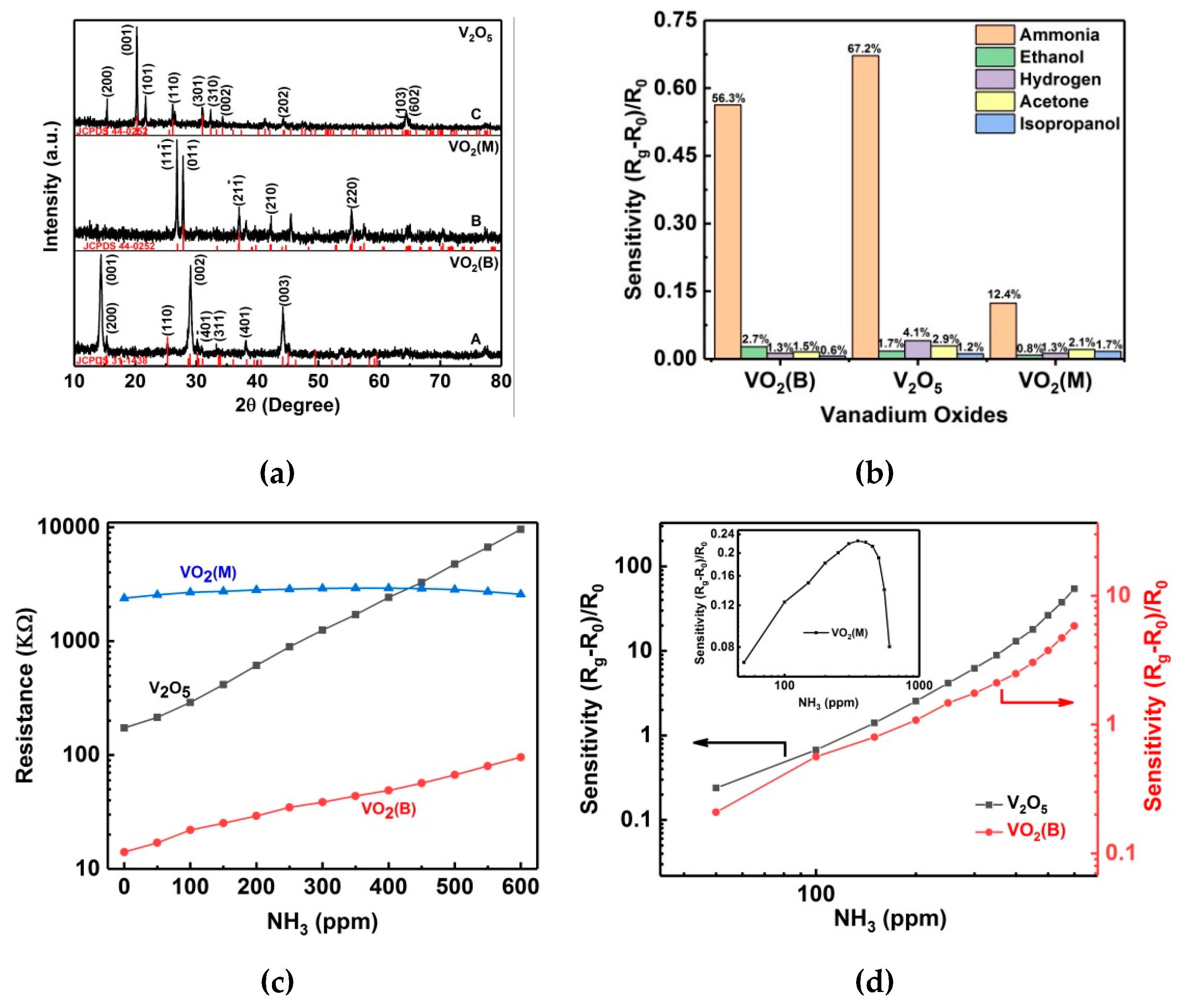
XRD patterns for the three specimens are shown in Figure 4a, where the patterns A, B and C correspond to the VO2(B), VO2(M), and V2O5 specimens, respectively. All peaks in pattern A are indexed to VO2(B) phase (space group: C2/m) with lattice constants of a = 12.03 Å, b = 3.693 Å, c = 6.42 Å, and β = 106.6° (JCPDS 31–1438); no any other phases or impurities were detected, revealing that the V5+ ions in V2O5 have been reduced to V4+ ions by the oxalic acid in the hydrothermal reaction and the products are mainly composed of VO2(B). After the calcination in either nitrogen or air, the B phase VO2 transformed to VO2(M) and V2O5, respectively. As shown in Figure 4a, all peaks in patterns B and C are indexed to VO2(M) phase (space group P21/c, JCPDS 44-0252) and orthorhombic V2O5 (space group Pmmn (59), JCPDS 41-1426), respectively, confirming the successful phase transform to VO2(M) and V2O5. No peaks of any other phases or impurities were detected in patterns B and C, revealing the high phase purity of VO2(M) and V2O5 products.
VO2(B), VO2(M), and V2O5 are the most famous compounds in the vanadium oxide family. In this work, their gas-sensing properties are investigated and compared. When sensors based on VO2(B), VO2(M), and V2O5 specimens are exposed in 100 ppm ammonia, ethanol, hydrogen, acetone, and isopropanol, they show different resistance response. As shown in Figure 4b, all vanadium oxide specimens exhibit higher response to ammonia, while no obvious response towards other target gases is observed. The resistance increment in 100 ppm ammonia are of 56.3%, 67.2%, and 12.4%, corresponding to VO2(B), V2O5, and VO2(M), respectively; thus, the three sensors show higher selectivity for ammonia than for other target gases. Figure 4c shows the resistance variation of VO2(B), VO2(M), and V2O5 sensors when they are exposed in 50–600 ppm ammonia at room temperature. Figure 4d and the inset show their corresponding sensitivity. It is noted that an approximated power-law dependence between the sensitivity and the NH3 concentration is observed for both V2O5 and VO2(B) nanoflakes, matching the sensor response law developed by Gurlo et al. [40]. Here, the sensitivity is defined as S = ( R g R 0 ) / R 0 , where R 0 and R g are the resistance of sensors in air and in ammonia, respectively. The resistance R 0 for sensors of VO2(B), VO2(M) and V2O5 are ( 1.41 ± 0.063 ) × 10 4 Ω, ( 2.38 ± 0.078 ) × 10 6 Ω, and ( 1.73 ± 0.059 ) × 10 5 Ω, respectively. The difference in resistance can be interpreted by their different intrinsic electrical properties. V2O5 is a semiconducting/insulating oxide with a band gap of 2.2 eV, and usually exhibits low conductivity due to the empty 3d orbital [41]. VO2(M) undergoes a near room-temperature MIT accompanied by a rapid change in resistivity, usually showing high resistivity at room temperature [41]. Although VO2(B) is generally regarded as an n-type semiconductor in gas sensing research [38], it is a theoretical semimetal/metal phase at room temperature, usually exhibiting high conductivity compared to VO2(M) and V2O5 [42]. As the ammonia concentration increases from 0 to 600 ppm, both the VO2(B) and V2O5 sensors show nonlinear increases of resistance. The sensor resistance of VO2(B) in 600 ppm ammonia is ( 9.58 ± 0.043 ) × 10 4 Ω, showing a ~5.80-fold increment, while the value for the V2O5 sensor is ( 9.58 ± 0.067 ) × 10 6 Ω, showing a ~54.4-fold increment. Obviously, the sensor sensitivity of V2O5 is superior, about one order of magnitude higher than the sensitivity of VO2(B). Unlike sensors of VO2(B) and V2O5, the resistance of the VO2(M) sensor first increases with the ammonia concentration, reaches a maximum value of ( 2.92 ± 0.033 ) × 10 6 Ω at 350 ppm, and then gradually decreases to ( 2.5 ± 0.028 ) × 10 6 Ω at 600 ppm. Therefore, the VO2(M) sensor whose resistance and sensitivity fluctuate in a narrow range (Figure 4c and the inset of Figure 4d) is not suitable for the ammonia detection.
To investigate the influence of the specific surface area on the ammonia sensing performance, we used the BET method of adsorption and desorption of nitrogen gas to measure the specific surface area of VO2(B) and V2O5 specimens. As shown in Figure 5, isotherms for both VO2(B) and V2O5 nanoflakes followed a typical IV-type curve with a clear hysteresis loop at p/p0 values of 0.55–0.99 for VO2(B) and 0.40–0.99 for V2O5. The BET specific surface area calculated from the nitrogen isotherms is 23.8 and 30.3 m2 g−1, corresponding to VO2(B) and V2O5 nanoflakes, respectively. Although V2O5 nanoflakes show a larger specific surface area, the two values are comparable, while the sensitivity of V2O5 in this work is about 10 times that of VO2(B). Moreover, V2O5 always showed higher sensing performance to ammonia than VO2(B) in the previous literature [38,43,44,45]. Therefore, one reasonable explanation is that the intrinsic properties of V2O5 determine its higher sensing performance. VO2(B) and V2O5 are generally regarded as n-type semiconductors in gas-sensing investigations [38]; however, VO2(B) is a theoretical semimetal/metal phase at room temperature [42]. Therefore, V2O5 should provide more absorbed oxygen ions on the surface, leading to high sensing performance.
The surface-depletion model is usually used to describe the sensing mechanism of the resistance-type metal-oxide semiconductor sensor. When the sensor is exposed to air, some oxygen molecules will be adsorbed on the surface, and then some oxygen ions, ( O ( ads ) ) , will be formed at the surface. When the sensor is put in the reducing gas, ammonia in this work, the reducing gas molecules will react with the oxygen ions on the surface as described below:
2 NH 3 + 3 ( O ( ads ) ) N 2 + 3 H 2 O + 3 e .
Thus the carrier concentration changes with the ammonia level and, consequently, causes the sensor resistance to change. It is well known that semiconductor metal oxides are usually classified as either n-type or p-type. Conductivity type plays an important role in sensor responses. For n-type semiconductor sensors, the reductive gas species reacts with the adsorbed oxygen ions and the electrons trapped by oxygen are released into the conduction band, leading to a decrease in resistance. With regard to p-type semiconductor sensors, the opposite change in the resistance is observed due to the combination of holes with electrons released from the surface reaction. Both VO2(B) and V2O5 are generally considered to be n-type semiconductors in gas-sensing investigations, and there are many studies reporting their n-type gas-sensing responses. In studies by Raj et al. [31] and Modafferi et al. [34], sensors based on V2O5 displayed a resistance decrease when they were exposed to an ascending ammonia level and the material was treated as n-type. However, our measured results indicate that the sensor resistance of VO2(B) and V2O5 displays a unidirectional increase as the ammonia level increases from 0 to 600 ppm, showing typical p-type resistance responses. Similar intriguing p-type gas-sensing behaviors have been reported previously. Qin et al. [29] and Evans et al. [38] reported p-type resistance responses in V2O5 hierarchical networks and VO2(B) nanoparticles, and attributed them to the inversion layer formed at the surface due to a larger quantity of surface oxygen vacancy [29]. These contrasting results suggest that the sensing response type of vanadium oxides at room temperature is highly dependent on the synthesis environment and the resultant surface species.
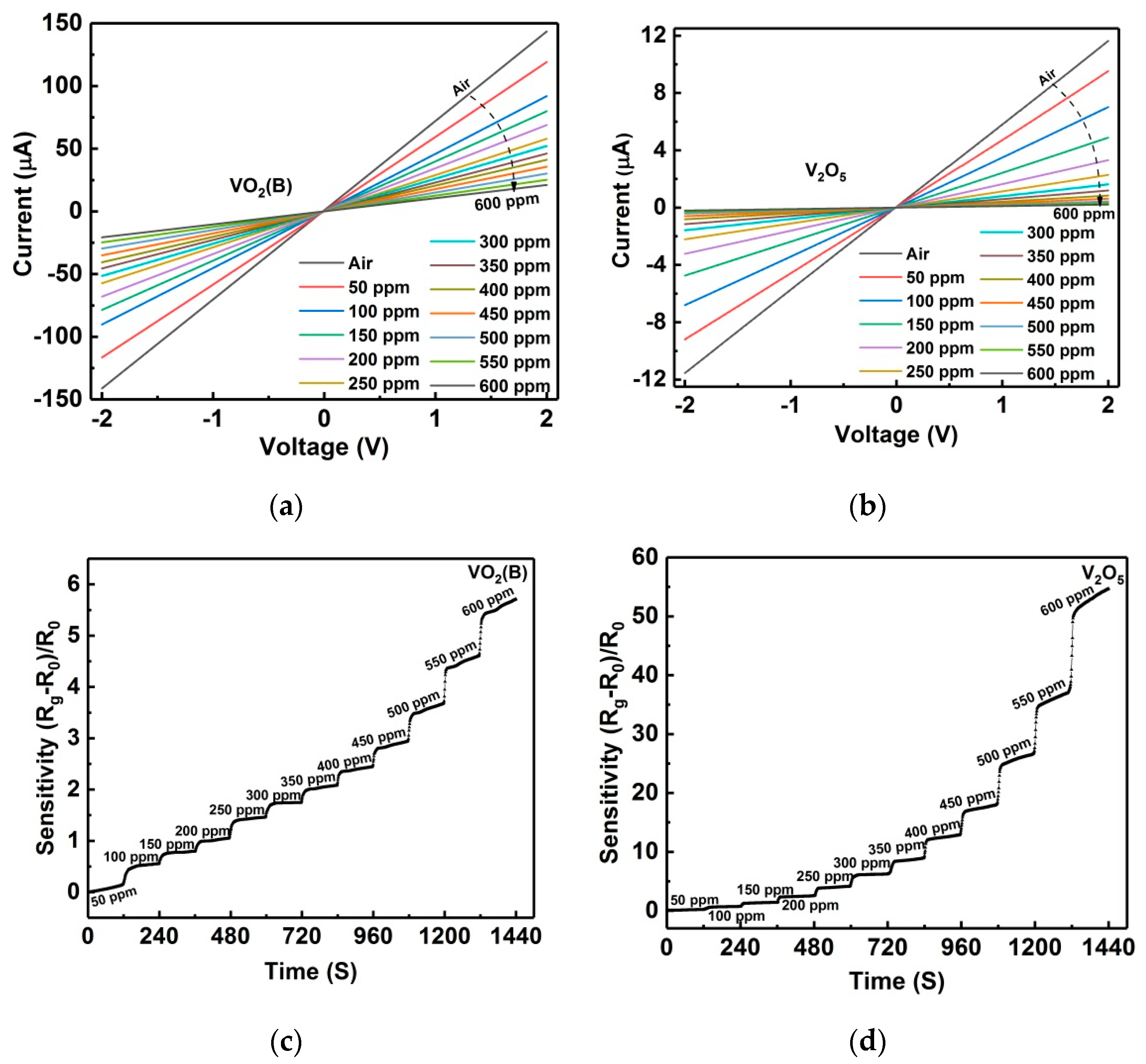
I‒V curves for sensors of VO2(B) and V2O5 in 0–600 ppm ammonia are measured at room temperature and shown in Figure 6a,b. All of the curves appear to be linear, indicating that good ohmic contacts formed between the metal electrodes and nanoflakes. From the I‒V curves, it is seen that the conduction of two devices progressively decays with the ascending ammonia level, showing p-type sensing responses and agreeing with the resistance measurement (Figure 4c). Figure 6c,d present the dynamic responses of VO2(B) and V2O5 sensors in the ascending ammonia levels. When the ammonia was continuously injected into the testing chamber with a step of 50 ppm, both sensors showed nonlinear increases in their resistances. From Figure 6c,d, it is obvious that in each ammonia level of both sensors the resistance ladder is very clear, revealing good dynamic response characteristics. Moreover, the V2O5 sensor shows a one order of magnitude higher sensitivity than the VO2(B) sensor. In both sensors, NH3 reacts with the surface adsorbed oxygen ions, releasing electrons and inducing an electrical response to the ascending ammonia level. However, VO2 and V2O5 are normally considered n-type semiconductors at room temperature, so the formation of a surface inversion layer is a reliable interpretation for the p-type behaviors [29,38], where the released electrons would reduce the number of holes (the majority charge carriers in the inversion layer) and result in a p-type increase of the sensor resistivity.
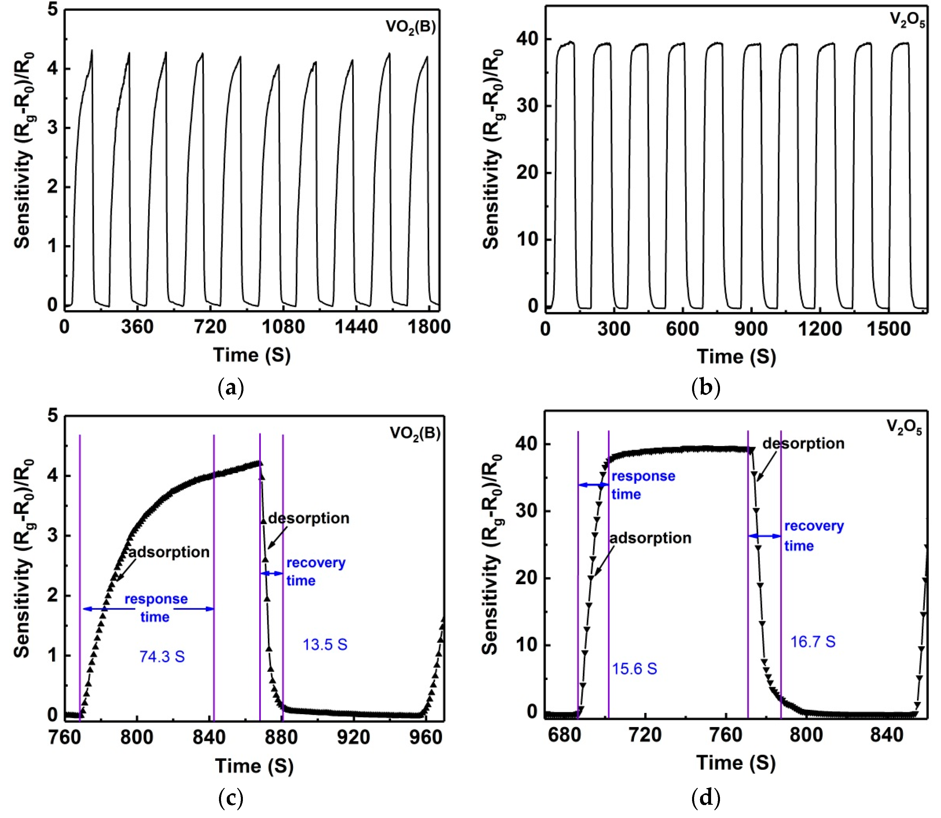
Further dynamic testing procedures were carried out, which provided more information on the most important parameters for a sensing device: sensitivity, response and recovery time, and reproducibility. Figure 7a,b show the dynamic responses of VO2(B) and V2O5 sensors for 10 cycles where the dynamic mesurement was performed between air and 550 ppm ammonia at room temperature. For clear observation, only 10 cycles are given. Similarly, both VO2(B) and V2O5 sensors indicate good reproducibility, as revealed in the repeated measurements (Figure 7a,b). When sensors of VO2(B) and V2O5 were exposed to 550 ppm ammonia, the ammonia adsorption was triggered, and simultaneously the sensor resistance increased abruptly and then reached a relative stable value. When the sensors were switched to air again, ammonia desorption happened, and the sensor resistance decreased abruptly and then reached a relative stable value. The response time and recovery time for the VO2(B) sensor (defined as 95% of the time between the peak value and the valley value) were 63–85 s and 11–16 s (Figure 7c), respectively. Comparatively, the V2O5 sensor indicated a faster response: the response time was 14–22 s and the recovery time was 14–20 s (Figure 7d). Compared with other ammonia sensors of VO2(B) and V2O5 reported previously, the sensors in this work showed a fast response/recovery rate (Table 2). As shown in Figure 7c, it is clear that the resistance of the VO2(B) sensor fluctuated significantly during the dynamic test and the response time fluctuated in a wide range of 63–85 s, while the V2O5 sensor showed a slight fluctuation in the resistance and a narrow range in the response time (14–22 s). Clearly, the V2O5 sensor showed superior reproducibility compared to the VO2(B) sensor. In addition, a mass of repeated measurements revealed the response sensitivity of both sensors decreasing after long-term work, and if the sensor was kept in air for about two hours, the response sensitivity would still recover to the original value. This phenomenon can be perfectly interpreted by interactions of surface hydroxyl groups with ammonia. As revealed in previous studies, the humidity in air can improve the ammonia adsorption and resultantly increase the sensor response [46,47]. A long-term work could decrease the site activities of adsorbed hydroxyl groups on the surface, leading to the sensitivity decreasing. After a few hours in air, the ammonia retained on hydroxyl groups should be removed during air purging, thus causing sensitivity recovery.

4. Conclusions

In summary, self-assembled VO2(B) nanoflakes were synthesized via a simple hydrothermal method, and VO2(M) and V2O5 nanoflakes were obtained through a high-temperature phase transition in nitrogen and air, respectively. Sensors based on the three famous vanadium oxide compounds were fabricated and their gas-sensing characteristics were comparatively investigated at room temperature. It was found that VO2(M) nanoflakes were gas-insensitive, while both VO2(B) and V2O5 nanoflakes were highly selective to ammonia. As an ammonia sensor, V2O5 nanoflakes showed higher sensitivity, faster response, and better reproducibility to ammonia than VO2(B) nanoflakes. The sensitivity was about one order of magnitude higher than that of VO2(B) nanoflakes, and the response time and recovery time were 14–22 s and 14–20 s, respectively. Interestingly, although vanadium oxides are generally regarded as n-type semiconductors, both VO2(B) and V2O5 nanoflakes showed p-type sensing responses to ammonia, which can be attributed to the surface inversion layer formation.

Author Contributions

H.Y. and Z.W. planned and supervised the study. H.Y. and C.S. performed the experiments and wrote this manuscript. All authors analyzed the data, discussed the results, and commented on the manuscript.

Funding

This work was funded by the National Natural Science Foundation of China under Grant Nos. 61574055, 61704093, and 61874168, the ‘The Six Top Talents’ of Jiangsu Province (Grant No. 2016-XCL-052), the Qing Lan Project of Jiangsu Province, and the Key NSF Program of Jiangsu Provincial Department of Education (Grant No. 15KJA510004).

Conflicts of Interest

The authors declare no conflict of interest.

References

  1. Wang, Y.; Zhou, Y.; Meng, C.; Gao, Z.; Cao, X.; Li, X.; Xu, L.; Zhu, W.; Peng, X.; Zhang, B.; et al. A high-response ethanol gas sensor based on one-dimensional TiO2/V2O5 branched nanoheterostructures. Nanotechnology 2016, 27, 425503. [Google Scholar] [CrossRef] [PubMed]
  2. Ju, D.-X.; Xu, H.-Y.; Qiu, Z.-W.; Zhang, Z.-C.; Xu, Q.; Zhang, J.; Wang, J.-Q.; Cao, B.-Q. Near Room Temperature, Fast-Response, and Highly Sensitive Triethylamine Sensor Assembled with Au-Loaded ZnO/SnO2 Core–Shell Nanorods on Flat Alumina Substrates. ACS Appl. Mater. Interfaces 2015, 7, 19163–19171. [Google Scholar] [CrossRef] [PubMed]
  3. Gurlo, A. Nanosensors: towards morphological control of gas sensing activity. SnO2, In2O3, ZnO and WO3 case studies. Nanoscale 2011, 3, 154–165. [Google Scholar] [CrossRef] [PubMed]
  4. Sakai, Y.; Kadosaki, M.; Matsubara, I.; Itoh, T. Preparation of total VOC sensor with sensor-response stability for humidity by noble metal addition to SnO2. J. Ceram. Soc. Jpn. 2009, 117, 1297–1301. [Google Scholar] [CrossRef]
  5. Wang, B.; Deng, L.; Sun, L.; Lei, Y.; Wu, N.; Wang, Y. Growth of TiO2 nanostructures exposed {001} and {110} facets on SiC ultrafine fibers for enhanced gas sensing performance. Sens. Actuat. B: Chem. 2018, 276, 57–64. [Google Scholar] [CrossRef]
  6. Seekaew, Y.; Wisitsoraat, A.; Phokharatkul, D.; Wongchoosuk, C. Room temperature toluene gas sensor based on TiO2 nanoparticles decorated 3D graphene-carbon nanotube nanostructures. Sens. Actuator B Chem. 2019, 279, 69–78. [Google Scholar] [CrossRef]
  7. Ganbavle, V.V.; Inamdar, S.I.; Agawane, G.L.; Kim, J.H.; Rajpure, K.Y. Synthesis of fast response, highly sensitive and selective Ni:ZnO based NO2 sensor. Chem. Eng. J. 2016, 286, 36–47. [Google Scholar] [CrossRef]
  8. Zhang, N.; Yu, K.; Li, Q.; Zhu, Z.Q.; Wan, Q. Room-temperature high-sensitivity H2S gas sensor based on dendritic ZnO nanostructures with macroscale in appearance. J. Appl. Phys. 2008, 103, 104305. [Google Scholar] [CrossRef]
  9. Mhamdi, A.; Labidi, A.; Souissi, B.; Kahlaoui, M.; Yumak, A.; Boubaker, K.; Amlouk, A.; Amlouk, M. Impedance spectroscopy and sensors under ethanol vapors application of sprayed vanadium-doped ZnO compounds. J. Alloy. Compd. 2015, 639, 648–658. [Google Scholar] [CrossRef]
  10. Zhang, H.; Wang, Y.; Zhu, X.; Li, Y.; Cai, W. Bilayer Au nanoparticle-decorated WO3 porous thin films: On-chip fabrication and enhanced NO2 gas sensing performances with high selectivity. Sens. Actuator B Chem. 2019, 280, 192–200. [Google Scholar] [CrossRef]
  11. Mehta, S.S.; Nadargi, D.Y.; Tamboli, M.S.; Chaudhary, L.S.; Patil, P.S.; Mulla, I.S.; Suryavanshi, S.S. Ru-Loaded mesoporous WO3 microflowers for dual applications: Enhanced H2S sensing and sunlight-driven photocatalysis. Dalton Trans. 2018, 47, 16840–16845. [Google Scholar] [CrossRef] [PubMed]
  12. Yan, S.; Li, Z.; Li, H.; Wu, Z.; Wang, J.; Shen, W.; Fu, Y.Q. Ultra-sensitive room-temperature H2S sensor using Ag-In2O3 nanorod composites. J. Mater. Sci. 2018, 53, 16331–16344. [Google Scholar] [CrossRef]
  13. Wei, D.; Jiang, W.; Gao, H.; Chuai, X.; Liu, F.; Liu, F.; Sun, P.; Liang, X.; Gao, Y.; Yan, X.; Lu, G. Facile synthesis of La-doped In2O3 hollow microspheres and enhanced hydrogen sulfide sensing characteristics. Sens. Actuator B Chem. 2018, 276, 413–420. [Google Scholar] [CrossRef]
  14. Chava, R.K.; Cho, H.-Y.; Yoon, J.-M.; Yu, Y.-T. Fabrication of aggregated In2O3 nanospheres for highly sensitive acetaldehyde gas sensors. J. Alloy. Compd. 2019, 772, 834–842. [Google Scholar] [CrossRef]
  15. Gebicki, J. Application of electrochemical sensors and sensor matrixes for measurement of odorous chemical compounds. TrAC Trends Anal. Chem. 2016, 77, 1–13. [Google Scholar] [CrossRef]
  16. Spinelle, L.; Gerboles, M.; Kok, G.; Persijn, S.; Sauerwald, T. Review of Portable and Low-Cost Sensors for the Ambient Air Monitoring of Benzene and Other Volatile Organic Compounds. Sensors 2017, 17, 1520. [Google Scholar] [CrossRef] [PubMed]
  17. Arshak, K.; Moore, E.; Lyons, G.M.; Harris, J.; Clifford, S. A review of gas sensors employed in electronic nose applications. Sens. Rev. 2004, 24, 181–198. [Google Scholar] [CrossRef]
  18. Röck, F.; Barsan, N.; Weimar, U. Electronic Nose:  Current Status and Future Trends. Chem. Rev. 2008, 108, 705–725. [Google Scholar] [CrossRef] [PubMed]
  19. Szulczyński, B.; Gębicki, J. Currently Commercially Available Chemical Sensors Employed for Detection of Volatile Organic Compounds in Outdoor and Indoor Air. Environments 2017, 4, 21. [Google Scholar] [CrossRef]
  20. Ureña-Begara, F.; Crunteanu, A.; Raskin, J.-P. Raman and XPS characterization of vanadium oxide thin films with temperature. Appl. Surf. Sci. 2017, 403, 717–727. [Google Scholar] [CrossRef] [Green Version]
  21. Yin, H.; Yu, K.; Peng, H.; Zhang, Z.; Huang, R.; Travas-Sejdic, J.; Zhu, Z. Porous V2O5 micro/nano-tubes: Synthesis via a CVD route, single-tube-based humidity sensor and improved Li-ion storage properties. J. Mater. Chem. 2012, 22, 5013–5019. [Google Scholar] [CrossRef]
  22. Lamsal, C.; Ravindra, N.M. Optical properties of vanadium oxides-an analysis. J. Mater. Sci. 2013, 48, 6341–6351. [Google Scholar] [CrossRef]
  23. Sathiya, M.; Prakash, A.S.; Ramesha, K.; Tarascon, J.M.; Shukla, A.K. V2O5-Anchored Carbon Nanotubes for Enhanced Electrochemical Energy Storage. J. Am. Chem. Soc. 2011, 133, 16291–16299. [Google Scholar] [CrossRef] [PubMed]
  24. Nethravathi, C.; Rajamathi, C.R.; Rajamathi, M.; Gautam, U.K.; Wang, X.; Golberg, D.; Bando, Y. N-Doped Graphene–VO2(B) Nanosheet-Built 3D Flower Hybrid for Lithium Ion Battery. ACS Appl. Mater. Interfaces 2013, 5, 2708–2714. [Google Scholar] [CrossRef] [PubMed]
  25. Yao, T.; Liu, L.; Xiao, C.; Zhang, X.; Liu, Q.; Wei, S.; Xie, Y. Ultrathin Nanosheets of Half-Metallic Monoclinic Vanadium Dioxide with a Thermally Induced Phase Transition. Angew. Chem. Int. Ed. 2013, 52, 7554–7558. [Google Scholar] [CrossRef] [PubMed]
  26. Li, Z.; Hu, Z.; Peng, J.; Wu, C.; Yang, Y.; Feng, F.; Gao, P.; Yang, J.; Xie, Y. Ultrahigh Infrared Photoresponse from Core–Shell Single-Domain-VO2/V2O5 Heterostructure in Nanobeam. Adv. Funct. Mater. 2014, 24, 1821–1830. [Google Scholar] [CrossRef]
  27. Mane, A.A.; Moholkar, A.V. Effect of film thickness on NO2 gas sensing properties of sprayed orthorhombic nanocrystalline V2O5 thin films. Appl. Surf. Sci. 2017, 416, 511–520. [Google Scholar] [CrossRef]
  28. Yan, W.; Hu, M.; Wang, D.; Li, C. Room temperature gas sensing properties of porous silicon/V2O5 nanorods composite. Appl. Surf. Sci. 2015, 346, 216–222. [Google Scholar] [CrossRef]
  29. Qin, Y.; Fan, G.; Liu, K.; Hu, M. Vanadium pentoxide hierarchical structure networks for high performance ethanol gas sensor with dual working temperature characteristic. Sens. Actuator B Chem. 2014, 190, 141–148. [Google Scholar] [CrossRef]
  30. Wang, R.; Yang, S.; Deng, R.; Chen, W.; Liu, Y.; Zhang, H.; Zakharova, G.S. Enhanced gas sensing properties of V2O5 nanowires decorated with SnO2 nanoparticles to ethanol at room temperature. RSC Adv. 2015, 5, 41050–41058. [Google Scholar] [CrossRef]
  31. Dhayal Raj, A.; Pazhanivel, T.; Suresh Kumar, P.; Mangalaraj, D.; Nataraj, D.; Ponpandian, N. Self assembled V2O5 nanorods for gas sensors. Curr. Appl. Phys. 2010, 10, 531–537. [Google Scholar] [CrossRef]
  32. Yang, X.H.; Xie, H.; Fu, H.T.; An, X.Z.; Jiang, X.C.; Yu, A.B. Synthesis of hierarchical nanosheet-assembled V2O5 microflowers with high sensing properties towards amines. RSC Adv. 2016, 6, 87649–87655. [Google Scholar] [CrossRef]
  33. Huotari, J.; Bjorklund, R.; Lappalainen, J.; Lloyd Spetz, A. Pulsed Laser Deposited Nanostructured Vanadium Oxide Thin Films Characterized as Ammonia Sensors. Sens. Actuator B Chem. 2015, 217, 22–29. [Google Scholar] [CrossRef]
  34. Modafferi, V.; Trocino, S.; Donato, A.; Panzera, G.; Neri, G. Electrospun V2O5 composite fibers: Synthesis, characterization and ammonia sensing properties. Thin Solid Films 2013, 548, 689–694. [Google Scholar] [CrossRef]
  35. Modafferi, V.; Panzera, G.; Donato, A.; Antonucci, P.L.; Cannilla, C.; Donato, N.; Spadaro, D.; Neri, G. Highly sensitive ammonia resistive sensor based on electrospun V2O5 fibers. Sens. Actuator B Chem. 2012, 163, 61–68. [Google Scholar] [CrossRef]
  36. Hakim, S.A.; Liu, Y.; Zakharova, G.S.; Chen, W. Synthesis of vanadium pentoxide nanoneedles by physical vapour deposition and their highly sensitive behavior towards acetone at room temperature. RSC Adv. 2015, 5, 23489–23497. [Google Scholar] [CrossRef]
  37. Yu, M.; Liu, X.; Wang, Y.; Zheng, Y.; Zhang, J.; Li, M.; Lan, W.; Su, Q. Gas sensing properties of p-type semiconducting vanadium oxide nanotubes. Appl. Surf. Sci. 2012, 258, 9554–9558. [Google Scholar] [CrossRef]
  38. Evans, G.P.; Powell, M.J.; Johnson, I.D.; Howard, D.P.; Bauer, D.; Darr, J.A.; Parkin, I.P. Room temperature vanadium dioxide–carbon nanotube gas sensors made via continuous hydrothermal flow synthesis. Sens. Actuator B Chem. 2018, 255, 1119–1129. [Google Scholar] [CrossRef]
  39. Yin, H.; Yu, K.; Zhang, Z.; Zhu, Z. Morphology-control of VO2(B) nanostructures in hydrothermal synthesis and their field emission properties. Appl. Surf. Sci. 2011, 257, 8840–8845. [Google Scholar] [CrossRef]
  40. Gurlo, A.; Bârsan, N.; Ivanovskaya, M.; Weimar, U.; Göpel, W. In2O3 and MoO3–In2O3 thin film semiconductor sensors: interaction with NO2 and O3. Sens. Actuator B Chem. 1998, 47, 92–99. [Google Scholar] [CrossRef]
  41. Lu, Q.; Bishop, S.R.; Lee, D.; Lee, S.; Bluhm, H.; Tuller, H.L.; Lee, H.N.; Yildiz, B. Electrochemically Triggered Metal–Insulator Transition between VO2 and V2O5. Adv. Funct. Mater. 2018, 28, 1803024. [Google Scholar] [CrossRef]
  42. Srivastava, A.; Rotella, H.; Saha, S.; Pal, B.; Kalon, G.; Mathew, S.; Motapothula, M.; Dykas, M.; Yang, P.; Okunishi, E.; Sarma, D.D.; Venkatesan, T. Selective growth of single phase VO2(A, B, and M) polymorph thin films. APL Mater. 2015, 3, 026101. [Google Scholar] [CrossRef]
  43. Amos Adeleke, A.; Augusto Goncalo Jose, M.; Bathusile, M.; George, C.; Boitumelo, M.; Kittessa, R.; Mart-Mari, D.; Hendrik, S.; Jayita, B.; Suprakas Sinha, R.; et al. Blue- and red-shifts of V2O5 phonons in NH3 environment by in situ Raman spectroscopy. J. Phys. D Appl. Phys. 2018, 51, 015106. [Google Scholar]
  44. Liu, J.; Wang, X.; Peng, Q.; Li, Y. Preparation and gas sensing properties of vanadium oxide nanobelts coated with semiconductor oxides. Sens. Actuator B Chem. 2006, 115, 481–487. [Google Scholar] [CrossRef]
  45. Fu, H.; Yang, X.; An, X.; Fan, W.; Jiang, X.; Yu, A. Experimental and theoretical studies of V2O5@TiO2 core-shell hybrid composites with high gas sensing performance towards ammonia. Sens. Actuator B Chem. 2017, 252, 103–115. [Google Scholar] [CrossRef]
  46. Bannov, A.G.; Prášek, J.; Jašek, O.; Shibaev, A.A.; Zajíčková, L. Investigation of Ammonia Gas Sensing Properties of Graphite Oxide. Procedia Eng. 2016, 168, 231–234. [Google Scholar] [CrossRef]
  47. Urasinska-Wojcik, B.; Gardner, J.W. H2S Sensing in Dry and Humid H2 Environment With p-Type CuO Thick-Film Gas Sensors. IEEE Sens. J. 2018, 18, 3502–3508. [Google Scholar] [CrossRef]
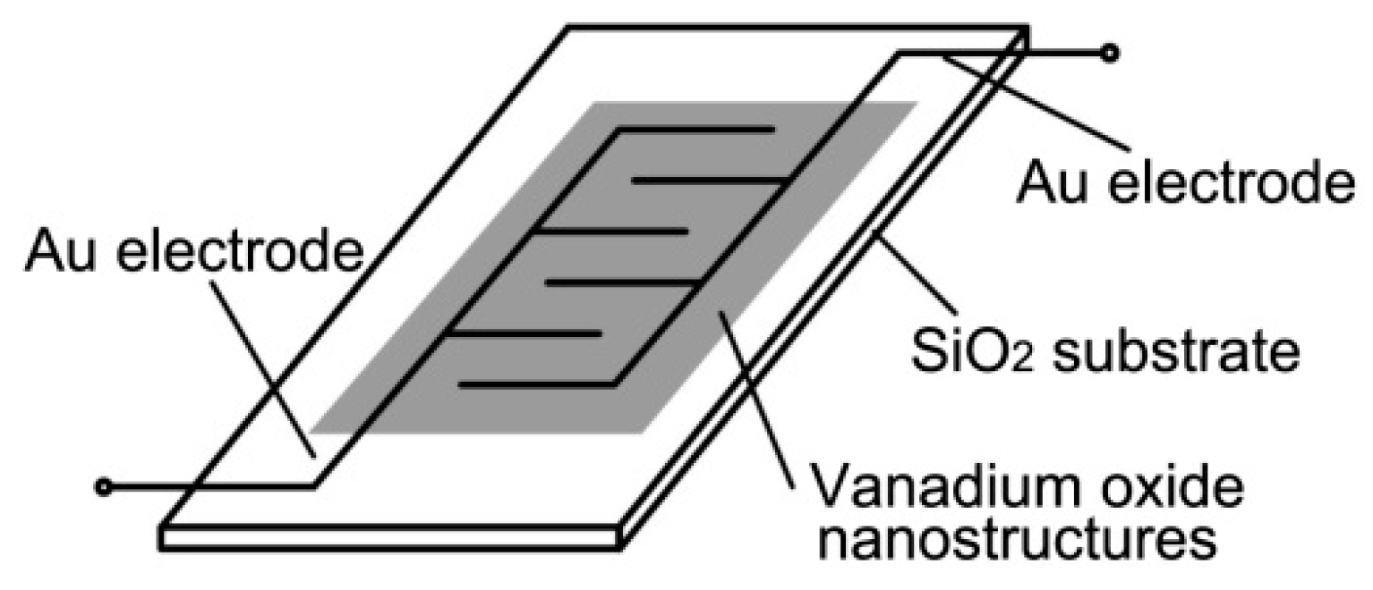
Figure 1. A schematic of the sensor substrate, the patterned gold electrodes and the sensing layer.
Nanomaterials 09 00317 g001
Figure 2. (a) Low-, (b) medium-, and (c) high-magnification SEM images of self-assembled VO2(B) nanoflakes synthesized by hydrothermal method. (d) TEM image of a single nanoflake. Upper inset: the high-resolution (HR) TEM image for the red square region. Lower inset: the corresponding fast Fourier transform (FFT) pattern.
Nanomaterials 09 00317 g002
Figure 3. (a) Low- and, (b) high-magnification SEM images of V2O5 specimen obtained after annealing VO2(B) nanoflakes in air. (c) TEM images of V2O5 nanoflakes. (d) Low- and, (e) high-magnification SEM images of VO2(M) specimen obtained after annealing VO2(B) nanoflakes in nitrogen atmosphere. (f) TEM images of VO2(M) nanoflakes. Upper inset in plane (c) and (f): the high-resolution TEM image for the red square region. Lower inset in plane (c) and (f): the corresponding fast Fourier transform (FFT) pattern.
Nanomaterials 09 00317 g003
Figure 4. (a) XRD patterns of self-assembled VO2(B), VO2(M) and V2O5 specimens. (b) Selectivity of sensors based on VO2(B), V2O5 and VO2(M) nanoflakes for 100 ppm ammonia, ethanol, hydrogen, acetone and isopropanol at room temperature. (c) Sensor resistance variation with 50–600 ppm ammonia gas for VO2(B), VO2(M) and V2O5 self-assembled nanoflakes. (d) The sensitivity of sensors based on VO2(B) and V2O5 self-assembled nanoflakes at room temperature. R0 and Rg are the resistances in the air and the measured ammonia level respectively. The inset in plane (d) is the sensitivity for the sensor of VO2(M) specimen.
Nanomaterials 09 00317 g004
Figure 5. N2 adsorption–desorption isotherms of self-assembled VO2(B) and V2O5 nanoflakes.
Nanomaterials 09 00317 g005
Figure 6. Room temperature I-V curves for sensors of (a) VO2(B), and (b) V2O5 nanoflakes measured in different static ammonia atmosphere from 0 to 600 ppm. Sensor responses of (c) VO2(B) and (d) V2O5 nanoflakes to ascending ammonia levels step-by-step.
Nanomaterials 09 00317 g006
Figure 7. Dynamic responses for sensors of (a) VO2(B), and (b) V2O5 nanoflakes. Dynamic switches are performed between air and 550 ppm ammonia. (c,d) High-magnification dynamic responses for one cycle, corresponding to VO2(B)- and V2O5-based sensors.
Nanomaterials 09 00317 g007
Table 1. Crystal structure, properties, and important applications of VO2(B), VO2(M), and V2O5.
Properties of Vanadium OxidesVO2(B)VO2(M)V2O5
Crystal StructureMonoclinic (C2/m)
a = 12.03, b = 3.693, c = 6.42, β = 106.6°
Monoclinic (P21/c)
a = 5.753, b = 4.526, c = 5.383, β = 122.6°
Orthorhombic (Pmmn)
a = 11.516, b = 3.566, c = 4.373
Structural/Physical/Chemical CharacteristicsLayer structureRapid reversible MIT (340 K) Drastic change in resistivity and optical transparency between MITLayer structure Oxidizing Enhanced surface reactivity
ApplicationsEnergy storage materials SensorsChromogenic materials High-speed electronicsChromogenic materials Catalysts Energy storage materials Sensors
Table 2. Comparison of various resistive sensor responses, response and recovery times to ammonia for VO2(B) and V2O5 sensing systems.
Vanadium OxidesResistive ResponseResponse TimeRecovery Time
VO2(B) nanoflakes *0.21 (50 ppm) 5.82 (600 ppm)63–85 s (550 ppm)11–16 s (550 ppm)
V2O5 nanoflakes *0.24 (50 ppm) 54.4 (600 ppm)14–22 s (550 ppm)14–20 s (550 ppm)
VO2(B) nanoparticles [38]0.1 (45 ppm)310 s40 min
V2O5 films [43]0.419 (40 ppm)59 s
V2O5 nanobelts [44]0.7 (100 ppm)32 s30 s
V2O5 fibers [35]50 s (0.85–45 ppm)350 s (0.85–45 ppm)
V2O5 nanoparticles [45]~2.0 (200 ppm)23 s13 s
* Indicates vanadium oxide sensor in this work.