3.1. Manufactured Smart Shirt
A manufactured smart shirt is shown in
Figure 3. The smart shirt in the figure is presented inside out to show the electrodes.
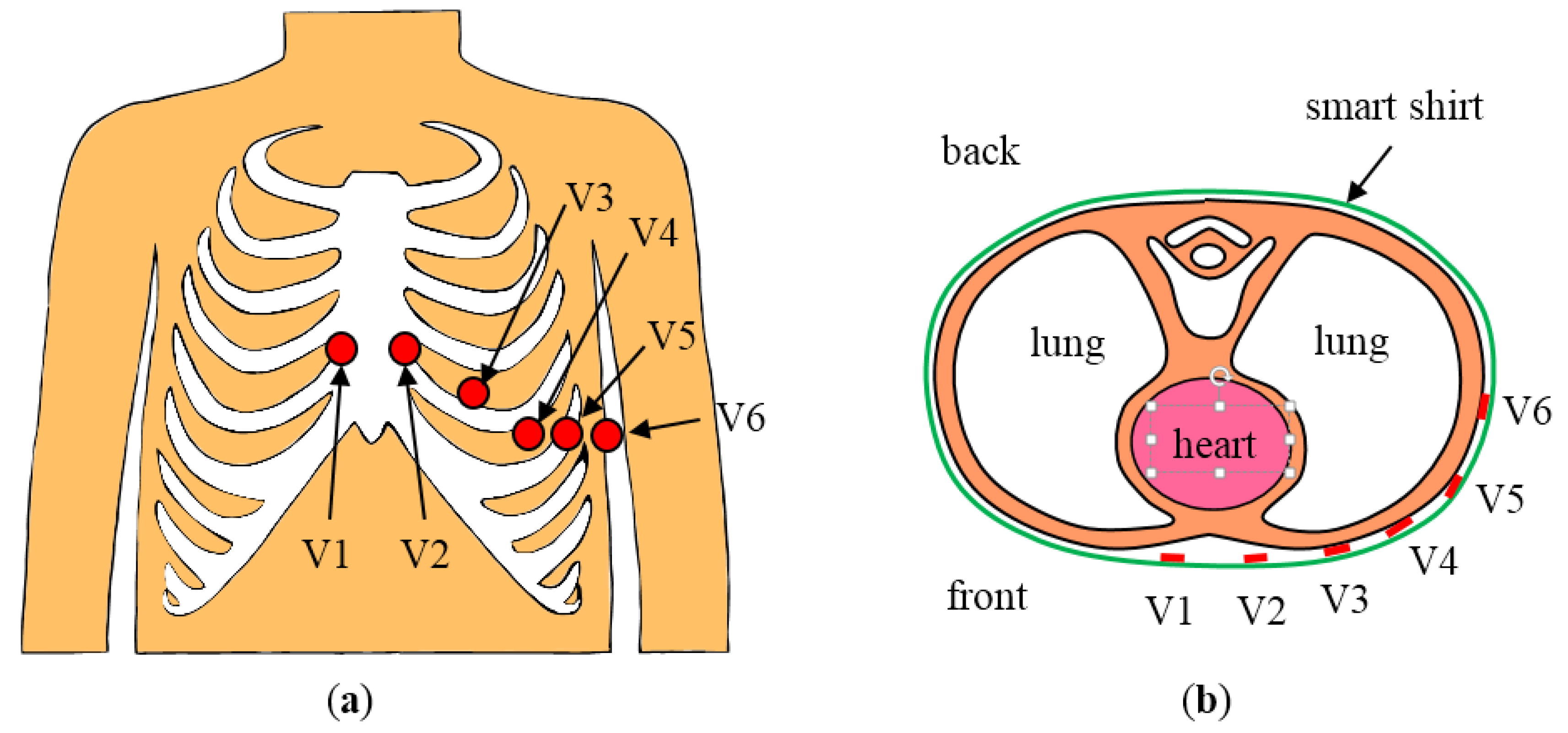
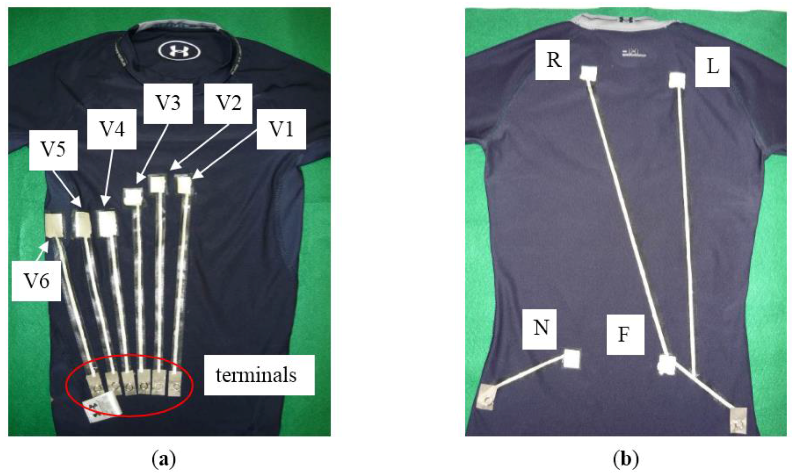
Figure 3a shows the front of the smart shirt. The dimensions of electrodes V1 and V2 were 20 mm × 20 mm, while the dimensions of the other electrodes were 20 mm × 30 mm. Positions of electrodes V1 to V6 corresponded to active electrodes with unipolar precordial leads. In the standard measurement using unipolar precordial leads, indifferent and neutral electrodes are placed on both wrists and both ankles. In this study, however, these electrodes were printed on the back of the smart shirt shown in
Figure 3b. The indifferent electrodes R, L, and F were printed on the scapulae and left lumbar part on the smart shirt. These electrodes were connected to one wire because the wiring was defined by the unipolar precordial lead configuration. The neutral electrode N was printed on the right lumbar part of the smart shirt. The indifferent and neutral electrodes were placed on the convex area of the scapulae and lumbar. These electrodes were therefore in stable contact with the subject’s body. The longest length of wire was 480 mm for electrode R. The electrical resistance of this wire was 7.0 Ω before the smart shirt was worn. The resistance increased to 17.1 Ω when the subject wore the smart shirt, but the resistance was low enough for measurement of the ECG signal.
Figure 3.
Manufactured smart shirt. The smart shirt is inside out in the photographs to show electrodes and wires: (a) front view; (b) back view.
Figure 3.
Manufactured smart shirt. The smart shirt is inside out in the photographs to show electrodes and wires: (a) front view; (b) back view.
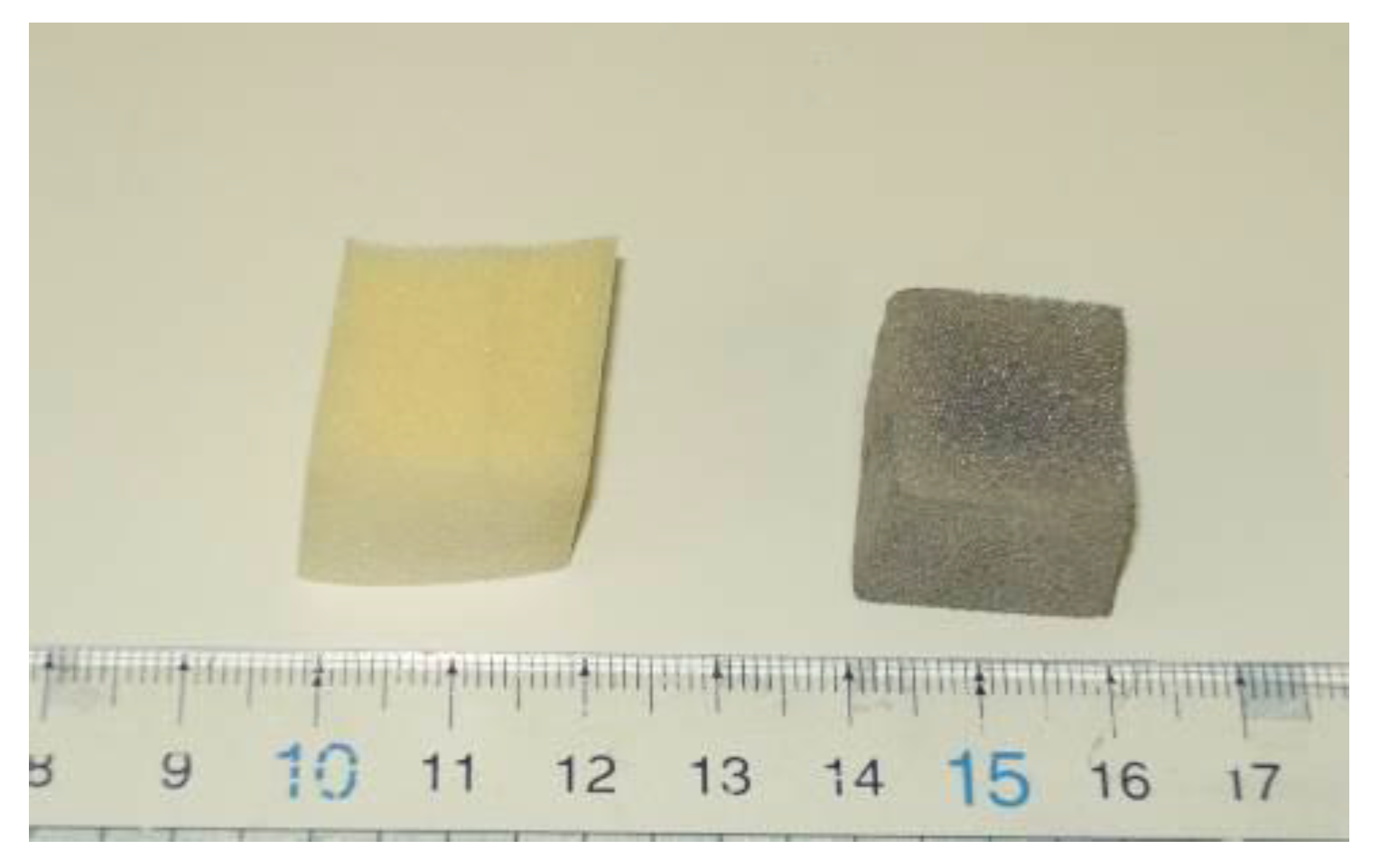
3.2. Manufactured Conductive Foam Block
A block of low-rebound urethane foam in reaction liquid was irradiated with ultrasound for 21 h. After irradiation, the urethane foam block was washed with ethanol and dried at 60 °C for 60 min.
Figure 4 shows the urethane foam block before and after irradiation. The irradiated urethane foam block changed color to dark gray because of the nanostructured Ag and became electrically conductive. A preliminary study of the nanostructured Ag showed that the nanostructured Ag was strongly attached to the urethane foam and did not peel off easily.
Figure 4.
(Left) Low-rebound urethane foam block before irradiation with ultrasound; (Right) Conductive foam block produced by irradiating the urethane foam block with ultrasound in the reaction liquid.
Figure 4.
(Left) Low-rebound urethane foam block before irradiation with ultrasound; (Right) Conductive foam block produced by irradiating the urethane foam block with ultrasound in the reaction liquid.
The initial electrical resistance of the conductive foam block was different for each pair of opposing faces and ranged from 22.2 to 460.0 Ω. In the measurement of electrical resistance, the block was not compressed by probes of the resistance meter. However, the block received pressure from the smart shirt and the subject’s body when the block was attached to the smart shirt. We investigated the resistance of the block when the block was pressed.
Figure 5 shows relationships between the compression pressure and resistance. The resistance was measured for pairs 1, 2, and 3 of opposing faces. The figure indicates that the resistance was different for each pair of faces but was reduced by pressure in each case. The difference in resistance for each pair of opposing faces was due to the unbalanced ultrasound irradiation; the ultrasound was transmitted in one direction, and the block in the reaction liquid was not rotated. However, the resistance at pressure of 2.0 kPa was less than 30 Ω even though the initial resistance was 460.0 Ω (pair 1); this was sufficient for the measurement of the ECG signal. Moreover, we measured the resistance of the other blocks manufactured by employing the same method. The resistance and characteristics of those blocks were similar to those mentioned above.
Figure 5.
Relationships between compression pressure and electrical resistance. The schematic on the right labels pairs of opposing faces in the measurement.
Figure 5.
Relationships between compression pressure and electrical resistance. The schematic on the right labels pairs of opposing faces in the measurement.
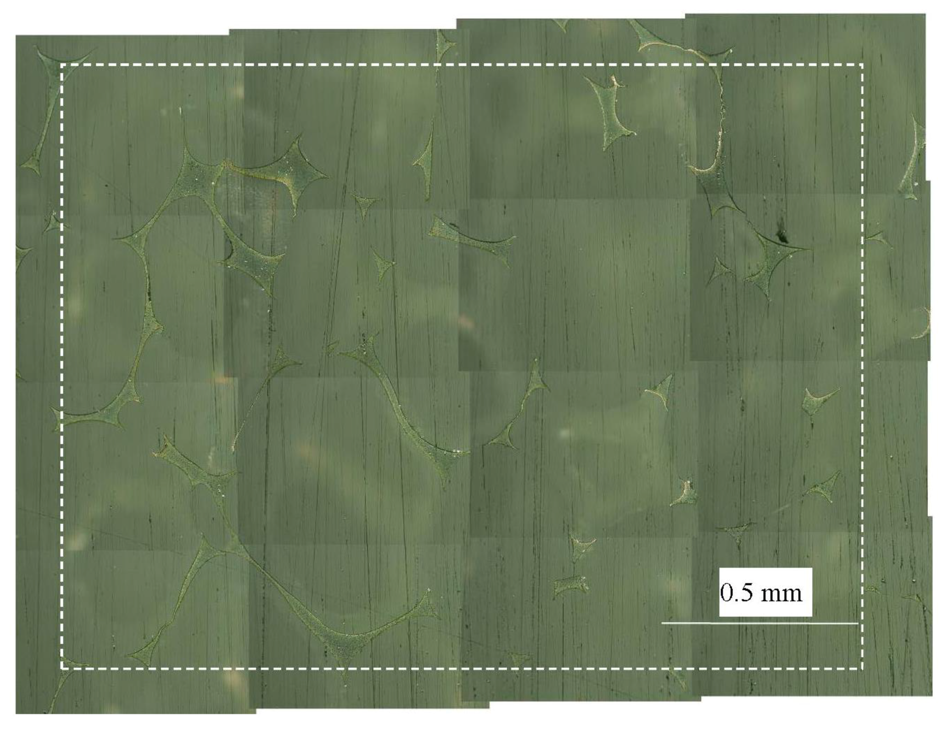
We here discuss the reason why the resistance was reduced by pressure. We assumed that the decrease in resistance was due to the change in contact area, and investigated the contact area before and after compressing the conductive foam block. The block before compression was filled with resin and polished. A surface of the block filled with resin was observed under a microscope.
Figure 6 shows the surface of the block. The figure is a composite of 16 images because the field of view of the microscope was narrow. The spiky long and thin features were fibers of conductive urethane foam. The broken-line rectangle (2.0 mm × 1.5 mm) was the observation area in this experiment. Moreover, we converted the image to the monochrome image shown in
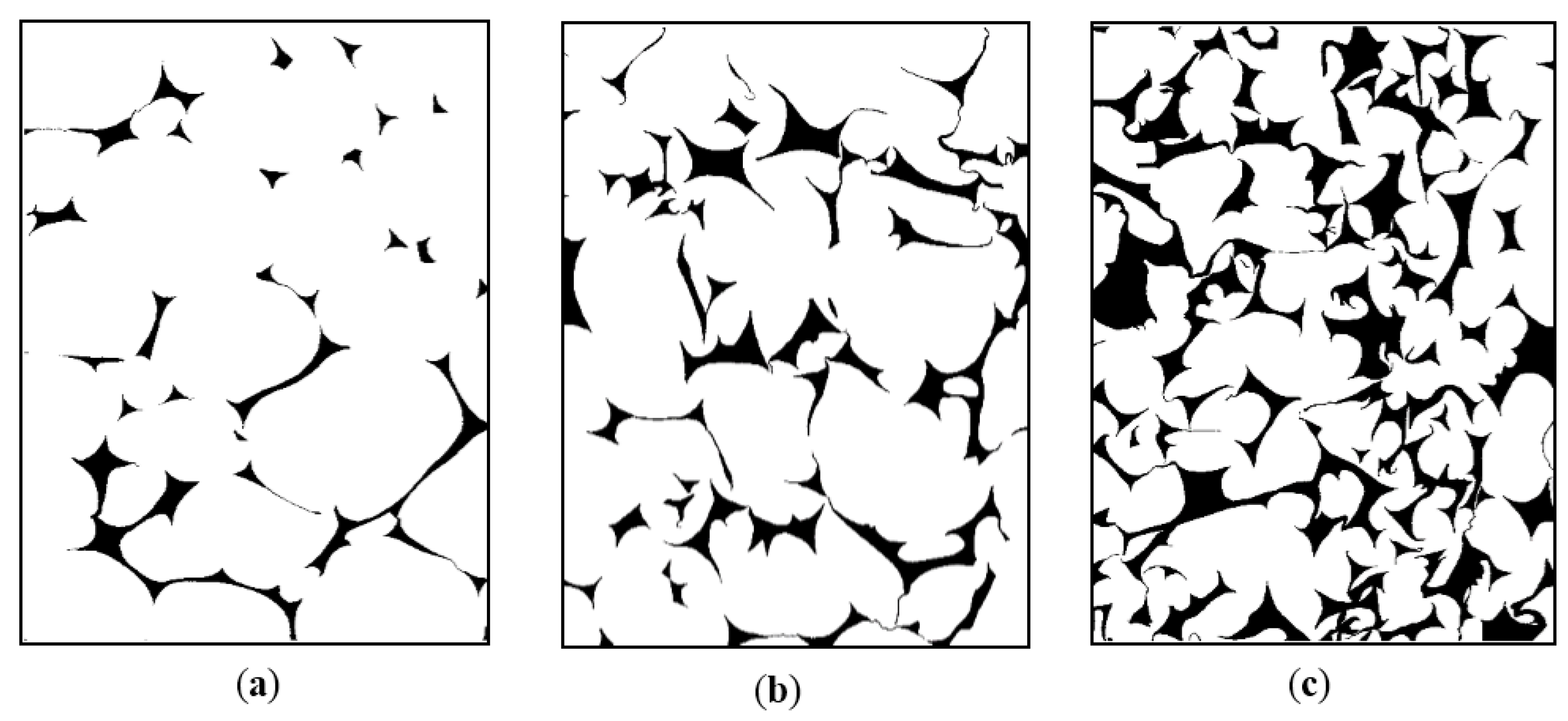
Figure 7a to improve visibility. The dimensions of each figure in
Figure 7 are 1.5 mm × 2.0 mm; the images are rotated for ease of comparison.
Black areas in the figure indicate the conductive foam;
i.e., the contact area. The combined area of contact was 0.16 mm
2. The pressed conductive foam blocks were observed employing the same method.
Figure 7b,c show surfaces of the blocks pressed at 2.0 and 3.0 kPa, respectively; summations of the contact area were 0.35 and 0.75 mm
2, respectively. It is thus seen that the contact area of the block was increased by compression pressure, and the resistance of the conductive foam block was decreased by increasing the contact area of the block.
Figure 6.
Surface of the conductive foam block. The compression pressure was 0.0 kPa. This figure is the combination of 16 images. Spiky long and thin features are fibers of the conductive foam. The broken-line rectangle shows the observation area (2.0 mm × 1.5 mm).
Figure 6.
Surface of the conductive foam block. The compression pressure was 0.0 kPa. This figure is the combination of 16 images. Spiky long and thin features are fibers of the conductive foam. The broken-line rectangle shows the observation area (2.0 mm × 1.5 mm).
Figure 7.
Comparison of contact areas of the conductive foam. Black areas indicate the surface of the conductive foam: (a) no compression (0.0 kPa); (b) compressed at 2.0 kPa; (c) compressed at 3.0 kPa.
Figure 7.
Comparison of contact areas of the conductive foam. Black areas indicate the surface of the conductive foam: (a) no compression (0.0 kPa); (b) compressed at 2.0 kPa; (c) compressed at 3.0 kPa.
3.3. Measured Contact Pressure at Each Electrode
We measured the contact pressure at each electrode when the subject wore the smart shirt. At first, the contact pressure was measured without the conductive foam blocks inserted between the shirt and body. We then measured the contact pressure when the conductive foam blocks were attached to the smart shirt (
Figure 8).
Measured pressures at each electrode are given in
Table 1. The upper row of the table gives the contact pressure when the blocks were not attached. The pressures at electrodes V1 and V2 have values of 0.0 kPa because these electrodes did not touch the body. The other pressures were approximately 2 to 3 kPa. In contrast, pressures were approximately 3 to 5 kPa when the blocks were attached to the shirt;
i.e., all electrodes were in contact with the body because of the conductive foam blocks.
Table 1.
Compression pressures at electrodes V1 to V6.
Table 1.
Compression pressures at electrodes V1 to V6.
| Electrode | V1 | V2 | V3 | V4 | V5 | V6 |
|---|
| without conductive foam blocks (kPa) | 0.0 | 0.0 | 2.7 | 2.4 | 1.8 | 2.0 |
| with conductive foam blocks (kPa) | 3.7 | 3.5 | 4.5 | 4.9 | 3.5 | 3.3 |
Figure 8.
Conductive foam blocks on the smart shirt.
Figure 8.
Conductive foam blocks on the smart shirt.
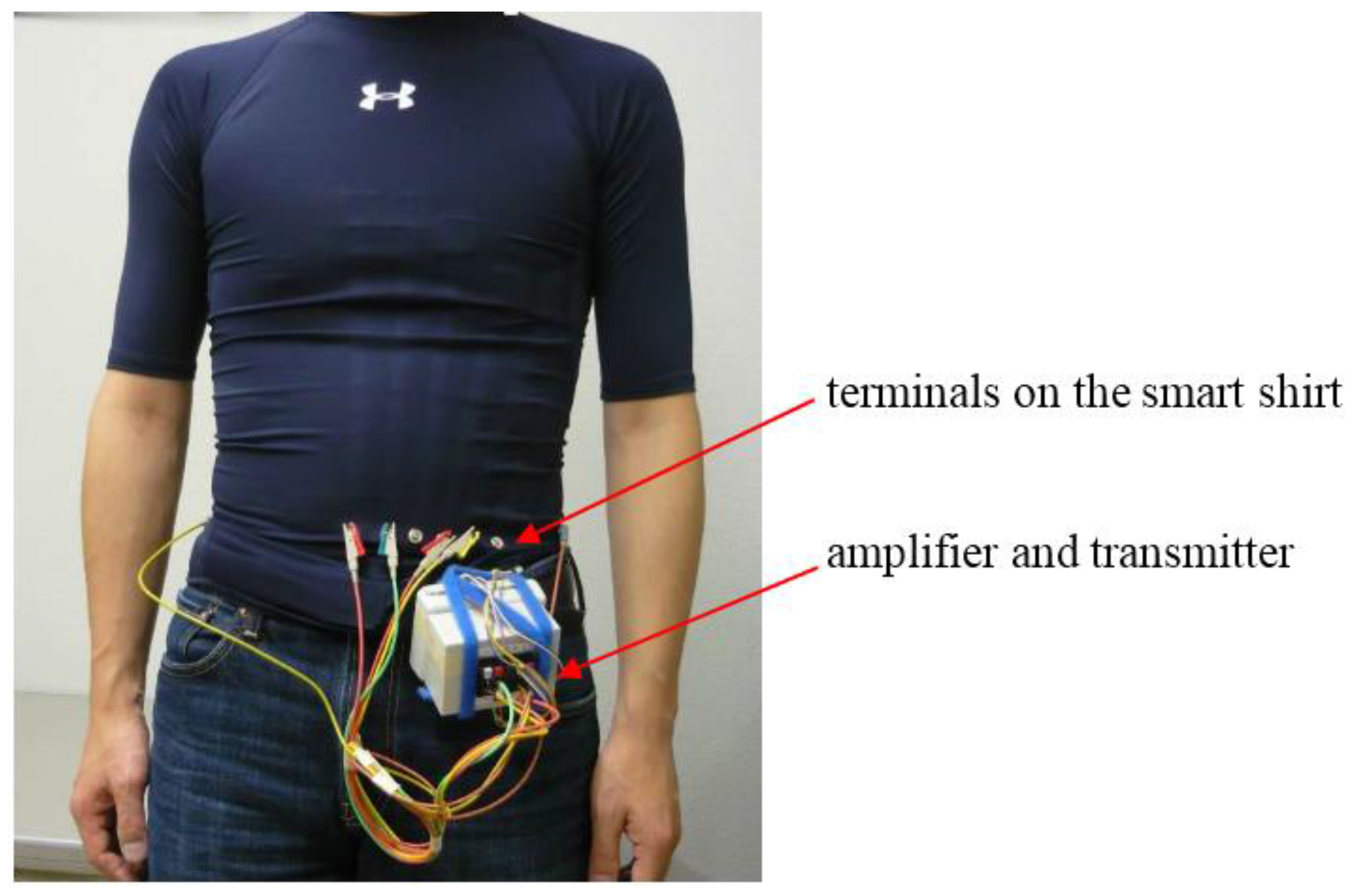
3.4. Measured ECG Signals
We measured the subject’s ECG signals using the smart shirt (
Figure 9). In experiments, we measured the signals from four electrodes—V1, V2, V4, and V5—because the amplifier had only four input channels. This selection was decided by the contact conditions of the electrodes. The contact conditions (noncontact or contact) of electrodes V1 and V2 changed drastically if the conductive foam blocks were used. In contrast, electrodes V4 and V5 were in contact with the body irrespective of whether the blocks were used.
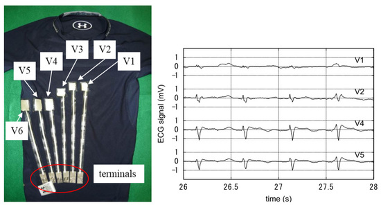
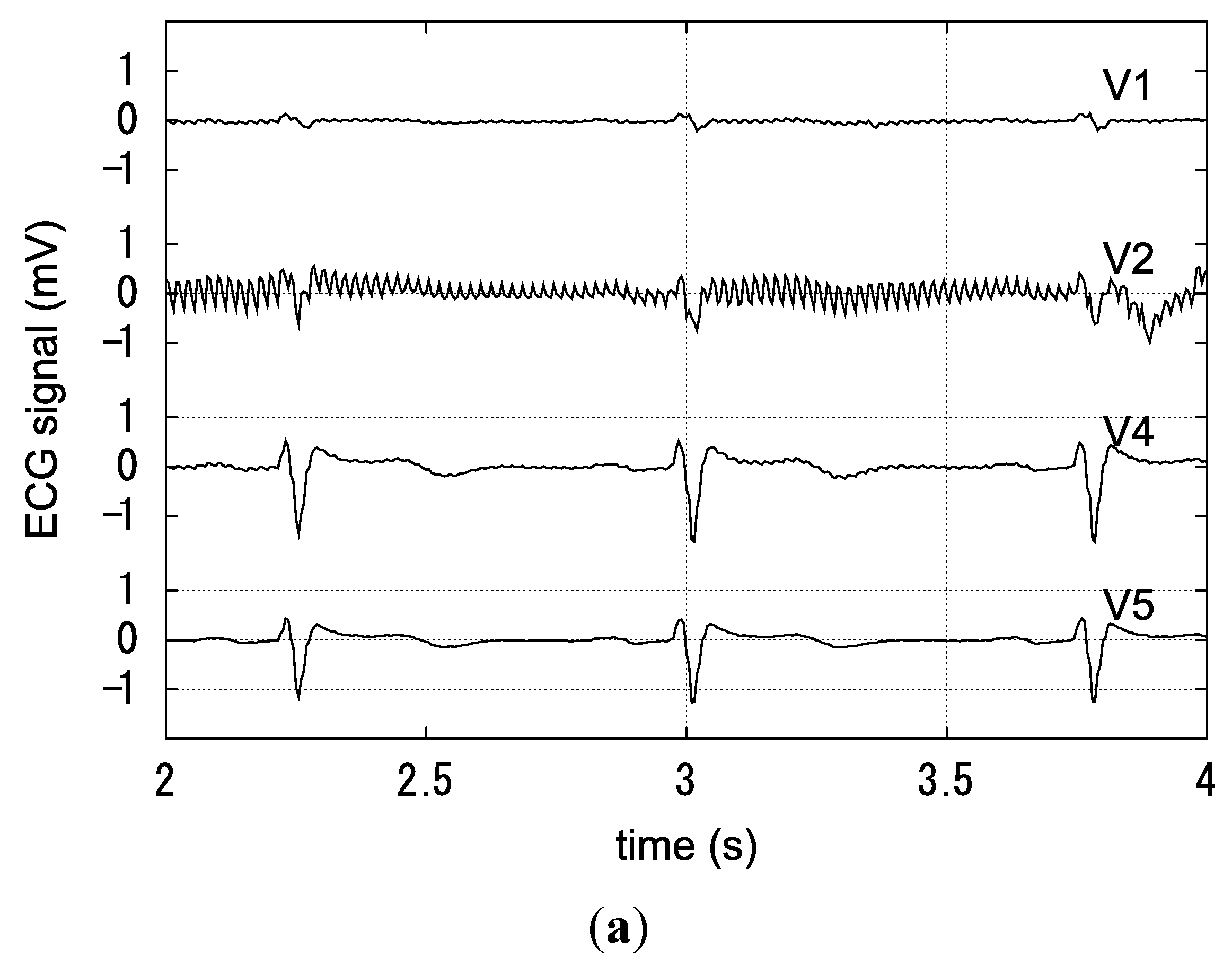
At first, the conductive foam blocks were not attached to the smart shirt. The subject was slightly sweaty; skin moisture was approximately 40%. The ECG measurement when the subject rested in a chair is shown in
Figure 10a. There was a humming noise in the V2 signal. In contrast, the V4 and V5 signals were stable. V1 and V2 signals appeared like the ECG signals even though the contact pressure was 0.0 kPa as described in section 3.3. This was because the electrodes were in slight contact with the body.
Figure 10b shows the ECG signals recorded while the subject walked. V1 and V2 signals were not discriminable as ECG signals because of swing noise and irregular peaks. In contrast, V4 and V5 signals were stable and discriminable as ECG signals. Moreover, we measured the ECG signals shown in
Figure 10c while the subject jogged slowly. V1 and V2 signals had much swing noise and could not be distinguished as ECG signals. There were slight swings in V4 and V5 signals, but peaks of the ECG signal were detectable.
Figure 9.
Subject wearing our smart shirt.
Figure 9.
Subject wearing our smart shirt.
Figure 10.
ECG signals measured by the smart shirt without the conductive foam blocks: (a) the subject resting in a chair; (b) the subject walking; (c) the subject slowly jogging.
Figure 10.
ECG signals measured by the smart shirt without the conductive foam blocks: (a) the subject resting in a chair; (b) the subject walking; (c) the subject slowly jogging.
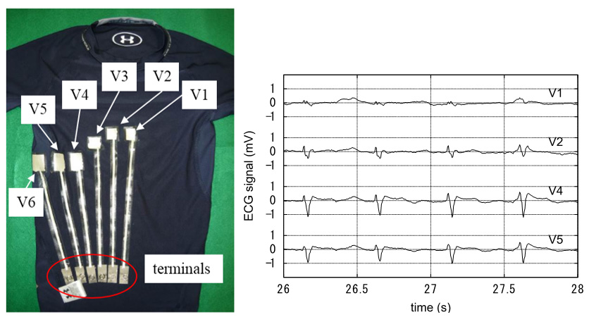
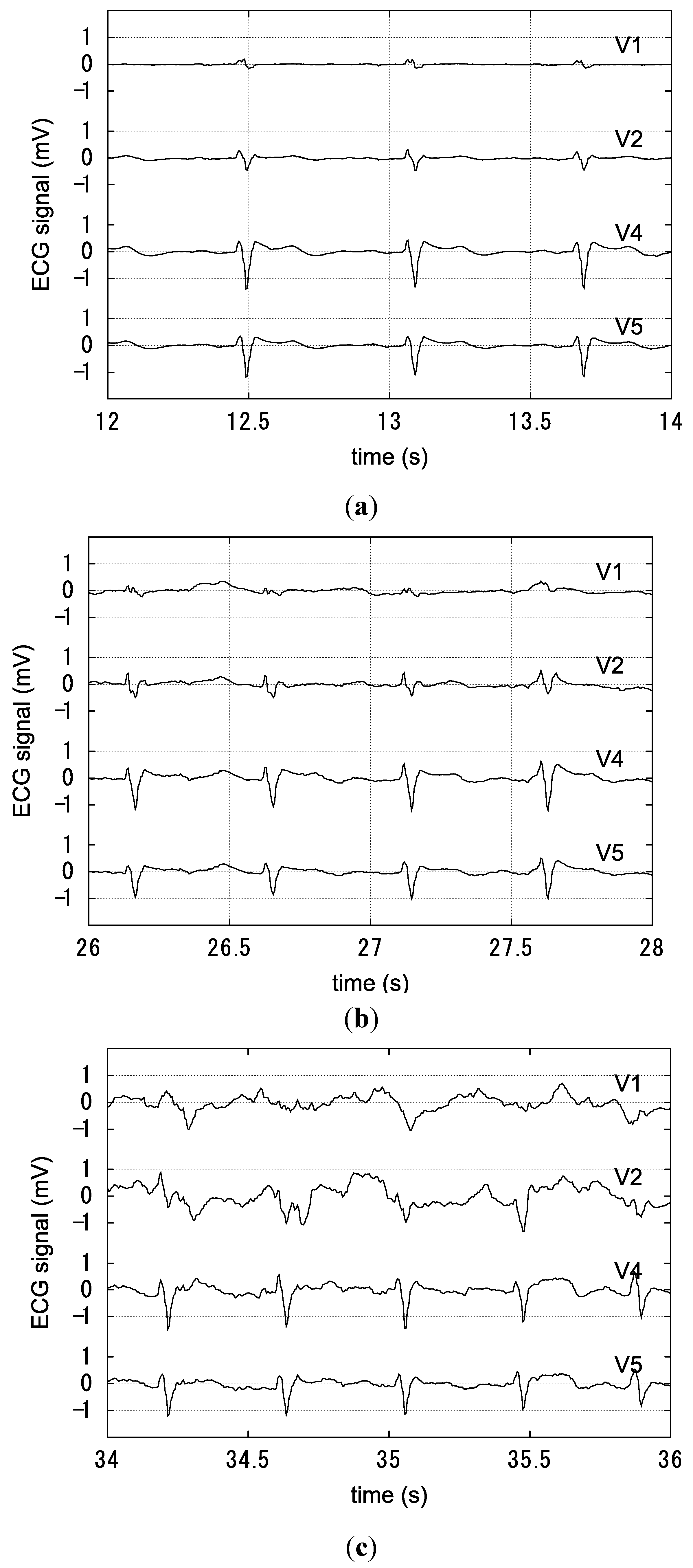
In the next experiments, conductive foam blocks were attached at electrodes V1, V2, V4 and V5 on the smart shirt. We measured the ECG signal while the subject rested, walked, and slowly jogged.
Figure 11a shows the ECG signals recorded while the subject rested. The ECG signals were very stable and easy to discriminate. The ECG signals recorded while the subject walked are shown in
Figure 11b. The ECG signal was easy to discriminate, but there was small swing noise. Finally,
Figure 11c shows the ECG signals recorded while the subject jogged slowly. V1 and V2 signals had much swing noise and could not be distinguished as ECG signals. V4 and V5 signals had small swing noise and distinguishable peaks of ECG signals.
The above experimental results are summarized as follows. The ECG signals from electrodes V1 and V2 that were difficult to measure stably without the conductive foam blocks could be measured with the conductive foam blocks. The ECG signals from all electrodes were measured stably even though the subject was walking. We thus considered that our smart shirt could measure ECG signals during light activities of daily life. However, measuring the ECG was difficult when the subject was exercising.
Figure 11.
ECG signals measured by the smart shirt with the conductive foam blocks: (a) the subject resting in a chair; (b) the subject walking; (c) the subject slowly jogging.
Figure 11.
ECG signals measured by the smart shirt with the conductive foam blocks: (a) the subject resting in a chair; (b) the subject walking; (c) the subject slowly jogging.
Figure 12 shows the ECG signals recorded when the subject jogged for 69 s and then rested in a standing position. The figure indicates that the measured ECG signals became stable immediately when the subject stopped exercising. Therefore, the smart shirt could measure the ECG signals intermittently when the subject was exercising.
Figure 12.
ECG signals measured by the smart shirt with the conductive foam blocks. The subject slowly jogged for 69 s and then rested in a standing position. The ECG signals became stable immediately when the subject stopped exercising.
Figure 12.
ECG signals measured by the smart shirt with the conductive foam blocks. The subject slowly jogged for 69 s and then rested in a standing position. The ECG signals became stable immediately when the subject stopped exercising.
The stability of signals from electrodes V1 and V2 has to be improved. The contact condition of these electrodes was greatly affected by the vibration of jogging. We considered that the change in the contact condition was a cause of the unstable signal. If the contact pressure increases, the electrodes are pushed close against the body and the ECG signal can be measured stably.


