Article

SARS-CoV-2 Infection Positively Correlates with Hyperglycemia and Inflammatory Markers in COVID-19 Patients: A Clinical Research Study

by
Prashanth Chikkahonnaiah
1,
Siva Dallavalasa
2,
SubbaRao V. Tulimilli
2,
Muskan Dubey
3,
Shashidhar H. Byrappa
4,
Raghavendra G. Amachawadi
5,
SubbaRao V. Madhunapantula
2,6,* and
Ravindra P. Veeranna
3,*
1
Department of Pulmonary Medicine, Mysore Medical College and Research Institute, Mysuru 570001, Karnataka, India
2
Center of Excellence in Molecular Biology and Regenerative Medicine (CEMR) Laboratory (DST-FIST Supported Centre and ICMR Collaborating Center of Excellence–ICMR-CCoE), Department of Biochemistry (DST-FIST Supported Department), JSS Medical College, JSS Academy of Higher Education & Research (JSS AHER), Mysuru 570015, Karnataka, India
3
Xavier University School of Medicine, Xavier University School of Veterinary Medicine, Santa Helenastraat #23, Oranjestad, Aruba
4
Department of Pathology, Mysore Medical College and Research Institute (MMC&RI), Mysuru 570001, Karnataka, India
5
Department of Clinical Sciences, College of Veterinary Medicine, Kansas State University, Manhattan, KS 66506, USA
6
Leader, Special Interest Group in Cancer Biology and Cancer Stem Cells (SIG-CBCSC), JSS Medical College, JSS Academy of Higher Education & Research (JSS AHER), Mysuru 570004, Karnataka, India
*
Authors to whom correspondence should be addressed.
Diseases 2024, 12(7), 143; https://doi.org/10.3390/diseases12070143
Submission received: 17 May 2024 / Revised: 22 June 2024 / Accepted: 1 July 2024 / Published: 4 July 2024
(This article belongs to the Section Infectious Disease)

Abstract

:
Diabetes mellitus (DM) is a common comorbidity in COVID-19 subjects. Hyperglycemia at hospital admission identified as a major risk factor and is responsible for poor prognosis. Hematological and inflammatory parameters have been recognized as predictive markers of severity in COVID-19. In this clinical study, we aimed to assess the impact of hyperglycemia at hospital admission on hematological and several inflammatory parameters in COVID-19 patients. A total of 550 COVID-19 subjects were primarily categorized into two major groups (normoglycemic and hyperglycemic) based on random blood sugar levels. On the first day of hospitalization, subjects’ oxygen saturation, random blood sugar, hematological variables, and inflammatory parameters were recorded. The hyperglycemic group exhibited higher levels of serum ferritin, total leukocyte count (TLC), lactate dehydrogenase (LDH), neutrophil count, and neutrophil-to-lymphocyte ratio (NLR). In contrast, oxygen saturation and lymphocyte count were lower compared to the normoglycemic group. Significantly elevated levels of hematological variables (TLC, neutrophil count, NLR) and inflammatory parameters (serum ferritin) were observed in the hyperglycemic group. Among inflammatory parameters, only serum ferritin levels showed statistical significance. This study supports the clinical association between hyperglycemia and an increased severity of COVID-19. Consequently, the identification of these parameters is a crucial and valuable prognostic indicator for assessing disease severity in hyperglycemic subjects.

1. Introduction

The COVID-19 pandemic was triggered mainly by severe acute respiratory syndrome coronavirus-2 (SARS-CoV-2), structurally a single-stranded, positive-sense, enveloped RNA-associated virus capable of infecting both humans and animals [1]. SARS-CoV-2 mainly enters the target cells with the help of angiotensin-converting enzyme (ACE)-2 receptors, utilizing the endosomal pathway. Following cell entry, the virus undergoes replication, leading to the apoptosis of the target cell and the release of mature virions. These virions subsequently infect neighboring cells, activating pro-inflammatory pathways that result in a cytokine storm, eventually leading to tissue fibrosis and loss of function in the affected organ. Numerous study reports have detailed that the severity of COVID-19 infection is heightened in individuals with several underlying comorbidities, like diabetes, obesity, hypertension, and cardiovascular diseases, among others [2]. Earlier studies from the Chinese Centre for Disease Control and Prevention have reported 72,314 new cases and revealed an increased mortality rate in individuals with diabetes (7.3% for diabetic subjects compared to 2.3% for non-diabetic subjects) [3]. In Hong Kong, among the total COVID-19-positive cases, 42.3% were diabetic subjects, with 26 fatalities reported in diabetic subjects aged 75 years and above [4]. A recent study conducted by Guan et al. (2020) demonstrated that of a total of 1099 subjects with COVID-19, 16.2% reported diabetes mellitus, 23.7% had hypertension, 5.8% reported coronary heart diseases, and 2.3% had cerebrovascular disease, indicating severity in cases with comorbidities [5]. In another study involving 140 COVID-19 subjects, 12% had diabetes, and 30% had hypertension [6].
In a multivariate analysis, it was found that diabetes independently correlates with increased mortality in individuals with COVID-19 infection [7]. The presence of insulin resistance and hyperglycemic conditions in diabetes stimulates pro-inflammatory cytokines synthesis, the advanced glycation process of end products (AGEs), and oxidative stress. Additionally, it mainly triggers the production of adhesion molecules, mediating inflammation in the tissue levels [8,9]. This pro-inflammatory response appears to be a potential underlying mechanism for the heightened infections and severity observed in subjects with diabetes, leading to worse outcomes.
Conversely, studies suggest that the SARS-CoV-2 infection alone can elevate blood glucose levels in individuals without a history of hyperglycemia. Subjects with COVID-19 have been observed to experience new-onset diabetic symptoms and severe metabolic complications, such as ketoacidosis and hyperosmolarity, etc. [10,11,12]. European pediatric populations reported an increase in type 1 diabetes among individuals under 18 years of age within 30 days of infection, while in adults it may manifest as a long-term consequence of the viral infection [13]. The virus infects cells through ACE-2 receptors, which are mainly expressed in key metabolic-associated organs, tissues like pancreatic beta cells, adipose tissue, the small intestine, and the kidney [14]. This suggests that SARS-CoV-2 may induce diverse changes in blood glucose levels, with glucose metabolism complicating the pathophysiology of preexisting diabetes or leading to new-onset diabetes. Furthermore, it has been observed that SARS-CoV-2 infection results in the release of high levels of glucocorticoids and catecholamines, further elevating blood glucose [15]. These findings highlight the bidirectional relationship between COVID-19 and diabetes. Several studies have previously reported that hematological and inflammatory parameters serve as predictive markers of severity in COVID-19 subjects, and assessing these parameters can aid in planning the management of COVID-19 subjects [16,17,18,19]. In this study, our goal was to investigate the impact of hyperglycemia at hospital admission on the hematological and inflammatory parameters in COVID-19 subjects.

2. Materials and Methods

2.1. Study Design, Ethical Considerations, Data Collection, and Case Enrollment

An observational cross-sectional cohort study was mainly conducted among subjects diagnosed with COVID-19, identified either through a rapid antigen test or RT-PCR; subjects were admitted to a designated COVID hospital in Mysuru, Karnataka, India. Preparatory approvals were taken from the institutional ethics committee (MMCEC36/2021). Written consent, RT-PCR test reports, and random blood sugar (RBS) levels were considered for patient enrollment. Subsequently, subjects were mainly categorized into two groups based on their random blood sugar levels: normoglycemic (RBS < 140 mg/dL) and hyperglycemic (RBS > 140 mg/dL).
The study involved the examination of patient blood for various parameters, including serum ferritin, lactate dehydrogenase, random blood sugar levels, total leukocyte count, neutrophil count, lymphocyte count, and neutrophil-to-lymphocyte ratio. The research was conducted at the Mysore Medical College and Research Institute (MMC&RI) (Department of Respiratory Medicine) and Princess Krishnajammanni Trauma Care Center (PKTCC) (which served as a designated COVID hospital), Mysore; both male and female participants were included in the present study. All the samples were processed at the central laboratory facility, Krishna Rajendra Hospital, Mysore, Karnataka, India.

2.2. Sample Collection and Processing

On the day of admission, after obtaining informed consent, about 5.0 mL of venous blood was collected from each subject aseptically and divided into two fractions, one for hematological (purple color-coded, EDTA anticoagulant tubes) and the other for serological (red color-coded, no anticoagulant tubes) examination. The blood collected for hematological examination was used for estimating random blood sugar levels, total leukocyte count, neutrophil count, lymphocyte count, and neutrophil-to-lymphocyte ratio. The blood collected for serological examination was allowed to clot by leaving it undisturbed at room temperature for 30 min. After coagulation, the serum was separated by centrifugation at 3000 rpm for 10 min at room temperature. Serum ferritin (determined using the chemiluminescence method, as detailed below) and serum LDH (estimated using a kit-based method-LDHI2 reagent kit Cat. No. 03004732122) were measured using a Cobas 6000 Chemistry Analyzer from Roche Diagnostics, Indianapolis, IN, USA, which were available at the central laboratory facility, Krishna Rajendra Hospital, Mysore, Karnataka, India.

2.3. Estimation of Serum Ferritin

Serum ferritin levels were determined using the chemiluminescence method (COBAS 6000 from Roche Diagnostics, Indianapolis, IN, USA). The assay works on a sandwich principle, where a ferritin-defined antibody and a specific labeled ferritin antibody form a sandwich complex in the primary step. In the second step, the specific microparticles were introduced to facilitate the sandwich for binding to a solid phase. Further, the reaction mixture was aspirated into the measuring cells and captured microparticles onto the electrode. After removing the unbound microparticles, a specific voltage was applied, inducing chemiluminescence; the emitted chemiluminescence was captured and measured by a photomultiplier, and readings were noted and used to quantify the serum ferritin through a calibration curve.

2.4. Statistical Analysis

The study findings encompass the both descriptive and inferential statistical analyses which were carried out. The results for continuous variables were presented as Mean ± SEM, while the results for categorical variables were expressed as frequency (percentage). A statistical analysis was performed using GraphPad Prism 5, and a p-value of <0.05 was considered statistically significant.

3. Results

3.1. Clinical Characteristics

Among the 550 subjects included in the study, 354 (64.36%) were males. The majority, comprising 368 (66.90%) of the subjects, fell within the age group of 30–60 years. A significant proportion, 442 (80.36%) of the subjects, had not received vaccination. More than 68% of the total subjects reported experiencing fever and cough, while 45% experienced breathlessness. In the normoglycemic group (202 subjects), 127 (62.87%) were males, and 136 (67.32%) were in the age group of 30–60 years. The majority, 159 (78.71%), were unvaccinated. More than 64% of these subjects reported symptoms of fever and cough, and 41% experienced breathlessness. Thirty-two of 202 (32/202 = 15.84%) normoglycemic participants had preexisting diabetes.
In the hyperglycemic group (n = 348), 227 (65.22%) were males. The majority (n = 232; 66.66%) of hyperglycemic participants were in the age group of 30–60. The number of unvaccinated participants was found to be very high in the hyperglycemic group (n = 283; 81.32%). More than 70% of these subjects reported symptoms of fever and cough, while 47% experienced breathlessness. Interestingly, whereas 225 (64.65%) of 348 hyperglycemic subjects developed new-onset hyperglycemia, 126 (36.20%) had preexisting diabetes (Table 1).

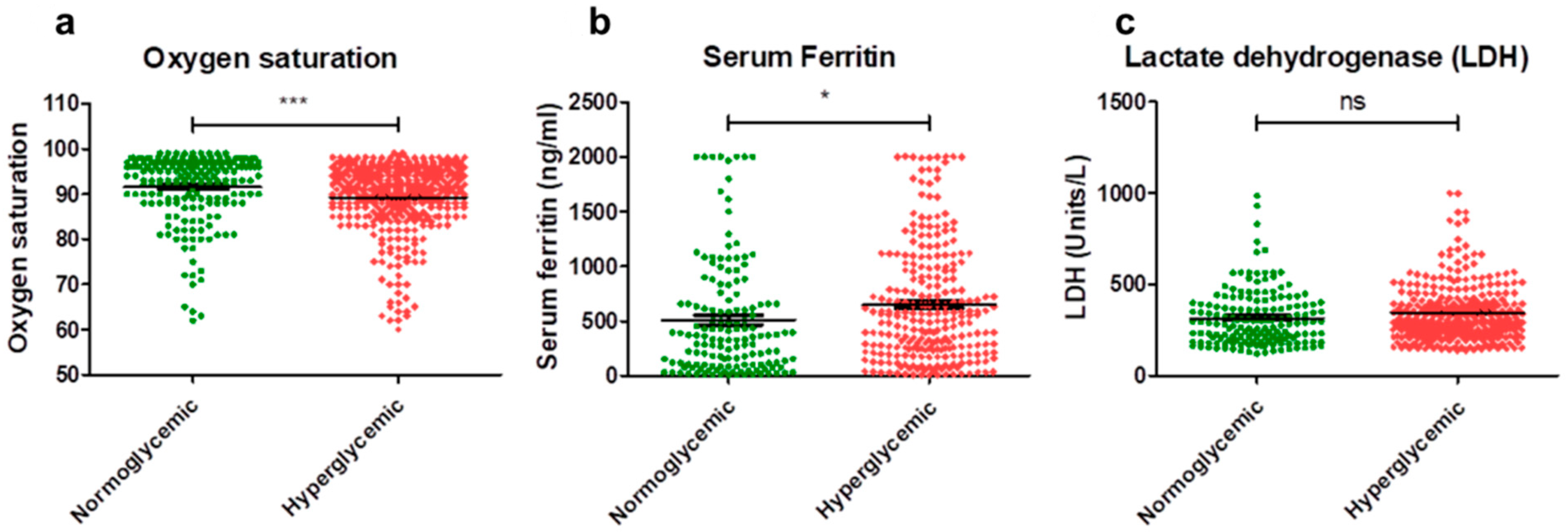
3.2. Inflammatory Parameters

In this current study, the mean oxygen saturation levels were 91.55% in normoglycemic COVID-19-infected subjects and 89.24% in hyperglycemic COVID-19-infected subjects. An analysis of the data revealed a significant difference, with normoglycemic subjects exhibiting a higher mean oxygen saturation of 91.55%, compared to hyperglycemic subjects, with a mean saturation of 89.24% (Figure 1a).
The mean serum ferritin levels were 509.5 ng/mL in normoglycemic subjects and 650.0 ng/mL in hyperglycemic subjects. An analysis of the data revealed a significant difference, with hyperglycemic subjects exhibiting a higher mean serum ferritin level of 650.0 ng/mL compared to normoglycemic subjects, who had a mean level of 509.5 ng/mL (Figure 1b).
The mean LDH levels were 315.9 units/L in normoglycemic COVID-19-infected subjects and 344.3 units/L in hyperglycemic subjects. An analysis of the data indicated that there was no significant difference observed between the mean LDH levels of normoglycemic and hyperglycemic COVID-19-infected subjects (Figure 1c).

3.3. Hematological Parameters

In this study, the mean TLC was 6814/microliter in normoglycemic COVID-19-infected subjects and 7699/microliter in hyperglycemic subjects. An analysis of the data revealed a significant difference, with hyperglycemic subjects showing a higher mean TLC of 7699/microliter compared to normoglycemic subjects with a mean TLC of 6814/microliter (Figure 2a).
The mean neutrophil count was 72.16% in normoglycemic COVID-19-infected subjects and 80.10% in hyperglycemic subjects. An analysis of the data revealed a significant difference, with hyperglycemic subjects demonstrating a higher mean neutrophil count of 80.10% compared to normoglycemic subjects with a mean count of 72.16% (Figure 2b).
In the present study, the mean lymphocyte count was 21.05% in normoglycemic COVID-19-infected subjects and 14.77% in hyperglycemic subjects. An analysis of the data revealed a significant difference, with hyperglycemic subjects demonstrating a lower mean lymphocyte count of 14.77% compared to normoglycemic subjects with a mean count of 21.05% (Figure 2c).
The mean neutrophil to lymphocyte ratio (NLR) was 6.034 in normoglycemic COVID-19-infected subjects and 8.769 in hyperglycemic subjects. An analysis of the data revealed a significant difference, with hyperglycemic subjects showing a higher mean NLR of 8.769 compared to normoglycemic subjects with a mean NLR of 6.034 (Figure 2d).

4. Discussion

Severe outcomes with higher morbidity and mortality rates in SARS-CoV-2 infected subjects have been associated with various factors, including age and comorbidities such as diabetes, hypertension, obesity, cardiovascular diseases, chronic kidney and liver diseases, low lymphocyte and albumin counts, elevated levels of lactate dehydrogenase, C-reactive protein, red blood cell distribution width, blood urea nitrogen, and direct bilirubin [20,21,22]. Neutrophilia, thrombocytopenia, higher lactate dehydrogenase, and D-dimer levels are more likely to develop Acute Respiratory Distress Syndrome (ARDS), particularly in older subjects [23,24].
Diabetes is one of the more frequent comorbidities of SARS-CoV-2 infection [4,25,26,27], and there is a bidirectional association between SARS-CoV-2 infection and diabetes. On one hand, pre-existing diabetes is associated with an increased risk of severe SARS-CoV-2 infection. On the other hand, severe SARS-CoV-2 infected subjects are prone to new-onset diabetes and severe metabolic complications of pre-existing diabetes, possibly due to adverse immune reactions and the destruction of pancreatic β-cells [10,11,12]. This bidirectional relationship between SARS-CoV-2 infection and diabetes poses challenges in clinical management and suggests a complex pathophysiology of COVID-19 disease.
SARS-CoV-2 binds to angiotensin-converting enzyme 2 (ACE2) receptors, expressed on the cells of key metabolic organs and tissues, including pancreatic beta cells, adipose tissue, small intestine, and kidneys [14]. Thus, SARS-CoV-2 may cause pleiotropic alterations in glucose metabolism that could complicate the pathophysiology of pre-existing diabetes or lead to new mechanisms of disease. Glycemic characteristics predict clinical outcomes, with higher blood glucose levels associated with more extended hospital stays and higher mortality rates in COVID-19 subjects [28]. Improved blood glucose monitoring and management have been shown to result in better outcomes and reduced mortality rates in subjects with SARS-CoV-2 infection [29,30].
Preliminary studies reported an increased prevalence of hyperglycemia in subjects affected by COVID-19 [31,32]. Similar results were reported previously in subjects affected by SARS-CoV-1, which increased fasting blood glucose levels compared to levels observed in subjects with non-SARS-CoV-2 infection pneumonia [33,34,35]. Severe hyperglycemia is common in critically ill subjects and is often considered a marker of disease severity [36]. Several studies over the course of the pandemic have reported that SARS-CoV-2 infection is associated with hyperglycemia in people with or without a history of diabetes [28,37].
In the present study, out of 550 COVID-19 subjects, 348 (63.27%) were found to have random blood sugar levels > 140 mg/dL (considered hyperglycemic), while 202 (36.72%) subjects had levels < 140 mg/dL (considered normoglycemic). The study demonstrated that 15% of the normoglycemic subjects and 36% of the hyperglycemic subjects had preexisting diabetes. Interestingly, 64% of the hyperglycemic subjects had new-onset hyperglycemia, indicating the effect of SARS-CoV-2 infection on the glycemic index. The study also observed, in COVID-19 subjects, a significant association of hyperglycemia with mean oxygen saturation, serum ferritin, total leukocyte count, neutrophil count, lymphocyte count, and neutrophil-to-lymphocyte ratio.
Several people infected with COVID-19 have low oxygen saturation levels, and maintaining oxygen saturation within the target range is crucial for patient management. In the present study, the mean oxygen saturation levels in the hyperglycemic subjects’ group (89.24%) were significantly lower compared to the normoglycemic subjects’ group (91.55%). Prior studies have reported that low blood oxygen saturation is associated with increased mortality, and individuals with diabetes have low blood oxygen saturation compared with non-diabetic controls [38,39]. Another study reported that hyperglycemia-induced cellular hypoxia and mtROS may promote hyperglycemic damage in a coordinated manner [40]. A recent study has shown an elevated ACE2 expression, and its glycated product, in COVID-19 patients who had hyperglycemia. This study has also reported that COVID-19 patients with sustained hyperglycemia had low oxygen saturation levels [41]. In summary, hyperglycemia is one of the critical factors contributing to the low oxygen saturation levels observed in diabetic individuals, and is responsible for various exacerbations reported in diabetic individuals suffering from COVID-19.
Serum ferritin, being an acute-phase reactant, reflects the degree of both chronic and acute inflammatory reactions in the body. High serum ferritin levels are associated with increased disease severity and a poor outcome in people infected with SARS-CoV-2 [42]. In the present study, mean serum ferritin levels in the hyperglycemic subjects’ group (650.0 ng/mL) were significantly higher compared to the normoglycemic subjects’ group (509.5 ng/mL). Although the exact mechanism is not known, a study reported that patients with type 2 diabetes had considerably higher serum ferritin levels, and there was a positive link found between serum ferritin and HbA1c [43].
Serum lactate dehydrogenase (LDH) catalyzes the conversion of pyruvate to lactate, the final step of aerobic glycolysis [44]. An elevated LDH indicates tissue hypoperfusion, which further indicates the extent of the disease [45,46] and is a potential prognostic biomarker in subjects with COVID-19 [47]. However, in the present study, no significant difference was observed between the mean LDH levels of normoglycemic and hyperglycemic COVID-19-infected subjects.
SARS-CoV-2 infection begins as an inflammatory disorder that progresses to severe pneumonia, cytokine storm, acute respiratory distress syndrome, and multi-organ failure, resulting in prolonged hospitalization with poor overall outcomes and potentially death [48]. Several studies have reported that hematological parameters are the predictive markers of severity in COVID-19 subjects [49,50,51]. Analyzing hematological parameters immediately after hospital admission may serve as an effective tool in the early identification of subjects at risk of developing severe disease [52]. Hematological parameters like total leukocyte count, neutrophil count, lymphocyte count, and neutrophil-to-lymphocyte ratio are known to predict the severity of SARS-CoV-2 infection in COVID-19 subjects [50,53,54,55]. These parameters are simple, readily available, and cost-effective prognostic indicators that can be utilized for diagnosis and predicting the severity of COVID-19 infection, particularly in developing countries [56,57,58]. In the present study, the mean total leukocyte count, neutrophil count, and neutrophil-to-lymphocyte ratio were significantly higher, and the lymphocyte count was significantly lower in the hyperglycemic subjects’ group compared to the normoglycemic subjects’ group. Hyperglycemia, along with elevated stress hormones such as cortisol and catecholamines, as well as cytokines, can cause bone marrow progenitor cell expansion, thereby promoting leukocytosis or increased leukocyte count [59]. Although the exact mechanism of the effects of hyperglycemia on lymphocytes is unclear, prior studies have reported that COVID-19 patients with diabetes mellitus do experience a faster decline in lymphocyte count, a lower lymphocyte number, and a longer hospital stay than non-diabetic patients [60,61].
According to a recent cohort study, the long-term persistence of COVID-19 induces hyperglycemia in patients without a previous history of diabetes [62]. The study identified that 77% of the participants continued to receive antidiabetic therapy (oral hypoglycemic agents or insulin) or had persistent hyperglycemia at the end of 6 months [62]. Hyperglycemia, with or without pre-existing diabetes, increases poor outcomes and mortality by aggravating the immune dysfunction (decreased lymphocytes, CD16+, 56+, NK cells, CD3+ T cells, CD8+ T cells, and increased neutrophil percentage, and serum concentration of IgA) [63], cytokine storm, elevating pro-inflammatory markers (C-reactive protein, procalcitonin) [64], and inflammatory cytokine production (interleukin 6, tumor necrosis factor-α) [65], as well as coagulation activation (as monitored by plasma D-dimer levels) [66], impaired beta cell function, worsened endothelial dysfunction, etc. [66,67,68].
Stress levels are generally high in critically ill, hospitalized, and COVID-19 patients [69]. Stress-induced hyperglycemia is a common observation seen in 50% of critically ill patients admitted to the hospital [70]. Counter-regulatory hormones (cortisol, catecholamines, glucagon, and growth hormone) disturb glucose hemostasis and pro-inflammatory cytokines (interleukin-1 (IL-1), interleukin-6 (IL-6), and tumor necrosis factor-α (TNF-α)) and worsen the metabolic milieu by causing insulin resistance in stress-induced hyperglycemia [70,71,72]. Glycosylated hemoglobin (HbA1c), which is used to determine an individual’s average blood sugar levels over the previous 2–3 months, is the most widely used monitoring and diagnosing biomarker for diabetes [73]. Along with the inflammatory and hematological parameters reported in the present study, future studies should focus on measuring the glycosylated hemoglobin (HbA1c), insulin, and glucocorticoids in the serum as markers to identify the diabetic individuals at higher risk and provide timely intervention as a priority to improve patient outcomes.

5. Conclusions

In conclusion, our study suggests that compared to normoglycemic subjects, hyperglycemic COVID-19-infected subjects showed an increase in lactate dehydrogenase, serum ferritin, total leukocyte count, neutrophil count, and neutrophil-to-lymphocyte ratio, and a decrease in oxygen saturation and lymphocyte count. These results indicate an association between an increased severity of SARS-CoV-2 infection and elevated random blood sugar levels. Whether elevated blood sugar levels in non-diabetic subjects are due to immune-mediated or inflammatory responses, the direct effects of SARS-CoV-2 on β-cells, or a complex combination of mechanisms, is not known. Further studies with long-term follow-up in large cohorts are warranted to confirm the potential association between SARS-CoV-2 infection and an increased risk of diabetes.

Author Contributions

R.P.V.: Data curation, conceptualization, design of the experiment, results analysis, and supervision. P.C., S.D. and S.V.T.: Data curation, original draft preparation. M.D.: editing and further improving the quality of manuscript through her scientific inputs. S.H.B.: Results analysis and draft preparation. S.V.M. and R.G.A.: Results analysis, draft reviewing, and editing. All authors have read and agreed to the published version of the manuscript.

Funding

This research received no external funding.

Institutional Review Board Statement

The study was conducted according to the guidelines of the Declaration of Helsinki and approved by the Institutional Ethics Committee of MMC&RI (Reference number: MMCEC36/2021), Mysore, Karnataka, India.

Informed Consent Statement

Informed consent was obtained from all subjects involved in the study.

Data Availability Statement

Data are available upon reasonable request. The dataset used to conduct the analyses is available from the corresponding author upon reasonable request.

Acknowledgments

Prashant Chikkahonnaiah (PC) would like to thank the Director/Dean, MMCRI, for providing the infrastructure support. RPV would like to thank the President, CAO and the Board, Xavier University, Aruba, and Director, CSIR-CFTRI, for the kind support for the research, the DBT Ramalingaswami Fellowship, Govt. of India for providing the fellowship during the course of the study and the Bharatiya Reserve Bank Note Mudran (P) Limited, Mysuru, and Govt. of Karnataka for providing infrastructure support during the study. SD would like to thank the Department of Biotechnology (DBT), New Delhi, and the Government of India for providing Senior Research Fellowship as part of the grant support (N0-BT/PR29598/PFN/20/1392/2018) sanctioned to SubbaRao V. Madhunapantula. SD would like to thank the Indian Council of Medical Research (ICMR), New Delhi, and the Government of India for providing Senior Research Fellowship (No. 3/2/284/2022/NCD3) under the supervision of SubbaRao V. Madhunapantula. SVT would like to thank the Indian Council of Medical Research (ICMR), New Delhi, and the Government of India for providing Senior Research Fellowship as part of an extramural research grant (No. 54/06/2019-HUM/BMS dated 22 November 2019) sanctioned to M.N. Suma, Department of Biochemistry, JSS Medical College, JSS Academy of Higher Education & Research, Mysore, Karnataka, India. SVM would like to thank the Special Interest Group in Cancer Biology and Cancer Stem Cells (SIG-CBCSC), JSS Medical College, JSS Academy of Higher Education & Research, Mysore, Karnataka, India. SVT, SD and SVM would like to acknowledge the infrastructure support provided by DST-FIST to the Center of Excellence in the Molecular Biology and Regenerative Medicine (CEMR) laboratory (CR-FST-LS-1/2018/178) and the Department of Biochemistry (SR/FST/LS-1-539/2012).

Conflicts of Interest

The authors declare no conflicts of interest.

References

  1. Pal, M.; Berhanu, G.; Desalegn, C.; Kandi, V. Severe acute respiratory syndrome coronavirus-2 (SARS-CoV-2): An update. Cureus 2020, 12, e7423. [Google Scholar] [CrossRef]
  2. Care, D. Care in diabetesd2019. Diabetes Care 2019, 42, S13–S28. [Google Scholar]
  3. Wu, Z.; McGoogan, J.M. Characteristics of and important lessons from the coronavirus disease 2019 (COVID-19) outbreak in China: Summary of a report of 72 314 cases from the Chinese Center for Disease Control and Prevention. JAMA 2020, 323, 1239–1242. [Google Scholar] [CrossRef] [PubMed]
  4. Deng, S.-Q.; Peng, H.-J. Characteristics of and public health responses to the coronavirus disease 2019 outbreak in China. J. Clin. Med. 2020, 9, 575. [Google Scholar] [CrossRef]
  5. Guan, W.-J.; Ni, Z.-Y.; Hu, Y.; Liang, W.-H.; Ou, C.-Q.; He, J.-X.; Liu, L.; Shan, H.; Lei, C.-L.; Hui, D.S. Clinical characteristics of coronavirus disease 2019 in China. N. Engl. J. Med. 2020, 382, 1708–1720. [Google Scholar] [CrossRef] [PubMed]
  6. Zhang, J.-J.; Dong, X.; Cao, Y.-Y.; Yuan, Y.-D.; Yang, Y.-B.; Yan, Y.-Q.; Akdis, C.A.; Gao, Y.-D. Clinical characteristics of 140 patients infected with SARS-CoV-2 in Wuhan, China. Allergy 2020, 75, 1730–1741. [Google Scholar] [CrossRef] [PubMed]
  7. Mehraeen, E.; Karimi, A.; Barzegary, A.; Vahedi, F.; Afsahi, A.M.; Dadras, O.; Moradmand-Badie, B.; Alinaghi, S.A.S.; Jahanfar, S. Predictors of mortality in patients with COVID-19—A systematic review. Eur. J. Integr. Med. 2020, 40, 101226. [Google Scholar] [CrossRef]
  8. Nowotny, K.; Jung, T.; Höhn, A.; Weber, D.; Grune, T. Advanced glycation end products and oxidative stress in type 2 diabetes mellitus. Biomolecules 2015, 5, 194–222. [Google Scholar] [CrossRef]
  9. Khalid, M.; Petroianu, G.; Adem, A. Advanced glycation end products and diabetes mellitus: Mechanisms and perspectives. Biomolecules 2022, 12, 542. [Google Scholar] [CrossRef]
  10. Chee, Y.J.; Ng, S.J.H.; Yeoh, E. Diabetic ketoacidosis precipitated by COVID-19 in a patient with newly diagnosed diabetes mellitus. Diabetes Res. Clin. Pract. 2020, 164, 108166. [Google Scholar] [CrossRef]
  11. Li, J.; Wang, X.; Chen, J.; Zuo, X.; Zhang, H.; Deng, A. COVID-19 infection may cause ketosis and ketoacidosis. Diabetes Obes. Metab. 2020, 22, 1935–1941. [Google Scholar] [CrossRef]
  12. Ren, H.; Yang, Y.; Wang, F.; Yan, Y.; Shi, X.; Dong, K.; Yu, X.; Zhang, S. Association of the insulin resistance marker TyG index with the severity and mortality of COVID-19. Cardiovasc. Diabetol. 2020, 19, 58. [Google Scholar] [CrossRef]
  13. Barrett, C.E. Risk for newly diagnosed diabetes 30 days after SARS-CoV-2 infection among persons aged 18 years—United States, March 1, 2020–June 28, 2021. MMWR Morb. Mortal. Wkly. Rep. 2022, 71, 59–65. [Google Scholar] [CrossRef]
  14. Hamming, I.; Timens, W.; Bulthuis, M.; Lely, A.; Navis, G.v.; van Goor, H. Tissue distribution of ACE2 protein, the functional receptor for SARS coronavirus. A first step in understanding SARS pathogenesis. J. Pathol. 2004, 203, 631–637. [Google Scholar] [CrossRef]
  15. Wang, A.; Zhao, W.; Xu, Z.; Gu, J. Timely blood glucose management for the outbreak of 2019 novel coronavirus disease (COVID-19) is urgently needed. Diabetes Res. Clin. Pract. 2020, 162, 108118. [Google Scholar] [CrossRef]
  16. Henry, B.M.; De Oliveira, M.H.S.; Benoit, S.; Plebani, M.; Lippi, G. Hematologic, biochemical and immune biomarker abnormalities associated with severe illness and mortality in coronavirus disease 2019 (COVID-19): A meta-analysis. Clin. Chem. Lab. Med. (CCLM) 2020, 58, 1021–1028. [Google Scholar] [CrossRef]
  17. Kumar, A.; Sepolia, S.; Shilpa, R.; Rezayani, G.; Kumari, S.; Gupta, S. Role of hematological and immunological parameters in COVID-19 patients. J. Pharm. Bioallied Sci. 2021, 13, 238–243. [Google Scholar]
  18. Usul, E.; Şan, İ.; Bekgöz, B.; Şahin, A. Role of hematological parameters in COVID-19 patients in the emergency room. Biomark. Med. 2020, 14, 1207–1215. [Google Scholar] [CrossRef]
  19. Elshazli, R.M.; Toraih, E.A.; Elgaml, A.; El-Mowafy, M.; El-Mesery, M.; Amin, M.N.; Hussein, M.H.; Killackey, M.T.; Fawzy, M.S.; Kandil, E. Diagnostic and prognostic value of hematological and immunological markers in COVID-19 infection: A meta-analysis of 6320 patients. PLoS ONE 2020, 15, e0238160. [Google Scholar] [CrossRef]
  20. Singh, A.K.; Singh, R. Is metformin ahead in the race as a repurposed host-directed therapy for patients with diabetes and COVID-19? Diabetes Res. Clin. Pract. 2020, 165, 108268. [Google Scholar] [CrossRef] [PubMed]
  21. Chen, Y.; Gong, X.; Wang, L.; Guo, J. Effects of hypertension, diabetes and coronary heart disease on COVID-19 diseases severity: A systematic review and meta-analysis. MedRxiv 2020. [Google Scholar] [CrossRef]
  22. Ji, D.; Zhang, D.; Xu, J.; Chen, Z.; Yang, T.; Zhao, P.; Chen, G.; Cheng, G.; Wang, Y.; Bi, J. Prediction for progression risk in patients with COVID-19 pneumonia: The CALL score. Clin. Infect. Dis. 2020, 71, 1393–1399. [Google Scholar] [CrossRef]
  23. Sahu, K.K.; Siddiqui, A.D. From Hematologistʼs desk: The effect of COVID-19 on the blood system. Am. J. Hematol. 2020, 95, E213. [Google Scholar] [CrossRef]
  24. Wu, C.; Chen, X.; Cai, Y.; Zhou, X.; Xu, S.; Huang, H.; Zhang, L.; Zhou, X.; Du, C.; Zhang, Y. Risk factors associated with acute respiratory distress syndrome and death in patients with coronavirus disease 2019 pneumonia in Wuhan, China. JAMA Intern. Med. 2020, 180, 934–943. [Google Scholar] [CrossRef] [PubMed]
  25. Xu, B.; Kraemer, M.U.; Gutierrez, B.; Mekaru, S.; Sewalk, K.; Loskill, A.; Wang, L.; Cohn, E.; Hill, S.; Zarebski, A.; et al. Open access epidemiological data from the COVID-19 outbreak. Lancet Infect. Dis. 2020, 20, 534. [Google Scholar] [CrossRef]
  26. Tay, M.Z.; Poh, C.M.; Rénia, L.; MacAry, P.A.; Ng, L.F. The trinity of COVID-19: Immunity, inflammation and intervention. Nat. Rev. Immunol. 2020, 20, 363–374. [Google Scholar] [CrossRef]
  27. Hu, B.; Guo, H.; Zhou, P.; Shi, Z.-L. Characteristics of SARS-CoV-2 and COVID-19. Nat. Rev. Microbiol. 2021, 19, 141–154. [Google Scholar] [CrossRef]
  28. Bode, B.; Garrett, V.; Messler, J.; McFarland, R.; Crowe, J.; Booth, R.; Klonoff, D.C. Glycemic characteristics and clinical outcomes of COVID-19 patients hospitalized in the United States. J. Diabetes Sci. Technol. 2020, 14, 813–821. [Google Scholar] [CrossRef] [PubMed]
  29. Zhu, L.; She, Z.-G.; Cheng, X.; Qin, J.-J.; Zhang, X.-J.; Cai, J.; Lei, F.; Wang, H.; Xie, J.; Wang, W. Association of blood glucose control and outcomes in patients with COVID-19 and pre-existing type 2 diabetes. Cell Metab. 2020, 31, 1068–1077.e3. [Google Scholar] [CrossRef]
  30. Gianchandani, R.; Esfandiari, N.H.; Ang, L.; Iyengar, J.; Knotts, S.; Choksi, P.; Pop-Busui, R. Managing hyperglycemia in the COVID-19 inflammatory storm. Diabetes 2020, 69, 2048–2053. [Google Scholar] [CrossRef]
  31. Chen, N.; Zhou, M.; Dong, X.; Qu, J.; Gong, F.; Han, Y.; Qiu, Y.; Wang, J.; Liu, Y.; Wei, Y. Epidemiological and clinical characteristics of 99 cases of 2019 novel coronavirus pneumonia in Wuhan, China: A descriptive study. Lancet 2020, 395, 507–513. [Google Scholar] [CrossRef] [PubMed]
  32. Fadini, G.; Morieri, M.; Longato, E.; Avogaro, D.A. Prevalence and impact of diabetes among people infected with SARS-CoV-2. J. Endocrinol. Investig. 2020, 43, 867–869. [Google Scholar] [CrossRef] [PubMed]
  33. Yang, J.; Feng, Y.; Yuan, M.; Yuan, S.; Fu, H.; Wu, B.; Sun, G.; Yang, G.; Zhang, X.; Wang, L.; et al. Plasma glucose levels and diabetes are independent predictors for mortality and morbidity in patients with SARS. Diabet. Med. 2006, 23, 623–628. [Google Scholar] [CrossRef] [PubMed]
  34. Yang, J.-K.; Lin, S.-S.; Ji, X.-J.; Guo, L.-M. Binding of SARS coronavirus to its receptor damages islets and causes acute diabetes. Acta Diabetol. 2010, 47, 193–199. [Google Scholar] [CrossRef] [PubMed]
  35. Apicella, M.; Campopiano, M.C.; Mantuano, M.; Mazoni, L.; Coppelli, A.; Del Prato, S. COVID-19 in people with diabetes: Understanding the reasons for worse outcomes. Lancet Diabetes Endocrinol. 2020, 8, 782–792. [Google Scholar] [CrossRef]
  36. Jivanji, C.J.; Asrani, V.M.; Windsor, J.A.; Petrov, M.S. New-onset diabetes after acute and critical illness: A systematic review. In Mayo Clinic Proceedings; Elsevier: Amsterdam, The Netherlands; pp. 762–773.
  37. Li, H.; Tian, S.; Chen, T.; Cui, Z.; Shi, N.; Zhong, X.; Qiu, K.; Zhang, J.; Zeng, T.; Chen, L.; et al. Newly diagnosed diabetes is associated with a higher risk of mortality than known diabetes in hospitalized patients with COVID-19. Diabetes Obes. Metab. 2020, 22, 1897–1906. [Google Scholar] [CrossRef] [PubMed]
  38. Laursen, J.C.; Jepsen, R.; Bruun-Rasmussen, N.E.; Frimodt-Møller, M.; Jørgensen, M.E.; Rossing, P.; Hansen, C.S. Blood oxygen saturation is lower in persons with pre-diabetes and screen-detected diabetes compared with non-diabetic individuals: A population-based study of the Lolland-Falster Health Study cohort. Front. Epidemiol. 2022, 2, 1022342. [Google Scholar] [CrossRef]
  39. Vold, M.L.; Aasebø, U.; Wilsgaard, T.; Melbye, H. Low oxygen saturation and mortality in an adult cohort: The Tromsø study. BMC Pulm. Med. 2015, 15, 9. [Google Scholar] [CrossRef]
  40. Sada, K.; Nishikawa, T.; Kukidome, D.; Yoshinaga, T.; Kajihara, N.; Sonoda, K.; Senokuchi, T.; Motoshima, H.; Matsumura, T.; Araki, E. Hyperglycemia induces cellular hypoxia through production of mitochondrial ROS followed by suppression of aquaporin-1. PLoS ONE 2016, 11, e0158619. [Google Scholar] [CrossRef]
  41. Vargas-Rodriguez, J.R.; Valdes Aguayo, J.J.; Garza-Veloz, I.; Martinez-Rendon, J.; del Refugio Rocha Pizaña, M.; Cabral-Pacheco, G.A.; Juarez-Alcala, V.; Martinez-Fierro, M.L. Sustained hyperglycemia and its relationship with the outcome of hospitalized patients with severe COVID-19: Potential role of ACE2 upregulation. J. Pers. Med. 2022, 12, 805. [Google Scholar] [CrossRef]
  42. Kaushal, K.; Kaur, H.; Sarma, P.; Bhattacharyya, A.; Sharma, D.J.; Prajapat, M.; Pathak, M.; Kothari, A.; Kumar, S.; Rana, S.; et al. Serum ferritin as a predictive biomarker in COVID-19. A systematic review, meta-analysis and meta-regression analysis. J. Crit. Care 2022, 67, 172–181. [Google Scholar] [CrossRef]
  43. Maheshwari, A.V.; Bhoi, B.K.; Sadariya, B.R.; Javia, H.N.; Gusani, J.K.; Sharma, H. Correlation between serum ferritin and glycaemic control in patients of type 2 diabetes mellitus: A case control study. Int. J. Res. Med. Sci. 2015, 3, 2327. [Google Scholar] [CrossRef]
  44. Lu, J.; Wei, Z.; Jiang, H.; Cheng, L.; Chen, Q.; Chen, M.; Yan, J.; Sun, Z. Lactate dehydrogenase is associated with 28-day mortality in patients with sepsis: A retrospective observational study. J. Surg. Res. 2018, 228, 314–321. [Google Scholar] [CrossRef]
  45. Kang, H.E.; Park, D.W. Lactate as a biomarker for sepsis prognosis? Infect. Chemother. 2016, 48, 252. [Google Scholar] [CrossRef] [PubMed]
  46. Duman, A.; Akoz, A.; Kapci, M.; Ture, M.; Orun, S.; Karaman, K.; Turkdogan, K.A. Prognostic value of neglected biomarker in sepsis patients with the old and new criteria: Predictive role of lactate dehydrogenase. Am. J. Emerg. Med. 2016, 34, 2167–2171. [Google Scholar] [CrossRef]
  47. Yang, X.; Yu, Y.; Xu, J.; Shu, H.; Liu, H.; Wu, Y.; Zhang, L.; Yu, Z.; Fang, M.; Yu, T. Clinical course and outcomes of critically ill patients with SARS-CoV-2 pneumonia in Wuhan, China: A single-centered, retrospective, observational study. Lancet Respir. Med. 2020, 8, 475–481. [Google Scholar] [CrossRef]
  48. Wu, Y.; Hou, B.; Liu, J.; Chen, Y.; Zhong, P. Risk factors associated with long-term hospitalization in patients with COVID-19: A single-centered, retrospective study. Front. Med. 2020, 7, 553604. [Google Scholar] [CrossRef]
  49. Yuan, X.; Huang, W.; Ye, B.; Chen, C.; Huang, R.; Wu, F.; Wei, Q.; Zhang, W.; Hu, J. Changes of hematological and immunological parameters in COVID-19 patients. Int. J. Hematol. 2020, 112, 553–559. [Google Scholar] [CrossRef]
  50. Saurabh, A.; Dey, B.; Raphael, V.; Barman, B.; Dev, P.; Tiewsoh, I.; Lyngdoh, B.S.; Dutta, K. Evaluation of hematological parameters in predicting intensive care unit admission in COVID-19 patients. SN Compr. Clin. Med. 2022, 4, 39. [Google Scholar] [CrossRef]
  51. Matin, S.; Safarzadeh, E.; Rezaei, N.; Negaresh, M.; Salehzadeh, H.; Matin, S.; Sharifiazar, A.H.; Abazari, M.; Dadkhah, M. Hematological parameters as diagnostic factors: Correlation with severity of COVID-19. Acta Bio Medica Atenei Parm. 2022, 93, e2022061. [Google Scholar]
  52. Liao, D.; Zhou, F.; Luo, L.; Xu, M.; Wang, H.; Xia, J.; Gao, Y.; Cai, L.; Wang, Z.; Yin, P.; et al. Haematological characteristics and risk factors in the classification and prognosis evaluation of COVID-19: A retrospective cohort study. Lancet Haematol. 2020, 7, e671–e678. [Google Scholar] [CrossRef]
  53. Anurag, A.; Jha, P.K.; Kumar, A. Differential white blood cell count in the COVID-19: A cross-sectional study of 148 patients. Diabetes Metab. Syndr. Clin. Res. Rev. 2020, 14, 2099–2102. [Google Scholar] [CrossRef]
  54. Ayalew, G.; Mulugeta, B.; Haimanot, Y.; Adane, T.; Bayleyegn, B.; Abere, A. Neutrophil-to-lymphocyte ratio and platelet-to-lymphocyte ratio can predict the severity in COVID-19 patients from Ethiopia: A retrospective study. Int. J. Gen. Med. 2022, 15, 7701. [Google Scholar] [CrossRef]
  55. Gujar, R.K.; Meena, A.; Chouhan, S.S.; Likhar, K. Hematological profiles of COVID-19 patients at the Ratlam district, Madhya Pradesh State, India. Bioinformation 2021, 17, 686. [Google Scholar]
  56. Gunardi, W.D. Corona Virus Disease 2019 dan Perkembangannya; PT. Scifintech Andrew Wijaya: South Jakarta, Indonesia, 2022. [Google Scholar]
  57. Zahorec, R.; Hulin, I.; Zahorec, P. Rationale Use of Neutrophil-to-lymphocyte ratio for early diagnosis and stratification of COVID-19. Bratisl. Med. J./Bratisl. Lek. Listy 2020, 121, 466–470. [Google Scholar] [CrossRef]
  58. Ramesh, J.; Reddy, S.S.; Rajesh, M.; Varghese, J. Evaluation of simple and cost-effective immuno-haematological markers to predict outcome in hospitalized severe COVID-19 patients, with a focus on diabetes mellitus-A retrospective study in Andhra Pradesh, India. Diabetes Metab. Syndr. Clin. Res. Rev. 2021, 15, 739–745. [Google Scholar] [CrossRef]
  59. Nagareddy, P.R.; Murphy, A.J.; Stirzaker, R.A.; Hu, Y.; Yu, S.; Miller, R.G.; Ramkhelawon, B.; Distel, E.; Westerterp, M.; Huang, L.-S.; et al. Hyperglycemia promotes myelopoiesis and impairs the resolution of atherosclerosis. Cell Metab. 2013, 17, 695–708. [Google Scholar] [CrossRef]
  60. Cheng, Y.; Yue, L.; Wang, Z.; Zhang, J.; Xiang, G. Hyperglycemia associated with lymphopenia and disease severity of COVID-19 in type 2 diabetes mellitus. J. Diabetes Its Complicat. 2021, 35, 107809. [Google Scholar] [CrossRef]
  61. Wu, D.; Gao, S. Analysis of the lymphocyte count in type 2 diabetic patients with coronavirus disease (COVID-19): A retrospective study in a centralized treatment center. Diabetes Res. Clin. Pract. 2020, 166, 108340. [Google Scholar] [CrossRef]
  62. Goel, V.; Raizada, A.; Aggarwal, A.; Madhu, S.; Kar, R.; Agrawal, A.; Mahla, V.; Goel, A. Long-Term Persistence of COVID-Induced Hyperglycemia: A Cohort Study. Am. J. Trop. Med. Hyg. 2024, 110, 512. [Google Scholar] [CrossRef]
  63. Wang, Y.; Yi, B.; Wang, S.; Chen, X.; Wen, Z. Effect of hyperglycemia on the immune function of COVID-19 patients with type 2 diabetes mellitus: A retrospective study. PeerJ 2022, 10, e14570. [Google Scholar] [CrossRef] [PubMed]
  64. Zhou, Y.; Chi, J.; Lv, W.; Wang, Y. Obesity and diabetes as high-risk factors for severe coronavirus disease 2019 (COVID-19). Diabetes/Metab. Res. Rev. 2021, 37, e3377. [Google Scholar] [CrossRef] [PubMed]
  65. Esposito, K.; Nappo, F.; Marfella, R.; Giugliano, G.; Giugliano, F.; Ciotola, M.; Quagliaro, L.; Ceriello, A.; Giugliano, D. Inflammatory cytokine concentrations are acutely increased by hyperglycemia in humans: Role of oxidative stress. Circulation 2002, 106, 2067–2072. [Google Scholar] [CrossRef] [PubMed]
  66. Nieuwdorp, M.; van Haeften, T.W.; Gouverneur, M.C.; Mooij, H.L.; van Lieshout, M.H.; Levi, M.; Meijers, J.C.; Holleman, F.; Hoekstra, J.B.; Vink, H.; et al. Loss of endothelial glycocalyx during acute hyperglycemia coincides with endothelial dysfunction and coagulation activation in vivo. Diabetes 2006, 55, 480–486. [Google Scholar] [CrossRef] [PubMed]
  67. Drucker, D.J. Diabetes, obesity, metabolism, and SARS-CoV-2 infection: The end of the beginning. Cell Metab. 2021, 33, 479–498. [Google Scholar] [CrossRef] [PubMed]
  68. Abu-Farha, M.; Al-Mulla, F.; Thanaraj, T.A.; Kavalakatt, S.; Ali, H.; Abdul Ghani, M.; Abubaker, J. Impact of diabetes in patients diagnosed with COVID-19. Front. Immunol. 2020, 11, 576818. [Google Scholar] [CrossRef]
  69. Zahedi, M.; Kordrostami, S.; Kalantarhormozi, M.; Bagheri, M. A Review of Hyperglycemia in COVID-19. Cureus 2023, 15, e37487. [Google Scholar] [CrossRef] [PubMed]
  70. Plummer, M.P.; Bellomo, R.; Cousins, C.E.; Annink, C.E.; Sundararajan, K.; Reddi, B.A.; Raj, J.P.; Chapman, M.J.; Horowitz, M.; Deane, A.M. Dysglycaemia in the critically ill and the interaction of chronic and acute glycaemia with mortality. Intensive Care Med. 2014, 40, 973–980. [Google Scholar] [CrossRef] [PubMed]
  71. Mehta, V.K.; Hao, W.; Brooks-Worrell, B.M.; Palmer, J.P. Low-dose interleukin 1 and tumor necrosis factor individually stimulate insulin release but in combination cause suppression. Eur. J. Endocrinol. 1994, 130, 208–214. [Google Scholar] [CrossRef]
  72. Vedantam, D.; Poman, D.S.; Motwani, L.; Asif, N.; Patel, A.; Anne, K.K. Stress-induced hyperglycemia: Consequences and management. Cureus 2022, 14, e26714. [Google Scholar] [CrossRef]
  73. Sherwani, S.I.; Khan, H.A.; Ekhzaimy, A.; Masood, A.; Sakharkar, M.K. Significance of HbA1c test in diagnosis and prognosis of diabetic patients. Biomark. Insights 2016, 11, 95–104. [Google Scholar] [CrossRef] [PubMed]
Figure 1. Inflammatory parameters in normoglycemic and hyperglycemic subjects infected with SARS-CoV-2. The dot plots illustrate the mean (Mean ± SEM) oxygen saturation levels (a), serum ferritin (b), and LDH levels (c) in admitted subjects, comparing normoglycemic and hyperglycemic groups. Statistical comparison between the two groups was conducted using an unpaired t-test (two-tailed), and a p-value < 0.05 was considered statistically significant. * p < 0.05 and *** p < 0.001, NS: Non-Significant.
Diseases 12 00143 g001
Figure 2. Hematological parameters in normoglycemic and hyperglycemic subjects infected with SARS-CoV-2. The dot plots illustrate the mean (Mean ± SEM) total leukocyte count (a), neutrophil count (b), lymphocyte count (c), and neutrophil to lymphocyte ratio (d) in admitted subjects, comparing normoglycemic and hyperglycemic groups. Statistical comparison between the two groups was conducted using an unpaired t-test (two-tailed), and a p-value < 0.05 was considered statistically significant. ** p < 0.01 and *** p < 0.001.
Diseases 12 00143 g002
Table 1. Clinical characteristics of subjects infected with SARS-CoV-2 infection (normoglycemic vs. hyperglycemic).
Clinical CharacteristicsTotalNormoglycemic
(RBS < 140 mg/dL)
Hyperglycemic
(RBS > 140 mg/dL)
Total number of subjectsn = 550n = 202n = 348
Gender
Male354 (64.36)127 (62.87)227 (65.22)
Female196 (35.63)75 (37.12)121 (34.77)
Age distribution
<3057 (10.36)28 (13.86)29 (8.33)
30–60368 (66.90)136 (67.32)232 (66.66)
>60125 (22.72)38 (18.81)87 (35.08)
Vaccination status
Unvaccinated442 (80.36)159 (78.71)283 (81.32)
Vaccinated108 (19.63)43 (21.28)65 (18.67)
Single Dose vaccination75 (69.44)31 (72.09)44 (67.69)
Double Dose vaccination33 (30.55)12 (27.90)21 (32.30)
Clinical features
Fever379 (68.90)131 (64.85)248 (71.26)
Cough417 (75.81)149 (73.76)268 (77.01)
Breathlessness249 (45.27)83 (41.08)166 (47.70)
Preexisting diabetes158 (28.72)32 (15.84)126 (36.20)
New onset hyperglycemia225 (40.90)None225 (64.65)
Data are presented as n (%).
Disclaimer/Publisher’s Note: The statements, opinions and data contained in all publications are solely those of the individual author(s) and contributor(s) and not of MDPI and/or the editor(s). MDPI and/or the editor(s) disclaim responsibility for any injury to people or property resulting from any ideas, methods, instructions or products referred to in the content.

