Article

Portable DNA Probe Detector and a New Dry-QCM Approach for SARS-CoV-2 Detection

1
Institute of Research and Development (IRD), Suranaree University of Technology, Nakhon Ratchasima 30000, Thailand
2
Synchrotron Light Research Institute (Public Organization), 111 University Avenue, Muang District, Nakhon Ratchasima 30000, Thailand
3
Centre for Interdisciplinary Research and Innovation (CIDRI), University of Petroleum and Energy Studies (UPES), Dehradun 248007, India
4
School of Health Sciences and Technology, University of Petroleum and Energy Studies (UPES), Dehradun 248007, India
5
Department of Physics, University of Petroleum and Energy Studies (UPES), Dehradun 248007, India
6
Institute of Medicine, Suranaree University of Technology, Nakhon Ratchasima 30000, Thailand
7
School of Ceramic Engineering, Suranaree University of Technology, Nakhon Ratchasima 30000, Thailand
*
Authors to whom correspondence should be addressed.
Technologies 2025, 13(3), 114; https://doi.org/10.3390/technologies13030114
Submission received: 28 December 2024 / Revised: 28 February 2025 / Accepted: 5 March 2025 / Published: 12 March 2025

Abstract

:
This work demonstrates the preliminary results of rapid and direct detection of severe acute respiratory syndrome coronavirus-2 (SARS-CoV-2) using the quartz crystal microbalance (QCM) method. Coronavirus Disease 2019 (COVID-19)-specific RNA-dependent RNA polymerase (RdRP) gene-dependent probe DNA was used as a selective agent toward target DNA, the inactivated SARS-CoV-2 virus, and RNAs extracted from clinical samples. This study developed and utilised a unique dry-QCM approach with a mitigated experimental procedure. Contact angle measurements, Atomic Force Microscopy (AFM) and X-ray Photoelectron Spectroscopy (XPS) measurements were employed to investigate the surface during probe immobilisation and target hybridisation. This study also investigates the effect of temperature on probe immobilisation and target hybridisation. The estimated probe density was 0.51 × 10¹² probes/cm2, which is below the critical limit. The estimated hybridisation efficiency was about 58.9%. The linear detection range with a Limit of Detection (LoD) was about ~1.22 nM with high selectivity toward SARS-CoV-2 target DNA. The sensor shelf-life was found to be extended to 25 days. The novelty of using a new dry-QCM approach for SARS-CoV-2 detection was proven with the results.

1. Introduction

The Coronavirus Disease 2019 (COVID-19) pandemic was caused by the severe acute respiratory syndrome coronavirus 2 (SARS-CoV-2) [1,2]. The disease was first identified in Wuhan, China, in December 2019 and quickly spread worldwide, resulting in over 775 million reported mortality cases by 2023 [3,4,5,6]. SARS-CoV-2 is a strain of the SARS-CoV mutated species, including SARS-CoV-1, the virus that caused the 2002–2004 SARS outbreak [5,7]. Such outbreaks are challenging to control and profoundly affect human lives and the global economic structure. Effective management and mitigation of these diseases rely on early detection, isolation, and vaccination. SARS-CoV-2 is a single-stranded RNA virus of the size of 29,903 nucleotides with a spherical structure of ~80–130 nm in diameter with characteristic spikes [8,9,10]. The World Health Organization (WHO) has recommended the RNA-dependent RNA Polymerase (RdRP), envelope (E), and nucleocapsid (N) genes as potential detecting elements for the novel coronavirus [11,12]. The first protocols targeting these E, N, and RdRP genes were introduced in 2020 [13]. Among these targets, the RdRP gene is nearly exclusive to SARS-CoV-2 and differs from other SARS-CoV families [14,15].
The COVID-19 pandemic and similar kinds of pandemics are highly contagious, and early detection is crucial as it enables prompt treatment and significantly reduces mortality rates. Currently, several diagnostic methods are employed for the detection of COVID-19 detection, such as molecular-based methods, sequencing-based methods, serological methods, antigen/antibody tastings, and biosensor-based methods [14,16,17,18,19]. The real-time reverse transcription-polymerase chain reaction (RT-PCR) test is a commonly used and gold standard method. Udugama et al. [14] reported that RT-PCR has high sensitivity and specificity for COVID-19 detection. Linares et al. [16] reported that antigen testing, such as commercial COVID-19 test kits, has higher specificity but lower sensitivity than RT-PCR. Serological testing has limitations in the early-stage detection of infections but can be helpful in the detection of past infections or in conjunction with other diagnostic methods. There are several diagnostic techniques available to detect COVID-19, but the choice of method depends on factors such as sensitivity, selectivity, availability, cost-effectiveness, and portability. Therefore, such point-of-care (POC) diagnostic tools are critically needed to address future pandemics. Portable sensor devices have revolutionised molecular diagnostics by offering rapid, sensitive, and on-site analysis of genetic material. These devices integrate advanced technologies, such as microfluidics, electrochemical detection, piezoelectricity, and fluorescence, to identify specific deoxyribonucleic acid (DNA) sequences with high precision. This feature significantly reduces the time and cost of sample transportation and centralised analysis.
Surface sensors are designed based on various transduction mechanisms, including mass-sensitive, electrochemical, optical, and acoustic wave-based methods. These transduction principles allow for the detection of biochemical interactions by converting physical or chemical changes into measurable signals. Recent advancements in sensor technology have explored diverse transduction modes, including surface acoustic wave (SAW), quartz crystal microbalance (QCM), and other novel strategies for enhancing sensitivity and specificity [20,21,22]. Quartz crystal microbalance (QCM) biosensing offers a sensitive, label-free detection method for detecting and quantifying biomolecules. The first report of QCM-based direct detection of nucleic acid interactions was reported by Fawcett et al. [23] in 1988. Advantages of QCM include sensitivity, selectivity, label-free detection, real-time monitoring, and small sample volumes [24,25,26,27,28,29]. Several studies have explored QCM-based detection for viruses, proteins, RNAs, peptide nucleic acids (PNAs), and DNAs [24,25,26,27,28,29]. The first report of QCM-based SARS-CoV-related coronavirus detection was published in 2004 by Boli Zuo et al. [30]. The developed QCM immunosensor was able to detect the SARS-associated coronavirus in sputum in the gas phase with a detection limit of 0.60 mg/mL. Later, Albino et al. [31] also reported a novel approach for the rapid and sensitive detection of the SARS-CoV helicase protein. This integrated approach enables the detection of the helicase protein within one minute, achieving a detection limit of 3.5 ng/mL and a linear range from 0.05 to 1 μg/mL. Also, there are few reports on QCM-based direct detection of SARS-CoV family-related influenza-A detection [32,33]. Recently and more specifically, Michala Forinová et al. [34,35] developed an antifouling quartz crystal microbalance (A-QCM) biosensor for the swift and accurate detection of SARS-CoV-2 in complex clinical specimens using a terpolymer-brush-based biointerface. The biosensor successfully identified N-proteinRNA complexes without requiring sample pre-treatment or amplification and achieved a detection limit of 1.3 × 10⁴ PFU/mL within 20 min, comparable to qRT-PCR. H. Nilsson et al. [36] developed a QCM-based label-free, flow-based sensor platform for the detection of SARS-CoV-2 antibodies. Lalit M. Pandey et al. [37] developed mixed self-assembled monolayers (SAMs) with hydrophobic (CH3) and negatively charged (COOH) groups to enhance specific interactions with the SARS-CoV-2 spike protein. These studies demonstrated the potential of QCM-based biosensors for detecting SARS-CoV-2 in oral swab samples with nanogram-level sensitivity. The combination of hydrophobic and electrostatic interactions improves selective detection, reducing false positives in complex biological samples. Nemčeková et al. [38] presented a comparative study of QCM, electrochemical sensors, and both designed for the detection of the SARS-CoV-2 spike receptor-binding domain (S-RBD). The QCM aptasensor demonstrated exceptional sensitivity, with a Limit of Detection (LOD) of 0.07 pg/mL and a linear detection range from 1 pg/mL to 0.1 µg/mL.
This study reports the preliminary results of a portable DNA sensor for the rapid, label-free detection of COVID-19-based SARS-CoV-2 synthetic DNA; patient RNA (from nasopharyngeal swab) samples were detected based on RdRP-specific genes. The development of the dry-QCM method and temperature-assisted immobilisation steps significantly helped improve the detection mechanism. The state-of-the-art dry-QCM technique represents a novel method and, to the best of our knowledge, is the first of its kind in QCM-based sensing. Unlike conventional liquid-phase QCM, which operates in viscous environments and requires complex modelling for mass loading corrections, dry-QCM provides a more direct and stable measurement of mass changes on the sensor surface. Dry-QCM is particularly advantageous for studying thin films, self-assembled monolayers (SAMs), and nucleic acid functionalisation, making it a powerful tool for biosensor development. Surface techniques like Atomic Force Microscopy (AFM) and X-ray Photoelectron Spectroscopy (XPS) measurements were utilised to confirm the self-assembled monolayer (SAM) formation and target hybridisations on the sensor surface. Finally, tracheal swab samples from COVID-19-infected patients were used to evaluate the affinity-based portable QCM biosensor.

2. Materials and Methods

2.1. Chemicals

Sulphuric acid (95% H2SO4, Anapure, New Zealand), hydrogen peroxide (30% H2O2, Qrec, Nong Khaem, Bangkok), and absolute ethanol (C2H5OH, Merck, Thailand) were purchased and used in the cleaning process of the quartz crystal (QC) electrodes. Sodium chloride (NaCl, Qrec, Nong Khaem, Bangkok, Thailand) and N-2-hydroxyethyl piperazine-N-2-ethane sulphonic acid (HEPES, EMD Millipore Corporation, Darmstadt, Germany) were used to dilute the probe and target single-stranded DNAs. Diethyl pyrocarbonate (DEPC, Apsalagen Co., LTD., Sathorn, Thailand)-treated water was used for the blank test. Autoclaved deionised water (DI water) and DEPC water were used to clean the QCs and to wash away unbound materials and chemicals.

2.2. Reagents

The specifically designed 21-base oligonucleotide thiol probe DNA (P-DNA) and complementary DNA (C-DNA) sequences of SARS-CoV-2 were obtained from Bionics Co., Ltd., Seoul, Republic of Korea. The P-DNA and C-DNA were designed based on the SARS-CoV-2 virus RdRP gene as the National Institute of Health (NIH) [39] recommended. Non-complementary DNA (NC-DNA), human papillomavirus 16 (HPV-16), and influenza-A were used for the control experiment. The inactivated SARS-CoV-2 whole virus swab was purchased from HELIX ELITE Microbiologics Pvt. Ltd., Saint Cloud, MN, USA. All reagents obtained were highly purified and were used directly without further purification. The COVID-19-affected RNA samples were collected from Suranaree University of Technology Hospital (SUT-H), Thailand.
P-DNA: 5′-HS-ATG AGC TTA GTC TGG TTT TGC AAG-3′
T-DNA: 5′-CTT GCA AAA CCA GAC TAA GCT CAT-3′
NC-DNA: 5′-TTG ATT TCG AGC AAC ATA AGC-3′
Each oligonucleotide DNA was made into 100 µM stock solutions, diluting with the respective buffer solutions, and stored at −20 °C. An aliquot of 10 µL of P-DNA was added to 990 µL of NaCl (0.5 M) and 10 µL of T-DNA and NC-DNA was added to 990 µL of HEPES (0.5 M) to make 1 µM working solutions. The SARS-CoV-2 virus swab was kept in a 300 µL hydrating fluid vial, shaken gently for 10 s, and rehydrated. A volume of 10 µL of the SARS-CoV-2 virus was added to 990 µL of HEPES (0.5 M) working solution.

2.3. Apparatus

A standard QCM kit and AT-cut quartz crystals (QCs) were purchased from Novaetech S.r.l. (Pompei, Italy) to conduct the proposed experiment. A standard and portable QCM instrument was purchased from Surazense Co., Ltd., Nakhon Ratchasima, Thailand. The fundamental resonance frequency of the purchased QCs is 10 MHz. A specially designed glass holder with a container and a shaker (Hercuva, 2D rocker-shaker, Shah Alam, Selangor, Malaysia) instrument was used to clean the QC and to remove the excess unbound DNA from QCs (as shown in Figure 1). An AFM instrument was used to estimate the root mean square (RMS) roughness value. Contact angle measurements were performed using a drop shape analyser (KRÜSS apparatus, Hamburg, Germany). XPS measurements were performed using the PHI5000 VersaProbe II (ULVAC-PHI, Chigasaki, Kanagawa, Japan) at the SUT-NANOTEC-SLRI Joint Research Facility, SLRI, Thailand. All binding energies were calibrated relative to the C1 peak binding energy of 284.8 eV.

2.4. Methods

Pristine QCs were thoroughly cleaned in DI water for 5 min, then piranha solution (70% of H2SO4, 30% of H2O2, and DI water in a 1:1:1 ratio) for 5 min, dried with nitrogen (N2) gas flow, followed by Argon (Ar) plasma cleaning/etching (300 W) for 5 min. Probe incubation was achieved by maintaining the probe working solution at 60 °C for 30 min and then at room temperature (RT) for 30 min. Target (DNA) hybridisation was achieved by maintaining a target solution at 60 °C for 30 min and then at room temperature (RT) for 30 min. Target (RNA) hybridisation was achieved by maintaining a target solution at 30 °C for 30 min. The excess unbound DNA/RNA from QCs was removed by rocking the QCs in a specially designed glass holder containing DPEC water for 15 min at 30 RPM (slow vibration) in a shaker instrument and drying with controlled N2 flow. Throughout the experiment, a 100 µL solution (probe, target, or virus) was chosen to incubate or hybridise (100 µL is the optimised quantity of liquid; see the supporting data-A1 in Appendix A). This shaker cleaning process was followed in every step to remove the excess unbound DNA/RNA from the QCs, and the setup is shown in Figure 1. The denaturation of T-DNA/RNA from P-DNA was achieved by rocking the QCs in 0.5 M NaOH and 3 M NaCl in a 1:1 ratio for 15 min, followed by DPEC water for 15 min, and drying with controlled N2 flow. The regeneration of QCs was achieved by cleaning the QCs in piranha solution for 5 min, followed by ultrasonication in DI water for 10 min, Ar plasma etching/cleaning for 5–30 min, ultrasonication in DI water for 15 min, and drying with N2 gas. The shelf-life of the biosensor was evaluated by storing the developed sensors in the refrigerator at 4 °C under desiccated (vacuum-sealed) conditions, and their stability was assessed at different intervals (every 5 days) for up to 45 days.

3. Results

3.1. Theory

The QCM system works on a basic piezoelectric principle, and a QC vibrates at its resonance frequency (f0) when an electric field is applied to its electrodes. The sensor electrode surface can be functionalised with a receptor to detect the specific complementary analyte material. The deposition of a certain amount of mass on the sensor surface causes a change in crystal resonance frequency, and the Sauerbrey equation explains the relationship between them [40]. The Sauerbrey model is applicable for a thin rigid film deposited on the sensor with minimum viscoelastic contributions; it remains valid for the nanometre thickness range and exhibits rigid mechanical properties [40,41,42].
Δ f = 2 f 0 2 μ p p q A Δ m
Δ f = 2.26 × 10 6 ( f 0 2 A ) Δ m
Here, f0 is the resonance frequency, δ f is a change in resonance frequency (or) frequency shift (Hz), Δ m is the change in mass (ng), A is the electrode area, quartz ( 2.648   g   c m 1 ), and μ q is the shear modulus of quartz (for AT-cut quartz, μ q = 2.947 × 10 11 g   c m 1 s 2 ) . From this equation, the frequency shift ( δ f ) is proportional to the addition of mass ( δ m ) and inversely proportional to the area under the working electrode. Similarly, the following equation can be used to calculate the immobilised probe (or hybridised target) density (d, probes/cm2) [43].
d = Δ m × N A ( l × C )
Here, Δ m is detected mass (5.81 n g   c m 2 ), N A is Avogadro’s number (6.022 × 10²³ number of copies mol−1), l is the P-DNA number of the base pair length (21), and C is the average DNA base pair mass (660 g   m o l 1 for double-stranded DNAs and 330 g   m o l 1 for single-stranded DNAs).
A series of experiments were conducted in both liquid-surface (measurements were taken when the liquid was in contact with the sensor surface) and air-surface (measurements were taken when the sensor was dried, also termed the dry-QCM method) media. Owing to the long stabilisation time and poor frequency stability observed in liquid–surface interactions (results are shown in supporting data-A2 and -A3 in Appendix A), a new method called dry-QCM was developed and optimised to mitigate these instabilities. In this method, probe (P-DNA) immobilisation and target (C-DNA, RNAs, and virus) hybridisations were prefabricated using liquid media, and sensor response was measured in the air-surface media. The excess unbound P-DNA and targets (C-DNA, RNAs, virus) were removed by the washing steps, as mentioned in the methods section. The schematics of the frequency response and measurements from the dry-QCM method are shown in Figure 2.

3.2. Effect of Temperature on Probe Incubation and Target Hybridisation

The probe incubation and target hybridisation processes are vital to DNA and/or RNA biosensors. Factors such as the potential of hydrogen (pH), ionic strength, temperature, and concentrations play a key role [44,45,46]. Surface suppression is likely caused by steric and electrostatic hindrance in the probe DNA SAM films. To validate the temperature effect, P-DNA was initially incubated at 5 °C, 30 °C, and 55 °C. Measured frequency shifts were −23.4 ± 19.1, −24.3 ± 12.6, and −97.2 ± 13.7 Hz, respectively, at 5, 30, and 55 °C, as shown in Figure 3. P-DNA incubation at an elevated temperature (55 °C) gave a higher frequency shift and hence better self-assembled P-DNA formation. It might have been due to the activation of Au-S bonds at elevated temperatures [47,48,49]. Similarly, T-DNA was hybridised on a prefabricated (incubated at 55 °C) P-DNA sensor at 5 °C, 30 °C, and 55 °C. The obtained hybridisation frequency shifts were −15.6 ± 12.2, −63.4 ± 14.8, and −74.6 ± 15.5 Hz, respectively, for 5, 30, and 55 °C, as shown in Figure 3. It is evident that T-DNA hybridisation is better at an elevated temperature (55 °C) compared to lower temperatures. All the measured frequency responses are shown in Figure 3, and corresponding frequency shifts are shown in Figure 4.

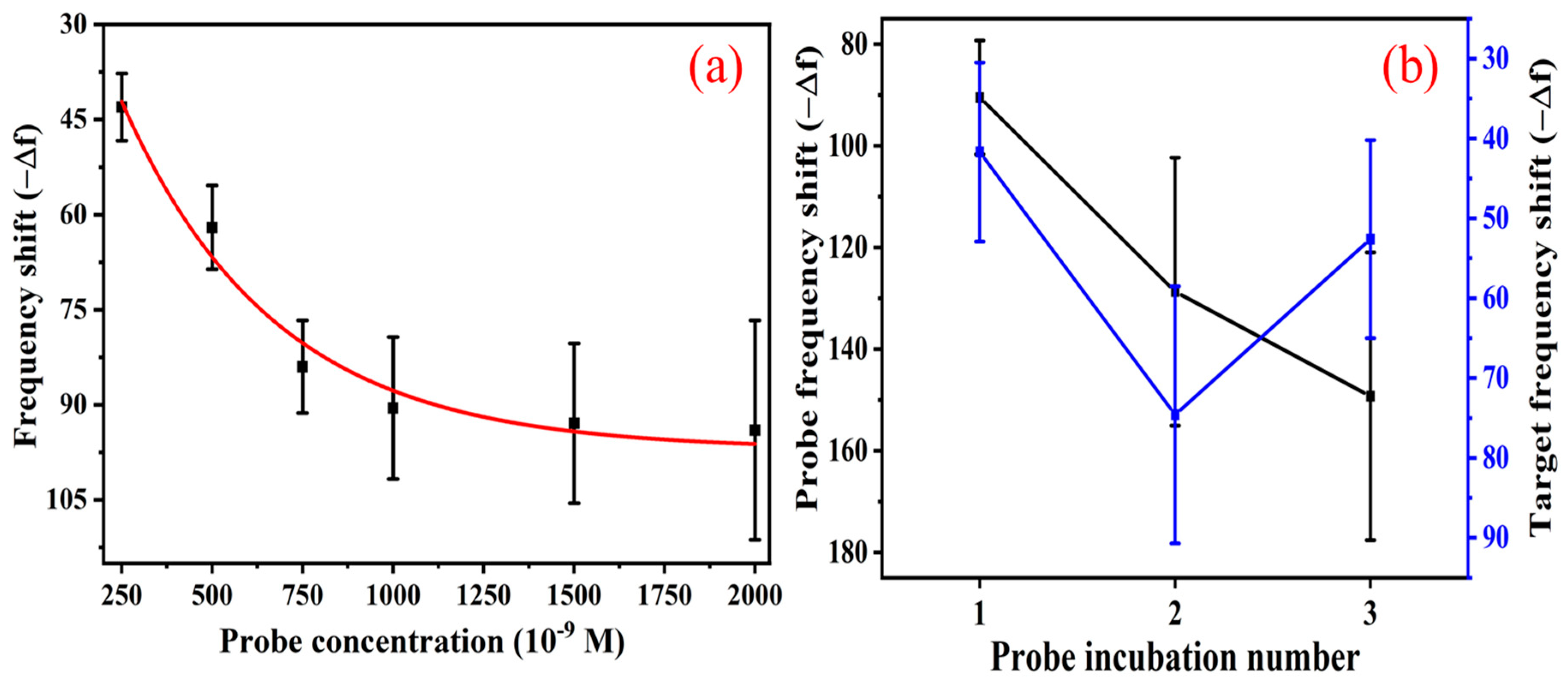
3.3. Effect of Probe Concentration and Double-Probe Approach

To optimise the probe concentration for effective target hybridisation in QCM measurements, probe concentrations were systematically varied at 250 nM, 500 nM, 750 nM, 1000 nM, 1500 nM, and 2000 nM while maintaining a constant target concentration of 1 µM. The corresponding frequency shifts recorded were −43 Hz ± 5.3, −62 Hz ± 6.6, −84 Hz ± 7.3, −90.5 Hz ± 11.2, −92.9 Hz ± 12.6, and −94 Hz ± 17.3, respectively, as shown in Figure 5a. The results demonstrated a progressive increase in the frequency shift with increasing probe concentration, indicating enhanced surface functionalisation and target binding efficiency. However, beyond 1000 nM, the frequency shift approached saturation, suggesting a maximum probe density threshold beyond which additional immobilisation did not significantly enhance hybridisation efficiency. Notably, at 1000 nM, a frequency shift of −90.5 Hz ± 11.2 was observed, indicating an optimal probe density that maximised hybridisation while minimising steric hindrance [50]. Higher probe concentrations resulted in only marginal frequency shifts with increased variability, potentially due to probe overcrowding or reduced accessibility for target binding. Based on these findings, 1000 nM was identified as the optimal probe concentration for maximising hybridisation efficiency and signal stability in subsequent QCM-based sensing applications.
The probe was incubated in consecutive steps to investigate the impact of sequential probe immobilisation and respective target hybridisation efficiency, and frequency shifts were recorded after each time. The corresponding frequency shifts after the single, double, and triple incubations were −90.5 Hz ± 11.2, −128.7 Hz ± 26.36, and −149.3 Hz ± 28.3, respectively, as shown in Figure 5b. These results indicate a progressive increase in surface mass loading with each additional probe deposition, suggesting an enhanced probe immobilisation. However, the increasing error bars at higher immobilisation steps imply variability in probe surface coverage, likely due to steric hindrance or probe aggregation effects. Following probe immobilisation, hybridisation efficiency was assessed by introducing a fixed target concentration in each case. The recorded frequency shifts upon hybridisation were −41.7 Hz ± 11.2, −74.6 Hz ± 16.1, and −52.6 Hz ± 12.4 for the single, double, and triple incubation steps. The hybridisation response exhibited a significant increase for double-probe incubation, suggesting an improved target binding due to a higher probe density. However, a decline in the frequency shift was observed for triple incubation, indicating a potential saturation effect or reduced accessibility of probe (hybridisation) sites due to steric hindrance. Based on these results, a double probe provides the most efficient balance between probe surface coverage and hybridisation efficiency.

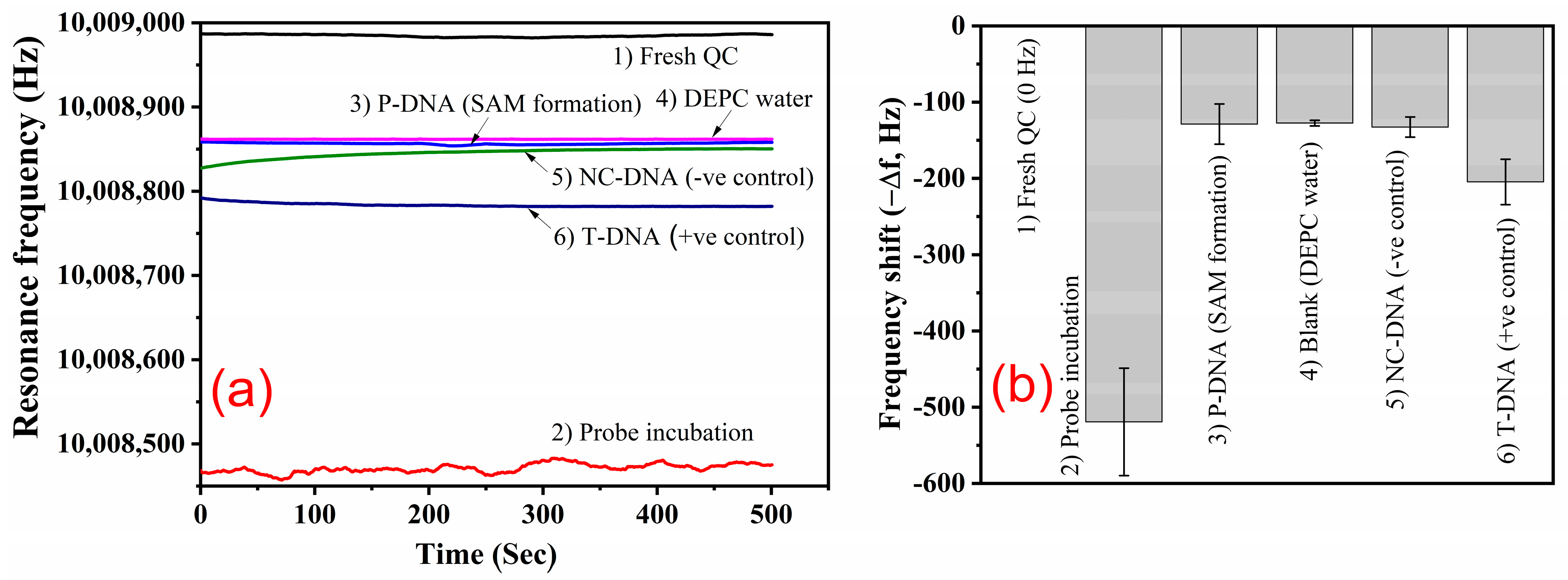
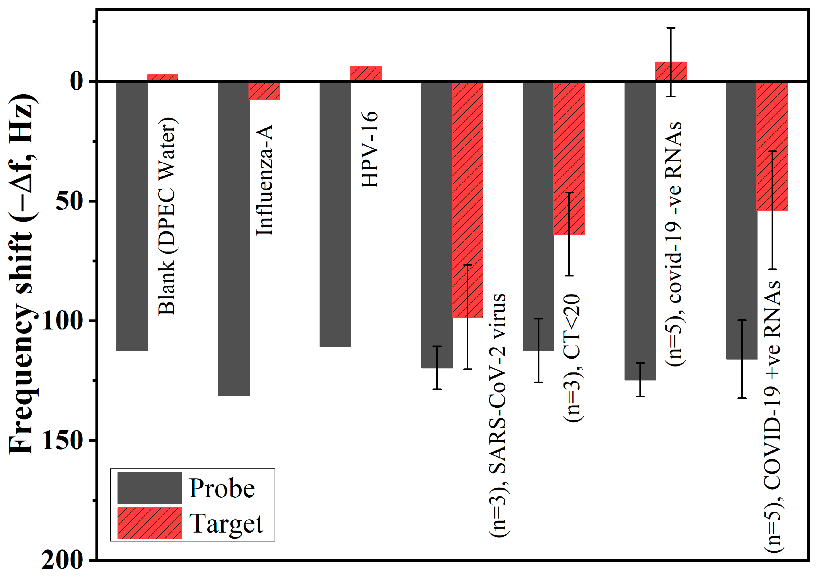
3.4. Target Hybridisation and Selectivity

Under optimised conditions based on the dry-QCM method, P-DNA SAM formation and the reactivity of synthetic T-DNA and NC-DNA were studied via the frequency response of the QCM sensor. P-DNA SAM formation was achieved by directly incubating the probe DNA in the QCM chamber at an elevated temperature of 55 °C using a double-probe approach. The frequency shift due to probe incubation (unwashed sensor) was around −519 ± 74.8 Hz from the baseline value (0 Hz). At this stage, the observed shift was due to both physisorbed and chemisorbed P-DNA. After washing the unbound P-DNA from the QC, the frequency shift due to the SAM was −128.7 ± 26.3 Hz. From Equation (2), the estimated amount of P-DNA attached to the sensor surface (change in mass, Δ m ) was around 0.59 ng/cm2. At this stage, the observed shift was due to chemisorbed P-DNA. The calculated probe (P-DNA) density was 0.505 × 10¹² probes/cm2. Excess probe density can cause less target hybridisation, and the recommended higher limit of probe density is 3 × 10¹² probes/cm2 [45,46,47,51]. The frequency shift for the blank from DEPC water was around ±6 Hz from the P-DNA level.
To assess the selectivity of the non-fouling target, NC-DNA diluted in 0.5 M HEPES solution was directly incubated in the QCM chamber for 30 min. The frequency shift due to the NC-DNA was around −13 ± 6 Hz from the P-DNA level, which could be due to the non-specific absorption of NC-DNA. To assess the selectivity of the complementary target, T-DNA diluted in 0.5 M HEPES solution was directly incubated in the QCM chamber for 30 min. The frequency shift due to T-DNA hybridisation was around −204.5 ± 29.7 Hz from the baseline and −75.8 Hz from the P-DNA reference level. From Equation (2), the estimated amount of T-DNA attached to the P-DNA was around 0.36 ng/cm2. The target hybridisation efficiency can be calculated by dividing the frequency shift due to the total number of probes with the frequency shift due to probe–target hybridisation, and the estimated hybridisation efficiency was about 58.9%. Probe density-dependent kinetics or steric constraints may alter the apparent stability and selectivity of probe–target binding in surface-immobilised oligonucleotides. Both the rate of target capture and the extent of hybridisation strongly depend on surface probe density [45,46]. The efficiency of hybridisation strongly depends on the availability and orientation of P-DNA. Respective time-dependent resonance frequency values are shown in Figure 6a, and frequency shift graphs are shown in Figure 6b.

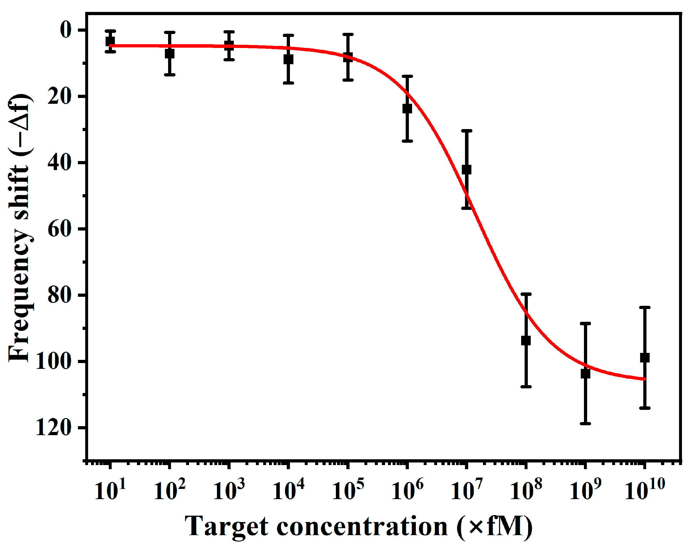
3.5. Sensitivity of Sensor and Limit of Detection (LoD)

To validate the developed sensor’s sensitivity and Limit of Detection (LoD), the P-DNA-functionalised sensor was tested across a broad T-DNA concentration range from 10 fM to 10 µM. The measured sensor frequency responses and corresponding shifts were plotted and analysed using different fitting models, as shown in Figure 7. In this study, we applied a logistic model to fit the relationship between concentration and frequency shift in our experiment. The logistic model is particularly suitable for biosensor response analysis as it effectively captures the characteristic saturation behaviour observed at low and high concentrations, with a transition phase in between the two. Given that the frequency shift does not follow a simple linear trend across several orders of magnitude of analyte concentration, a log-linear transformation (log concentration vs. frequency shift) was used for accurate fitting. The logistic function used for modelling is expressed as follows:
f ( c ) = A + C m a x C m i n 1 + e ( l o g 10 ( C ) C 0 ) / k
where C m a x and C m i n are the lower and upper asymptotes of the frequency shifts. C0 is the inflexion point, and k controls the steepness of the transition. Based on this model, we determined the Limit of Detection (LoD), which is defined as follows:
LoD = C m i n + 3 σ b a s e l i n e
where C m i n is the lowest detectable concentration, and σ b a s e l i n e is the standard deviation of the baseline signal. Using the lowest concentration measurement (10 fM) as the baseline, we estimated σ b a s e l i n e was 3.1 Hz by solving for the concentration corresponding to a frequency shift equal to A + σ b a s e l i n e . The calculated LoD was approximately 1.22 nM. This value represents the minimum concentration at which the sensor provides a distinguishable response from the noise level, demonstrating our system’s high sensitivity.

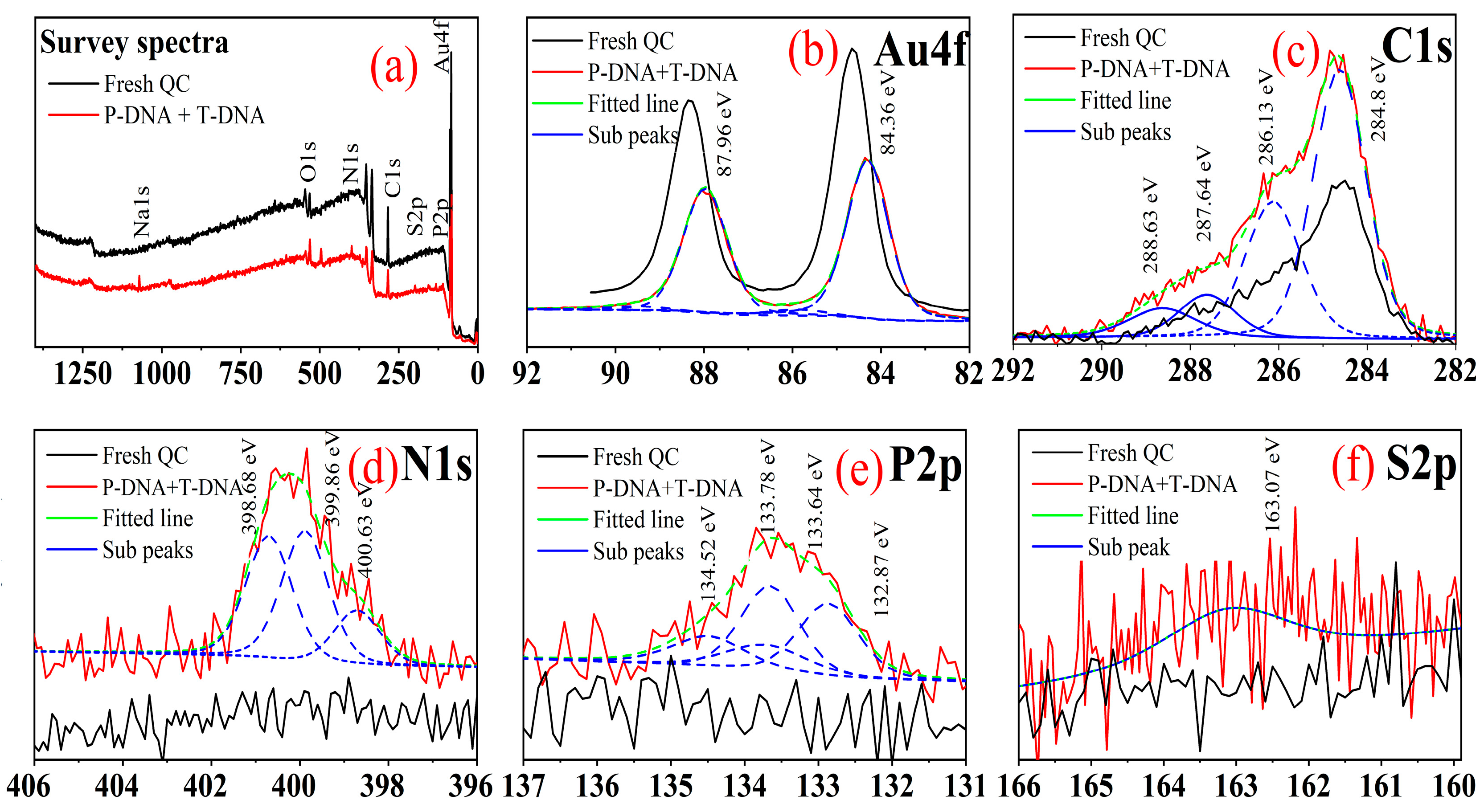
3.6. Surface Characterisations

The formation of SAMs on the sensor surface was investigated using contact angle measurements via the sessile drop method. DI water was used to characterise P-DNA immobilisation and T-DNA hybridisation. By analysing the change in contact angle values, the hydrophobic/hydrophilic nature of the surface due to the immobilisation of the thiolated probe and hybridisation with genomic DNA molecules can be revealed. The initial contact angle of the fresh QC surface was measured at 67.8°, which decreased to 46.1° following P-DNA immobilisation, further reduced to 36.5° upon T-DNA hybridisation, and ultimately declined to 27.5° after hybridisation with COVID-19 RNA, as illustrated in Figure 8. The decrease in contact angle values is attributed to the presence of a hydrophilic group (OH) and charged nucleotides in P-DNA. The hydrophilic nature is further increased after the T-DNA hybridisation, and it might be due to the formation of a duplex and more charged moieties being present in a double-stranded nucleotide sequence. These findings indicate that the nucleic acid was successfully bound to the Au electrode surface. The results further confirm the successful deposition of the probe and T-DNA hybridisation to the P-DNA.
Atomic Force Microscopy (AFM) was employed to characterise the surface topography of the QC electrode following P-DNA immobilisation, T-DNA hybridisation, and COVID-19 RNA hybridisation. Two-dimensional morphological images of 10 × 10 µm2 area scans are presented in Figure 9. The initial root mean square (RMS) roughness of the fresh QC electrode was measured at 2.77 nm, which increased to 3.17 nm after P-DNA immobilisation, further rising to 3.60 nm following T-DNA hybridisation, and ultimately reaching 4.17 nm after COVID-19 RNA hybridisation. The observed increase in RMS values indicates significant surface morphological changes upon probe and target DNA/RNA incubation. This increase can be attributed to the adsorption of single-stranded DNA (ssDNA) during probe immobilisation, the formation of a more complex double-stranded DNA (dsDNA) structure upon target hybridisation, and the additional hybridisation of COVID-19 RNA, which further enhances molecular crowding and structural complexity. The AFM results confirm the successful deposition and hybridisation of probe and target DNAs/RNA on the QC electrode surface, aligning well with existing literature reports.
XPS is an effective method for analysing the SAM formation on the sensor surface and allows a detailed examination of the surface composition and the chemical states of elements within the SAM by identifying functional groups or bonding environments on the surface. XPS characterisation of P-DNA SAM formation and T-DNA hybridisations are shown in Figure 10. Figure 10a describes the survey spectra of all the detected elements before and after hybridisation. High-resolution scans were acquired using a 45° take-off angle for the peak regions typical for Au4f, C1s, N1s, P2p, and S2p peaks. The observed negative shift in the Au 4f peak from 84.6 to 84.1 eV (as shown in Figure 10b) after DNA immobilisation is primarily due to the charge transfer from the negatively charged DNA to the gold surface rather than the expected positive shift from Au–S bond formation. Ar plasma cleaning played a crucial role by removing surface oxides, exposing more reactive gold sites, and enhancing electron density, which further contributed to the negative shift. Additionally, the presence of high-density DNA coverage and electrostatic interactions modified the local work function and dipole potential, reinforcing the shift toward lower binding energy. The C1s peak can be deconvoluted into the four most prominent peaks, as shown in Figure 10c. These peaks at 284.9, 286.13, 287.6, and 288.63 eV are attributed to C=C, C–O, C=O, and O-C=O bindings, respectively [48,49,52]. The N1s region has three broad peaks at 398.6, 399.86, and 400.63 eV, which can be attributed to C–N or –C–NH, –C=N–, and –NH bonds, respectively (as shown in Figure 10d) [53,54,55,56]. Deconvolution of the P2p signal should be a doublet with BEs of 134.84 and 134.00 eV. However, a single peak was able to fill due to a very weak signal (Figure 10e). To a better extent, the presence of both N and P together is an excellent indicator of the probe/target DNA adsorbing to the electrode, because N and P surface contamination is typically very rare during sample preparation and handling. The peak at 163.01 eV in the S2p spectra is specifically attributed to the S-Au bond, as shown in Figure 10f. Obtaining the S peak is very difficult since its abundance is very low in SAM, and the existence of the S peak is direct evidence of SAM formation [44,55]. There was not any peak between 163 and 164 eV associated with unbound thiol groups, and the peak at 167.4 eV was assigned to fully oxidised sulphur. However, both peak regions were observed in unwashed sensor chips XPS spectra. This means that the unbound and oxidised sulphur-containing probe DNA was washed away during the cleaning steps and only remained bound to thiol oligonucleotides.

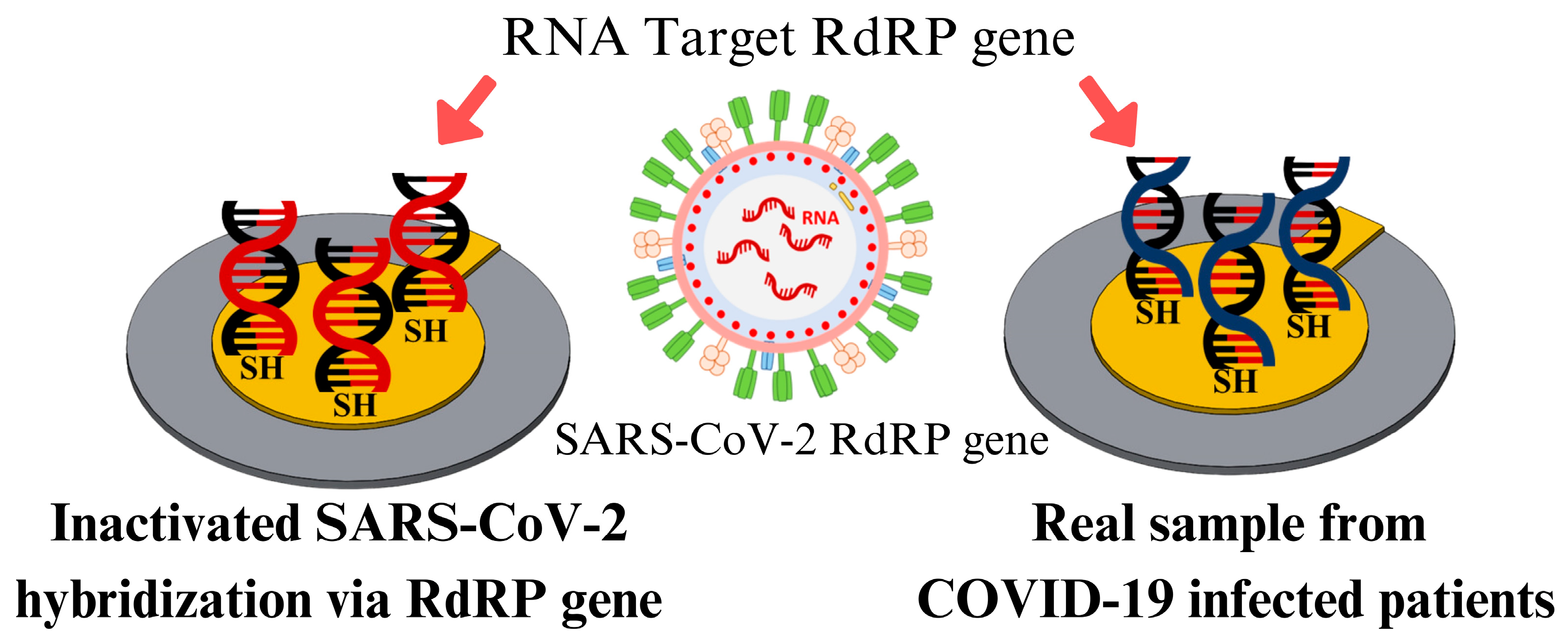
3.7. Specificity and Clinical Validation of Real Samples

A sensor’s specificity can be significantly enhanced with nonspecific target controls for its point-of-care diagnosis. Cross-reactivity was determined using the influenza-A sample with no absorbable frequency shift, indicating non-reactivity with the developed assay. In fact, influenza-A is in the same family as the SARS-CoV-2 virus, and the anticipated result was non-reactive. Similarly, cross-reactivity with a cervical HPV-infected (non-specific target control) sample was tested and found to be non-reactive with the developed assay. It can be seen that the frequency shift values for the blank (DEPC water), influenza-A, and HPV-16 are +2.72 Hz, −7.53 Hz, and 6.19 Hz, respectively, as shown in Figure 11. The utilised oligonucleotide RdRP P-DNA gene is non-complementary to any SARS family and human mRNAs [57].
However, there was a large negative shift in cross-reactivity with inactivated SARS-CoV-2 virus (−98.4 ± 19.8), COVID-19 CT<20-valued RNAs (−63.5 ± 17.6), and COVID-19-infected RNA (−53.8 ± 24.7) samples, indicating the hybridisation of a specific RdRP gene with P-DNA. Further, its non-interference is also seen with COVID-19 −ve RNA samples (−8.61 ± 24.67). The respective results with their frequency shifts are plotted as shown in Figure 11. The developed sensor is highly specific and selective for the COVID-19-infected patients. The SARS-CoV-2 virus is found to be highly stable at 4 °C, stable for 1 day at 37 °C, and unstable (less than 5 min) at 70 °C [58]. Since the RNA and virus are unstable at higher temperatures, hybridisation was performed at 35 °C. The hybridisation strategy of the DNA probe with the SARS-CoV-2 virus and real RNA samples from patients is shown in Figure 12.

3.8. Re-Use and Regeneration of QCM Sensor

To explore the repeatability and stability, the developed QCM sensor was hybridised and de-hybridised between P-DNA and T-DNA. The experiment was repeated using the same concentrations and circumstances. There are some reports published on the de-hybridisation of T-DNA, including dipping the sensor in 1 mM HCl twice, each time for 30 s [51] and dipping the sensor in 0.1 M NaOH for 10 s followed by DI water rinsing [59]. However, this experiment used 0.5 M NaOH (in 3 M NaCl at a 1:1 ratio) for de-hybridisation [60]. As shown in Figure 13, after three cycles of de-hybridisation and re-hybridisation, the second and third hybridisation produced 82% and 80% of the first hybridisation signal, respectively.
In this study, regeneration steps removed the DNA SAM layer, thiolate surface and oxidised Au sensor surface. The regenerated sensor frequency shifted positively (~+10 Hz) compared to the pristine QC, as shown in Figure 13. The proclaimed efficiency of sensor regeneration using NaBH4 is about 90% [61]. Ultrasonication can remove the physisorbed contaminants and break the covalent bonds by cavitation (20–70 K Pascals and localised temperature up to 5000 K) [62]. Plasma cleaning/etching (here we used Ar gas plasma) is an effective and efficient technique to remove metal oxide layers (a few nanometres) [63]. The energised Ar ions are enough to knock out a few atomic layers of the sensor surface (Au surface), which might be thiolated Au (Au-S) and oxidised Au, and those exhibit a positive shift in the sensor frequency. This developed protocol has 100% efficiency in sensor regeneration and creates a highly hydrophilic Au surface (supposed to have +ve charge), which is essential for probe (−ve charged Au-S DNA) incubation. In fact, each sensor could regenerate more than 15 times in our case study, and the generated sensor could be used as new for further experiments.

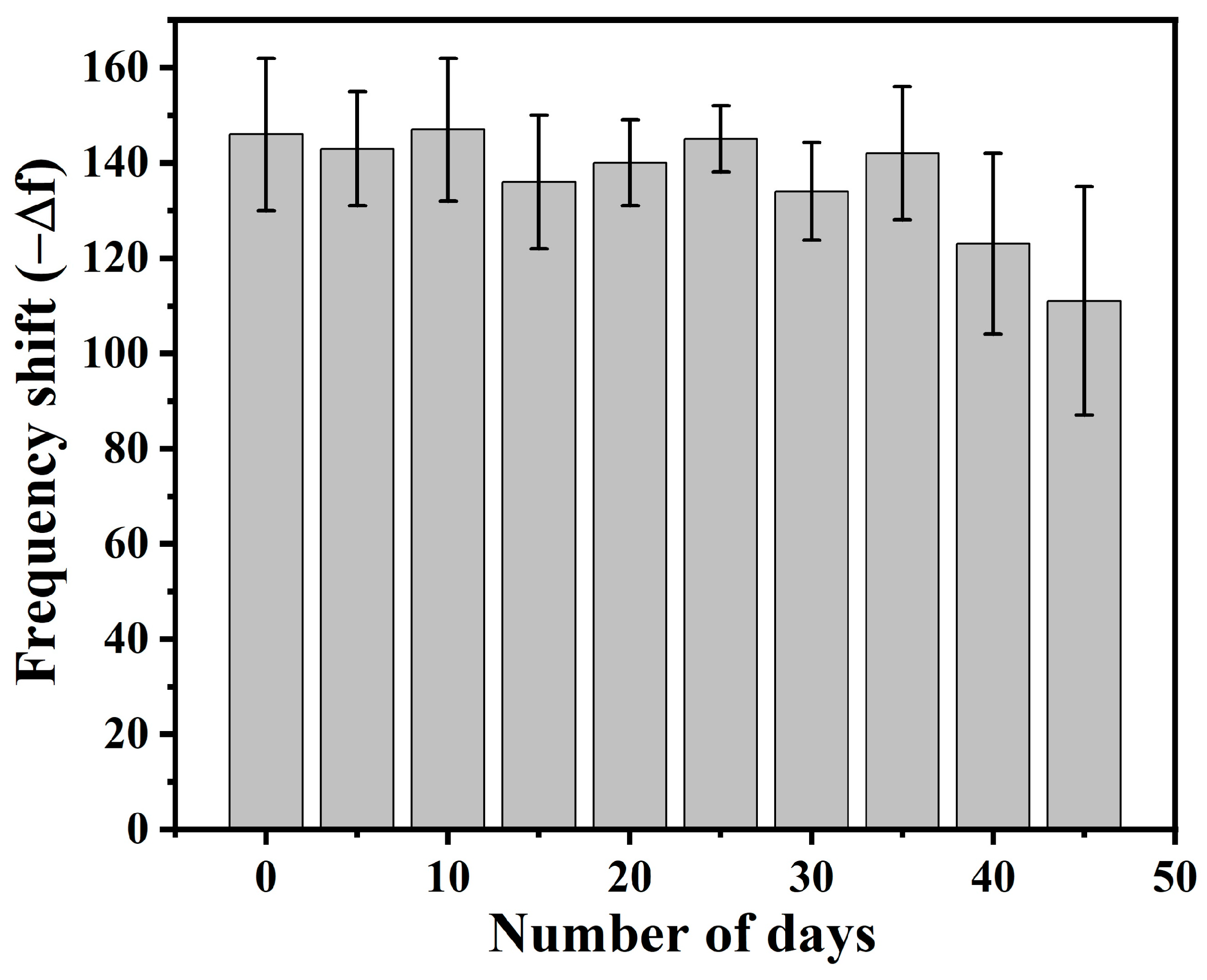
3.9. Shelf-Life of the Developed Sensor

The analytical performance of the developed sensor was also evaluated over a month, as shown in Figure 14. Sensor stability was assessed every 5 days for 45 days, and frequency responses were measured. The results showed that the sensors were effectively preserved for a certain period and later degraded gradually. The biosensor generated a stable response over 25 days with a deviation of −12 Hz, the value of which corresponds to the blank QC variation with DPEC water, and further degradation might be attributed to the denaturalisation of P-DNA strands from the sensor. This establishes the shelf-life of the biosensor over the specified duration.

4. Conclusions

In summary, the developed QCM-based biosensors were able to detect the SARS-CoV-2-related DNA (T-DNA) and RNAs (samples from the hospitals). The proposed dry-QCM method brought high stability, a fast response time and mitigated the usage of wet reagents during the measurement. Temperature-assisted incubation enhanced the P-DNA SAM formation on the sensor surface. Contact angle measurements, AFM, and XPS provided adequate evidence of P-DNA SAM formation, thiol interactions via Au-S bond formation, and probe–target hybridisation. The developed sensor is particular to the T-DNA and COVID-19 RNA (from nasopharyngeal swab) samples. The obtained LoD was 1.03 PM for T-DNA, and the shelf life of the developed sensor was 25 days. Re-use of the developed sensor is possible for up to 5 times, and in terms of regeneration, the sensor is able to be extended more than 15 times.

Author Contributions

D.M.—Conceptualisation, formal analysis, data curation, writing—original draft, writing—review and editing. T.S.—Data curation. N.C.—Data curation. D.K.A.—Resources and funding acquisition. A.M.—Investigation, funding acquisition, and writing—review and editing. S.R.—Data curation. S.S.—Formal analysis, supervision, and methodology. S.P.—Supervision, funding acquisition, and writing—review and editing. All authors have read and agreed to the published version of the manuscript.

Funding

This research has received funding support from (i) Suranaree University of Technology (SUT) and (ii) the NSRF via the Program Management Unit for Human Resources & Institutional Development, Research and Innovation (PMU-B) [grant number B13F660067].

Institutional Review Board Statement

This work was approved by the Human Research Ethics Committee (HREC) from Suranaree University of Technology (SUT), Thailand, on 21 December 2022 (EC_64-136). All biosafety work was carried out after approval from the HREC (COA. No. 138/2564). Suranaree University of Technology Hospital (SUTH), Thailand, provided the RT-PCR data of the COVID-19 patients.

Informed Consent Statement

Patient consent was waived due to the retrospective nature of the study, as it involved the use of pre-existing, de-identified medical records, and obtaining individual consent was impractical. The study posed minimal risk to participants, and all data were handled in accordance with ethical guidelines to ensure confidentiality and privacy. The waiver was approved by the Institutional Review Board (IRB) in compliance with relevant regulations.

Data Availability Statement

The original contributions presented in this study are included in the article. Further inquiries can be directed to the corresponding author(s).

Acknowledgments

Authors acknowledge the funding support from NSRF via the Program Management Unit for Human Resources & Institutional Development, Research and Innovation (PMU-B) [grant number B13F660067]. The authors also acknowledge the Indo-ASEAN collaboration project (grant no CRD/2021/000472).

Conflicts of Interest

The authors declare no conflicts of interest.

Abbreviations

The following abbreviations are used in this manuscript:
DNADeoxyribonucleic acids
RNARibonucleic acid
RdRPRNA-dependent RNA Polymerase
SAMSelf-assembled monolayer
QCMQuartz crystal microbalance

Appendix A

Figure A1. Amount of liquid optimisation in the QCM chamber: (a) frequency response of QC at different liquid quantities, (b) frequency shift due to different liquid quantities.
Technologies 13 00114 g0a1
Supporting data-A1: Amount of liquid optimisation in the QCM chamber. The frequency shift is invariant after 80 μL (DIW). The unexpected spike (around 1500 s) after 20 μL is due to the relocation of liquid droplets to the edges and the inability to control the size until the total sensor surface is covered. The bigger +ve shifts (around 3000 and 5000 s) are due to the removal of liquid from the chamber.
Figure A2. QCM measurement in liquid media without P-DNA at different target concentrations. Inset shows the enlarged view of different concentrations.
Technologies 13 00114 g0a2
Supporting data-A2: Different concentrations of target DNA without probe DNA immobilisation. This is a continuous measurement of sequential additions to the sensor surface. The signal is highly unstable in the liquid media. The spikes are due to the removal of liquid from the chamber. The frequency shift seems to be invariant or very small for all the concentrations.
Figure A3. QCM measurement in liquid media with P-DNA at different target concentrations. Inset image shows the enlarged view of different concentrations.
Technologies 13 00114 g0a3
Supporting data-A3: Different concentrations of target DNA with the probe DNA immobilisation. This is continuous measurement of sequential additions to the sensor surface. The signal is highly unstable in the liquid media. The spikes are due to the removal of liquid from the chamber. However, there is an observable shift for different concentrations.

References

  1. Spiteri, G.; Fielding, J.; Diercke, M.; Campese, C.; Enouf, V.; Gaymard, A.; Bella, A.; Sognamiglio, P.; Sierra Moros, M.J.; Riutort, A.N.; et al. First Cases of Coronavirus Disease 2019 (COVID-19) in the WHO European Region, 24 January to 21 February 2020. Eurosurveillance 2020, 25, 1–6. [Google Scholar] [CrossRef] [PubMed]
  2. Li, X.; Cui, W.; Zhang, F. Who Was the First Doctor to Report the COVID-19 Outbreak in Wuhan, China? J. Nucl. Med. 2020, 61, 782–783. [Google Scholar] [CrossRef] [PubMed]
  3. LeDuc, J.W.; Barry, M.A. SARS, the First Pandemic of the 21st Century. Emerg. Infect. Dis. 2004, 10, e26. [Google Scholar] [CrossRef]
  4. World Health Organization (WHO). Tracking SARS-CoV-2 Variants. 2024. Available online: https://www.who.int/emergencies/overview/tracking-SARS-CoV-2-Variants (accessed on 1 September 2023).
  5. COVID-19 Map Johns Hopkins: Coronavirus Resource Center, COVID-19 Dashboard. 2023. Available online: https://coronavirus.jhu.edu/map.html (accessed on 4 September 2023).
  6. Zhu, H.; Wei, L.; Niu, P. The Novel Coronavirus Outbreak in Wuhan, China. Glob. Health Res. Policy 2020, 5, 6. [Google Scholar] [CrossRef]
  7. Haque, S.M.; Ashwaq, O.; Sarief, A.; Azad John Mohamed, A.K. A Comprehensive Review About SARS-CoV-2. Future Virol. 2020, 15, 625–648. [Google Scholar] [CrossRef]
  8. Lu, R.; Zhao, X.; Li, J.; Niu, P.; Yang, B.; Wu, H.; Wang, W.; Song, H.; Huang, B.; Zhu, N.; et al. Genomic Characterisation and Epidemiology of 2019 Novel Coronavirus: Implications for Virus Origins and Receptor Binding. Lancet 2020, 395, 565–574. [Google Scholar] [CrossRef] [PubMed]
  9. Cui, J.; Li, F.; Shi, Z.-L. Origin and Evolution of Pathogenic Coronaviruses. Nat. Rev. Microbiol. 2019, 17, 181–192. [Google Scholar] [CrossRef]
  10. Dutta, D.; Naiyer, S.; Mansuri, S.; Soni, N.; Singh, V.; Bhat, K.H.; Singh, N.; Arora, G.; Mansuri, M.S. COVID-19 Diagnosis: A Comprehensive Review of the RT-qPCR Method for Detection of SARS-CoV-2. Diagnostics 2022, 12, 1503. [Google Scholar] [CrossRef]
  11. Munne, K.; Bhanothu, V.; Bhor, V.; Patel, V.; Mahale, S.D.; Pande, S. Detection of SARS-CoV-2 Infection by RT-PCR Test: Factors Influencing Interpretation of Results. VirusDisease 2021, 32, 187–189. [Google Scholar] [CrossRef]
  12. Zhang, W.; Xu, X.; Qi, L.; Liu, M.; Zhao, X.; Kong, L.; Wang, Y.; Chen, F.; Zhang, C.; Cheng, J.; et al. Clinical Evaluation of a New COVID-19 Antigen Rapid Test Kit for Detection of SARS-CoV-2. Diagn. Microbiol. Infect. Dis. 2024, 108, 116136. [Google Scholar] [CrossRef]
  13. Mak, G.C.; Lau, S.S.; Wong, K.K.; Chow, N.L.; Lau, C.-S.; Lam, E.T.; Ng, K.H.; Chan, R.C. Evaluation of Rapid Antigen Detection Kits for the Detection of SARS-CoV-2 B.1.617.2 Virus. Future Virol. 2022, 17, 387–392. [Google Scholar] [CrossRef] [PubMed]
  14. Udugama, B.; Kadhiresan, P.; Kozlowski, H.N.; Malekjahani, A.; Osborne, M.; Li, V.Y.C.; Chen, H.; Mubareka, S.; Gubbay, J.B.; Chan, W.C.W. Diagnosing COVID-19: The Disease and Tools for Detection. ACS Nano 2020, 14, 3822–3835. [Google Scholar] [CrossRef] [PubMed]
  15. Vashist, S.K.; Vashist, P. Recent Advances in Quartz Crystal Microbalance-Based Sensors. J. Sens. 2011, 2011, 571405. [Google Scholar] [CrossRef]
  16. Linares, M.; Pérez-Tanoira, R.; Carrero, A.; Romanyk, J.; Pérez-García, F.; Gómez-Herruz, P.; Arroyo, T.; Cuadros, J. Panbio Antigen Rapid Test Is Reliable to Diagnose SARS-CoV-2 Infection in the First 7 Days After the Onset of Symptoms. J. Clin. Virol. 2020, 133, 104659. [Google Scholar] [CrossRef] [PubMed]
  17. Guo, L.; Ren, L.; Yang, S.; Xiao, M.; Chang, D.; Yang, F.; Dela Cruz, C.S.; Wang, Y.; Wu, C.; Xiao, Y.; et al. Profiling Early Humoral Response to Diagnose Novel Coronavirus Disease (COVID-19). Clin. Infect. Dis. 2020, 71, 778–785. [Google Scholar] [CrossRef]
  18. Baldanti, F.; Ganguly, N.K.; Wang, G.; Möckel, M.; O’Neill, L.A.; Renz, H.; Dos Santos Ferreira, C.E.; Tateda, K.; Van Der Pol, B. Choice of SARS-CoV-2 Diagnostic Test: Challenges and Key Considerations for the Future. Crit. Rev. Clin. Lab. Sci. 2022, 59, 445–459. [Google Scholar] [CrossRef]
  19. Ravina; Kumar, A.; Manjeet; Twinkle; Subodh; Narang, J.; Mohan, H. Analytical Performances of Different Diagnostic Methods for SARS-CoV-2 Virus—A Review. Sens. Int. 2022, 3, 100197. [Google Scholar] [CrossRef]
  20. Philip, A.; Kumar, A.R. The Performance Enhancement of Surface Plasmon Resonance Optical Sensors Using Nanomaterials: A Review. Coord. Chem. Rev. 2022, 458, 214424. [Google Scholar] [CrossRef]
  21. Melo, A.F.A.A.; Singh, S.J.; Chinnamuthu, P.; Crespilho, F.N.; Rydzek, G. Magnetically Stimulated Bio- and Electrochemical Systems: State-of-the-Art, Applications, and Future Directions. ChemNanoMat 2023, 9, e202300192. [Google Scholar] [CrossRef]
  22. Sun, C.; Wang, X.; Auwalu, M.A.; Cheng, S.; Hu, W. Organic Thin Film Transistors-Based Biosensors. EcoMat 2021, 3, e12094. [Google Scholar] [CrossRef]
  23. Fawcett, N.C.; Evans, J.A.; Chien, L.-C.; Flowers, N. Nucleic Acid Hybridization Detected by Piezoelectric Resonance. Anal. Lett. 1988, 21, 1099–1114. [Google Scholar] [CrossRef]
  24. Cadoni, E.; Manicardi, A.; Madder, A. PNA-Based MicroRNA Detection Methodologies. Molecules 2020, 25, 1296. [Google Scholar] [CrossRef] [PubMed]
  25. Chen, J.Y.; Penn, L.S.; Xi, J. Quartz Crystal Microbalance: Sensing Cell-Substrate Adhesion and Beyond. Biosens. Bioelectron. 2018, 99, 593–602. [Google Scholar] [CrossRef]
  26. Zuo, C.; Wen, Y.; Chen, D.; Ouyang, J.; Li, P.; Dong, T. Dynamic Monitoring of Biomolecular Hydrodynamic Dimensions by Magnetization Motion on Quartz Crystal Microbalance. Anal. Chem. 2024, 96, 7421–7428. [Google Scholar] [CrossRef] [PubMed]
  27. Rawle, R.J.; Selassie, C.R.D.; Johal, M.S. Creation of Mammalian Single- and Double-Stranded DNA Surfaces: A Real-Time QCM-D Study. Langmuir 2007, 23, 9563–9566. [Google Scholar] [CrossRef] [PubMed]
  28. Hampitak, P.; Jowitt, T.A.; Melendrez, D.; Fresquet, M.; Hamilton, P.; Iliut, M.; Nie, K.; Spencer, B.; Lennon, R.; Vijayaraghavan, A. A Point-of-Care Immunosensor Based on a Quartz Crystal Microbalance with Graphene Biointerface for Antibody Assay. ACS Sens. 2020, 5, 3520–3532. [Google Scholar] [CrossRef]
  29. Nilebäck, E.; Feuz, L.; Uddenberg, H.; Valiokas, R.; Svedhem, S. Characterization and Application of a Surface Modification Designed for QCM-D Studies of Biotinylated Biomolecules. Biosens. Bioelectron. 2011, 28, 407–413. [Google Scholar] [CrossRef]
  30. Zuo, B.; Li, S.; Guo, Z.; Zhang, J.; Chen, C. Piezoelectric Immunosensor for SARS-Associated Coronavirus in Sputum. Anal. Chem. 2004, 76, 3536–3540. [Google Scholar] [CrossRef]
  31. Albano, D.R.B.; Shum, K.; Tanner, J.A.; Fung, Y.S. BS5.3—Piezoelectric Quartz Crystal Aptamer Biosensor for Detection and Quantification of SARS CoV Helicase Protein. In Proceedings of the 17th International Meeting on Chemical Sensors—IMCS 2018, Vienna, Austria, 15–19 July 2018; AMA Service GmbH: Wunstorf, Germany, 2018; pp. 211–213. [Google Scholar] [CrossRef]
  32. Owen, T.W.; Al-Kaysi, R.O.; Bardeen, C.J.; Cheng, Q. Microgravimetric Immunosensor for Direct Detection of Aerosolized Influenza A Virus Particles. Sens. Actuators B Chem. 2007, 126, 691–699. [Google Scholar] [CrossRef]
  33. Wang, R.; Wang, L.; Callaway, Z.T.; Lu, H.; Huang, T.J.; Li, Y. A Nanowell-Based QCM Aptasensor for Rapid and Sensitive Detection of Avian Influenza Virus. Sens. Actuators B Chem. 2017, 240, 934–940. [Google Scholar] [CrossRef]
  34. Forinova, M.; Pilipenco, A.; Visova, I.; Kuncak, J.; Lynn, N.S.; Yudin, P.; Dostalek, J.; Honig, V.; Palus, M.; Maskova, H.; et al. Biosensor for Rapid Detection of SARS-CoV-2 in Real-World Samples. In Proceedings of the 2021 IEEE Sensors, Sydney, Australia, 31 October–3 November 2021; IEEE: New York, NY, USA, 2021; pp. 1–4. [Google Scholar] [CrossRef]
  35. Forinová, M.; Pilipenco, A.; Víšová, I.; Lynn, N.S.; Dostálek, J.; Mašková, H.; Hönig, V.; Palus, M.; Selinger, M.; Kočová, P.; et al. Functionalized Terpolymer-Brush-Based Biointerface with Improved Antifouling Properties for Ultra-Sensitive Direct Detection of Virus in Crude Clinical Samples. ACS Appl. Mater. Interfaces 2021, 13, 60612–60624. [Google Scholar] [CrossRef] [PubMed]
  36. Nilsson, P.H.; Al-Majdoub, M.; Ibrahim, A.; Aseel, O.; Suriyanarayanan, S.; Andersson, L.; Fostock, S.; Aastrup, T.; Tjernberg, I.; Rydén, I.; et al. Quartz Crystal Microbalance Platform for SARS-CoV-2 Immuno-Diagnostics. Int. J. Mol. Sci. 2023, 24, 16705. [Google Scholar] [CrossRef] [PubMed]
  37. Pandey, L.M. Design of Engineered Surfaces for Prospective Detection of SARS-CoV-2 Using Quartz Crystal Microbalance-Based Techniques. Expert Rev. Proteom. 2020, 17, 425–432. [Google Scholar] [CrossRef] [PubMed]
  38. Nemčeková, K.; Korčeková, J.; Svitková, V.; Baraniak, D.; Domšicová, M.; Melníková, E.; Hornychová, M.; Szebellaiová, V.; Gál, M.; Poturnayová, A. Comparative Analysis of QCM and Electrochemical Aptasensors for SARS-CoV-2 Detection. Biosensors 2024, 14, 431. [Google Scholar] [CrossRef]
  39. Jeong, H.; Lee, S.; Ko, J.; Ko, M.; Seo, H.W. Identification of Conserved Regions from 230,163 SARS-CoV-2 Genomes and Their Use in Diagnostic PCR Primer Design. Genes Genom. 2022, 44, 899–912. [Google Scholar] [CrossRef]
  40. Sauerbrey, G. Verwendung von Schwingquarzen zur Wägung Dünner Schichten und zur Mikrowägung. Z. Phys. 1959, 155, 206–222. [Google Scholar] [CrossRef]
  41. Kanazawa, K.K.; Gordon, J.G. Frequency of a Quartz Microbalance in Contact with Liquid. Anal. Chem. 1985, 57, 1770–1771. [Google Scholar] [CrossRef]
  42. Rodahl, M.; Höök, F.; Fredriksson, C.; Keller, C.A.; Krozer, A.; Brzezinski, P.; Voinova, M.; Kasemo, B. Simultaneous Frequency and Dissipation Factor QCM Measurements of Biomolecular Adsorption and Cell Adhesion. Faraday Discuss. 1997, 107, 229–246. [Google Scholar] [CrossRef]
  43. Researcher Formula for Copy Number Calculation|IDT. Available online: https://sg.idtdna.com/pages/education/decoded/article/calculations-converting-from-nanograms-to-copy-number (accessed on 25 June 2024).
  44. Xu, J.; Craig, S.L. Thermodynamics of DNA Hybridization on Gold Nanoparticles. J. Am. Chem. Soc. 2005, 127, 13227–13231. [Google Scholar] [CrossRef]
  45. Wetmur, J.G.; Davidson, N. Kinetics of Renaturation of DNA. J. Mol. Biol. 1968, 31, 349–370. [Google Scholar] [CrossRef]
  46. Morrison, L.E.; Stols, L.M. Sensitive Fluorescence-Based Thermodynamic and Kinetic Measurements of DNA Hybridization in Solution. Biochemistry 1993, 32, 3095–3104. [Google Scholar] [CrossRef] [PubMed]
  47. Li, Q.; Gu, Y.; Xie, B. Hybrid Temperature Effect on a Quartz Crystal Microbalance Resonator in Aqueous Solutions. Chin. Phys. B 2017, 26, 067704. [Google Scholar] [CrossRef]
  48. Petrovykh, D.Y.; Kimura-Suda, H.; Whitman, L.J.; Tarlov, M.J. Quantitative Analysis and Characterization of DNA Immobilized on Gold. J. Am. Chem. Soc. 2003, 125, 5219–5226. [Google Scholar] [CrossRef]
  49. Roelfs, B.; Bunge, E.; Schröter, C.; Solomun, T.; Meyer, H.; Nichols, R.J.; Baumgärtel, H. Adsorption of Thymine on Gold Single-Crystal Electrodes. J. Phys. Chem. B 1997, 101, 754–765. [Google Scholar] [CrossRef]
  50. Suvanasuthi, R.; Cheewasatheinchaiyaporn, T.; Wat-aksorn, K.; Promptmas, C. Nucleic Acid Amplification Free-QCM-DNA Biosensor for Burkholderia Pseudomallei Detection. Curr. Microbiol. 2023, 80, 376. [Google Scholar] [CrossRef]
  51. Peterson, A.W.; Wolf, L.K.; Georgiadis, R.M. Hybridization of Mismatched or Partially Matched DNA at Surfaces. J. Am. Chem. Soc. 2002, 124, 14601–14607. [Google Scholar] [CrossRef] [PubMed]
  52. Senthilnathan, J.; Sanjeeva Rao, K.; Lin, W.-H.; Liao, J.-D.; Yoshimura, M. Low Energy Synthesis of Nitrogen Functionalized Graphene/Nanoclay Hybrid via Submerged Liquid Plasma Approach. Carbon 2014, 78, 446–454. [Google Scholar] [CrossRef]
  53. Dementjev, A.P.; De Graaf, A.; Van De Sanden, M.C.M.; Maslakov, K.I.; Naumkin, A.V.; Serov, A.A. X-Ray Photoelectron Spectroscopy Reference Data for Identification of the C3N4 Phase in Carbon–Nitrogen Films. Diam. Relat. Mater. 2000, 9, 1904–1907. [Google Scholar] [CrossRef]
  54. Johansson, Å.; Stafström, S. A Δ-Self-Consistent-Field Study of the Nitrogen 1s Binding Energies in Carbon Nitrides. J. Chem. Phys. 1999, 111, 3203–3208. [Google Scholar] [CrossRef]
  55. Senthilnathan, J.; Rao, K.S.; Lin, W.-H.; Ting, J.-M.; Yoshimura, M. Formation of Reusable Au-Acetonitrile Polymers and N-Doped Graphene Catalyst Under UV Light via Submerged Liquid Plasma Process. J. Mater. Chem. A 2015, 3, 3035–3043. [Google Scholar] [CrossRef]
  56. Zhang, B.; Kaziz, S.; Li, H.; Wodka, D.; Malola, S.; Safonova, O.; Nachtegaal, M.; Mazet, C.; Dolamic, I.; Llorca, J.; et al. Pd2 Au36 (SR)24 Cluster: Structure Studies. Nanoscale 2015, 7, 17012–17019. [Google Scholar] [CrossRef] [PubMed]
  57. Moitra, P.; Alafeef, M.; Dighe, K.; Frieman, M.B.; Pan, D. Selective Naked-Eye Detection of SARS-CoV-2 Mediated by N Gene Targeted Antisense Oligonucleotide Capped Plasmonic Nanoparticles. ACS Nano 2020, 14, 7617–7627. [Google Scholar] [CrossRef] [PubMed]
  58. Chin, A.W.H.; Chu, J.T.S.; Perera, M.R.A.; Hui, K.P.Y.; Yen, H.-L.; Chan, M.C.W.; Peiris, M.; Poon, L.L.M. Stability of SARS-CoV-2 in Different Environmental Conditions. Lancet Microbe 2020, 1, e10. [Google Scholar] [CrossRef]
  59. Mannelli, I.; Minunni, M.; Tombelli, S.; Wang, R.; Michela Spiriti, M.; Mascini, M. Direct Immobilisation of DNA Probes for the Development of Affinity Biosensors. Bioelectrochemistry 2005, 66, 129–138. [Google Scholar] [CrossRef]
  60. Lazerges, M.; Perrot, H.; Zeghib, N.; Antoine, E.; Compere, C. In Situ QCM DNA-Biosensor Probe Modification. Sens. Actuators B Chem. 2006, 120, 329–337. [Google Scholar] [CrossRef]
  61. Dupont-Filliard, A.; Billon, M.; Livache, T.; Guillerez, S. Biotin/Avidin System for the Generation of Fully Renewable DNA Sensor Based on Biotinylated Polypyrrole Film. Anal. Chim. Acta 2004, 515, 271–277. [Google Scholar] [CrossRef]
  62. Le, H.T.N.; Phan, L.M.T.; Cho, S. Removal of Thiol-SAM on a Gold Surface for Re-Use of an Interdigitated Chain-Shaped Electrode. Materials 2022, 15, 2218. [Google Scholar] [CrossRef]
  63. Dehghani, M.H.; Karri, R.R.; Koduru, J.R.; Manickam, S.; Tyagi, I.; Mubarak, N.M. Suhas Recent Trends in the Applications of Sonochemical Reactors as an Advanced Oxidation Process for the Remediation of Microbial Hazards Associated with Water and Wastewater: A Critical Review. Ultrason. Sonochem. 2023, 94, 106302. [Google Scholar] [CrossRef]
Figure 1. A shaker instrument with a specially designed glass holder (to hold the QCs) was used to clean the QCs, remove the unbound DNA, and de-hybridise the target. Inset image shows the QC holder with QCs.
Technologies 13 00114 g001
Figure 2. Schematic representation of each step of the dry-QCM technique: (a) fresh QC, (b) probe DNA incubation, (c) target DNA hybridisation, and (d) frequency response in each step.
Technologies 13 00114 g002
Figure 3. Effect of temperature on probe immobilisation and target hybridisation: (a) P-DNA incubation at 5 °C, (b) P-DNA incubation at 30 °C, (c) P-DNA incubation at 55 °C, (d) T-DNA hybridisation at 5 °C, (e) T-DNA hybridisation at 30 °C, and (f) T-DNA hybridisation at 55 °C. For all the graphs, X-axis represents Time (s) and Y-axis represents Resonance frequency (Hz).
Technologies 13 00114 g003
Figure 4. Effect of temperature on probe immobilisation, target hybridisation and corresponding frequency shifts.
Technologies 13 00114 g004
Figure 5. (a) Optimisation of probe concentration and (b) effect of sequential probe incubations (black colour) for better target hybridisation (blue colour) and corresponding frequency shifts.
Technologies 13 00114 g005
Figure 6. Frequency response of the portable device by using the dry-QCM method. (a) Frequency response of probe, non-complementary and complementary target DNAs, and (b) corresponding frequency shifts.
Technologies 13 00114 g006
Figure 7. Established calibration curve of a QCM sensor for T-DNA in log-linear scale.
Technologies 13 00114 g007
Figure 8. Contact angle measurement on pristine QC, with P-DNA immobilisation, after T-DNA and ssRNA hybridisation.
Technologies 13 00114 g008
Figure 9. AFM surface morphological 2D images of (a) fresh QC electrode, (b) with P-DNA immobilisation, (c) with T-DNA hybridisation, and (d) with COVID-19 RNA hybridisation.
Technologies 13 00114 g009
Figure 10. XPS characterisation of probe (P-DNA) self-assembled monolayer formation and target (T-DNA) hybridisation. (a) survey spectra, (b) Au4f spectra, (c) C1s spectra, (d) N1s spectra, (e) P2p spectra, and (f) S2p spectra. The X-axis represents binding energy (eV), and the Y-axis represents intensity (arb.units). Solid line represents raw data and dashed line represents fitted spectra.
Technologies 13 00114 g010
Figure 11. Effect of several interfering nucleic acids on frequency response.
Technologies 13 00114 g011
Figure 12. Schematic representation of RdRP gene hybridisation and duplex formation with P-DNA in SARS-CoV-2 virus and with COVID-19 RNA samples.
Technologies 13 00114 g012aTechnologies 13 00114 g012b
Figure 13. Re-use and regeneration of QCM sensor. (a) Frequency response of different stages of QCM sensor and (b) corresponding frequency shifts. Here, PI—probe incubation, TH—target hybridisation, RT—removal of target, and RG—regeneration of sensor.
Technologies 13 00114 g013
Figure 14. Shelf-life analysis of developed QCM sensor for 45 days.
Technologies 13 00114 g014
Disclaimer/Publisher’s Note: The statements, opinions and data contained in all publications are solely those of the individual author(s) and contributor(s) and not of MDPI and/or the editor(s). MDPI and/or the editor(s) disclaim responsibility for any injury to people or property resulting from any ideas, methods, instructions or products referred to in the content.

