Review

Progress of Microfluidics Combined with SERS Technology in the Trace Detection of Harmful Substances

1
College of Metrology and Measurement Engineering, China Jiliang University, Hangzhou 310018, China
2
College of Optical and Electronic Technology, China Jiliang University, Hangzhou 310018, China
3
State Key Laboratories of Transducer Technology, Shanghai Institute of Microsystem and Information Technology, Chinese Academy of Sciences, Shanghai 200050, China
4
Chinese Academy of Inspection and Quarantine (CAIQ), Beijing 100123, China
*
Authors to whom correspondence should be addressed.
These authors contributed equally to this work.
Chemosensors 2022, 10(11), 449; https://doi.org/10.3390/chemosensors10110449
Submission received: 22 September 2022 / Revised: 15 October 2022 / Accepted: 24 October 2022 / Published: 28 October 2022
(This article belongs to the Special Issue Nanocomposites for SERS Sensing)

Abstract

:
The combination of microfluidic technology and surface-enhanced Raman spectroscopy (SERS) has the advantages of being label-free, fingerprint spectroscopy, and high sensitivity, which giving the combination great potential for rapid trace-level biological and environmental analysis. In this review, we summarized the recent progress in these two fields, e.g., microfluidics and SERS, including the basic strategies of a simple and versatile microfluidic-SERS detection system and its wide-ranging applications. Moreover, we listed the main challenges and future directions of the microfluidic-SERS systems; proposed on-chip applications beyond SERS; developed a more efficient, more sensitive, and more convenient microfluidic-SERS system; and formed a more complete on-site real-time detection technology.

1. Introduction

Trace analysis refers to the analysis method in which the content of the component to be measured in the sample is less than one part per million. In recent years, with the great progress in analytical technology, the ability to detect trace components and the ability to remove interference from complex samples has been enhanced; for example, spectrophotometry, high-performance liquid chromatography (HPLC), liquid chromatography (LC-MS), and gas chromatography coupled with mass spectrometry (GC/MS) are strong and effective methods for trace detection of hazardous substances [1,2,3,4]. However, it is inevitable that these analytical methods not only require complex operating procedures, expensive equipment, and trained staff, but also require a long time to process and analyze the entire sample, which undoubtedly cannot meet the needs of rapid on-site testing. On the other hand, expensive equipment maintenance and reagent use costs are not friendly to some economically underdeveloped areas. Therefore, how to use a small amount of samples to achieve fast, simple, and accurate detection results has become a difficult problem that researchers need to solve urgently. Among many emerging technologies, the application of microfluidic chip analysis technology has become a new development trend in the analysis and detection of trace harmful substances [5,6,7]. Microfluidic chips have the advantages of small size, portability, simple operation, and reusability. They are widely used in medical diagnosis [8], material synthesis [9,10,11], food safety [12], sensors [13], environmental monitoring [14,15], organ chips [16], and other fields, as shown in Figure 1.
In terms of detection, the analysis of harmful substances based on microfluidic technology has the advantages of efficient detection, trace amounts of samples, low reagent consumption, ease of use, and reduced risk of contamination that may lead to false positives [17]. The microfluidic chip can integrate all of the operations that previously needed to be performed manually in the laboratory into the chip and automatically complete them, so that the contamination of the sample is reduced to a minimum. In addition, the microfluidic chip can be designed as multi-channel; through the multi-channel network, the sample can be split to multiple reaction units simultaneously. As the reaction units are isolated from each other, the reactions in each reaction unit do not interfere with each other, so multiple items can be detected as needed. Currently, many strategies including electrochemistry, fluorescence, and SERS have been applied in microfluidic systems for molecular sensing [18,19]. Among these detection methods, new advances in surface-enhanced Raman scattering (SERS) spectroscopy offer great opportunities for the advancement of microfluidic detection chips. SERS is essentially an integrative technique that combines Raman spectroscopy with nanotechnology. Raman spectroscopy is an inelastic light scattering phenomenon first observed by Raman and Krishnan in 1928; the Raman scattering effect provides molecular fingerprint specificity based on the different vibrations and rotations of each analyte and applies to molecular structure studies [20]. The advantages of Raman spectroscopy make it possible to identify different molecules through different scattering spectra, but the Raman spectra of general matter molecules are very weak. In trace analysis, it is difficult to achieve large-scale application of Raman spectroscopy owing to the low content of the measured element and the weak Raman spectral signal [21]. Fortunately, SERS can enhance the ordinary Raman scattering signal of analytes adsorbed on or near the surface of rough noble metal nanoparticles by a factor of 103–108, which was discovered by Fleischmann et al. in 1974 [22]. Since then, SERS spectroscopy has grown rapidly with the development of nano-sensors with engineered surface properties and advances in laser technology [23,24,25,26].
SERS is an excellent optical detection method with many advantages. For example, it can be used as a molecular fingerprint spectrum for non-destructive detection, its ultra-high sensitivity can realize single-molecule detection, and its narrow peak width can realize multiplex detection, among others [27,28,29]; it has a wide range of applications in many sensing fields such as pollutant detection in water, air, and soil [30,31,32,33]; biosensing [34]; in vitro and in vivo drug tracking [35,36]; and on-site monitoring of chemical reactions [37] (Figure 2). The advanced technology of SERS has been introduced as an alternative to traditional methods for the detection and analysis of trace samples, which has the advantages of relatively low cost, fast and simple use, and suitability for field monitoring. However, in practical applications, the reproducibility and detection limit of SERS technology have always been difficult to achieve. Among the numerous solution strategies, the microfluidic-SERS system offers great potential for rapid and ultrasensitive detection of small-molecule contaminants. The microfluidic-SERS detection system formed by the combination of SERS technology and microfluidic chip technology has become a new research trend [38,39]. The ideal microfluidic SERS analysis chip and its system should have the characteristics of fast detection speed, high sensitivity, good repeatability, and high integration. Microfluidic technology, as a technology for rapidly processing small volumes of fluid, when combined with SERS technology to form a microfluidic-SERS system, can not only detect ultra-low concentration samples, but also realize SERS under dynamic flow conditions. The continuous acquisition of spectra can improve the repeatability of SERS detection and increase the detection efficiency [40]. The multi-channel substrate design of the microfluidic chip can also realize multiple SERS detection and the problem of irregular signal fluctuation during SERS detection, thereby greatly improving the reliability of SERS detection. On the other hand, microfluidic systems have also used by many researchers to synthesize SERS substrates with different morphologies. In microfluidic systems, a high mass transfer, heat transfer, and high mixing efficiency can be achieved during the synthesis of nanoparticles. Nanoparticle nucleation and growth stages can be precisely controlled to achieve excellent control over particle size, size distribution, and morphology, and thus increase reproducibility [28,41]. The combination of SERS and microfluidic chips provides many new opportunities for the development of both technologies, and there is a lot of room for development in the detection of trace harmful substances.
So far, there have been some reviews and papers on microfluidic-SERS detection systems [43,44]. There are two main types of analysis systems for the combination of microfluidic technology and SERS technology: The first type of system is to use the advantages of microfluidic systems to synthesize metal nanoparticle sol as a reinforcing medium, and the sample molecular solution and nanometer metal sol are in the microchannel. After internal mixing, the SERS test is performed on the sample molecules irradiated by the laser, or the sample molecule solution is mixed with the nano metal solution and then added dropwise to the silicon wafer for SERS detection. The advantages of this detection scheme are a simple operation and low cost. However, the aggregation degree of nanoparticles is not controllable, resulting in poor reproducibility of the detection results. The second type of system integrates microfluidic technology and SERS technology, using nano-metal particles or nano-metal structures fixed on the surface of the microchannel as the reinforcing medium and, after injecting the sample molecule solution, SERS detection is performed in a specific detection area. This type of detection method has the advantage of system stability because the enhancement medium is fixed on the surface of the microchannel. The design of the specific channel system can also enhance the SERS detection results. In addition, the relatively closed detection system also reduces the risk of false positive detection. The current work of our group has certain explorations in the integration of nanomaterial preparation and trace object detection and has obtained certain results and published papers [45,46,47,48,49]. Moreover, in the processing of the Raman detection results, we have also performed a series of intelligent algorithm analyses and published related papers [50,51].
In this review, we will introduce a series of new ideas and achievements including microfluidic-SERS detection methodology, substrates, comprehensive technologies, and advanced intelligent algorithms. In this paper, the progress of microfluidic detection technology and SERS in the detection of trace substances will be briefly reviewed, and then the structural progress of microfluidic-SERS detection system, the latest application prospects, and the SERS detection results will be reviewed briefly. Intelligent algorithms and visualization of the results are discussed and comments are provided. Finally, we will summarize the future challenges of the microfluidic-SERS detection system and prospect the multi-functional microfluidic-SERS detection system, in order to propose suggestions and future directions for the development of microfluidic-SERS chips.

2. Microfluidic and SERS

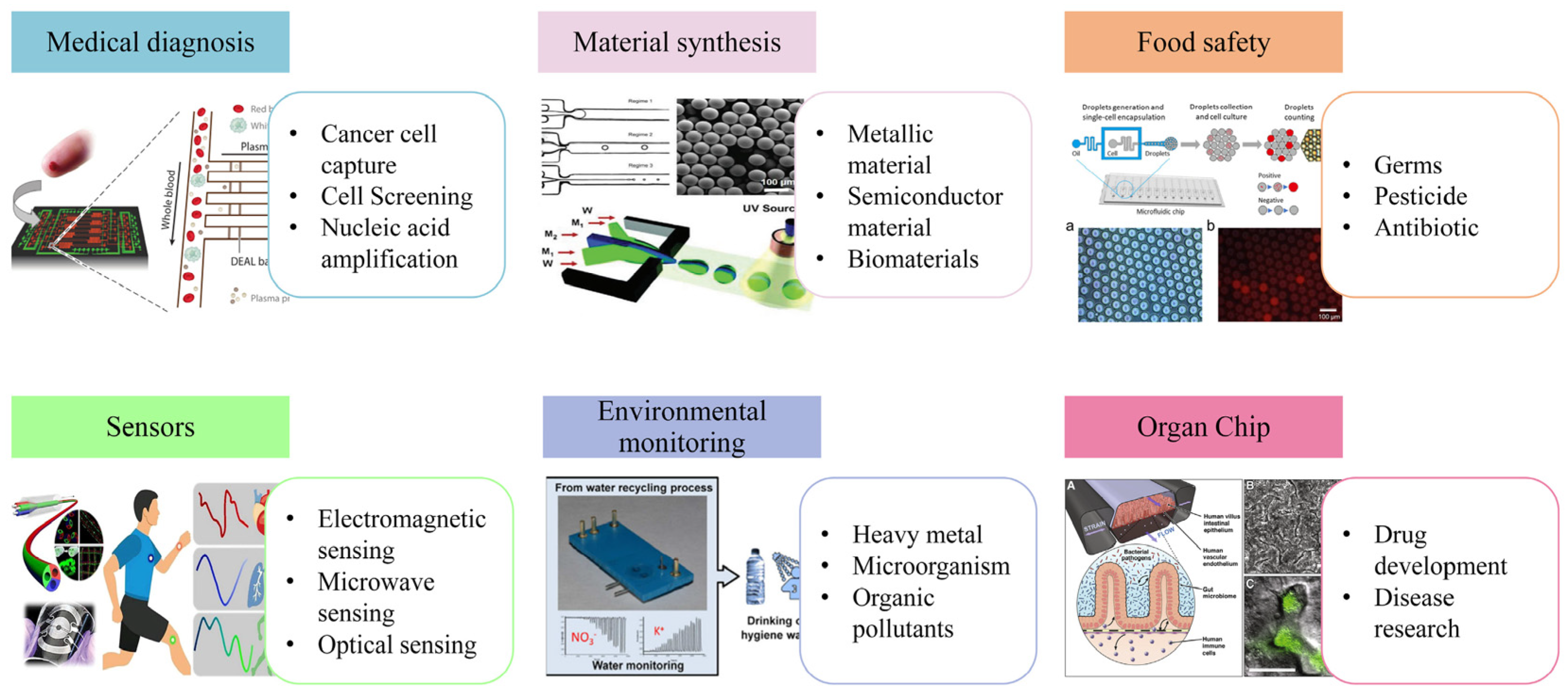
2.1. Microfluidics: An Excellent Detection Platform

Microfluidic chips are also known as lab-on-a-chip (LOC) and micro-total analytical systems (µTASs) [52]. By controlling the fluid flow in the microfluidic chip, the basic operation units such as sample preparation, reaction, separation, and detection in the biological, chemical, and medical analysis process are integrated into a micron-scale chip, and the entire analysis process is automatically completed. Microfluidic chips are usually portable devices of a few centimeters in size, and these devices consume low sample volumes (≤few microliters) [53]. Owing to the microscale structure of the chip, the fluid flow of these microscale fluids in the channel is completely different from the fluid flow behavior in the macroscopic channel. For instance, laminar flow, surface tension and capillary effect, fast heat conduction, and microfluidic diffusion, among others, are conducive to precise fluid control and a rapid response [54]. Numerous microfluidic devices have been developed to address specific scientific problems that cannot be easily solved by conventional techniques.
A microfluidic chip is a flexible platform, because its structure and function can be designed according to different target materials. Fluid control and sample control can be carried out through hydraulic pressure, electric field, micro-valve and micro-dam structure, oil pressure, and so on. In addition, the microfluidic chip also has the characteristics of openness, optical, electrical, magnetic, and other detection technologies and can be easily combined with microfluidic technology. For specific detection needs, the appropriate technique can be selected or several techniques can be combined according to the purpose of analysis and the nature of the sample; As shown in Figure 3e–h, PCR, LAMP, mass spectrometry, fluorescence spectroscopy, spectrophotometry, electrochemistry, and so on, in the form of on-chip or post-chip detection, for the detection and analysis of trace amounts of harmful substances.
PCR is commonly used to detect microbial genes in just a few hours and can be performed on a microfluidic chip by carefully controlling the reaction conditions, such as temperature, reaction time, and so on. Microfluidic chips that combine PCR and sample preparation can serve as point-of-care devices for fast and accurate microbial analysis [57]. Salman et al. fabricated a polycarbonate microfluidic PCR chip for DNA amplification and fluorescence detection using micromilling and thermal fusion bonding processes. This method can be used as a low-cost, simple PCR device for rapid bacterial detection [61]. Zhu et al. proposed an integrated lab-on-a-chip device that enables DNA extraction, solid-phase PCR, and genotyping detection functions. Using the chip, five high-risk types of human papillomavirus (HPV) and cervical swab samples from HPV patients can be analyzed within 1 h, which verifies the actual effectiveness of the integrated chip [62].
Compared with PCR, which usually requires changes in three or two temperature zones to achieve amplification requirements, LAMP only needs a constant temperature to achieve amplification, which solves the problem of temperature control in PCR. The LAMP method has been used to detect various pathogens, such as influenza virus, SARS, avian influenza, HIV, and so on. Wang et al. developed a LAMP-integrated microfluidic chip system for multiplex respiratory virus detection (LMCS-MRVA). The system can use magnetic beads to extract nucleic acid to realize the pretreatment of the sample. The microfluidic chip system used can identify influenza A virus subtypes, influenza B virus, and human adenovirus with high specificity and sensitivity within 1 h [58].
Mass spectrometry (MS) can identify a wider range of bacterial types. A unique advantage of MS is its low cost, which can avoid expensive gene amplification and biochemical experimental reagents. Bian et al. designed a microfluidic chip that can efficiently capture and concentrate airborne bacteria. They used liquid chromatography mass spectrometry (LC-MS) to identify a variety of bacteria from samples collected on the chip [59]. Huang et al. describe a microfluidic system for in situ extraction of a single cell and analyze its phosphatidylcholine (PC) compositions through MS [63]. Zhang et al. proposed a chip-based array monolithic microextraction system combined with inductively coupled plasma mass spectrometry (ICPMS) for the online analysis of trace amounts of Hg, Pb, and Bi in HepG2 cells [64].
The fluorescence method has the advantages of high sensitivity, simple and easy integration, and so on. The most important thing is that fluorescence detection and analysis can perform real-time fluorescence signal acquisition, providing easier-to-observe visual signals. Using a fluorescence microscope, the bacteria stained with fluorescent dyes can be directly observed, and the movement trajectories of the bacteria can be visualized and recorded. Another advantage is that the fluid flow inside the microfluidic chip is observed in real time, and the fluid flow state can be adjusted in time as needed. Zhang et al. developed a simple microfluidic immunomagnetic fluorescence analysis method for pathogenic bacteria, which combines a fiber optic spectrometer with a microfluidic chip and captures and aggregates the H9N2 virus through an immunomagnetic target in the microfluidic chip. A fiber optic spectrometer was used to detect the fluorescence intensity for quantitative analysis of the virus [60]. Guo et al. designed a magnetron microfluidic device that combines dynamic magnetophoretic separation and immobilized magnetic traps to detect Salmonella typhimurium (S. typhimurium) in a complex matrix with high sensitivity and selectivity. Captured target pathogens identified by streptavidin-modified QDs (SA-QDs) were detected by inverted fluorescence microscopy [65]. Lin et al. developed a centrifugal microchip fluorescent immunoassay platform that uses fluorescent microspheres (FMSs) to label targets for wash-free, rapid, quantitative, and point-of-care (POC) detection of proteins [66].
Although trace detection based on the above techniques is regarded as an effective analytical method, it requires a large initial capital investment and high maintenance costs. The longer analysis process also determines that these methods are not suitable for on-site instant detection. On the contrary, optical detection techniques have attracted a lot of interest owing to their advantages of high sensitivity, fast, efficient, and non-destructive imaging, and their potential in a variety of applications has been demonstrated (Figure 2). Raman spectroscopy is especially known for exploiting the vibrational modes of molecules to provide molecular fingerprints. The combination of microfluidics and Raman has become a new research direction, and the highly integrated microfluidic systems make possible the emergence of a portable Raman device for on-site detection without any contamination and waste [67].

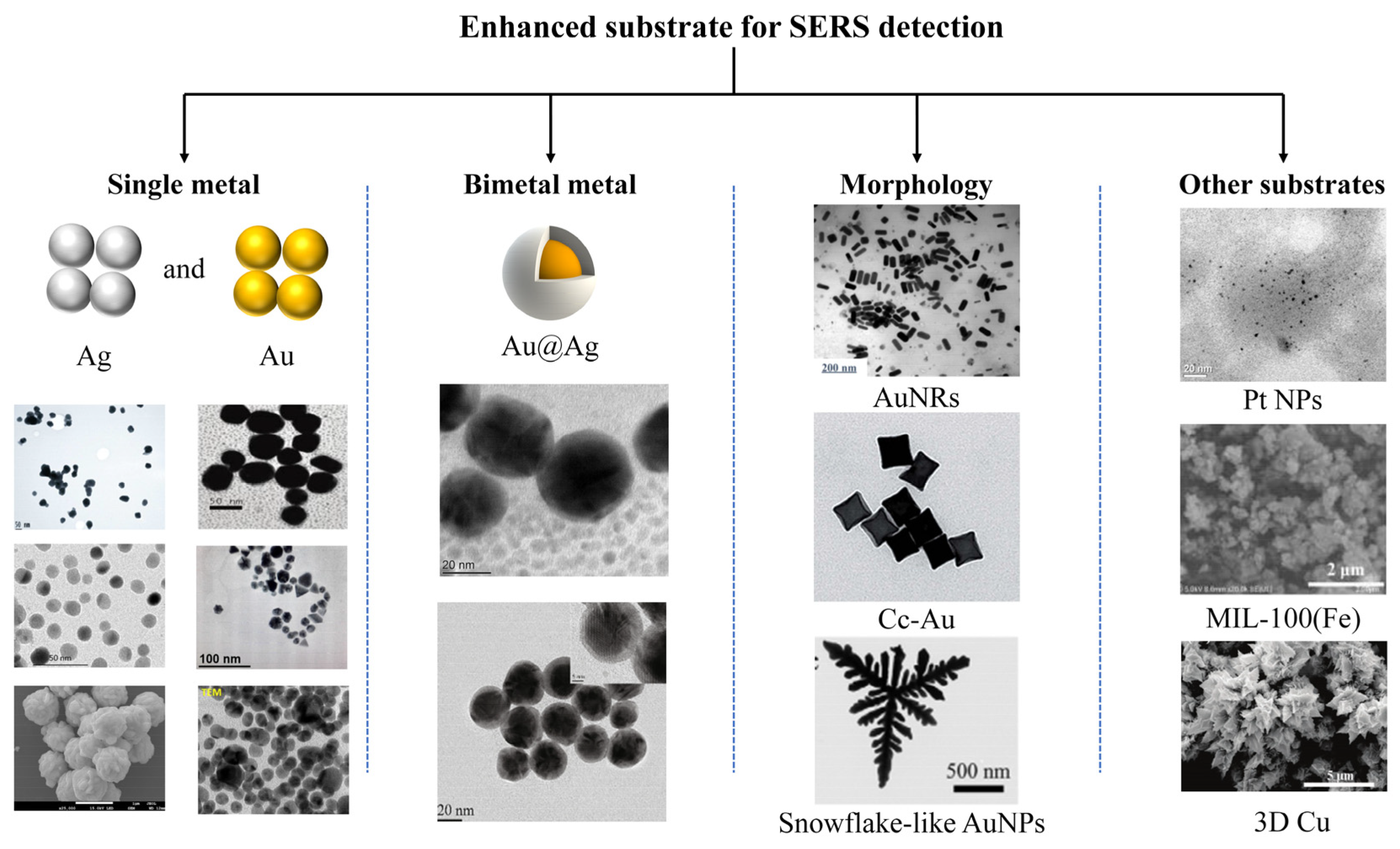
2.2. SERS: Principles and Substrate

Raman spectroscopy is based on the phenomenon of inelastic light scattering, whereby light scattered by molecules changes in frequency as it travels through a transparent medium, so that differences in different vibrational energy levels can be precisely matched. This property allows Raman spectroscopy to provide its molecular fingerprint spectrum based on the different vibrations and rotations of each analyte, which is of great advantage in applications for the detection of single or multiple analytes or compounds [20]. However, when light strikes the analyte surface, inelastic scattering accounts for only a small fraction of the incident light. The remaining elastic scattering is called Rayleigh scattering, which results in the Raman signal being very weak and not suitable for trace detection [68].
SERS can enhance the common Raman scattering signal of analytes absorbed on or near the surface of metal particles, and a variety of substrates can obtain a Raman signal amplification ability ranging from 106 to 108 [25]. The enhancement mechanism is mainly attributed to two widely accepted mechanisms: The electromagnetic enhancement mechanism (EM) and chemical enhancement mechanism (CM) [69]. The former relies on localized surface plasmon resonance (LSPR) at the nanoparticle surface. The resonance phenomenon is mainly determined by the substrate size, morphology, and distribution pattern [70]. The latter is thought to be the chemisorption of the analyte on the SERS substrate, which will cause the electronic state of the synthesized complex to change and resonate with the laser excitation frequency, ultimately enhancing the Raman signal [71]. The EM is generally considered to be the main enhancement mechanism. Owing to the collective oscillation of electrons (i.e., plasmon modes) on the surface of the metal substrate caused by the incident light, the local electromagnetic field strength of the metal substrate is greatly increased, which leads to a greatly enhanced Raman signal. While the mechanism does not require direct chemical interactions between the analyte and the substrate, the amplification of the field is a local effect, so SERS requires close proximity of the analyte to the substrate. The need for adsorption or any type of stable interaction between the analyte and the metal surface is an important feature of SERS; it dictates the limitations and advantages of the technique [72].
The main challenge in obtaining reliable SERS spectra is represented by the quality of the SERS substrate. In SERS active substrates, regions of strong local field enhancement caused by LSPR are called “hot spots”, which lead to significant enhancement of the SERS signal [73,74]. For analyte detection, the preparation of these substrates must be simple, reproducible, and cost-effective, while providing high signal enhancement. With the development of nanotechnology, from single metal substrates such as gold and silver, bimetallic composite substrates, semiconductor substrates, composite material substrates, and inorganic material substrates to nanoparticles with different morphologies have been reported for SERS detection; we present a summary of these SERS substrates in Figure 4. Among these SERS substrate materials, noble metal nanoparticles are most commonly used. At present, the most widely used SERS substrates are Au, Ag, and Cu. Among these three metals, Ag has the best performance as a SERS substrate. González et al. synthesized AgNPs with an average diameter of 49.5 nm and an error of 5% using glucose in a simple, rapid, and low-cost method, proved to be adequate for the SERS-based detection of trace-level oxytetracycline (OTC) in honey samples [75]. Kamran et al. synthesized AgNPs using the reduction procedure and used as a SERS substrate for the detection of valeric acid (VA). TEM image showed the spherical structure of the AgNPs. Using the synthesized silver nanoparticles as a SERS substrate, the trace amount of VA in aqueous medium was identified, and a detection limit of 10 × 10−1 M of VA was successfully achieved [76]. Hassan et al. synthesized signal-optimized flower-like AgNPs at 25 °C using AgNO3, PVP, and ascorbic acid as reaction solutions. The average size of the AgNPs was about 800 nm. Crystal violet (CV) was used as the SERS probe to evaluate the enhancement of the synthesized AgNPs. As a result, the highest enhancement factor (EF) of AgNPs synthesized at 25 °C was 1.39 × 10⁶. The synthesized AgNPs were fabricated into a SERS-based sensing platform for the detection of methomyl, acetamidoacetic acid (AC), and 2,4-dichlorophenoxyacetic acid-(2,4-D) residual levels in green tea. The detection limits were 5.58 × 10⁻⁴, 1.88 × 10⁻⁴, and 4.72 × 10⁻³ µg/mL, respectively, and the results showed high stability and accuracy [77]. Compared with silver materials, gold materials can produce more stable and reproducible spectra, so gold nanoparticle (AuNPs) substrates have been used for testing complex matrices, especially in vegetables and fruits. Dasary et al. successfully identified TNT in aqueous solution at the 2 picomolar (pM) level using gold nanoparticles (AuNPs) as SERS substrates. Gold nanoparticles of different sizes and shapes were synthesized by controlling the ratio of HAuCl4, 3H2O, and sodium citrate concentrations. The surface modification of gold nanoparticles with cysteine can recognize TNT with high selectivity and ultrasensitivity. The AuNPs after recognizing TNT aggregated and formed more hot spots owing to electrostatic interactions, which enhanced the Raman signal by nine orders. The method also has excellent discrimination for other nitro compounds and heavy metals [78]. Brittod et al. proposed a method that can transiently synthesize AuNPs at room temperature, using ascorbic acid and sucrose as reducing agents with the advantage of non-toxicity; the size of the synthesized nanoparticles is 10–20 nm and AuNPs with different morphologies were synthesized. The SERS effect was evaluated with natural zeolite (chabazite) as the analyte, and the results showed that the study could be applied to soil analysis [79]. Al-Saadi et al. used AuNPs as SERS substrates to detect procaine in aqueous media. The AuNPs were prepared by the reduction method and the synthesized AuNPs were uniform in shape with an average size of about 40 nm. Experiments showed that the SERS signal intensity was linearly correlated with the logarithmic concentration of procaine solution. The lowest detectable concentration for this method is 10−10 M [80].
Nano-silver has strong surface plasmon resonance, but the chemical stability of nano-silver is weak. Gold nanoparticles have stable chemical properties, but the SERS enhancement effect is weaker than that of silver nanoparticles [81,82]. If two metals are used and designed into a core–shell structure, not only can the stability problem be solved, but the core and shell can be designed and controllably prepared, such as Au@Ag, Au@Pb, or other assembled structures. This composite metal-type nanomaterial can well expand the applicable scope of SERS. Hussain et al. employed silver-coated gold nanoparticles (Au@AgNPs) as SERS active platforms for rapid assessment of trace contaminants of agrochemicals in agricultural production. The synthesized Au@AgNPs with a gold core size of 28 nm and a silver shell thickness of 6 nm were able to sterilize tricyclazole (TCZ) and thiram in pear fruit samples after the nanoparticles were modified with mercaptooctane (MCO). The detection limits were 0.005 and 0.003 ppm, respectively [83]. Similarly, Yaxin et al. also chose to use Au@AgNPs as SERS substrates to detect thiacloprid (carbamate), profenofos (organophosphate), and oxamyl (neonicotinoid) in fruits. The lowest detection limit can reach 0.01 mg/kg. The Au@Ag NPs with 26 nm Au core size and 6 nm Ag shell thickness can precisely identify the Raman characteristic peaks of pesticides (thiacloprid, profenofos, and oxamyl). It has great potential in the field of food testing [84]. Other metals, such as Pt, Fe, and Cu [85,86,87], can also be used as SERS substrates, but this type of material has not been used more frequently for SERS detection because of their limited enhancement of the Raman signal.
SERS signals depend largely on LSPR performance. The LSPR intensities at each position of the shaped nanoparticles are the same. For nanoparticles with other morphologies, such as rod-like, polyhedral, and flake-like nanoparticles, different intensities of LSPRs are generated, thus affecting the repeatability of SERS signals [88,89]. It is obvious that sphere nanoparticles can obtain highly reproducible SERS signal acquisition. Unfortunately, sphere nanoparticles cannot achieve both high repetition and high sensitivity [90]. Nanoparticles with higher roughness, such as flower-shaped or star-shaped nanoparticles, have significantly higher sensitivity than sphere nanoparticles. According to finite difference time domain (FDTD) calculations, flower-like nanoparticle arrays with various protrusions on the surface have more and denser hot spots than spherical nanoparticle arrays, which can exhibit higher Raman enhancement effects [91]. Hot spots play a decisive role in the strength of SERS. Substrates that are favorable for generating hot spots are more likely to obtain high-intensity SERS signals. In order to achieve lower detection limits, some nanoparticles of other structures have also been used for SERS detection. Using gold nanorods as SERS substrates to detect carbaryl residues in juice and milk, it was possible to detect carbaryl at 50 ppb levels extracted from juice and milk samples [92]. Concave cubic gold (cc-Au) nanoparticles as SERS substrates also have a high Raman enhancement effect, which is much higher than that of spherical nanoparticles of similar size [93]. Snowflake-like AuNPs prepared by reducing chloroauric acid (HAuCl4) with ascorbic acid in aqueous solution of tetradecylpiperidine (C14PDB) surfactant also exhibited excellent Raman enhancement. The detection limit of rhodamine 6G (R6G) in aqueous solution is about 3 × 10⁻⁹ mol/L and the detection limit of organophosphorus pesticides in solution is 1 × 10⁻⁸ mol/L. In the actual detection, trace pesticide residues on apple peels were successfully detected, which indicated that snowflake-like Au NPs have great prospects in the detection of trace organophosphorus pesticides [94].
In conclusion, SERS is an ideal tool for the detection of trace substances, suitable for rapid and on-site detection. For different types of samples to be tested, corresponding methods can be combined with SERS technology to achieve ideal detection results. Table 1 also lists SERS detection information for different types of substrates. Owing to the extensive research on various SERS substrates, it can effectively improve the enhancement effect and promote the development of SERS. At present, SERS-enhanced substrates have been widely used in the detection of solid samples (such as vegetables, fruits, and grains) and liquid samples (such as milk, juice, and water) in combination with other advanced technologies (such as sample pretreatment technology). By combining with other advanced technologies, the reliability of real sample detection is enhanced and the use of SERS is expanded.

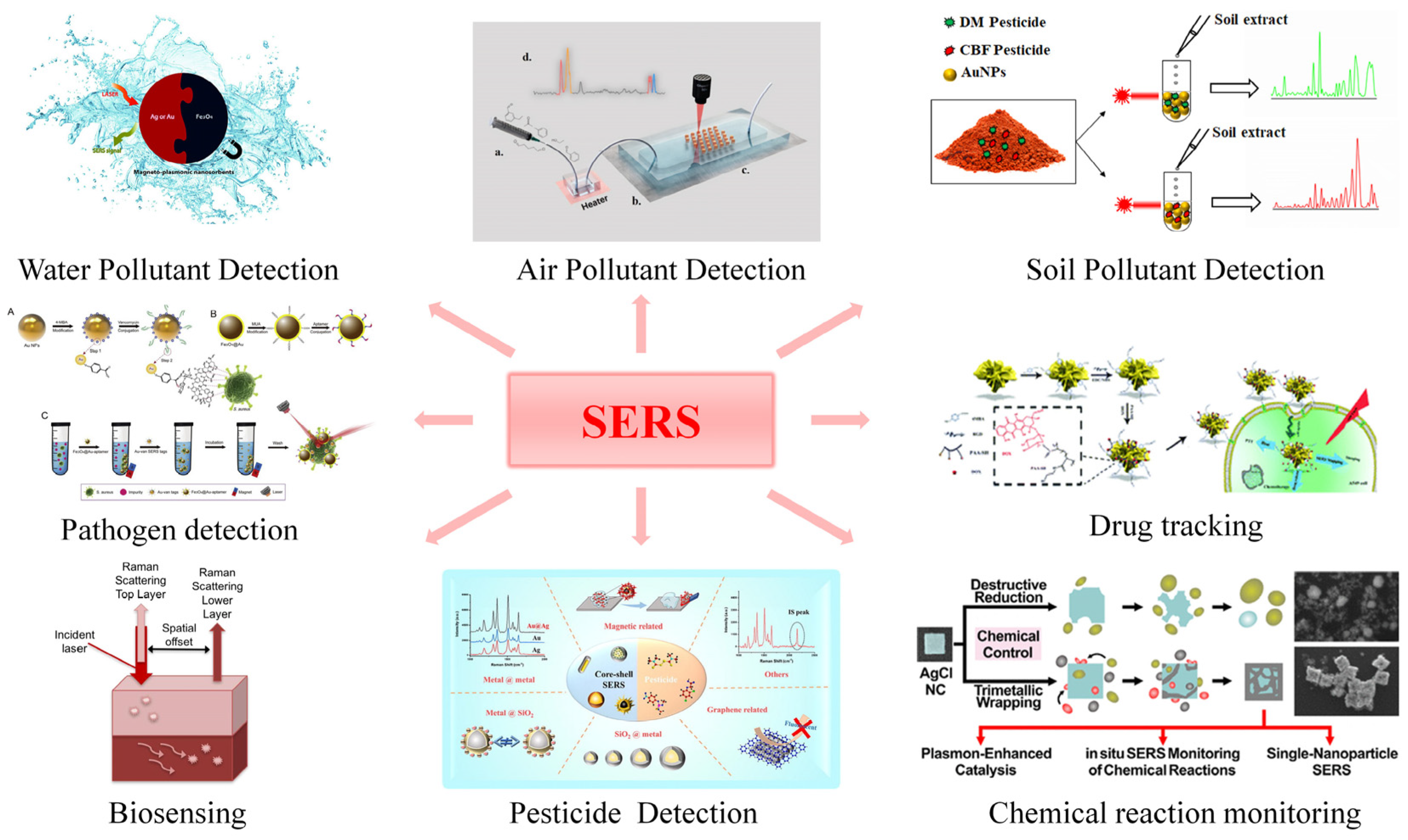
3. Microfluidics Combined with SERS

3.1. Key Factors for Combining Microfluidics with SERS

The key factors affecting the detection efficiency of the microfluidic-SERS platform can be divided into the following categories: (1) the mixing efficiency of the SERS active substrate and the analyte in the microchannel—excellent mixing efficiency can provide more SERS hot spots; (2) synthesis or modification of SERS-active substrates in microfluidic devices, which provide more reproducible SERS detection results and more convenient detection; and (3) target analyte trapping in microfluidic channels for better SERS specific detection. Some analytical results on these influencing factors are listed in Table 2.

3.1.1. Mixing of Nanocolloids and Analytes in Microchannels

Mixing efficiency is a key requirement to achieve reproducible detection in microfluidic-SERS platforms. Microfluidic-SERS platforms are usually designed to handle liquid samples and, to obtain more hot spots, the analytes are required to be thoroughly mixed with colloidal nanoparticles. Generally, colloidal nanoparticles are used as the SERS-active substrates. Nanoparticles are injected through a microfluidic channel where they encounter and mix with the analyte at specific locations for SERS detection. Several different passive channel designs for fast and efficient mixing of nanoparticles with analytes exist, such as T-shaped channels, which can realize simple diffusion mixing at the fluid interface [95]; z-shaped channels, which can achieve chaotic mixing at high Reynolds numbers [96]; and a staggered herringbone mixer (SHM) for solution mixing at low Reynolds numbers [97].
Compared with conventional mixing channels, alligator-toothed microchannels show higher mixing efficiency, a zigzag-shaped microfluidic channel is used to mix analytes and colloidal nanoparticles, and the triangular structures are located on the upper and lower surfaces of the channel in a zigzag pattern. When the nanocolloids and analytes were introduced into the mixing channel, the analyte molecules were effectively adsorbed on the surface of silver nanoparticles and reproducible SERS signals were obtained at the laser detection point (Figure 5a) [39]. Yasui et al. fabricated a microfluidic baker’s transformation (MBT) mixer as a three-dimensional passive-type mixer for the efficient mixing of solutions. They also quantitatively evaluated the mixing performance of the 3D microfluidic mixer with a confocal microscopic method. The results showed that the MBT mixer could increase the mixing efficiency by 20% after two cycles and was able to mix the fluorescein isothiocyanate solution with water within 51 ms, which is 70-fold faster than using straight channel mixing [98].
This continuous SERS microfluidic system enables repeatable measurements and is relatively simple to use. However, in microchannels, the mixing of analyte molecules with colloidal nanoparticles occurs by molecular diffusion, which results in weak SERS detection signals and high signal changes, resulting in low reproducibility. To solve the problem of poor signal stability of SERS detection in microchannels, Yazdi et al. designed a special microfluidic channel based on surface-enhanced resonance Raman spectroscopy (SERRS), which can achieve high sensitivity detection of DNA sequences through competitive displacement assays, as shown in Figure 5b. The platform combines a competitive displacement method with a specially designed microchannel structure that allows the Raman-labeled reporter sequence to be replaced by the target DNA sequence, enabling the detection of unlabeled target DNA sequences through a simple one-step procedure. The diffusion limitations present in open-channel microfluidic assays are overcome by the special microchannel structure filled with silica particles, enabling aggregation of nanoparticles within the detection volume and a significant increase in the SERS signal of analyte molecules, thereby enhancing the sensitivity of the assay. Detection of target DNA sequences down to 100 pM can be achieved using this device [108]. The above two continuous flow microfluidic-SERS systems still have certain limitations. For example, nanocolloids and sample molecules need to be mixed for a long time. Besides, with continuous flow, the analyte–nanoparticle conjugates will become enriched on the microfluidic channel, which can lead to the so-called memory effect. The above shortcomings can be overcome by droplet microfluidics, in which the mixture of sample molecules and nanocolloids forms discrete droplets to avoid direct contact with the channel walls, which can reduce the memory effect. In addition, samples and colloidal nanoparticles can be mixed more efficiently within the microdroplets, allowing reliable handling of microsamples. Zhang et al. designed a SERS microfluidic chip to generate droplets for the detection of 6-thioguanine (6-TG) in real human serum, as shown in Figure 5c. 6-TG mixed with gold nanoparticles efficiently in each droplet. The SERS signal of each droplet is acquired from a fixed position in the channel. The droplets can provide a stable microenvironment, reduce the interference of external factors, and improve the repeatability of the detection signal, with a detection limit of 0.032 μM [109].

3.1.2. Synthesis and Integration of SERS Active Substrates in Microfluidic Chips

On the other hand, microfluidic systems are also used by many researchers to synthesize SERS substrates with different morphologies. In microfluidic systems, a high mass transfer, heat transfer, and high mixing efficiency can be achieved during the synthesis of nanoparticles. Nanoparticle nucleation and growth stages can be precisely controlled to achieve excellent control over particle size, size distribution, and morphology, and thus increase reproducibility.
Adamo et al. first used cotton thread to form a microfluidic device to rapidly and directly synthesize AuNPs. Depending on the synthesis conditions, spherical AuNPs with sizes ranging from 20 nm to 40 nm can be synthesized. The synthesized AuNPs were successfully used for SERS detection of crystal violet and nicotine molecules. The limit of detection for nicotine was 0.18 mg L−1 [99]. A simple and fast method for the synthesis of (Au nanorods (NR)@Ag)-polyaniline (PANI) Janus nanoparticles (JNPs) using a droplet microfluidic platform was proposed by Wang et al. The excellent dispersion and uniform size of (AuNR@Ag)-PANI JNPs can be achieved by controlling the droplet volume and reliably manipulating individual droplets. (AuNR@Ag)-PANI JNP as a SERS substrate can be used to detect Hg2+ ions with high sensitivity and good selectivity, and the detection limit of Hg2+ ions concentration was 0.97 nM [100]. Lawanstiend et al. synthesized in situ nanoporous silver microstructures (np-AgMSs) using a microfluidic system and to study the effect of reactant concentration on nanostructure by simply adjusting the flow rate. Finally, the multipod np-AgMSs were successfully used as on-chip SERS substrates for the determination of thiocyanate in human saliva. The LOD and LOQ were 0.1 and 1.5 μM, respectively. It is worth noting that, using np-AgMSs as the Raman substrate, the relative standard deviations of the measured Raman signals are all less than 9%. This indicates that the synthesized np-AgMSs have high precision and reproducibility [101].
Although there are different microfluidic channels that can improve the mixing performance of nanoparticles and analytes, and microfluidic systems can also be used to synthesize SERS substrates, the introduction of many analytes with SERS substrates into microfluidics seems to be a big challenge, which is clearly not the most efficient strategy for microfluidic-SERS systems. Therefore, another microfluidic-SERS integration approach was proposed, in which metal nanoparticles or nanostructures with fine morphologies were immobilized on the surface of microchannels as solid-state SERS active substrates [110,111]. In this method of integration, the sample molecules only generate SERS signals when they are adsorbed on the area where the SERS active substrate is immobilized during the detection process. The patterned SERS active substrate has a fixed and ordered structure, which can integrate a hotter spot for more sensitive SERS detection. Xu et al. fabricated 50 silver microflora arrays (SMAs) composed of nanoparticles within microchannels by femtosecond laser processing. The SMAs can achieve in situ SERS monitoring, and the on-chip catalytic reduction of 4-nitrophenol (4-NP) to 4-aminophenol (4-AP) shows that the SMAs have high catalytic activity and SERS enhancement (Figure 5d) [102]. Parisi et al. designed a microfluidic chip decorated with a highly active Raman substrate, first by in situ electrodeposition of Cu core/C-seath nanowalls on the channel surface and then by the galvanic replacement method in the channel. AgNPs decorated nanowall structures were fabricated within the channel (Figure 5e). The microfluidic chip has excellent sensitivity and Raman enhancement and can easily detect crystal violet down to 50 pM [103]. Yan et al. proposed a method to synthesize Ag nanostructures in microfluidic channels via a chemical-electro-displacement reaction between Ag and Cu, as shown in Figure 5f. The microfluidic-SERS system enables continuous, real-time sensing of targets in a microfluidic chip. Experiments have shown that the chip can perform label-free sensing of chemical molecules (i.e., methylene blue) and biomolecules (i.e., urea), and can be used to detect Hg ions in aqueous solutions with an LOD of 1 × 10⁻⁷ M [104].

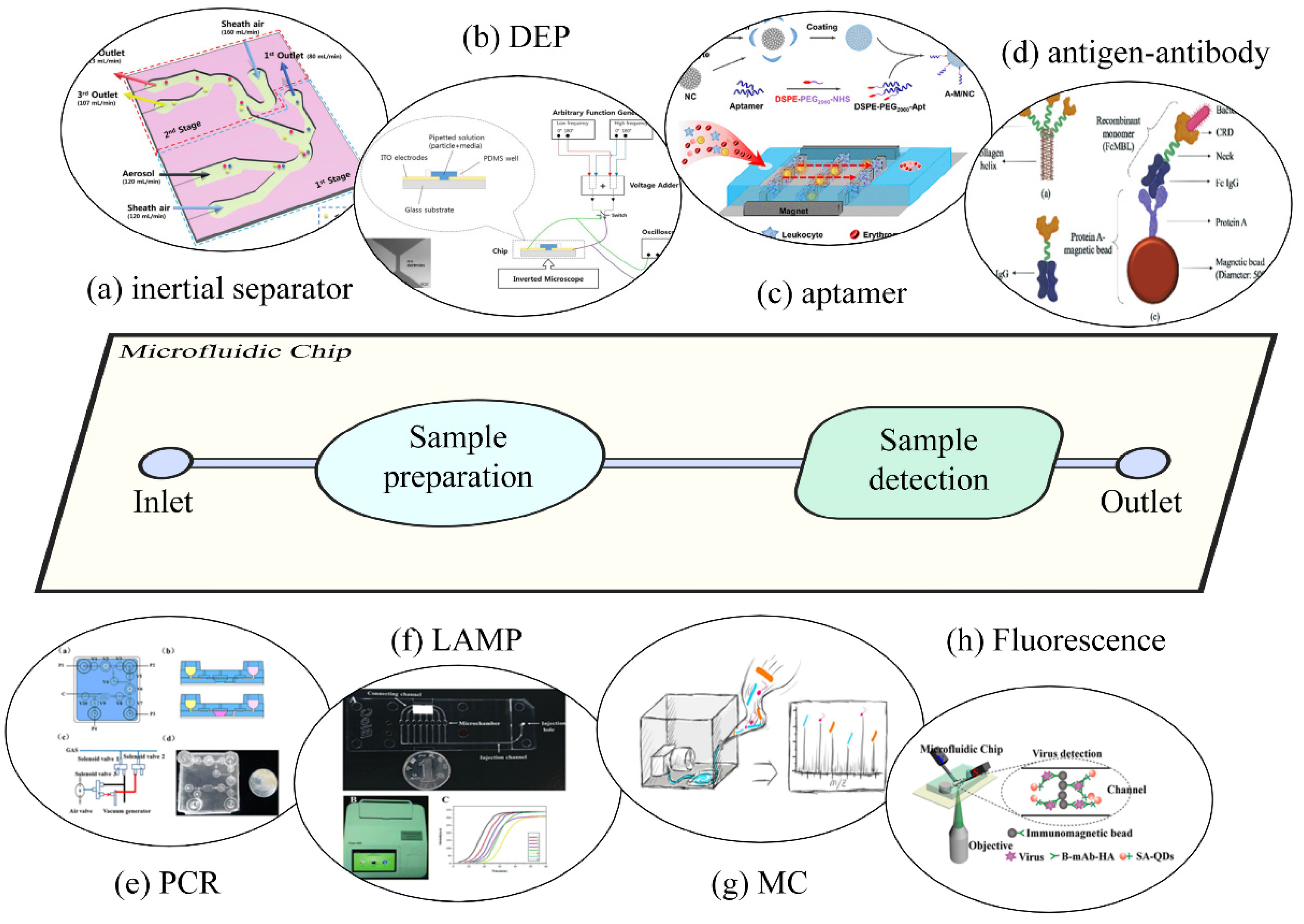
3.1.3. Target Trapping

One outstanding advantage of integrating microfluidic systems with SERS analysis is that, as hazardous substances are present in trace (parts per million to parts per billion) amounts in a sample, capture or preconcentration of the analyte is often required before detection. Microfluidic technology can remove and concentrate trace amounts of harmful substances from air, water, food samples, clinical samples, and so on, using physical, chemical, and biochemical methods, thereby eliminating any tedious operations and preventing sample processing loss or contamination. According to different chip designs and different operating methods, sample pretreatment can be roughly divided into two categories: physical and chemical methods.
Curved microchannels have been shown to be an effective physical means to achieve continuous multi-particle pretreatment [112,113,114,115]. In this context, Hong et al. developed a microchannel-based inertial separator consisting of two 90° curved microchannels and three outlets for the separation of viruses, bacteria, and larger particles. Two 90° curved microchannels can use centrifugal force to simultaneously separate airborne microorganisms and virus particles in bacterial cells by size. As shown in Figure 3a, when the fluid passes through a 90° curved channel, the centrifugal force of particles with different particle sizes and masses is different. At the same flow rate, the particles with larger mass move more radially and flow into the outlet channel from the outside, while the small particles maintain their streamlines and pass through the inner channel until the next curved channel to achieve the second separation [55]. The combination of microelectrodes and microchambers is also commonly used for microbial capture [116,117,118]. Figure 3b shows the experimental setup of Han et al. to selectively capture bacteria, viruses, and proteins by controlling the voltage and frequency of dielectrophoresis (DEP) and electroosmosis (EO) applied between two coplanar electrodes. The method of schematic illustration proved to be an excellent biosensor for rapid detection of biological particles [119]. The above-mentioned inertial force and dielectrophoresis (DEP)-based extraction methods for trace substances are fast, convenient, and accurate, and are suitable for the detection of multi-substance mixed samples.
For the specific identification and separation of microorganisms, the enrichment and purification of specific trace harmful substances, chemical and biochemical methods have greater advantages. The trace amount of harmful substances and the probe molecules will be connected through specific interaction and the probe molecules can be immobilized in magnetic beads or microchannels, so as to realize the specific capture of the trace amount of harmful substances [120,121,122,123]. Compared with physical methods, biochemical methods can extract target substances from particles of similar size and density. Zhang et al. showed that modification of Fe3O4 magnetic nanoclusters (NCs) with aptamers can improve the specific capture efficiency of circulating tumor cells (CTCs), which can capture and detect more than 90% of rare tumor cells in blood within 20 min [56]. Fang et al., using protein-coated magnetic beads, also captured five target bacteria at a rate of 56% to 85% in only 20 min [57]. Rodoplu et al., using a combination of external static and dynamic magnetic fields, captured ultra-low concentrations of bacteria by antibody-modified superparamagnetic magnetic beads (Ab@SPM) [124].
Cheng et al. designed a compact hybrid electric mechanism-simultaneous using dielectrophoresis (DEP), electrophoresis, and electrohydrodynamics (EHD) to achieve bacterial concentration from human blood. Three bacteria, Staphylococcus aureus, Escherichia coli, and Pseudomonas aeruginosa, were successfully identified from human blood cells in less than 1 min. The device enables on-chip SERS measurement and analysis without the use of any antibodies/chemicals immobilized in the on-chip microfluidic device [105]. Krafft et al. reported a PDMS drinking water bacterial concentration device composed of two vertical channels, using a porous membrane for the concentration and detection of pathogenic microorganisms in drinking water. The device combines filtration and electrokinetic flow to enable concentration and surface-enhanced Raman spectroscopy analysis of pathogens in tap water [106]. Zheng et al. designed a microfluidic chip for breast cancer biomarker detection in real samples with high sensitivity. AgNPs were immobilized on microfluidic channels to form SERS-active substrates and then specific antibodies were attached to different regions of the SERS substrate to capture target biomarkers. The qualitative and quantitative analysis of breast cancer biomarkers can be achieved by comparing the SERS intensities of different regions of the SERS substrate. The device not only has a good surface-enhanced Raman spectroscopy effect, but also has broad application prospects in clinical diagnosis [107].

3.2. Application Prospect of the Microfluidic-SERS Detection System

The microfluidic-SERS detection system, as an emerging technology that has attracted wide attention, has broad application prospects in the fields of biomedical sensing, environmental monitoring, and food safety detection. We provide a brief summary of the application prospects of the microfluidic SERS detection system in Figure 6. In biomedical sensing, microfluidic-SERS detection systems have the characteristics of being fast, on-site, providing fingerprint detection, and high sensitivity. Overcoming the limitations of traditional methods, it has been applied to various clinical samples, including nucleic acids, proteins, viruses, bacteria, cells, and others. A microfluidic chip consisting of a magnetic enrichment chamber, a serpentine fluid mixer, and a SERS substrate for capturing functionalized probes was used to detect exosomes miRNAs online. Exosome enrichment, online lysis, and miRNA detection are integrated into a microfluidic platform. The limit of detection decreased to 1 pmol/L [125]. Saha et al. report a simple paper-based microfluidic system for highly reproducible and sensitive detection of proteins within microfluidic channels. Using 4-mercaptopyridine and glucose (or biotin) functionalized Ag@Au nanoparticles as the SERS substrate, the protein can be reproducibly detected from pico to femtomolar concentrations in the reaction zone [126]. Jadhav et al. proposed a microfluidic device with integrated SERS, where the microchannels were functionalized using vertically aligned Au/Ag-coated carbon nanotubes or disposable electrospun micro/nanofiltration membranes. This device can successfully capture viruses from various biological fluids/secretions including saliva, nasopharynx, tears, and so on, and can accurately identify viruses from Raman signatures. It will facilitate rapid screening of both symptomatic and asymptomatic individuals with COVID-19 [127]. Similarly, microfluidic-SERS detection systems have also been applied to the detection of bacteria, cells, and ions [106,128,129,130].
Environment pollution is one of the major challenges in human society. Environmental pollutants are generally released into the environment through volcanic emissions, solid waste incineration, coal combustion, chemical and electronic products, and other natural activities and human industrial activities. These pollutants are widely present in air, water resources, and even soil. Human absorption of pollutants can cause them to accumulate in the human body, posing a threat to human health. Therefore, the monitoring of environmental pollutants is crucial. In order to achieve instant and rapid detection of environmental pollutants, Yang et al. proposed a novel array-assisted SERS microfluidic chip (array-SERS chip), which combines the advantages of ultra-high sensitivity and multiplex sensing capability. Using MOF materials (Zeolitic Imidazolate framework-8 (ZIF-8)) and Au@Ag nanocubes as SERS substrates, and cysteamine (CA) as the gas capturing agent, the microstructure of micro-structured triangular arrays can be greatly improved, increasing the collision probability between the gas molecules and the sensing interface in the channel and improving the detection sensitivity. Taking acetaldehyde gas as a typical air pollutant model, the chip realizes the detection of air pollutants with a detection limit as low as 1 ppb [42]. Lafuente et al. presented a simple method for the preparation of SERS-active regions within PDMS microchannels 50 cm in length. Au@POM (POM: H3PW12O40(PW) and H3PMo12O40(PMO)) modified with polyoxometalate nanostructures were formed on the channel surface driven by UV light. The structure self-assembles in situ on the PDMS microchannel surface without any additional functionalization. The Au@POM-coated microfluidic-SERS analysis platform can detect pollutants in water with high sensitivity, as can analytical platforms for real-time detection of the organophosphorous esticide Paraoxon-methyl at 10−6 M concentration level [111]. Lin et al. fabricated a paper-based SERS substrate by assembling Au@Ag nanocubes onto patterned paper using a liquid–liquid interface self-assembly technique. A new paper-based laboratory SERS platform was constructed by pasting paper-based SERS substrates onto glass slides. The SERS sensor has a sample injection area and a SERS detection area, which can perform sample pretreatment and detection at the same time. Based on the advantages and functions of this SERS platform, the quantitative determination and analysis of the representative insecticide thiram in soil was successfully carried out without any pretreatment [132].
The pollutants that endanger food safety mainly include chemical pollutants such as pesticides, antibiotic residues, mycotoxins, and melamine, and microbial pollutants such as food-borne pathogens and viruses. Rapid and on-site analysis of these contaminants is difficult because of their low concentrations and presence in complex food samples. The microfluidic-SERS detection system can provide flexible sample preparation and fast and sensitive detection, which has great advantages in the field of food safety detection. Asgari et al. developed an on-site analysis technique for food samples that integrates filtration technology into SERS microchips. The chip incorporates a filter membrane at the chip inlet to remove interfering particles and enrich for target analytes. Using Au@Ag nanoparticles as SERS substrates, the microchannel design was optimized by the finite element method, which facilitated the efficient mixing of samples and nanoparticles. Four pesticides (thiabendazole, thiram, endosulfan, and malathion) were successfully detected in strawberries individually or in combination using this sensor. The detection limits of these four pesticides were in the range of 44–88 μg/kg, indicating that the sensor has good sensitivity to the target analytes [133]. Wang et al. used a simple and fast method to construct a high-sensitivity SERS substrate based on Ag dendrites in a T-type microfluidic device to fabricate a microfluidic SERS sensor. The analytical performance of this sensor was investigated with different concentrations of amoxicillin aqueous solution and the detection limit was up to 1.0 ng/mL [134]. Rodríguez-Lorenzo et al. demonstrated the potential of SERS combined with microfluidics for the detection and identification of foodborne pathogens. Gold nanostars (GNSs) were used as SERS tags to report for the presence of the pathogen of interest. SERS-tagged GNSs were functionalized with a monoclonal antibody specific for Listeria monocytogenes. In the presence of L. monocytogenes, the SERS signal corresponding to the antibody-paired SERS marker was detected in real time and continuously, in flow using a flow focusing microfluidic device that allows concentrating samples on-chip while reducing the detection time. It is worth noting that the designed in-flow detection strategy is able to distinguish the target species down to 1 × 10⁵ CFU/mL of L. monocytogenes [131].
The microfluidic-SERS detection system conforms to the development trend of modern analysis technology and has a good application prospect in the detection of food contaminants. However, to achieve on-site detection of contaminants in real food, it is also necessary to improve the portability of microfluidic-SERS detection systems and to develop portable or even hand-held Raman spectrometers for SERS detection.

3.3. Smart Algorithms and Results’ Visualization for SERS Data

The data detected by SERS are usually spectral data and the usual practice is to directly analyze the Raman shift of the characteristic peaks of the SERS spectrum to identify substances. However, in the actual testing process, complex detection objects cannot be directly analyzed by spectrograms. On the one hand, sample molecules with similar structures have similar SERS spectrograms, which causes great difficulties in the analysis of SERS spectrograms; on the other hand, in the actual detection in complex matrices, the signal of trace substances is very weak, it is difficult to separate from the matrix background, and the signal of the target cannot be clearly observed. In order to analyze complex SERS data, researchers usually use intelligent algorithms to intelligently analyze SERS spectra.
Commonly used analysis methods for SERS spectral data include principal component analysis (PCA), partial least squares (PLS), and support vector machines (SVMs) [135,136]. Dong et al. used the method of SVM to detect 3,4-methylenedioxy methamphetamine (MDMA) in human urine and judged the real samples with verified concentrations; the accuracy rate exceeded 90%, demonstrating that this method is effective when dealing with data from targets in complex matrices [137]. Dies et al., using the PCA-SVM model for data analysis, successfully quantified and characterized structurally similar drugs in aqueous solution. The accuracy of the qualitative determination was 100% and the accuracy of the quantification of cocaine in saliva was 98.3% [138]. Deep learning networks such as multilayer perceptron (MLP), fully connected network (FNN), convolutional neural network (CNN), recurrent neural network (RNN), fully convolutional network (FCN), and principal component analysis network (PCA) Net), among others, have also been applied to the analysis of SERS spectra [139,140,141]. Lussier et al. reported the application of ANN combined with SERS. The application of ANN can extract various features in the SERS spectra associated with the different orientations molecules can adopt on the SERS nano-sensors, characteristic of specific metabolites. This method was used to analyze metabolite gradients near various cell lines, including HeLa and HUVEC [142].
Like most spectroscopic techniques, SERS spectroscopy requires advanced data processing to extract meaningful information from the spectra. SERS spectra are easily changed by concentration, enhancement substrate, target matrix, integration time, and so on. Therefore, how to establish a suitable SERS data analysis method is also an important issue to realize the large-scale application of SERS technology.

4. Conclusions and Perspectives

In this review, we briefly introduce the concept and application of the microfluidic SERS detection system. Microfluidic technology can integrate the separation and purification of biological and environmental samples in the same chip. SERS technology has great advantages in probing the structural properties of compounds at the microscopic scale. By introducing the SERS technology into the microfluidic chip, the formed microfluidic-SERS detection system can effectively expand the existing optical detection means. The microfluidic-SERS detection system is a portable, miniaturized, modern analytical platform that enables highly sensitive, rapid, and high-throughput analysis while using a smaller sample. The microfluidic-SERS detection system conforms to the development trend of modern analysis technology and meets today’s detection needs. Although significant progress has been made in microfluidic SERS chips, higher levels of detection selectivity and reproducibility, multifunctionality, and integration of chips are still the direction of future research. However, it is foreseeable that the microfluidic chip and SERS technology will be further developed and the promotion of perfect on-site real-time detection technology will greatly benefit the fields of life and health science, chemistry, biology, and medicine. The highly integrated and automated microfluidic-SERS detection system will surely become a very important technology in the field of sensing.
The advantages of microfluidic systems for diagnostic purposes are rapid detection, ease of use, cost-effectiveness, and high precision in identifying trace amounts of harmful substances. The use of microfluidic chips in detection can significantly shorten the time between detection and prevention, which is critical for disease and pollution. The micro-channel design of the microfluidic chip minimizes the sample volume and the customized and complex micro-channel allows the control of fluid flow, improving the mixing efficiency of reagents and the reproducibility of results. In addition, the multi-channel parallel microchannel realizes the separation of reagent flow and simultaneously detects a variety of trace substances on a single chip to improve the analysis efficiency. The detection of trace substances is an obvious progress in analytical technology, but at the same time, it puts forward higher requirements on the sensitivity of detection methods. Therefore, it is a win–win option to combine SERS technology, which can realize ultra-low-concentration solution detection, with microfluidic technology.
To date, microfluidic-SERS detection systems have been successfully adapted to detect a variety of analytes, including drugs, pesticides, hormones, antibiotics, disease markers, nucleic acids, whole cells, and others. The microfluidic-SERS detection system is highly suitable for the development needs of POCT. The high performance, small size, and portability of the microfluidic system, coupled with the high-performance optical detection capability of the SERS technology, enable the realization of optimized diagnostic methods. However, several obvious limitations hinder the further development of microfluidic-SERS detection systems. For example, SERS substrates exhibit the disadvantage of weak selectivity for target signals, resulting in limited SERS applications. The high degree of order and high enhancement factor of the SERS substrate in the microfluidic chip is difficult to achieve, which limits the reproducibility of the SERS microfluidic system. Microchip materials and complex samples may generate unwanted Raman signals that interfere with the Raman signal spectrum, limiting their use in complex sample detection. Furthermore, nanoparticles used as SERS substrates are difficult to recycle and reuse, which makes many portable microfluidic SERS devices very expensive. Once the above challenges are solved, microfluidic-SERS has the potential to serve as a next-generation detection technology, which not only retains the advantages of conventional POCT, such as rapidity, excellent performance of small and large-scale equipment, and a high degree of automation, but also solves the problem of poor performance of conventional POCT and large-scale equipment. The shortcomings of a large size, complex hardware, and high cost can solve difficult problems such as automatic inspection, rapid and accurate detection, intelligent diagnosis of pathology, early prevention of pollution, and early diagnosis of diseases, and speed up single-molecule detection, automated nucleic acid detection, intelligent biosensing, and other technologies. It can better meet the detection requirements of early, fast, fast, accurate, and other detection. It can further accelerate the development of new testing instruments; develop high-performance testing equipment and core components; and realize wider applications in cytology research, clinical diagnosis, and food safety.

Author Contributions

Conceptualization, Q.C., P.L., J.C. and S.L.; methodology, J.C. and S.L.; software, J.C., F.Y. and F.B.; validation, J.C., S.L., F.Y. and Y.G.; formal analysis, J.C., S.L., Q.C. and P.L.; investigation, F.Y. and M.Z.; resources, J.C., S.L., F.Y. and F.B.; data curation, J.C., S.L. and F.Y.; writing—original draft preparation, J.C., S.L. and Q.C.; writing—review and editing, Q.C., P.L. and M.Z.; visualization, F.B. and Y.G.; supervision, Q.C. and P.L.; project administration, Q.C. and P.L.; funding acquisition, Q.C., P.L., Y.G. and M.Z. All authors have read and agreed to the published version of the manuscript.

Funding

This work was supported by the National Key Research and Development Program of China (Nos. 2021YFD2000204). The work was also supported by the NMPA Key Laboratory for POCT Technology Transforming and Quality Control.

Institutional Review Board Statement

Not applicable.

Informed Consent Statement

Not applicable.

Data Availability Statement

Data sharing not applicable.

Conflicts of Interest

The authors declare no conflict of interest.

References

  1. Samsidar, A.; Siddiquee, S.; Shaarani, S.M. A review of extraction, analytical and advanced methods for determination of pesticides in environment and foodstuffs. Trends Food Sci. Technol. 2018, 71, 188–201. [Google Scholar] [CrossRef]
  2. Jin, B.; Xie, L.; Guo, Y.; Pang, G. Multi-residue detection of pesticides in juice and fruit wine: A review of extraction and detection methods. Food Res. Int. 2012, 46, 399–409. [Google Scholar] [CrossRef]
  3. Sulaiman, N.S.; Rovina, K.; Joseph, V.M. Classification, extraction and current analytical approaches for detection of pesticides in various food products. J. Consum. Prot. Food Saf. 2019, 14, 209–221. [Google Scholar] [CrossRef]
  4. Reyzer, M.L.; Brodbelt, J.S. Analysis of fire ant pesticides in water by solid-phase microextraction and gas chromatography/mass spectrometry or high-performance liquid chromatography/mass spectrometry. Anal. Chim. Acta 2001, 436, 11–20. [Google Scholar] [CrossRef]
  5. Yogarajah, N.; Tsai, S.S.H. Detection of trace arsenic in drinking water: Challenges and opportunities for microfluidics. Environ. Sci. Water Res. Technol. 2015, 1, 426–447. [Google Scholar] [CrossRef]
  6. Dong, J.; Ueda, H. ELISA-type assays of trace biomarkers using microfluidic methods. Wiley Interdiscip Rev. Nanomed. Nanobiotechnol. 2017, 9, e1457. [Google Scholar] [CrossRef] [PubMed]
  7. Piorek, B.D.; Lee, S.J.; Moskovits, M.; Meinhart, C.D. Free-surface microfluidics/surface-enhanced Raman spectroscopy for real-time trace vapor detection of explosives. Anal. Chem. 2012, 84, 9700–9705. [Google Scholar] [CrossRef]
  8. Cui, F.; Rhee, M.; Singh, A.; Tripathi, A. Microfluidic Sample Preparation for Medical Diagnostics. Annu. Rev. Biomed. Eng. 2015, 17, 267–286. [Google Scholar] [CrossRef]
  9. Zhao, Q.; Cui, H.; Wang, Y.; Du, X. Microfluidic Platforms toward Rational Material Fabrication for Biomedical Applications. Small 2020, 16, e1903798. [Google Scholar] [CrossRef]
  10. Seo, M.; Nie, Z.; Xu, S.; Mok, M.; Lewis, P.C.; Graham, R.; Kumacheva, E. Continuous microfluidic reactors for polymer particles. Langmuir 2005, 21, 11614–11622. [Google Scholar] [CrossRef]
  11. Nie, Z.; Li, W.; Seo, M.; Xu, S.; Kumacheva, E. Janus and ternary particles generated by microfluidic synthesis: Design, synthesis, and self-assembly. J. Am. Chem. Soc. 2006, 128, 9408–9412. [Google Scholar] [CrossRef] [PubMed]
  12. An, X.; Zuo, P.; Ye, B.C. A single cell droplet microfluidic system for quantitative determination of food-borne pathogens. Talanta 2020, 209, 120571. [Google Scholar] [CrossRef] [PubMed]
  13. Guo, J.; Yu, Y.; Cai, L.; Wang, Y.; Shi, K.; Shang, L.; Pan, J.; Zhao, Y. Microfluidics for flexible electronics. Mater. Today 2021, 44, 105–135. [Google Scholar] [CrossRef]
  14. Pol, R.; Céspedes, F.; Gabriel, D.; Baeza, M. Microfluidic lab-on-a-chip platforms for environmental monitoring. TrAC Trends Anal. Chem. 2017, 95, 62–68. [Google Scholar] [CrossRef]
  15. Calvo-Lopez, A.; Arasa-Puig, E.; Puyol, M.; Casalta, J.M.; Alonso-Chamarro, J. Biparametric potentiometric analytical microsystem for nitrate and potassium monitoring in water recycling processes for manned space missions. Anal. Chim. Acta 2013, 804, 190–196. [Google Scholar] [CrossRef]
  16. Bein, A.; Shin, W.; Jalili-Firoozinezhad, S.; Park, M.H.; Sontheimer-Phelps, A.; Tovaglieri, A.; Chalkiadaki, A.; Kim, H.J.; Ingber, D.E. Microfluidic Organ-on-a-Chip Models of Human Intestine. Cell Mol. Gastroenterol. Hepatol. 2018, 5, 659–668. [Google Scholar] [CrossRef] [Green Version]
  17. Baker, C.A.; Duong, C.T.; Grimley, A.; Roper, M.G. Recent advances in microfluidic detection systems. Bioanalysis 2009, 1, 967–975. [Google Scholar] [CrossRef] [Green Version]
  18. Lin, S.; Yu, Z.; Chen, D.; Wang, Z.; Miao, J.; Li, Q.; Zhang, D.; Song, J.; Cui, D. Progress in Microfluidics-Based Exosome Separation and Detection Technologies for Diagnostic Applications. Small 2020, 16, e1903916. [Google Scholar] [CrossRef]
  19. Patabadige, D.E.; Jia, S.; Sibbitts, J.; Sadeghi, J.; Sellens, K.; Culbertson, C.T. Micro Total Analysis Systems: Fundamental Advances and Applications. Anal. Chem. 2016, 88, 320–338. [Google Scholar] [CrossRef]
  20. Raman, C.V.; Krishnan, K.S. A New Type of Secondary Radiation. Nature 1928, 121, 501–502. [Google Scholar] [CrossRef]
  21. Liu, G.-k.; Zheng, H.; Lu, J.-l. Recent progress and perspective of trace antibiotics detection in aquatic environment by surface-enhanced Raman spectroscopy. Trends Environ. Anal. Chem. 2017, 16, 16–23. [Google Scholar] [CrossRef]
  22. Fleischmann, M.; Hendra, P.J.; McQuillan, A.J. Raman spectra of pyridine adsorbed at a silver electrode. Chem. Phys. Lett. 1974, 26, 163–166. [Google Scholar] [CrossRef]
  23. Sharma, B.; Frontiera, R.R.; Henry, A.-I.; Ringe, E.; Van Duyne, R.P. SERS: Materials, applications, and the future. Mater. Today 2012, 15, 16–25. [Google Scholar] [CrossRef]
  24. Yang, B.; Jin, S.; Guo, S.; Park, Y.; Chen, L.; Zhao, B.; Jung, Y.M. Recent Development of SERS Technology: Semiconductor-Based Study. ACS Omega 2019, 4, 20101–20108. [Google Scholar] [CrossRef]
  25. Bantz, K.C.; Meyer, A.F.; Wittenberg, N.J.; Im, H.; Kurtulus, O.; Lee, S.H.; Lindquist, N.C.; Oh, S.H.; Haynes, C.L. Recent progress in SERS biosensing. Phys. Chem. Chem. Phys. 2011, 13, 11551–11567. [Google Scholar] [CrossRef]
  26. Yuan, Y.; Panwar, N.; Yap, S.H.K.; Wu, Q.; Zeng, S.; Xu, J.; Tjin, S.C.; Song, J.; Qu, J.; Yong, K.-T. SERS-based ultrasensitive sensing platform: An insight into design and practical applications. Coord. Chem. Rev. 2017, 337, 1–33. [Google Scholar] [CrossRef]
  27. Votteler, M.; Carvajal Berrio, D.A.; Pudlas, M.; Walles, H.; Stock, U.A.; Schenke-Layland, K. Raman spectroscopy for the non-contact and non-destructive monitoring of collagen damage within tissues. J. Biophotonics 2012, 5, 47–56. [Google Scholar] [CrossRef]
  28. Guo, J.; Zeng, F.; Guo, J.; Ma, X. Preparation and application of microfluidic SERS substrate: Challenges and future perspectives. J. Mater. Sci. Technol. 2020, 37, 96–103. [Google Scholar] [CrossRef]
  29. Lin, Z.; He, L. Recent advance in SERS techniques for food safety and quality analysis: A brief review. Curr. Opin. Food Sci. 2019, 28, 82–87. [Google Scholar] [CrossRef]
  30. Homola, J. Surface plasmon resonance sensors for detection of chemical and biological species. Chem. Rev. 2008, 108, 462–493. [Google Scholar] [CrossRef]
  31. Pinheiro, P.C.; Daniel-da-Silva, A.L.; Nogueira, H.I.S.; Trindade, T. Functionalized Inorganic Nanoparticles for Magnetic Separation and SERS Detection of Water Pollutants. Eur. J. Inorg. Chem. 2018, 2018, 3443–3461. [Google Scholar] [CrossRef]
  32. Sun, J.; Zhang, Z.; Liu, C.; Dai, X.; Zhou, W.; Jiang, K.; Zhang, T.; Yin, J.; Gao, J.; Yin, H.; et al. Continuous in situ portable SERS analysis of pollutants in water and air by a highly sensitive gold nanoparticle-decorated PVDF substrate. Anal. Bioanal. Chem. 2021, 413, 5469–5482. [Google Scholar] [CrossRef] [PubMed]
  33. He, Y.; Xiao, S.; Dong, T.; Nie, P. Gold Nanoparticles for Qualitative Detection of Deltamethrin and Carbofuran Residues in Soil by Surface Enhanced Raman Scattering (SERS). Int. J. Mol. Sci. 2019, 20, 1731. [Google Scholar] [CrossRef] [PubMed] [Green Version]
  34. Pang, Y.; Wan, N.; Shi, L.; Wang, C.; Sun, Z.; Xiao, R.; Wang, S. Dual-recognition surface-enhanced Raman scattering(SERS)biosensor for pathogenic bacteria detection by using vancomycin-SERS tags and aptamer-Fe3O4@Au. Anal. Chim. Acta 2019, 1077, 288–296. [Google Scholar] [CrossRef]
  35. Song, C.; Dou, Y.; Yuwen, L.; Sun, Y.; Dong, C.; Li, F.; Yang, Y.; Wang, L. A gold nanoflower-based traceable drug delivery system for intracellular SERS imaging-guided targeted chemo-phototherapy. J. Mater. Chem B 2018, 6, 3030–3039. [Google Scholar] [CrossRef]
  36. Moore, T.J.; Moody, A.S.; Payne, T.D.; Sarabia, G.M.; Daniel, A.R.; Sharma, B. In Vitro and In Vivo SERS Biosensing for Disease Diagnosis. Biosens 2018, 8, 46. [Google Scholar] [CrossRef] [Green Version]
  37. Ryu, H.J.; Shin, H.; Oh, S.; Joo, J.H.; Choi, Y.; Lee, J.S. Wrapping AgCl Nanostructures with Trimetallic Nanomeshes for Plasmon-Enhanced Catalysis and in Situ SERS Monitoring of Chemical Reactions. ACS Appl. Mater. Interfaces 2020, 12, 2842–2853. [Google Scholar] [CrossRef]
  38. Kim, D.; Campos, A.R.; Datt, A.; Gao, Z.; Rycenga, M.; Burrows, N.D.; Greeneltch, N.G.; Mirkin, C.A.; Murphy, C.J.; Van Duyne, R.P.; et al. Microfluidic-SERS devices for one shot limit-of-detection. Analyst 2014, 139, 3227–3234. [Google Scholar] [CrossRef] [Green Version]
  39. Chen, L.; Choo, J. Recent advances in surface-enhanced Raman scattering detection technology for microfluidic chips. Electrophoresis 2008, 29, 1815–1828. [Google Scholar] [CrossRef]
  40. Wang, C.; Yu, C. Analytical characterization using surface-enhanced Raman scattering (SERS) and microfluidic sampling. Nanotechnology 2015, 26, 092001. [Google Scholar] [CrossRef]
  41. Visaveliya, N.; Lenke, S.; Kohler, J.M. Composite Sensor Particles for Tuned SERS Sensing: Microfluidic Synthesis, Properties and Applications. ACS Appl. Mater. Interfaces 2015, 7, 10742–10754. [Google Scholar] [CrossRef] [PubMed]
  42. Yang, K.; Zong, S.; Zhang, Y.; Qian, Z.; Liu, Y.; Zhu, K.; Li, L.; Li, N.; Wang, Z.; Cui, Y. Array-Assisted SERS Microfluidic Chips for Highly Sensitive and Multiplex Gas Sensing. ACS Appl. Mater. Interfaces 2020, 12, 1395–1403. [Google Scholar] [CrossRef] [PubMed]
  43. Pu, H.; Xiao, W.; Sun, D.-W. SERS-microfluidic systems: A potential platform for rapid analysis of food contaminants. Trends Food Sci. Technol. 2017, 70, 114–126. [Google Scholar] [CrossRef]
  44. Ochoa-Vazquez, G.; Kharisov, B.; Arizmendi-Morquecho, A.; Cario, A.; Aymonier, C.; Marre, S.; Lopez, I. Microfluidics and Surface-Enhanced Raman Spectroscopy: A Perfect Match for New Analytical Tools. IEEE Trans. Nanobiosci. 2019, 18, 558–566. [Google Scholar] [CrossRef] [PubMed]
  45. Chen, W.; Li, C.; Yu, Z.; Song, Y.; Zhang, X.; Ni, D.; Zhang, D.; Liang, P. Optimum synthesis of cactus-inspired SERS substrate with high roughness for paraquat detection. Spectrochim. Acta A Mol. Biomol. Spectrosc. 2022, 268, 120703. [Google Scholar] [CrossRef]
  46. Li, S.; Liang, P.; Chen, Q.; Sun, B.; Shang, Z.; Huang, J.; Zou, M.; Qi, X.; Wu, J. One-pot fabrication of Mo1-xWxS2 alloy nanosheets as SERS substrates with highly Raman enhancement effect and long-term stability. Spectrochim. Acta A Mol. Biomol. Spectrosc. 2022, 279, 121465. [Google Scholar] [CrossRef] [PubMed]
  47. Shao, Q.; Zhang, X.; Liang, P.; Chen, Q.; Qi, X.; Zou, M. Fabrication of magnetic Au/Fe3O4/MIL-101(Cr) (AF-MIL) as sensitive surface-enhanced Raman spectroscopy (SERS) platform for trace detection of antibiotics residue. Appl. Surf. Sci. 2022, 596, 153550. [Google Scholar] [CrossRef]
  48. Song, Y.; Chen, J.; Yang, X.; Zhang, D.; Zou, Y.; Ni, D.; Ye, J.; Yu, Z.; Chen, Q.; Jin, S.; et al. Fabrication of Fe3O4@Ag magnetic nanoparticles for highly active SERS enhancement and paraquat detection. Microchem. J. 2022, 173, 107019. [Google Scholar] [CrossRef]
  49. Zhang, X.; Jin, Y.; Wang, Y.; Liang, P.; Zou, M.; Li, S.; Liu, J.; Qi, X.; Zhang, X.; Shang, Z.; et al. Measurement of trace bisphenol A in drinking water with combination of immunochromatographic detection technology and SERS method. Spectrochim. Acta A Mol. Biomol. Spectrosc. 2022, 267, 120519. [Google Scholar] [CrossRef]
  50. Hu, J.; Zhang, D.; Zhao, H.; Sun, B.; Liang, P.; Ye, J.; Yu, Z.; Jin, S. Intelligent spectral algorithm for pigments visualization, classification and identification based on Raman spectra. Spectrochim. Acta A Mol. Biomol. Spectrosc. 2021, 250, 119390. [Google Scholar] [CrossRef]
  51. Hu, J.; Zou, Y.; Sun, B.; Yu, X.; Shang, Z.; Huang, J.; Jin, S.; Liang, P. Raman spectrum classification based on transfer learning by a convolutional neural network: Application to pesticide detection. Spectrochim. Acta A Mol. Biomol. Spectrosc. 2022, 265, 120366. [Google Scholar] [CrossRef]
  52. Manz, A.; Graber, N.; Widmer, H.M. Miniaturized total chemical analysis systems: A novel concept for chemical sensing. Sens. Actuators B: Chem. 1990, 1, 244–248. [Google Scholar] [CrossRef]
  53. Kovarik, M.L.; Ornoff, D.M.; Melvin, A.T.; Dobes, N.C.; Wang, Y.; Dickinson, A.J.; Gach, P.C.; Shah, P.K.; Allbritton, N.L. Micro total analysis systems: Fundamental advances and applications in the laboratory, clinic, and field. Anal. Chem. 2013, 85, 451–472. [Google Scholar] [CrossRef] [PubMed] [Green Version]
  54. Novotny, J.; Foret, F. Fluid manipulation on the micro-scale: Basics of fluid behavior in microfluidics. J. Sep. Sci. 2017, 40, 383–394. [Google Scholar] [CrossRef]
  55. Hong, S.C.; Kang, J.S.; Lee, J.E.; Kim, S.S.; Jung, J.H. Continuous aerosol size separator using inertial microfluidics and its application to airborne bacteria and viruses. Lab Chip 2015, 15, 1889–1897. [Google Scholar] [CrossRef] [PubMed]
  56. Zhang, F.; Wu, L.; Nie, W.; Huang, L.; Zhang, J.; Li, F.; Xie, H.Y. Biomimetic Microfluidic System for Fast and Specific Detection of Circulating Tumor Cells. Anal. Chem. 2019, 91, 15726–15731. [Google Scholar] [CrossRef]
  57. Fang, Y.L.; Wang, C.H.; Chen, Y.S.; Chien, C.C.; Kuo, F.C.; You, H.L.; Lee, M.S.; Lee, G.B. An integrated microfluidic system for early detection of sepsis-inducing bacteria. Lab Chip 2021, 21, 113–121. [Google Scholar] [CrossRef]
  58. Wang, R.; Zhao, R.; Li, Y.; Kong, W.; Guo, X.; Yang, Y.; Wu, F.; Liu, W.; Song, H.; Hao, R. Rapid detection of multiple respiratory viruses based on microfluidic isothermal amplification and a real-time colorimetric method. Lab Chip 2018, 18, 3507–3515. [Google Scholar] [CrossRef]
  59. Bian, X.; Lan, Y.; Wang, B.; Zhang, Y.S.; Liu, B.; Yang, P.; Zhang, W.; Qiao, L. Microfluidic Air Sampler for Highly Efficient Bacterial Aerosol Collection and Identification. Anal. Chem. 2016, 88, 11504–11512. [Google Scholar] [CrossRef]
  60. Zhang, R.Q.; Liu, S.L.; Zhao, W.; Zhang, W.P.; Yu, X.; Li, Y.; Li, A.J.; Pang, D.W.; Zhang, Z.L. A simple point-of-care microfluidic immunomagnetic fluorescence assay for pathogens. Anal. Chem. 2013, 85, 2645–2651. [Google Scholar] [CrossRef]
  61. Salman, A.; Carney, H.; Bateson, S.; Ali, Z. Shunting microfluidic PCR device for rapid bacterial detection. Talanta 2020, 207, 120303. [Google Scholar] [CrossRef] [PubMed]
  62. Zhu, C.; Hu, A.; Cui, J.; Yang, K.; Zhu, X.; Liu, Y.; Deng, G.; Zhu, L. A Lab-on-a-Chip Device Integrated DNA Extraction and Solid Phase PCR Array for the Genotyping of High-Risk HPV in Clinical Samples. Micromachines 2019, 10, 537. [Google Scholar] [CrossRef] [Green Version]
  63. Huang, Q.; Mao, S.; Khan, M.; Li, W.; Zhang, Q.; Lin, J.M. Single-cell identification by microfluidic-based in situ extracting and online mass spectrometric analysis of phospholipids expression. Chem. Sci. 2019, 11, 253–256. [Google Scholar] [CrossRef] [Green Version]
  64. Zhang, J.; Chen, B.; Wang, H.; He, M.; Hu, B. Facile Chip-Based Array Monolithic Microextraction System Online Coupled with ICPMS for Fast Analysis of Trace Heavy Metals in Biological Samples. Anal. Chem. 2017, 89, 6878–6885. [Google Scholar] [CrossRef]
  65. Guo, P.L.; Tang, M.; Hong, S.L.; Yu, X.; Pang, D.W.; Zhang, Z.L. Combination of dynamic magnetophoretic separation and stationary magnetic trap for highly sensitive and selective detection of Salmonella typhimurium in complex matrix. Biosens. Bioelectron. 2015, 74, 628–636. [Google Scholar] [CrossRef] [PubMed]
  66. Lin, Q.; Wu, J.; Fang, X.; Kong, J. Washing-free centrifugal microchip fluorescence immunoassay for rapid and point-of-care detection of protein. Anal. Chim. Acta 2020, 1118, 18–25. [Google Scholar] [CrossRef]
  67. Nge, P.N.; Rogers, C.I.; Woolley, A.T. Advances in microfluidic materials, functions, integration, and applications. Chem. Rev. 2013, 113, 2550–2583. [Google Scholar] [CrossRef] [Green Version]
  68. Pang, S.; Yang, T.; He, L. Review of surface enhanced Raman spectroscopic (SERS) detection of synthetic chemical pesticides. TrAC Trends Anal. Chem. 2016, 85, 73–82. [Google Scholar] [CrossRef] [Green Version]
  69. Trivedi, D.J.; Barrow, B.; Schatz, G.C. Understanding the chemical contribution to the enhancement mechanism in SERS: Connection with Hammett parameters. J. Chem. Phys. 2020, 153, 124706. [Google Scholar] [CrossRef]
  70. Zhang, D.; Liang, P.; Chen, W.; Tang, Z.; Li, C.; Xiao, K.; Jin, S.; Ni, D.; Yu, Z. Rapid field trace detection of pesticide residue in food based on surface-enhanced Raman spectroscopy. Mikrochim. Acta 2021, 188, 370. [Google Scholar] [CrossRef]
  71. Wang, T.; Wang, S.; Cheng, Z.; Wei, J.; Yang, L.; Zhong, Z.; Hu, H.; Wang, Y.; Zhou, B.; Li, P. Emerging core–shell nanostructures for surface-enhanced Raman scattering (SERS) detection of pesticide residues. Chem. Eng. J. 2021, 424, 130323. [Google Scholar] [CrossRef]
  72. Fornasaro, S.; Cialla-May, D.; Sergo, V.; Bonifacio, A. The Role of Surface Enhanced Raman Scattering for Therapeutic Drug Monitoring of Antimicrobial Agents. Chemosensors 2022, 10, 128. [Google Scholar] [CrossRef]
  73. Wang, X.; Huang, S.-C.; Hu, S.; Yan, S.; Ren, B. Fundamental understanding and applications of plasmon-enhanced Raman spectroscopy. Nat. Rev. Phys. 2020, 2, 253–271. [Google Scholar] [CrossRef]
  74. Langer, J.; Jimenez de Aberasturi, D.; Aizpurua, J.; Alvarez-Puebla, R.A.; Auguie, B.; Baumberg, J.J.; Bazan, G.C.; Bell, S.E.J.; Boisen, A.; Brolo, A.G.; et al. Present and Future of Surface-Enhanced Raman Scattering. ACS Nano 2020, 14, 28–117. [Google Scholar] [CrossRef] [Green Version]
  75. González Fá, A.; Pignanelli, F.; López-Corral, I.; Faccio, R.; Juan, A.; Di Nezio, M.S. Detection of oxytetracycline in honey using SERS on silver nanoparticles. TrAC Trends Anal. Chem. 2019, 121, 115673. [Google Scholar] [CrossRef]
  76. Kamran, M.; Haroon, M.; Popoola, S.A.; Almohammedi, A.R.; Al-Saadi, A.A.; Saleh, T.A. Characterization of valeric acid using substrate of silver nanoparticles with SERS. J. Mol. Liq. 2019, 273, 536–542. [Google Scholar] [CrossRef]
  77. Hassan, M.M.; Zareef, M.; Jiao, T.; Liu, S.; Xu, Y.; Viswadevarayalu, A.; Li, H.; Chen, Q. Signal optimized rough silver nanoparticle for rapid SERS sensing of pesticide residues in tea. Food Chem. 2021, 338, 127796. [Google Scholar] [CrossRef]
  78. Dasary, S.S.; Singh, A.K.; Senapati, D.; Yu, H.; Ray, P.C. Gold nanoparticle based label-free SERS probe for ultrasensitive and selective detection of trinitrotoluene. J. Am. Chem. Soc. 2009, 131, 13806–13812. [Google Scholar] [CrossRef]
  79. Britto Hurtado, R.; Cortez-Valadez, M.; Ramírez-Rodríguez, L.P.; Larios-Rodriguez, E.; Alvarez, R.A.B.; Rocha-Rocha, O.; Delgado-Beleño, Y.; Martinez-Nuñez, C.E.; Arizpe-Chávez, H.; Hernández-Martínez, A.R.; et al. Instant synthesis of gold nanoparticles at room temperature and SERS applications. Phys. Lett. A 2016, 380, 2658–2663. [Google Scholar] [CrossRef]
  80. Al-Saadi, A.A.; Haroon, M.; Popoola, S.A.; Saleh, T.A. Sensitive SERS detection and characterization of procaine in aqueous media by reduced gold nanoparticles. Sens. Actuators B Chem. 2020, 304, 127057. [Google Scholar] [CrossRef]
  81. Kim, K.; Ryoo, H.; Shin, K.S. Adsorption and aggregation characteristics of silver nanoparticles onto a poly(4-vinylpyridine) film: A comparison with gold nanoparticles. Langmuir 2010, 26, 10827–10832. [Google Scholar] [CrossRef] [PubMed]
  82. Gupta, A.; Moyano, D.F.; Parnsubsakul, A.; Papadopoulos, A.; Wang, L.S.; Landis, R.F.; Das, R.; Rotello, V.M. Ultrastable and Biofunctionalizable Gold Nanoparticles. ACS Appl. Mater. Interfaces 2016, 8, 14096–14101. [Google Scholar] [CrossRef] [PubMed] [Green Version]
  83. Hussain, N.; Pu, H.; Sun, D.W. Core size optimized silver coated gold nanoparticles for rapid screening of tricyclazole and thiram residues in pear extracts using SERS. Food Chem. 2021, 350, 129025. [Google Scholar] [CrossRef] [PubMed]
  84. Yaseen, T.; Pu, H.; Sun, D.W. Fabrication of silver-coated gold nanoparticles to simultaneously detect multi-class insecticide residues in peach with SERS technique. Talanta 2019, 196, 537–545. [Google Scholar] [CrossRef]
  85. Sankar, S.S.; Sangeetha, K.; Karthick, K.; Anantharaj, S.; Ede, S.R.; Kundu, S. Pt nanoparticle tethered DNA assemblies for enhanced catalysis and SERS applications. New. J. Chem. 2018, 42, 15784–15792. [Google Scholar] [CrossRef]
  86. Fu, J.H.; Zhong, Z.; Xie, D.; Guo, Y.J.; Kong, D.X.; Zhao, Z.X.; Zhao, Z.X.; Li, M. SERS-Active MIL-100(Fe) Sensory Array for Ultrasensitive and Multiplex Detection of VOCs. Angew. Chem. Int. Ed. Engl. 2020, 59, 20489–20498. [Google Scholar] [CrossRef]
  87. Zhao, X.; Deng, M.; Rao, G.; Yan, Y.; Wu, C.; Jiao, Y.; Deng, A.; Yan, C.; Huang, J.; Wu, S.; et al. High-Performance SERS Substrate Based on Hierarchical 3D Cu Nanocrystals with Efficient Morphology Control. Small 2018, 14, e1802477. [Google Scholar] [CrossRef]
  88. Mustafa, D.E.; Yang, T.; Xuan, Z.; Chen, S.; Tu, H.; Zhang, A. Surface Plasmon Coupling Effect of Gold Nanoparticles with Different Shape and Size on Conventional Surface Plasmon Resonance Signal. Plasmonics 2010, 5, 221–231. [Google Scholar] [CrossRef]
  89. Mahmoud, M.A.; El-Sayed, M.A. Substrate effect on the plasmonic sensing ability of hollow nanoparticles of different shapes. J. Phys. Chem. B 2013, 117, 4468–4477. [Google Scholar] [CrossRef]
  90. McLellan, J.M.; Li, Z.Y.; Siekkinen, A.R.; Xia, Y. The SERS activity of a supported Ag nanocube strongly depends on its orientation relative to laser polarization. Nano Lett. 2007, 7, 1013–1017. [Google Scholar] [CrossRef]
  91. Fang, J.; Yi, Y.; Ding, B.; Song, X. A route to increase the enhancement factor of surface enhanced Raman scattering (SERS) via a high density Ag flower-like pattern. Appl. Phys. Lett. 2008, 92, 131115. [Google Scholar] [CrossRef]
  92. Alsammarraie, F.K.; Lin, M. Using Standing Gold Nanorod Arrays as Surface-Enhanced Raman Spectroscopy (SERS) Substrates for Detection of Carbaryl Residues in Fruit Juice and Milk. J. Agric. Food Chem. 2017, 65, 666–674. [Google Scholar] [CrossRef] [PubMed]
  93. Krajczewski, J.; Kedziora, M.; Kolataj, K.; Kudelski, A. Improved synthesis of concave cubic gold nanoparticles and their applications for Raman analysis of surfaces. RSC Adv. 2019, 9, 18609–18618. [Google Scholar] [CrossRef] [PubMed] [Green Version]
  94. Huang, D.; Zhao, J.; Wang, M.; Zhu, S. Snowflake-like gold nanoparticles as SERS substrates for the sensitive detection of organophosphorus pesticide residues. Food Control. 2020, 108, 106835. [Google Scholar] [CrossRef]
  95. Kamholz, A.E.; Weigl, B.H.; Finlayson, B.A.; Yager, P. Quantitative analysis of molecular interaction in a microfluidic channel: The T-sensor. Anal. Chem. 1999, 71, 5340–5347. [Google Scholar] [CrossRef]
  96. Mengeaud, V.; Josserand, J.; Girault, H.H. Mixing processes in a zigzag microchannel: Finite element simulations and optical study. Anal. Chem. 2002, 74, 4279–4286. [Google Scholar] [CrossRef]
  97. Stroock, A.D.; Dertinger, S.K.; Ajdari, A.; Mezic, I.; Stone, H.A.; Whitesides, G.M. Chaotic mixer for microchannels. Science 2002, 295, 647–651. [Google Scholar] [CrossRef] [Green Version]
  98. Yasui, T.; Omoto, Y.; Osato, K.; Kaji, N.; Suzuki, N.; Naito, T.; Okamoto, Y.; Tokeshi, M.; Shamoto, E.; Baba, Y. Confocal microscopic evaluation of mixing performance for three-dimensional microfluidic mixer. Anal. Sci. 2012, 28, 57–59. [Google Scholar] [CrossRef] [Green Version]
  99. Adamo, C.B.; Junger, A.S.; Bressan, L.P.; da Silva, J.A.F.; Poppi, R.J.; de Jesus, D.P. Fast and straightforward in-situ synthesis of gold nanoparticles on a thread-based microfluidic device for application in surface-enhanced Raman scattering detection. Microchem. J. 2020, 156, 104985. [Google Scholar] [CrossRef]
  100. Wang, Y.; Shang, M.; Wang, Y.; Xu, Z. Droplet-based microfluidic synthesis of (Au nanorod@Ag)–polyaniline Janus nanoparticles and their application as a surface-enhanced Raman scattering nanosensor for mercury detection. Anal. Methods 2019, 11, 3966–3973. [Google Scholar] [CrossRef]
  101. Lawanstiend, D.; Gatemala, H.; Nootchanat, S.; Eakasit, S.; Wongravee, K.; Srisa-Art, M. Microfluidic approach for in situ synthesis of nanoporous silver microstructures as on-chip SERS substrates. Sens. Actuators B Chem. 2018, 270, 466–474. [Google Scholar] [CrossRef]
  102. Xu, B.B.; Zhang, R.; Liu, X.Q.; Wang, H.; Zhang, Y.L.; Jiang, H.B.; Wang, L.; Ma, Z.C.; Ku, J.F.; Xiao, F.S.; et al. On-chip fabrication of silver microflower arrays as a catalytic microreactor for allowing in situ SERS monitoring. Chem. Commun. (Camb) 2012, 48, 1680–1682. [Google Scholar] [CrossRef]
  103. Parisi, J.; Su, L.; Lei, Y. In situ synthesis of silver nanoparticle decorated vertical nanowalls in a microfluidic device for ultrasensitive in-channel SERS sensing. Lab Chip 2013, 13, 1501–1508. [Google Scholar] [CrossRef]
  104. Yan, S.; Chu, F.; Zhang, H.; Yuan, Y.; Huang, Y.; Liu, A.; Wang, S.; Li, W.; Li, S.; Wen, W. Rapid, one-step preparation of SERS substrate in microfluidic channel for detection of molecules and heavy metal ions. Spectrochim. Acta A Mol. Biomol. Spectrosc. 2019, 220, 117113. [Google Scholar] [CrossRef]
  105. Cheng, I.F.; Chang, H.C.; Chen, T.Y.; Hu, C.; Yang, F.L. Rapid (<5 min) identification of pathogen in human blood by electrokinetic concentration and surface-enhanced Raman spectroscopy. Sci. Rep. 2013, 3, 2365. [Google Scholar] [CrossRef] [Green Version]
  106. Krafft, B.; Tycova, A.; Urban, R.D.; Dusny, C.; Belder, D. Microfluidic device for concentration and SERS-based detection of bacteria in drinking water. Electrophoresis 2021, 42, 86–94. [Google Scholar] [CrossRef]
  107. Zheng, Z.; Wu, L.; Li, L.; Zong, S.; Wang, Z.; Cui, Y. Simultaneous and highly sensitive detection of multiple breast cancer biomarkers in real samples using a SERS microfluidic chip. Talanta 2018, 188, 507–515. [Google Scholar] [CrossRef]
  108. Yazdi, S.H.; Giles, K.L.; White, I.M. Multiplexed detection of DNA sequences using a competitive displacement assay in a microfluidic SERRS-based device. Anal. Chem. 2013, 85, 10605–10611. [Google Scholar] [CrossRef]
  109. Zhang, W.-S.; Wang, Y.-N.; Wang, Y.; Xu, Z.-R. Highly reproducible and fast detection of 6-thioguanine in human serum using a droplet-based microfluidic SERS system. Sens. Actuators B: Chem. 2019, 283, 532–537. [Google Scholar] [CrossRef]
  110. Ma, W.; Liu, L.; Zhang, X.; Liu, X.; Xu, Y.; Li, S.; Zeng, M. A microfluidic-based SERS biosensor with multifunctional nanosurface immobilized nanoparticles for sensitive detection of MicroRNA. Anal. Chim. Acta 2022, 1221, 340139. [Google Scholar] [CrossRef]
  111. Lafuente, M.; Pellejero, I.; Clemente, A.; Urbiztondo, M.A.; Mallada, R.; Reinoso, S.; Pina, M.P.; Gandia, L.M. In Situ Synthesis of SERS-Active Au@POM Nanostructures in a Microfluidic Device for Real-Time Detection of Water Pollutants. ACS Appl. Mater Interfaces 2020, 12, 36458–36467. [Google Scholar] [CrossRef]
  112. Hou, H.W.; Warkiani, M.E.; Khoo, B.L.; Li, Z.R.; Soo, R.A.; Tan, D.S.; Lim, W.T.; Han, J.; Bhagat, A.A.; Lim, C.T. Isolation and retrieval of circulating tumor cells using centrifugal forces. Sci. Rep. 2013, 3, 1259. [Google Scholar] [CrossRef] [Green Version]
  113. Shiriny, A.; Bayareh, M. Inertial focusing of CTCs in a novel spiral microchannel. Chem. Eng. Sci. 2021, 229, 116102. [Google Scholar] [CrossRef]
  114. Warkiani, M.E.; Guan, G.; Luan, K.B.; Lee, W.C.; Bhagat, A.A.; Chaudhuri, P.K.; Tan, D.S.; Lim, W.T.; Lee, S.C.; Chen, P.C.; et al. Slanted spiral microfluidics for the ultra-fast, label-free isolation of circulating tumor cells. Lab Chip 2014, 14, 128–137. [Google Scholar] [CrossRef] [Green Version]
  115. Kuntaegowdanahalli, S.S.; Bhagat, A.A.; Kumar, G.; Papautsky, I. Inertial microfluidics for continuous particle separation in spiral microchannels. Lab Chip 2009, 9, 2973–2980. [Google Scholar] [CrossRef] [Green Version]
  116. Choi, W.; Min, Y.W.; Lee, K.Y.; Jun, S.; Lee, H.G. Dielectrophoresis-based microwire biosensor for rapid detection of Escherichia coli K-12 in ground beef. Lwt 2020, 132, 109230. [Google Scholar] [CrossRef]
  117. Yang, L.; Banada, P.P.; Chatni, M.R.; Seop Lim, K.; Bhunia, A.K.; Ladisch, M.; Bashir, R. A multifunctional micro-fluidic system for dielectrophoretic concentration coupled with immuno-capture of low numbers of Listeria monocytogenes. Lab Chip 2006, 6, 896–905. [Google Scholar] [CrossRef]
  118. Hashimoto, M.; Kaji, H.; Nishizawa, M. Selective capture of a specific cell type from mixed leucocytes in an electrode-integrated microfluidic device. Biosens. Bioelectron. 2009, 24, 2892–2897. [Google Scholar] [CrossRef]
  119. Han, C.H.; Woo, S.Y.; Bhardwaj, J.; Sharma, A.; Jang, J. Rapid and selective concentration of bacteria, viruses, and proteins using alternating current signal superimposition on two coplanar electrodes. Sci. Rep. 2018, 8, 14942. [Google Scholar] [CrossRef] [Green Version]
  120. Castillo-Torres, K.Y.; McLamore, E.S.; Arnold, D.P. A High-Throughput Microfluidic Magnetic Separation (microFMS) Platform for Water Quality Monitoring. Micromachines 2019, 11, 16. [Google Scholar] [CrossRef]
  121. Armbrecht, L.; Rutschmann, O.; Szczerba, B.M.; Nikoloff, J.; Aceto, N.; Dittrich, P.S. Quantification of Protein Secretion from Circulating Tumor Cells in Microfluidic Chambers. Adv. Sci. (Weinh) 2020, 7, 1903237. [Google Scholar] [CrossRef] [PubMed] [Green Version]
  122. Chen, H.; Zhang, Z.; Liu, H.; Zhang, Z.; Lin, C.; Wang, B. Hybrid magnetic and deformability based isolation of circulating tumor cells using microfluidics. AIP Adv. 2019, 9, 025023. [Google Scholar] [CrossRef] [Green Version]
  123. Zhuo, S.; Xun, M.; Li, M.; Kong, X.; Shao, R.; Zheng, T.; Pan, D.; Li, J.; Li, Q. Rapid and label-free optical assay of S-layer protein with high sensitivity using TiO2-coated porous silicon-based microfluidic biosensor. Sens. Actuators B: Chem. 2020, 321, 128524. [Google Scholar] [CrossRef]
  124. Rodoplu, D.; Chang, C.S.; Kao, C.Y.; Hsu, C.H. A simple magnetic-assisted microfluidic method for rapid detection and phenotypic characterization of ultralow concentrations of bacteria. Talanta 2021, 230, 122291. [Google Scholar] [CrossRef]
  125. Zhao, Y.; Fang, X.; Bai, M.; Zhang, J.; Yu, H.; Chen, F.; Zhao, Y. A microfluidic surface-enhanced Raman scattering (SERS) sensor for microRNA in extracellular vesicles with nucleic acid-tyramine cascade amplification. Chin. Chem. Lett. 2022, 33, 2101–2104. [Google Scholar] [CrossRef]
  126. Saha, A.; Jana, N.R. Paper-based microfluidic approach for surface-enhanced raman spectroscopy and highly reproducible detection of proteins beyond picomolar concentration. ACS Appl. Mater. Interfaces 2015, 7, 996–1003. [Google Scholar] [CrossRef]
  127. Jadhav, S.A.; Biji, P.; Panthalingal, M.K.; Murali Krishna, C.; Rajkumar, S.; Joshi, D.S.; Sundaram, N. Development of integrated microfluidic platform coupled with Surface-enhanced Raman Spectroscopy for diagnosis of COVID-19. Med. Hypotheses 2021, 146, 110356. [Google Scholar] [CrossRef]
  128. Wang, J.; Kao, Y.C.; Zhou, Q.; Wuethrich, A.; Stark, M.S.; Schaider, H.; Soyer, H.P.; Lin, L.L.; Trau, M. An Integrated Microfluidic-SERS Platform Enables Sensitive Phenotyping of Serum Extracellular Vesicles in Early Stage Melanomas. Adv. Funct. Mater. 2021, 32, 2010296. [Google Scholar] [CrossRef]
  129. Lin, H.Y.; Huang, C.H.; Hsieh, W.H.; Liu, L.H.; Lin, Y.C.; Chu, C.C.; Wang, S.T.; Kuo, I.T.; Chau, L.K.; Yang, C.Y. On-line SERS detection of single bacterium using novel SERS nanoprobes and a microfluidic dielectrophoresis device. Small 2014, 10, 4700–4710. [Google Scholar] [CrossRef]
  130. He, X.; Zhou, X.; Liu, Y.; Wang, X. Ultrasensitive, recyclable and portable microfluidic surface-enhanced raman scattering (SERS) biosensor for uranyl ions detection. Sens. Actuators B: Chem. 2020, 311, 127676. [Google Scholar] [CrossRef]
  131. Rodríguez-Lorenzo, L.; Garrido-Maestu, A.; Bhunia, A.K.; Espiña, B.; Prado, M.; Diéguez, L.; Abalde-Cela, S. Gold Nanostars for the Detection of Foodborne Pathogens via Surface-Enhanced Raman Scattering Combined with Microfluidics. ACS Appl. Nano Mater. 2019, 2, 6081–6086. [Google Scholar] [CrossRef]
  132. Lin, X.; Lin, S.; Liu, Y.; Zhao, H.; Liu, B.; Wang, L. Lab-on-paper surface-enhanced Raman spectroscopy platform based on self-assembled Au@Ag nanocube monolayer for on-site detection of thiram in soil. J. Raman Spectrosc. 2019, 50, 916–925. [Google Scholar] [CrossRef]
  133. Asgari, S.; Wu, G.; Aghvami, S.A.; Zhang, Y.; Lin, M. Optimisation using the finite element method of a filter-based microfluidic SERS sensor for detection of multiple pesticides in strawberry. Food Addit. Contam. Part. A Chem. Anal. Control. Expo. Risk Assess. 2021, 38, 646–658. [Google Scholar] [CrossRef] [PubMed]
  134. Wang, L.; Zhou, G.; Guan, X.L.; Zhao, L. Rapid preparation of surface-enhanced Raman substrate in microfluidic channel for trace detection of amoxicillin. Spectrochim. Acta A Mol. Biomol. Spectrosc. 2020, 235, 118262. [Google Scholar] [CrossRef]
  135. Zhang, L.; Li, Q.; Tao, W.; Yu, B.; Du, Y. Quantitative analysis of thymine with surface-enhanced Raman spectroscopy and partial least squares (PLS) regression. Anal. Bioanal. Chem. 2010, 398, 1827–1832. [Google Scholar] [CrossRef]
  136. Shin, H.; Jeong, H.; Park, J.; Hong, S.; Choi, Y. Correlation between Cancerous Exosomes and Protein Markers Based on Surface-Enhanced Raman Spectroscopy (SERS) and Principal Component Analysis (PCA). ACS Sens. 2018, 3, 2637–2643. [Google Scholar] [CrossRef] [PubMed]
  137. Dong, R.; Weng, S.; Yang, L.; Liu, J. Detection and direct readout of drugs in human urine using dynamic surface-enhanced Raman spectroscopy and support vector machines. Anal. Chem. 2015, 87, 2937–2944. [Google Scholar] [CrossRef] [PubMed]
  138. Dies, H.; Raveendran, J.; Escobedo, C.; Docoslis, A. Rapid identification and quantification of illicit drugs on nanodendritic surface-enhanced Raman scattering substrates. Sens. Actuators B Chem. 2018, 257, 382–388. [Google Scholar] [CrossRef]
  139. Shao, X.; Zhang, H.; Wang, Y.; Qian, H.; Zhu, Y.; Dong, B.; Xu, F.; Chen, N.; Liu, S.; Pan, J.; et al. Deep convolutional neural networks combine Raman spectral signature of serum for prostate cancer bone metastases screening. Nanomedicine 2020, 29, 102245. [Google Scholar] [CrossRef]
  140. Das, S.K.; Bhattacharya, T.S.; Ghosh, M.; Chowdhury, J. Probing blood plasma samples for the detection of diabetes using SERS aided by PCA and LDA multivariate data analyses. New. J. Chem. 2021, 45, 2670–2682. [Google Scholar] [CrossRef]
  141. Zhu, J.; Sharma, A.S.; Xu, J.; Xu, Y.; Jiao, T.; Ouyang, Q.; Li, H.; Chen, Q. Rapid on-site identification of pesticide residues in tea by one-dimensional convolutional neural network coupled with surface-enhanced Raman scattering. Spectrochim Acta A Mol. Biomol. Spectrosc. 2021, 246, 118994. [Google Scholar] [CrossRef] [PubMed]
  142. Lussier, F.; Missirlis, D.; Spatz, J.P.; Masson, J.F. Machine-Learning-Driven Surface-Enhanced Raman Scattering Optophysiology Reveals Multiplexed Metabolite Gradients Near Cells. ACS Nano 2019, 13, 1403–1411. [Google Scholar] [CrossRef] [PubMed]
Figure 1. Microfluidic chips are widely used in medical diagnosis, material synthesis (adapted with permission from [10]. Copyright {2005} American Chemical Society) (adapted with permission from [11]. Copyright {2006} American Chemical Society), food safety, sensors, environmental monitoring, and organ chips, among others.
Chemosensors 10 00449 g001
Figure 2. Surface-enhanced Raman scattering has a wide range of applications in many sensing fields, such as water, air (adapted with permission from [42]. Copyright {2020} American Chemical Society), and soil pollutant detection; biosensing; drug tracking; pathogen detection; and chemical reaction monitoring (adapted with permission from [37]. Copyright {2020} American Chemical Society).
Chemosensors 10 00449 g002
Figure 3. Schematic illustration of on-microchip microsample analysis, combining (ad) on-chip purification of target microsample and (eh) detection methods for applications ((a) Adapted with permission from [55]. Copyright {2015} American Chemical Society. (c) Adapted with permission from [56]. Copyright {2019} American Chemical Society. (d) Adapted with permission from [57]. Copyright {2021} American Chemical Society. (f) Adapted with permission from [58]. Copyright {2018} American Chemical Society. (g) Adapted with permission from [59]. Copyright {2016} American Chemical Society. (h) Adapted with permission from [60]. Copyright {2013} American Chemical Society).
Chemosensors 10 00449 g003
Figure 4. Schematics of various types of substrates used for SERS detection. These substrates with different materials and different morphologies are used to improve SERS performance such as sensitivity, selectivity, reproducibility, and stability (adapted with permission from [78]. Copyright {2009} American Chemical Society).
Chemosensors 10 00449 g004
Figure 5. Integration of SERS-active substrates into the microfluidic system. (a) A zigzag microfluidic channel was used to mix analytes and colloidal nanoparticles and for SERS detection. (b) The special microchannel structure filled with silica particles enables the aggregation of nanoparticles within the detection volume and a significant increase in the SERS signal of analyte molecules (adapted with permission from [108]. Copyright {2013} American Chemical Society). (c) The application of droplet microfluidics avoids the direct contact between the sample and the channel wall, reduces the memory effect, and achieves more efficient mixing between the sample and colloidal nanoparticles. (d) Femtosecond laser processing fabricated 50 silver microflora arrays composed of nanoparticles within microchannels to serve as SERS substrates. (e) Fabrication of a novel Ag NP-decorated nanowall structure within a microfluidic channel for use as a SERS substrate (adapted with permission from [103]. Copyright {2013} American Chemical Society). (f) Synthesis of Ag nanostructures in a microfluidic channel via a chemical-electrical displacement reaction between Ag and Cu.
Chemosensors 10 00449 g005
Figure 6. Application prospect of the microfluidic-SERS detection system. Microfluidic-SERS detection systems have broad applications in biomedical sensing, environmental monitoring, and food safety testing. In biomedical sensing, the detection of nucleic acid, protein (adapted with permission from [126]. Copyright {2015} American Chemical Society), virus, and other clinical samples can be realized. In environmental monitoring, pollutants in water (adapted with permission from [111]. Copyright {2020} American Chemical Society), air (adapted with permission from [42]. Copyright {2020} American Chemical Society), and soil can be detected. In the field of food safety testing, rapid detection and analysis of pesticides, antibiotic residues, food-borne pathogens (adapted with permission from [131]. Copyright {2019} American Chemical Society), and so on can be performed.
Chemosensors 10 00449 g006
Table 1. SERS detection information of different types of substrates.
TypeSubstratesDetectorLODRef
Single metalAgNPsOxytetracycline5 ppb[75]
Valeric acid10 × 10−1 M[76]
Methomyl5.58 × 10−4 µg/mL[77]
Acetamiprid-(AC)1.88 × 10⁻⁴ µg/mL
2,4-dichlorophenoxyacetic acid-(2,4-D)4.72 × 10⁻³ µg/mL
AuNPs2,4,6-Trinitrotoluene2 pM[78]
Chabazite-[79]
Procaine10−10 M[80]
Bimetal metalAu@AgNPsTricyclazole0.005 ppm[83]
Thiram0.003 ppm
Thiacloprid0.1 mg/kg[84]
Profenofos0.01 mg/kg
Oxamyl0.01 mg/kg
MorphologyAuNRsCarbaryl391 ppb[92]
cc-Aup-MBA-[93]
SnowflakeR6G3 × 10−9 M[94]
Organophosphorus
Pesticides
10−8 M
Other
substrates
PtNPsMethylene blue10−5 M[85]
MIL-100(Fe)Toluene2.5 ppm[86]
3D Cu4-MBA10−5 M[87]
Table 2. Key factors for combining microfluidics with SERS.
TypeYearChannel StructureResultRef
Mix1999T-shapedDiffusion mixing[95]
2002Z-shapedMixing at high Reynolds numbers[96]
2002Staggered herringbone mixer (SHM)Mixing at low Reynolds numbers[97]
2008Alligator-toothedHigher mixing efficiency[39]
2012Baker’s transformation (MBT)70-fold faster than straight channel[98]
Synthetic Nanoparticles2020Cotton microfluidic deviceAuNPs[99]
2019Droplet microfluidics(AuNR@Ag)-PANI JNP[100]
2018In situnp-AgMS[101]
Substrate integration2012SMAsHigh catalytic activity and SERS enhancement[102]
2013AgNPs’ nanowallsLOD: 50 pM[103]
2019Ag nanostructuresLOD: 10−7 M[104]
Target trapping2013DEPTarget trapping within 1 min[105]
2021Filtration and electrokinetic flowBacterial concentration[106]
2018Antibody/antigenLOD:
0.0001 U/mL
[107]
Publisher’s Note: MDPI stays neutral with regard to jurisdictional claims in published maps and institutional affiliations.

