Review

Aptamer-Based Sensors for Thrombin Detection Application

1
Key Laboratory of Functional Nanomaterial and Medical Theranostics, Henan Joint International Research Laboratory of Green Construction of Functional Molecules and Their Bioanalytical Applications, College of Chemistry, Institute of Analytical Chemistry for Life Science, Zhengzhou University, Zhengzhou 450001, China
2
Office of Linshu County, Linyi Ecological Environmental Bureau, Linyi 276700, China
*
Author to whom correspondence should be addressed.
Chemosensors 2022, 10(7), 255; https://doi.org/10.3390/chemosensors10070255
Submission received: 19 May 2022 / Revised: 25 June 2022 / Accepted: 28 June 2022 / Published: 30 June 2022
(This article belongs to the Collection Advances of Chemical and Biosensors in China)

Abstract

:
Thrombin facilitates the aggregation of platelet in hemostatic processes and participates in the regulation of cell signaling. Therefore, the development of thrombin sensors is conducive to comprehending the role of thrombin in the course of a disease. Biosensors based on aptamers screened by SELEX have exhibited superiority for thrombin detection. In this review, we summarized the aptamer-based sensors for thrombin detection which rely on the specific recognitions between thrombin and aptamer. Meanwhile, the unique advantages of different sensors including optical and electrochemical sensors were also highlighted. Especially, these sensors based on electrochemistry have the potential to be miniaturized, and thus have gained comprehensive attention. Furthermore, concerns about aptamer-based sensors for thrombin detection, prospects of the future and promising avenues in this field were also presented.

1. Introduction

Thrombin is a versatile enzyme and has its irreplaceable position in the hemostatic process of injured vessels [1,2]. Furthermore, sufficient research results suggest that thrombin, as activating protease of multiple receptors, exerts its effects on the occurrence and development of many vascular diseases, cancer cell migration and Alzheimer’s disease [3,4,5]. Hence, the accurate determination of thrombin content is conducive to ascertaining the course of diseases and formulating the treatment plan. Currently, there are some assay methods to measure clot formation in the clinical practical, such as prothrombin time (PT), activated partial thromboplastin time (APTT) and thrombin time (TT). However, these methods can only reflect the overall clotting capacity of blood but fail to the content of thrombin alone. Therefore, to achieve quantitation for thrombin, neotype detection methods need to be introduced.
Aptamers, a category of functional molecules, are single-stranded artificial oligonucleotides (DNA or RNA) in the range of 10–100 nucleotides, which selectively have remarkable affinities to various targets, such as amino acids [6], adenosine triphosphate (ATP) [7], proteins [8] and even cells [9]. Aptamers for specific targets can be artificially isolated from combinatorial oligonucleotides libraries by the systematic evolution of ligands by the exponential enrichment (SELEX) technique [10]. Compared with antibodies, aptamers have high affinities for binding to the target as well. In addition, low immunogenicity and penetrability of deep tissue enable aptamers to be used in areas where antibodies cannot be achieved. Due to their advantages such as ease of synthesis and modification, high stability, affinity and specificity, aptamers are becoming attractive alternatives for the commonly used antibodies as can be corroborated by their application in the detection of biotoxins, metal ions, cancer markers and other harmful substances [11,12,13,14,15,16,17,18,19]. The utilization of the specific recognition ability of aptamer combined with traditional methods and nanomaterials extremely enhanced the performance of thrombin detection [20,21,22].
Although there have been many published strategies using aptamer-based sensors for thrombin detection, reviews focusing on thrombin aptamer biosensors are relatively rare. Therefore, we present this review on the application of aptamer sensors for the detection of thrombin, especially in combination with other materials (shown in Scheme 1). The contents include the status of current research, the problems presented in the research process and the future of these aptamer-based biosensors for detection of thrombin.

2. Thrombin

Thrombin belongs to the chymotrypsin family of serine proteases, which was discovered in the 19th century. It is produced from prothrombin and plays an indispensable role in the regulation of hemostatic and non-hemostatic processes [23,24,25,26,27]. There are three specific sites on the surface of thrombin, including the active site, fibrinogen-recognition exosite (exosite I) and heparin-binding exosite (exosite II). Combining targets with specific sites in different ways, thrombin performs different functions.
In normal conditions, the injured vessel will rapidly activate the hemostasis to initiate the formation of a platelet plug [28]. Platelets adhere to extracellular matrix components at the injured site one by one plugging the vascular cut and stopping bleeding, which are activated by thrombin recognition of specific protease receptors on platelets and involvement of the feedback amplification of the coagulation factors V, VIII and XIII. This process is called primary hemostasis [29]. Meanwhile, thrombin catalyzes the conversion of soluble fibrinogens into insoluble fibrin strands that aggregate in the site of a vascular wound and help activated platelets prevent continuous bleeding. This process is called secondary hemostasis [29]. In pathological conditions, aberrant levels of thrombin in the body can lead to anomalous hemostatic processes and serious clinical disorders including heart attack [30], stroke [31,32], thrombus, atherosclerosis [33] and hemophilia [27].
As a fundamental factor, thrombin participates in numerous non-hemostasis processes as well as those, whose functions are mediated through protease-activated receptors (PARs) [34]. PARs are G protein-coupled receptors on the cell surface including PAR-1, PAR-2, PAR-3 and PAR-4. They can activate many kinds of cell transcription factors by mediating extracellular signal-regulated kinase signal transduction pathway and inducing nuclear reaction. PAR-1, PAR-3 and PAR-4 are thrombin receptors that can be activated directly, while PAR-2 can be transactivated in the form of heterodimers with PAR-1 or activated by other factors [35]. PARs are expressed by varieties of human body cell types, which have a wide range of physiological functions such as inducing hemostasis, promoting cell proliferation, division and migration, as well as regulating local inflammation and vascular tension [1,26,36,37]. There is substantial evidence that exogenous thrombin acts through PAR-1 to induce tumor growth and angiogenesis, as well as adhesion of tumor cells to platelets, endothelial cells and fibronectin, with the cumulative result of promoting metastasis [37]. PAR-3 has a hirudin-like N-terminal domain and high affinity for thrombin [38], which plays its special function in the regulation of cellular processes of PAR-1 under physiologic conditions. Whereas PAR-4 is only activated in the presence of higher thrombin concentration [39], which is in close contact with pathophysiologic conditions.
As mentioned above, the role of thrombin in physiological processes depends on its own concentration. Under normal conditions, the concentration of thrombin in blood changes from nM to μM level, which corresponds to the normal state and blood coagulation separately. However, under pathological conditions, the amount of thrombin varies from pM to μM level. Therefore, quantitative detection of thrombin has always been an important task in the field of blood disease, cardiovascular disease, and cancer metastasis.

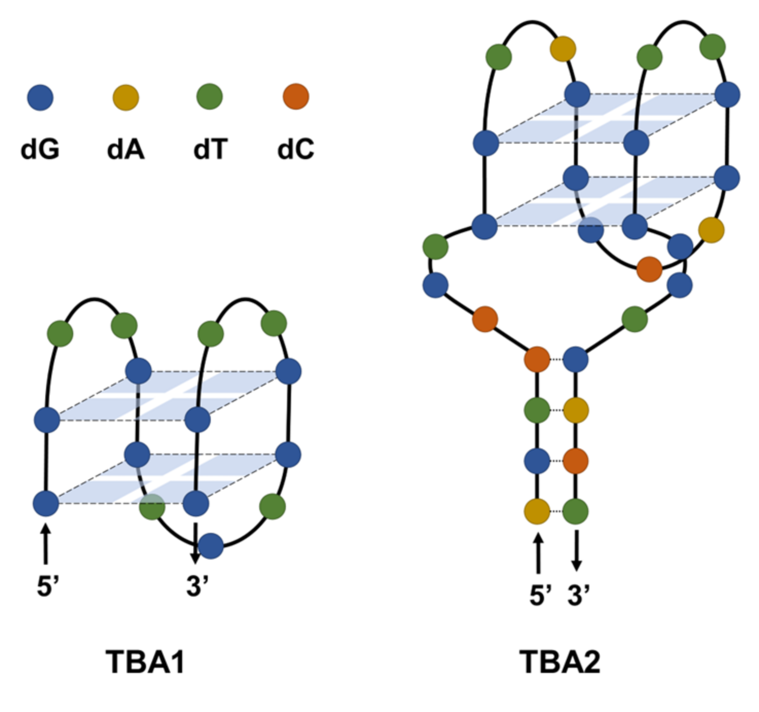
3. Thrombin Binding Aptamer (TBA)

In 1992, Bock et al., reported a nucleic acid aptamer for the first time that binds to human thrombin [40]. Since then, a variety of thrombin aptamers have been reported [41,42,43,44,45,46,47]. Among them, two of the most widely used aptamers are TBA1 and TBA2 (shown in Figure 1). TBA1 is a 15-mer DNA aptamer (5′-GGTTGGTGTGGTTGG-3′) binding to the fibrinogen-recognition exosite of thrombin with a Kd ≈ 100 nM, and TBA2 is a 29-mer DNA aptamer (5′-AGTCCGTGGTAGGGCAGGTTGGGGTGACT-3′) binding to the heparin-binding exosite of thrombin with higher affinity and a Kd ≈ 0.5 nM. Thrombin aptamers fold to form G-quadruplex structures in the process of recognition with thrombin [48,49], which can bond hemin to form hemin/G-quadruplex DNAzyme [50]. Wu et al., conjugated a short peptide to TBA1 and bond hemin to develop a highly sensitive electrochemical thrombin sensor [51]. Based on the optical properties of gold nanoparticles (AuNPs), Li et al., developed an assay platform that realized simply but efficiently detection of thrombin with ultrahigh sensitivity [52]. Simultaneously using TBA1 as the detection probe and TBA2 as the capture probe, Liu et al., constructed a click chemistry-based electrochemical sensor for the simple and fast detection of thrombin, which had great potential for practical applications [53].

4. Optical Aptamer Sensors for Thrombin Detection

Due to their advantages of high sensitivity and accuracy, visualization and easy to read, optical signals (including visual light, ultraviolet and fluorescence), have been widely used in substance detection [54,55]. There are many modifiable sites in the nucleobases and skeleton of the aptamer, which endow it with more functions without significantly changing the binding ability between the aptamer and its target. This builds an ideal platform for the construction of optical thrombin sensors.

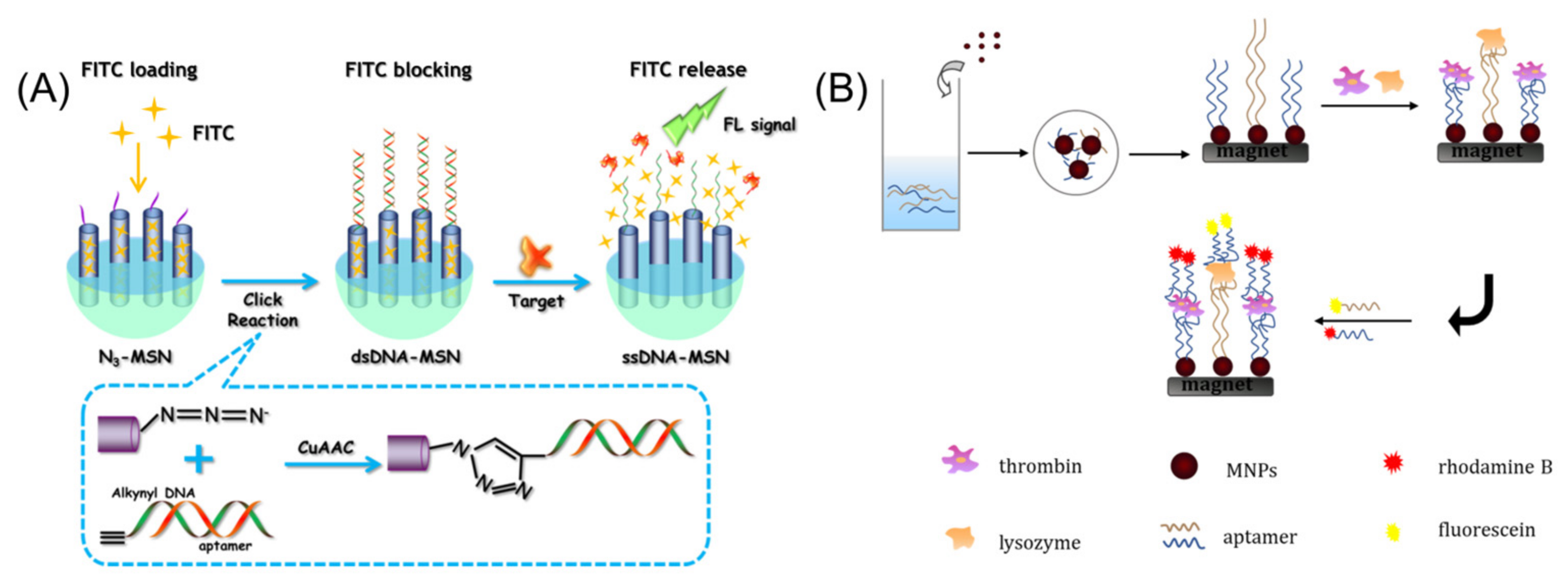
4.1. Fluorescent Sensors

In general, the fluorescent detection methods lie in the detection of variations in fluorescence spectral characteristics consisting of fluorescence intensity, wavelength, and lifetime caused by the direct or indirect interactions of fluorophores with analytes. In this regard, the strategy based on the variation of fluorescence intensity is one of the most accurate methods in biosensing. Limitations in the sensitivity of traditional fluorescent aptamer sensors still exist, thus it is usually essential to combine nucleic acid signal amplification strategies to improve the detection sensitivity. These strategies include rolling circle amplification (RCA), hybrid chain reaction (HCR), catalytic hairpin assembly (CHA) and DNAzyme and CRISPR-Cas9 assisted amplification [56]. At present, there have been plenty of aptamer-based fluorescence sensors established for the detection of thrombin sensitively [57,58,59,60].
For instance, by combining aptamer proximity recognition-dependent strand translocation and CHA-mediated signal amplification, Li et al., reported a simple and sensitive thrombin sensor utilizing fluorescent signal as an indicator [61]. In addition, exonuclease (Exo) catalyzed target circulation is also a common method of signal amplification [62]. Li et al., exploited a tactic for the aptamer-MoS2 sensor based upon the strategy of fluorescence resonance energy transfer (FRET) to detect thrombin [63]. First, the double- stranded DNA (dsDNA) formed by TBA2 modified with carboxyfluorescein (FAM) and its complementary sequence modified with thiol group was immobilized on the Au nanoparticles (AuNPs) adsorbed on the surface of MoS2, and AuNPs@MoS2 nanocomposites quenched the fluorescence of FAM. Then the binding of thrombin to TBA2 induced the change of the aptamer conformation, causing the fluorophore to leave the surface of MoS2 and resulting in the recovery of the fluorescence. In this case, Exo degraded the aptamer in the TBA2-thrombin complex making thrombin to be released into the solution for target recycling.
In general, there is the need to modify the fluorophore on the aptamer of aptamer-based thrombin fluorescence sensors, which are bound to increase the cost of the sensors as well as complicate the purification process. To ameliorate this challenge, the label-free fluorescence sensors have attracted extensive attention. Chen et al., realized fluorescence detection of thrombin through functionalizing mesoporous silica nanoparticles (MSNs) by aptamer (Figure 2A) [64]. TBA2 was hybridized with alkynyl modified complement DNA to form dsDNA firstly and followed with alkyne-azide cycloaddition reaction of the N3-MSN by Cu(I) catalyzing so that the combined ds-DNA could efficiently block fluorescein isothiocyanate (FITC) in the pores of the MSN. In the presence of thrombin, aptamer identified and combined with it, which was released from the surface of MSN leaving single stranded DNA (ssDNA). Since ssDNA had poor sealing ability for the pores of MSN, the FITC redispersed into the solution and a detectable fluorescent signal was provided. The enhanced fluorescence intensity possessed a well linear relation with the amount of thrombin. Based on [Ru(bpy)2(o-mopip)]2+ (OMO) and graphene oxide (GO), Li et al., developed three label-free sensors for the detection of thrombin [65]. Above all, the fluorescent signal of OMO would be extremely enhanced on account of the binding between OMO and TBA1 or TBA2. GO could differentiate various DNA structures and adsorb ssDNA but not G-quadruplex structure. Hence, the added GO adsorbed ssDNA aptamers and OMO and quenched the fluorescence of OMO via π-π interactions and electrostatic interactions. Due to the addition of thrombin, TBA1 and TBA2 folded into G-quadruplex structures efficiently and selectively bound to different exosites on the thrombin surface. Meanwhile, the π–π stacking between OMO and the newly formed G-quadruplex weakened the adsorption of GO resulting in the release of the G-quadruplex and OMO complex from the surface of GO and the recovery of fluorescent signal. On the basis of the performance of these three sensors, all of them were general-purpose sensing platforms with immense sensitivity and selectivity. Among them, the sensing platform utilizing aptamer pair TBA1 and TBA2 simultaneously exhibited outstanding detection performance. These outcomes indicated that the aptamer pair-assisted sensors have broader application prospects in the field of clinical diagnosis.
Sensors that can detect multiple biomarkers simultaneously have important implications for proteome analysis, clinical diagnosis in the exploration of their physiological and pathological relationships. Wang et al., described a simple aptamer sensor with high sensitivity able to simultaneously detect thrombin and lysozyme through different fluorescence signal changes [66]. One thrombin aptamer (TBA1) and one lysozyme aptamer were firstly modified on the surface of magnetic nanoparticles (MNPs) to capture the target proteins, thrombin and lysozyme, respectively (Figure 2B). Whereafter, thrombin was bound with another thrombin aptamer (TBA2) that were labeled with rhodamine B, and lysozyme was bound with another lysozyme aptamer that was labeled with fluorescein individually. This converted target-identifying events into fluorescence signals for detection. Utilizing the magnetic field not only eliminated interferences of coexistence proteins but also concentrated trace target proteins enhancing fluorescence signals.

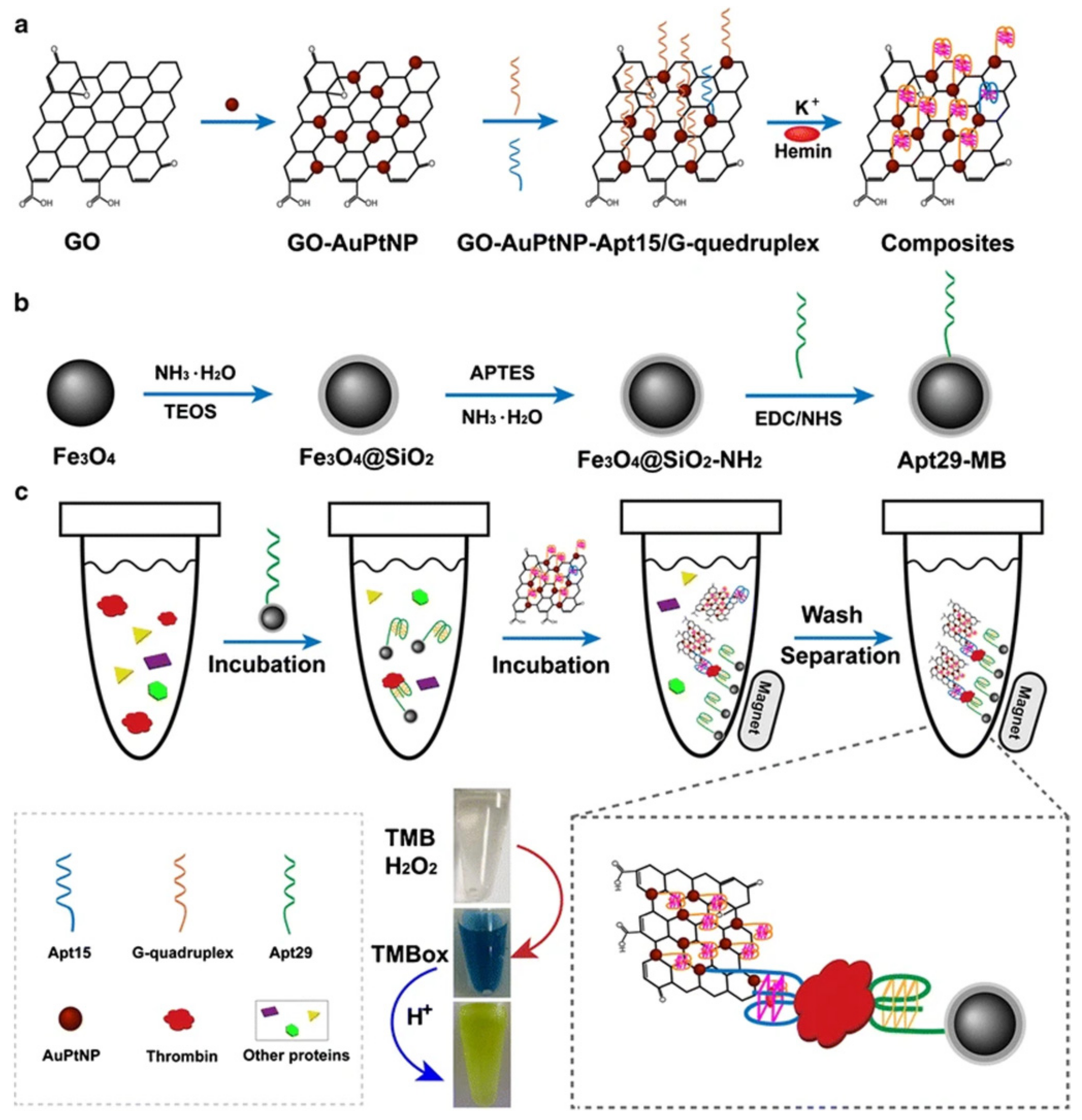
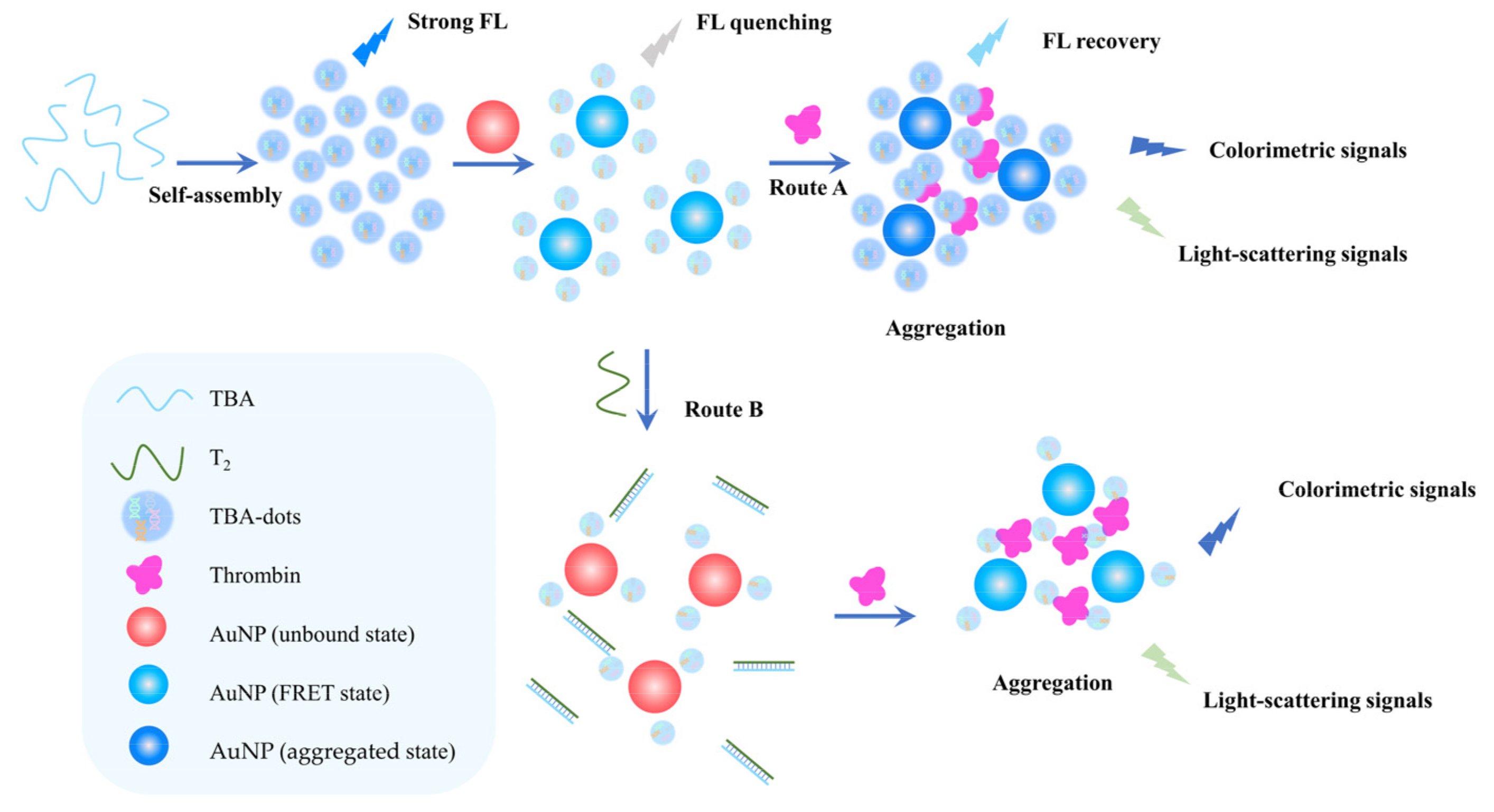
4.2. Colorimetric Sensors

Colorimetric sensors are a series of devices to achieve qualitative or quantitative detection of targets by analyzing the color variations of the sensing system caused by analytes. In addition to not relying on the expensive instrument and rapid detection, the most unique characteristic of these sensors is visualizing by the naked eye directly, so they are favored by a growing number of researchers. With the introduction of AuNP, graphene, Fe3O4 and other nanomaterials, the colorimetric sensors have been promoted rapidly and their detection performance and stability have been improved. The powerful combination of aptamers with nanomaterials enhances the detection ability of colorimetric sensors and therefore is widely utilized in environmental monitoring, food safety, clinical diagnostics and other fields [67,68,69,70].
Li et al., reported a straightforward thrombin colorimetric sensor in the light of the property of G-quadruplex [71]. The G-quadruplex structure of TBA formed by the combination of K+ with low stability had weak peroxidase-mimetic activity when bound hemin. However, when TBA coordinated thrombin, the formed complex of thrombin-G-quadruplex-hemin was highly stable and had the strong ability to catalyze the color change reaction of the 2,2′-azinobis (3-ethylbenzthiazoline-6-sulfonic acid) (ABTS)–H2O2 substrate which indicated the content of thrombin. Wang et al., described a neotype thrombin sensing strategy combining GO enzyme-like activities and a pair of thrombin binding aptamers [72]. As the capture probe (Figure 3), the carboxylated TBA2 was first modified on the surface of the magnetic bead by the EDC and NHS coupling reaction and then captured thrombin from the complex matrix. In addition, the thiolated TBA1 conjugated the AuPtNP via Au-S bond, then the composite was adsorbed on the surface of GO. However, TBA1 bound hemin to form a complex with weak peroxidase-like activity in the absence of thrombin. Thrombin could be captured by the TBA1-MB under the magnetic field, and then the composites formed by GO-AuPtNP-TBA1, G-quadruplex and hemin were forward brought to prepare the sandwich structure as the detection platform. Through oxidizing TMB (3,3′,5,5′-tetramethylbenzidine) to cause a color change in the system, the colorimetric thrombin detection was realized. In this design, the signal amplification strategy relied on the peroxidase-like abilities of GO, AuPtNP and G-quadruplex/hemin complex that catalyzed substrate TMB color change for colorimetric detection, and the separative ability of magnetic beads that were functionalized by thrombin aptamer.
In addition to G-quadruplex/hemin composite, a series of colorimetric thrombin aptamer biosensors have been constructed by combining with nanozymes that mimic oxidase, peroxidase or superoxide oxidase [73]. The structure of cationic polymers, such as poly [3-(3′-N, N, N-triethylamino-1′-propyloxy)-4-methyl-2, 5-thiophene hydrochloride] (PMNT), is very susceptible to interference from negative ions in solution. PMNT has a peroxidase-like catalytic activity that is capable of accelerating the color changing of TMB. As one of the cationic polythiophene derivatives, PMNT exhibits differentiated catalysis capabilities in the presence of discrepant structures of thrombin aptamer. Based on this phenomenon, Liu et al., presented an ultrasensitive colorimetric sensing platform of thrombin taking advantage of cationic polythiophene derivative as a tool [74]. The TBA1 played an auxiliary role in the TMB-H2O2 reaction raising the absorbance peak, and this effect was weakened by thrombin binding to TBA1. The obtained results showed that the displayed platform was doing pretty well in thrombin sensing and offered a new perspective strategy for the determination of clinical biomolecules. Zafar et al., constructed a solid-phase colorimetric thrombin sensor based on color changes of AuNPs caused by binding between thrombin and TBA that are modified on the surface of AuNP [75]. TBA-dots are kinds of nanoparticles formed by aggregation of thrombin aptamer, which retain the recognition ability to thrombin originating from the structure of aptamer while obtaining optical properties similar to that of carbon quantum dots. It was first reported by Kuang et al., and composed with AuNPs to construct a versatile sensor for the detection of thrombin that had three different signal reading modes including colorimetric, fluorometric and light-scattering signals (Figure 4) [76]. In addition to part of TBA-dots being covalently linked to AuNPs through Au-N bonds, there were abundant TBA-dots adsorbed on the surface of AuNPs forming a shell whose charge accumulation effectively dispersed AuNPs. Meanwhile, the fluorescence intensity of TBA-dots was impaired because of fluorescence resonance energy transfer (FRET) with AuNPs. On the one hand (route A), since thrombin had multiple binding sites for the aptamers, TBA-dots/AuNPs were linked together then inducing AuNPs aggregation followed by the maximum absorption peak with redshift. In this issue, the FRET efficiency was weakened and then the fluorescence of TBA-dots recovered. Consequently, thrombin could be detected by the extent of fluorescence intensity recovery of TBA-dots and colorimetric and light-scattering signals caused by the aggregation of AuNPs. On the other hand (route B), T2 was fully complementary to TBA and hybridized with it, which brought about the destruction of the TBA-dots shell on the periphery of AuNPs and thus these AuNPs were decentralized. Under the circumstances, thrombin played the role of the bond linking TBA-dots/AuNPs closely and leading increscent AuNPs aggregation degree. Therefore, thrombin could be detected by colorimetric signal and light scattering signal simultaneously according to different aggregation extent of AuNPs.
Due to the advantages of small physical dimensions, reduced sample demand and high integration, condensing the detection system on a microfluidic chip has evoked increasing research enthusiasm in biological and chemical analysis fields. In the last few years, researchers have built numerous microfluidic chip systems to detect proteins [77,78]. Zhao et al., designed an aptamer-based microfluidic chip modified by Ag nanoparticles (AgNPs) realizing colorimetric thrombin detecting [79]. The microfluidic chip system consisted of several parallel sample detection channels, and their glass substrates were modified with streptavidin (SA) to immobilize the biotinylated TBA1 by the affinity between biotin and streptavidin for thrombin capture (Figure 5). The samples were mixed with the TBA2 functionalized AgNPs and then injected into the microfluidic channels. If there was thrombin in the sample, it would be captured to form a sandwich structure composed of TBA1, thrombin and TBA2 functionalized AgNPs. The color variation due to the capture of yellow AgNPs was in line with the concentration of thrombin in the sample. Thrombin concentration could be read out approximately according to the color difference and quantitative analysis could be realized by the grayscale value measured by a flatbed scanner.

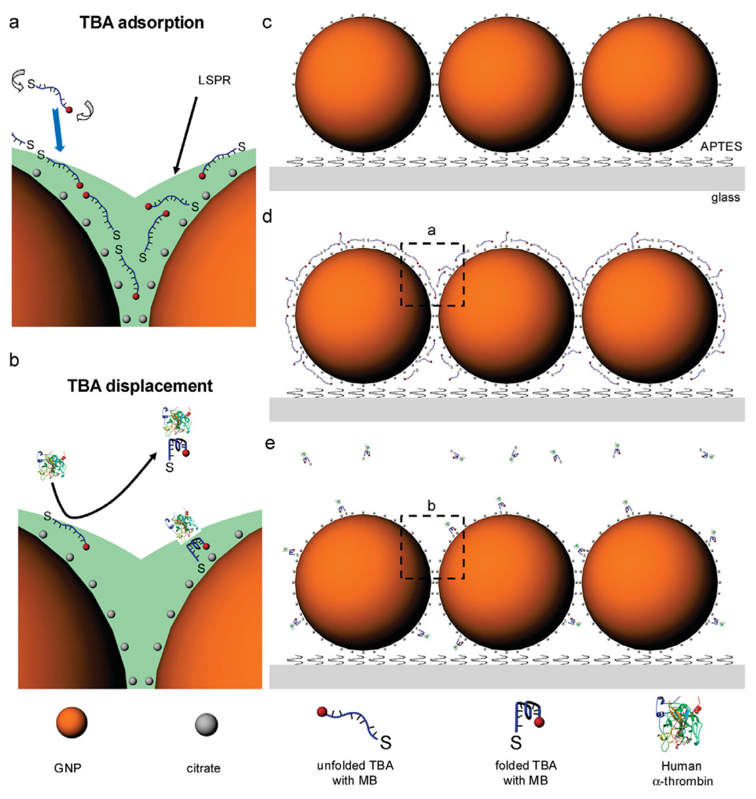
4.3. SERS-Based Sensors

Surface-enhanced Raman scattering (SERS) is a phenomenon whereby the photoexcitation of unitary vibrations of electrons that are dispersed in the metallic nanostructure surface causes a large Raman scattering signal [80]. This was discovered in 1974 by Fleischmann et al., for the first time [81]. Due to the superiorities such as strong spectral signature and simultaneous detection of different analytes, the detection tools based on SERS enjoy great popularity in biochemical and life science. By combining surface-enhanced resonance Raman scattering (SERRS) with aptamer, Cho et al., constructed an ingenious sensor (Figure 6) with relatively simple principles for thrombin detection [82]. First of all, single-stranded TBA1, which was modified with photosensitive molecule methylene blue, readily adsorbed onto AuNP surfaces. In the absence of thrombin, TBA1 was unfolded. This effectively shortened the distance between methylene blue and AuNP, causing to the generation of strong SERS signal of methylene blue. Due to the target protein, thrombin, the spatial conformational of TBA1 folding was transformed into G-quadruplex on account of specific recognition. Removal of TBA1 from the surface of AuNP decreased the methylene blue SERS signal intensity.
DNA pyramids are a class of three-dimensional DNA nanostructures formed by the hybridization of multiple single strands. They have the programmable size and multiple modification sites, so they receive extensive attention and have been widely used in the study of cell interaction and sensing fields. In order to detect different targets with different signaling modes simultaneously, Hao et al., designed a multifunction aptamer sensor by modifying AuNP and UCNP (upconversion nanoparticle) at the DNA pyramid vertexes [83]. As shown in Figure 7, first, the pyramid framework consisted of two unmodified oligonucleotide chains and two complementary chains separately modified with AuNP and UCNP. Then the thrombin aptamer modified with AuNP and prostate-specific antigen (PSA) aptamer modified with UCNP were combined with the frame and then the intact pyramid structure was successfully spliced. When the aptamer modified with nanoparticle encountered its target (thrombin or PSA), the specific recognition process between aptamers and corresponding targets and the competitions of aptamers with the collapse of the pyramid occurred. When thrombin was present in the detection system, the AuNP modified on TBA1 left the another causing a weakened SERS signal that was transduced by 4-mercaptophenylacetic acid as the Raman reporter. When PSA identified its aptamer, the quenched fluorescence of UCNP would be restored by leaving the pyramid. This DNA structure based on aptamers and nanoparticles assembly has imponderable potential for precise differentiation of disease markers in specific clinical applications.

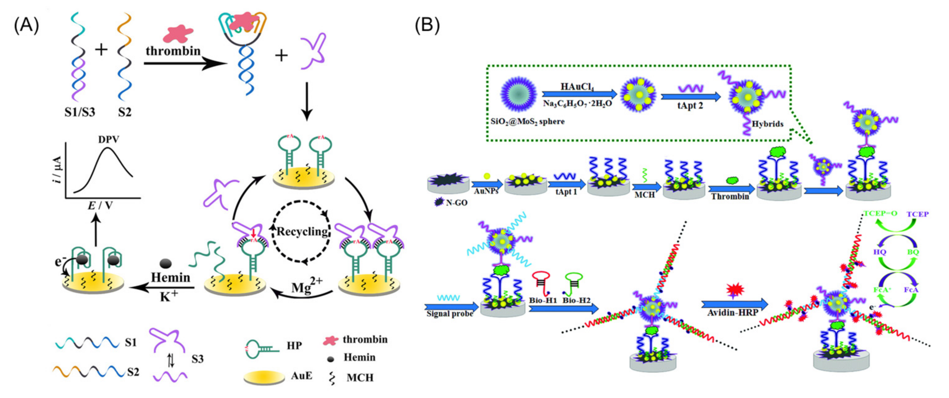
5. Electrochemical Aptamer Sensors for Thrombin Detection

Another kind of commonly used biosensor is the electrochemical biosensor which quantifies the transferred electrons or ions during the electrochemical reactions induced by the analyte to calculate the unknown concentration. Compared with optical sensors, electrochemical sensors are likely to achieve miniaturization and portability, which have attracted much attention as well [84,85]. Combining molecular recognition ability of aptamer with signal transduction ability of electron, electrochemical sensors transform the recognition process of aptamer and target molecule into electrical signals [20,86,87,88,89,90,91].
Yang et al., ingeniously cascaded strand displacement and DNAzyme signal amplification to build an electrochemical sensor for thrombin detection that was label-free without the need for enzymes [92]. There were four oligonucleotides (S1, S2, S3, and a hairpin-structured substrate sequence named HP) involved in this detecting system shown in Figure 8A. TBA1 and TBA2 were contained in S1 and S2, respectively, and were designed to recognize the thrombin target in different exosites, respectively. Initially, the DNAzyme sequence (S3) was hybridized to S1 to form a stabilized S1/S3 dsDNA. The G-quadruplex forming sequence inserted within the thiol-functionalized HP that was assembled on Au electron through the Au−S bond was segmentally blocked in the stem region to prohibit the HP from forming the stable G-quadruplex structure. S3 possessed a similar sequence as S2 that complemented S1. Therefore, if there was no thrombin, the presence of S2 would not interfere with hybridization of S3 with S1. However, if there was thrombin in the system, the two aptamers could bind thrombin at the same time enriching S1/S3 and S2 in the solution, which accelerated the proximity-dependent strand displacement reaction by shortening the distance between S3 and S2. Whereafter, the released DNAzyme cleaved the substrate sequence in HP to unlock the G-quadruplex forming sequence with the assistance of Mg2+. In the stabilization of K+, the G-quadruplex forming sequences transformed into antiparallel G-quadruplex and bound hemins to produce a megascopic current signal during the potential scan process which realized extremely sensitive thrombin sensing.
In the above experimental scheme, the cyclic cleaving ability of DNAzyme achieves one-step signal amplification, however, that only goes so far as to elevate the detection ability. The ingenious cascade of multiple amplification strategies in constructing thrombin sensors can decrease the limit of detection (LOD) to a lower level. Shuai et al., reported the development of an ultrasensitive sandwich-type electrochemical sensor based on dual signal amplification of HCR and horseradish peroxidases (HRP) catalysis for thrombin detection [93]. Firstly, AuNPs and N-GO (N-doped GO) were immobilized on the electrode surface and modified with thiolated tApt1 (TBA1 with poly T) to capture thrombin (Figure 8B). Then, the unbonded sites of the surface of the electrode were sealed by 6-mercapto-1-hexanol (MCH) followed by the introduction of tApt2 (TBA1) functionalized SiO2@MoS2 that specifically recognized and bound thrombin to produce a sandwich architecture biosensor. Under the circumstances, a good deal of signal probes that were nailed to the surface of AuNPs induced the HCR of biotin functionalized H1 and H2 oligonucleotides. Streptavidin-modified HRPs were concatenated on the biotinylated nanowire of H1 and H2 to catalyze the redox reaction of substrates producing a magnified electrochemical signal. Likewise, employing enzymatic biofuel cell (EBFC) as electrical signal source and capacitor as a signal amplifier, Wang et al., fabricated a self-energized electrochemical detecting device for the sensitive thrombin detection [94]. In terms of the cathode, bilirubin oxidase (BOD) was adsorbed on the carbon paper (CP) that was modified with carbon nanotubes (CNTs) and AuNPs in advance (Figure 9A). In terms of the anode, the CP electrode pretreated with SnS2 and AuNPs was availed as electrode matrix to immobilize TBA1, and then the free site was blocked by MCH. Glucose oxidase (GOD) and TBA2 were deposited on the surface of CNTs and AuNPs to obtain bioactive carriers. In the presence of thrombin, the sandwich structure formed through thrombin connecting bioactive carrier to the anode was used to accelerate the redox reaction of glucose. As a cathodic enzyme, BOD was utilized as an accelerator of the redox reaction of O2 resulting in an electrochemical signal. The charge accumulation and instantaneous discharge capabilities of the capacitor amplified the transient current from the electrode at a higher rate improving the detection capability of EBFC availably. This amplified electrical signal could be facilely read by an electricity meter. This sensor, therefore, integrated the intelligent specialties of the self-energized detector, oligonucleotide aptamers and capacitance amplifier exhibiting greater prospects in biomarker analysis fields.
The vigorous advancement of nanotechnology and microelectronics fields has promoted the miniaturization of biosensors, therefore, some biosensors such as microfluidic chips and paper strips have attracted the attention of many researchers. Li et al., first came up with a self-energized rotatable microfluidic chip designed with paper for sensing thrombin [95]. The microfluidic chip contained a top layer and bottom layer shown in Figure 9B. The former consisted of two sets of reaction areas for signal generation and the latter incorporated a detection area for analyte identification, the supercapacitor for signal amplification, washing channels and the hollow holes for electrochemical detection. The detection disc of paper was coated with carbon ink, polyvinyl alcohol (PVA) and silver/silver chloride ink in turn on two sides. The redox activity of the detection area was endowed by the addition of K3[Fe(CN)6] and glucose. By Au-S interaction, AuNPs deposited on the reaction area of the reaction disc were modified with DNA1 that could partially hybridize with thrombin aptamer. Next, TBA2 connected the DNA2 modified with glucose oxidase to the reaction disc. Then, the reaction disc and detection disc were assembled. The “ON” and “OFF” states could be switched, and other processes could be carried out independently by rotating the top layer. When the reaction area was rotated to the top of the detection area to add thrombin and elute by phosphate buffer solution, Fe3+ would be reduced to Fe2+ in the reaction area by glucose oxidase (Gox)-DNA2 that mixed within the buffer during the recognition process. The transient release of charge that was stored in the self-energized supercapacitor created an enhanced signal that was readily scouted by the digital multimeter.

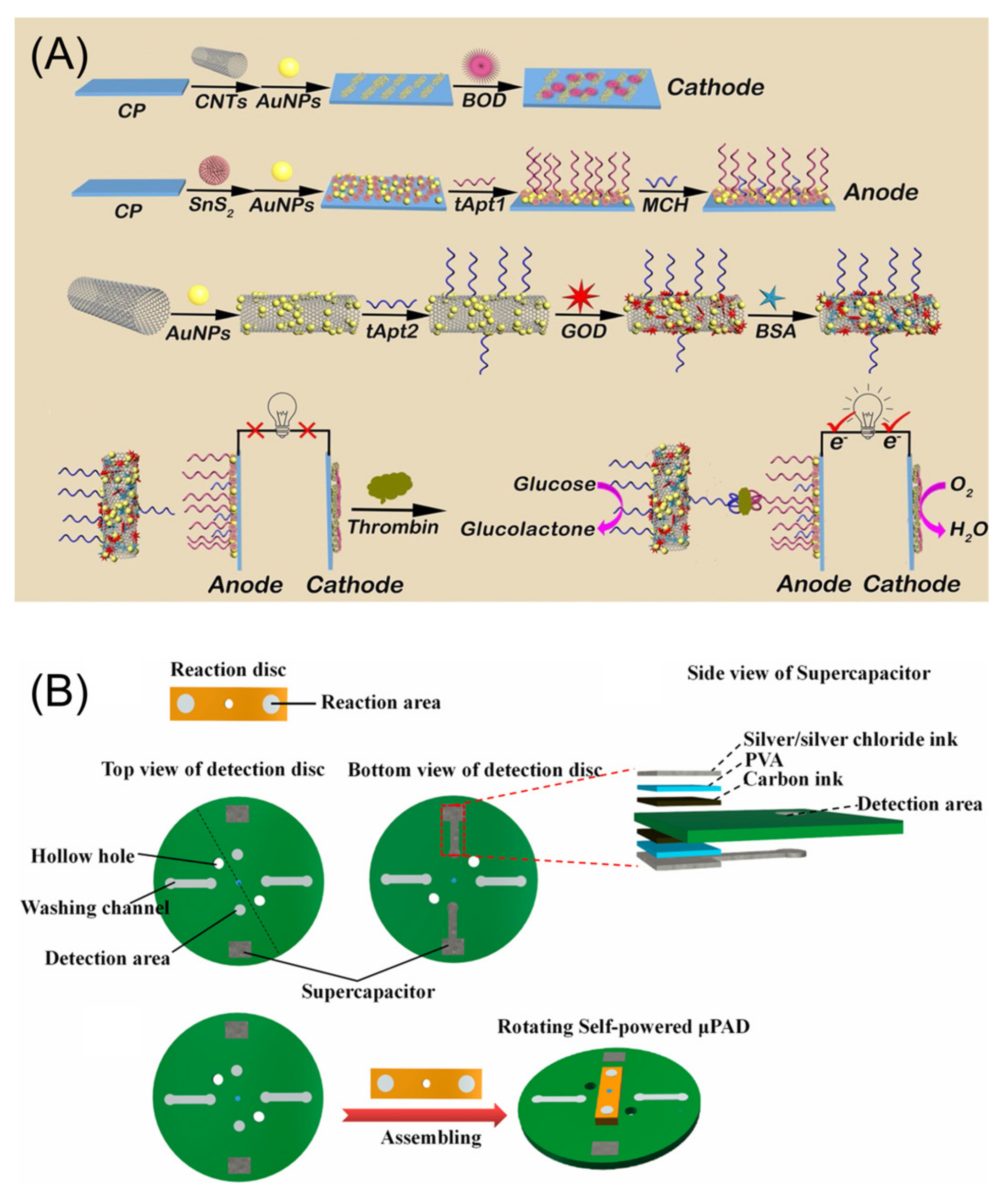
6. Other Aptamer Sensors for Thrombin Detection

In addition to various sensing strategies for thrombin detection which are based on fluorescent, colorimetric and electrochemical methods (as listed in Table 1), there are many researchers that have creatively developed multifarious neotype aptamer sensors [96,97,98,99]. For example, Ma et al., developed a liquid crystals detector on the basis of the phenomenon that the binding process of thrombin and aptamer would affect the orientational transition of liquid crystals due to the electrostatic interaction between the aptamer and octadecyl-trimethylammonium bromide [100].
Since thrombin could simultaneously bind TBA1 and TBA2 at two different exosites, Bai et al., designed a sensing platform that took surface plasmon resonance (SPR) of gold nanoparticles as the signal output through the recognition of aptamer and thrombin [101]. Prior to proceeding sample testing, distinct functionalization of TBA1 and TBA2 were implemented. TBA1 was fixed on the sensor chip by the interaction between biotin and streptavidin, and TBA2 was fixed on the surface of AuNP by sulfhydryl (Figure 10A). When thrombin showed up, two aptamers identified the target and combined at both poles of it forming the sandwich structure. The distance between AuNP and SPR Au film was shortened and the SPR signal was amplified. There was a good linear relationship between enhanced SPR signal and thrombin concentration. Therefore, highly sensitive and accurate detection of thrombin was achieved.
Fu et al., constructed an economically friendly and handy sensing strategy (Figure 10B) for detecting thrombin by combining a barometer [102]. The design employed Au@PtNPs with peroxidase activity to decompose H2O2 to release O2 that accumulated in the chamber producing a pressure signal that was measurable just by using a barometer. Firstly, thrombin connected TBA1 modified Au@PtNP to TBA2 modified MB to form a sandwich structure. The complex was then resuspended in H2O2 in the chamber and all valves were closed. After the reaction was complete, valve A was opened followed by the measurement of the pressure in the chamber to quantify thrombin. Furthermore, the barometer-modified detector for the convenient test of carcinoembryonic antigen, ractopamine and Hg2+ were realized as well.
Supramolecular assembly is a crucial means to create new substances and vest materials with new functions via the bottom-up approach. By using this method, multistage assembly structures can be constructed and supramolecular materials with dynamic, multi-functional and high performance obtained. Shen et al., designed a cyanine dyes probe DMSB (3-ethyl-2-[3-(3-ethyl-3H-benzoselenazol-2-ylidene)-2-methylprop-1-enyl] benzoselenazolium bromide) to detect thrombin [103]. In the presence of thrombin, the formation of TBA G-quadruplex induced DMSB monomers to assemble into J-aggregates. Through this unique supramolecular self-assembly strategy, the detection signal could be amplified, and the sensitivity of thrombin detection enhanced. In addition, the distinct signals caused by different TBA G-quadruplex could eliminate the interference caused by K+ and improve the accuracy of detection (Figure 10C).
Since its advent in the 19th century, optical fibers based on various materials have been developed and optimized. The intriguing characteristics of optical fiber such as ease to miniaturize and relatively low cost make biosensors based on the optical fiber of particular interest to researchers in the recent decade. Sypabekova et al., developed a miniature biosensing strategy on the basis of MgO nanomaterial functionalized optical fiber for thrombin detection [101]. After being pretreated with hydrofluoric acid, the fiber was decorated with a thin Au layer by using the Au electroless plating method. Then the thiol-modified TBA1 was immobilized on the surface of the Au layer by the Au-S bond to capture thrombin. By monitoring the Rayleigh scattering spectra shift monitored by the optical backscatter reflectometry during the specific binding of thrombin and TBA, the sensor could detect thrombin. This kind of surface-modified optical fiber biosensor with outstanding performance have great application prospects in the detection of thrombin in clinical samples.

7. Conclusions and Future Perspective

With the in-depth study of physiological and pathological processes in blood, the role of thrombin, a serine protease, in blood coagulation and cellular signaling pathway activation has been continuously revealed. The relationships between cardiovascular disease, inflammation, cancer migration and thrombin are gradually strengthened. At present, although methods such as PT, APTT and TT have been widely used in clinical aspect to evaluate thrombin activity, some drawbacks associated with these methods limit their general application. In addition, the lack of methods for quantitative and accurate determination of thrombin concentration necessitates the need to develop new sensitive, accurate and simple qualitative and quantitative detection strategies of thrombin to address these challenges.
The emergence of SELEX technology has greatly promoted the development of various nucleic acid aptamers, and the thrombin aptamer is no exception. The introduction of aptamers endows biosensors with recognition and targeting ability. Compared with traditional antibodies, aptamers are not only cheaper, more stable and more accessible, but they have many modification sites on their nucleic acid skeleton to facilitate binding with signal indicator molecules and new-fashioned nanomaterials. Moreover, nucleic acid signal amplification strategies for example RCA, HCR, CHA and DNAzyme have been developed to further elevate the detection sensitivity of oligonucleotide aptamer sensors. Therefore, in recent years, thrombin biosensors based on nucleic acid aptamers have been vigorously developed.
The detection principle of thrombin aptamer sensor based on fluorescence is basically that the combination of thrombin and aptamer causes the change of spatial distance between the fluorescence group and quenching group, which enhances or weakens the fluorescence signal to realize the quantitative detection of thrombin. The detection of thrombin aptamer sensor based on colorimetry relies on the conformational change of thrombin aptamer, which varies the nanozyme activity of other materials that will catalyze reactions to cause a color change, or the conformational change of thrombin aptamer, which leads to the change of aggregation state of nanoparticles, resulting in the nanoparticles to emit different colors. The thrombin aptamer sensor based on SERS uses the biometric interaction between thrombin aptamer and thrombin to cause the change of Raman scattering light on the surface of metal nanoparticles for thrombin detection. The electrochemical thrombin aptamer sensors mostly shorten the distance between electroactive materials and electrodes by combining different aptamers on two surficial exosites of thrombin and then convert chemical signals into electrical signals. In addition, there are some other methods with their own characteristics to achieve highly sensitive detections of thrombin. In the sensors mentioned in Table 1, compared with the optical sensors, the electrochemical aptamer sensors for thrombin detection tend to have wider linear ranges and lower limit of detection, some even as low as aM level. The stronger serum sample detection capability and rapid test capability conferred by electrochemical methods are conducive to point-of-care testing (POCT) of thrombin in clinical samples. In addition, the sensors based on electrochemistry are likely to be miniaturized and combined with microfluidic chips and test strips to construct integrated thrombin detection devices.
Although the thrombin aptamer sensors are relatively mature, there are still some directions for us to future development. First of all, the materials used to bind aptamers at present, such as gold nanoparticles, are common materials in nucleic acid detection. Exploiting materials that can specifically bind to the thrombin aptamer G-quadruplex structure deserve more endeavors. Secondly, the content of thrombin in the human body keeps changing with the development of physiological processes. The development of aptamer-based probe which can be used to monitor thrombin content in vivo is of great help to the further understanding of the pathogenesis of the disease. Then, in order to reveal the relationship between disease and biomarkers, the detection of a single target often cannot satisfy the demand. Thus, achieving multitarget signal demonstration based on the logic gate is not only beneficial to avoiding the generation of false positive signals for the precision thrombin detection but to analyzing the association between multiple biomarkers for a better understanding of pathogenesis. Finally, the instrumentalization of thrombin aptamer sensors and the construction of detection devices have a far-reaching impact for the clinical detection of thrombin. As aptamer technology and chemical detection strategy continuously develop, aptamer-based thrombin sensors have broad application prospects in clinical detection and physiological and pathological research.

Author Contributions

Writing—original draft preparation, H.S.; writing—review and editing, N.W., L.Z., H.M. and Z.L.; supervision, H.M. and Z.L. All authors have read and agreed to the published version of the manuscript.

Funding

This work was supported by the Program for Science Technology Innovation Teams in Universities of Henan Province (22IRTSTHN002), the Key Project of Science and Technology of Henan Province (212102310334).

Institutional Review Board Statement

Not applicable.

Informed Consent Statement

Not applicable.

Data Availability Statement

Not applicable.

Acknowledgments

We thank Yanan Wu and Xinxin Shi for their kind help in the revision of the manuscript.

Conflicts of Interest

The authors declare no conflict of interest.

References

  1. Posma, J.J.N.; Posthuma, J.J.; Spronk, H.M.H. Coagulation and non-coagulation effects of thrombin. J. Thromb. Haemost. 2016, 14, 1908–1916. [Google Scholar] [CrossRef] [Green Version]
  2. Negrier, C.; Shima, M.; Hoffman, M. The central role of thrombin in bleeding disorders. Blood Rev. 2019, 38, 100582. [Google Scholar] [CrossRef] [PubMed]
  3. Krenzlin, H.; Lorenz, V.; Alessandri, B. The involvement of thrombin in the pathogenesis of glioblastoma. J. Neurosci. Res. 2017, 95, 2080–2085. [Google Scholar] [CrossRef] [PubMed]
  4. Yin, X.; Wright, J.; Wall, T.; Grammas, P. Brain Endothelial Cells Synthesize Neurotoxic Thrombin in Alzheimer’s Disease. Am. J. Pathol. 2010, 176, 1600–1606. [Google Scholar] [CrossRef] [PubMed] [Green Version]
  5. Tsopanoglou, N.E.; Maragoudakis, M.E. Role of Thrombin in Angiogenesis and Tumor Progression. Semin. Thromb. Hemost. 2004, 30, 63–69. [Google Scholar]
  6. Mannironi, C.; Scerch, C.; Fruscoloni, P.; Tocchini-Valentini, G.P. Molecular recognition of amino acids by RNA aptamers: The evolution into an L-tyrosine binder of a dopamine-binding RNA motif. RNA 2000, 6, 520–527. [Google Scholar] [CrossRef] [Green Version]
  7. Huizenga, D.E.; Szostak, J.W. A DNA Aptamer That Binds Adenosine and ATP. Biochemistry 1995, 34, 656–665. [Google Scholar] [CrossRef]
  8. Xu, W.; Ellington, A.D. Anti-peptide aptamers recognize amino acid sequence and bind a protein epitope. Proc. Natl. Acad. Sci. USA 1996, 93, 7475–7480. [Google Scholar] [CrossRef] [Green Version]
  9. Xiao, Z.; Shangguan, D.; Cao, Z.; Fang, X.; Tan, W. Cell-Specific Internalization Study of an Aptamer from Whole Cell Selection. Chem. Eur. J. 2008, 14, 1769–1775. [Google Scholar] [CrossRef]
  10. Gopinath, S.C.B. Methods developed for SELEX. Anal. Bioanal. Chem. 2007, 387, 171–182. [Google Scholar] [CrossRef]
  11. Mukherjee, M.; Sistla, S.; Veerabhadraiah, S.R.; Bettadaiah, B.; Thakur, M.; Bhatt, P. DNA aptamer selection and detection of marine biotoxin 20 Methyl Spirolide G. Food Chem. 2021, 363, 130332. [Google Scholar] [CrossRef]
  12. Zhao, L.; Huang, Y.; Dong, Y.; Han, X.; Wang, S.; Liang, X. Aptamers and Aptasensors for Highly Specific Recognition and Sensitive Detection of Marine Biotoxins: Recent Advances and Perspectives. Toxins 2018, 10, 427. [Google Scholar] [CrossRef] [Green Version]
  13. Guo, W.; Zhang, C.; Ma, T.; Liu, X.; Chen, Z.; Li, S.; Deng, Y. Advances in aptamer screening and aptasensors’ detection of heavy metal ions. J. Nanobiotechnol. 2021, 19, 166. [Google Scholar] [CrossRef]
  14. Jolly, P.; Formisano, N.; Estrela, P. DNA aptamer-based detection of prostate cancer. Chem. Pap. 2015, 69, 77–89. [Google Scholar] [CrossRef] [Green Version]
  15. Bakhtiari, H.; Palizban, A.A.; Khanahmad, H.; Mofid, M.R. Aptamer-based approaches for in vitro molecular detection of cancer. Res. Pharm. Sci. 2020, 15, 107–122. [Google Scholar]
  16. Zhao, X.; Dai, X.; Zhao, S.; Cui, X.; Gong, T.; Song, Z.; Meng, H.; Zhang, X.; Yu, B. Aptamer-based fluorescent sensors for the detection of cancer biomarkers. Spectrochim. Acta Part A 2021, 247, 119038. [Google Scholar] [CrossRef]
  17. Ronda, L.; Tonelli, A.; Sogne, E.; Autiero, I.; Spyrakis, F.; Pellegrino, S.; Abbiati, G.; Maffioli, E.; Schulte, C.; Piano, R.; et al. Rational Design of a User-Friendly Aptamer/Peptide-Based Device for the Detection of Staphylococcus Aureus. Sensors 2020, 20, 4977. [Google Scholar] [CrossRef]
  18. Cunha, I.; Biltes, R.; Sales, M.; Vasconcelos, V. Aptamer-Based Biosensors to Detect Aquatic Phycotoxins and Cyanotoxins. Sensors 2018, 18, 2367. [Google Scholar] [CrossRef] [Green Version]
  19. Hwang, J.Y.; Kim, S.T.; Han, H.-S.; Kim, K.; Han, J.S. Optical Aptamer Probes of Fluorescent Imaging to Rapid Monitoring of Circulating Tumor Cell. Sensors 2016, 16, 1909. [Google Scholar] [CrossRef] [Green Version]
  20. Zhang, L.; Zhang, X.; Feng, P.; Han, Q.; Liu, W.; Lu, Y.; Song, C.; Li, F. Photodriven Regeneration of G-Quadruplex Aptasensor for Sensitively Detecting Thrombin. Anal. Chem. 2020, 92, 7419–7424. [Google Scholar] [CrossRef]
  21. Malecka, K.; Ferapontova, E.E. Femtomolar Detection of Thrombin in Serum and Cerebrospinal Fluid via Direct Electrocatalysis of Oxygen Reduction by the Covalent G4-Hemin-Aptamer Complex. ACS Appl. Mater. Interfaces 2021, 13, 37979–37988. [Google Scholar] [CrossRef]
  22. Qiu, F.; Gan, X.; Jiang, B.; Yuan, R.; Xiang, Y. Electrode immobilization-free and sensitive electrochemical sensing of thrombin via magnetic nanoparticle-decorated DNA polymers. Sens. Actuators B Chem. 2021, 331, 129395. [Google Scholar] [CrossRef]
  23. Motta, J.-P.; Palese, S.; Giorgio, C.; Chapman, K.; Denadai-Souza, A.; Rousset, P.; Sagnat, D.; Guiraud, L.; Edir, A.; Seguy, C.; et al. Increased mucosal thrombin is associated with Crohn’s disease and causes inflammatory damage through protease-activated receptors activation. J. Crohn’s Colitis 2021, 15, 787–799. [Google Scholar] [CrossRef] [PubMed]
  24. Ebrahimi, S.; Jaberi, N.; Avan, A.; Ryzhikov, M.; Keramati, M.R.; Parizadeh, M.R.; Hassanian, S.M. Role of thrombin in the pathogenesis of central nervous system inflammatory diseases. J. Cell. Physiol. 2017, 232, 482–485. [Google Scholar] [CrossRef] [PubMed]
  25. Reddel, C.J.; Tan, C.W.; Chen, V.M. Thrombin Generation and Cancer: Contributors and Consequences. Cancers 2019, 11, 100. [Google Scholar] [CrossRef] [PubMed] [Green Version]
  26. Danckwardt, S.; Hentze, M.W.; Kulozik, A.E. Pathologies at the nexus of blood coagulation and inflammation: Thrombin in hemostasis, cancer, and beyond. J. Mol. Med. 2013, 91, 1257–1271. [Google Scholar] [CrossRef] [PubMed] [Green Version]
  27. Young, G.; Sørensen, B.; Dargaud, Y.; Negrier, C.; Brummel-Ziedins, K.; Key, N.S. Thrombin generation and whole blood viscoelastic assays in the management of hemophilia: Current state of art and future perspectives. Blood 2013, 121, 1944–1950. [Google Scholar] [CrossRef] [Green Version]
  28. Monroe, D.M.; Hoffman, M. What does it take to make the perfect clot? Arterioscler. Thromb. Vasc. Biol. 2006, 26, 41–48. [Google Scholar] [CrossRef]
  29. Monroe, D.M.; Hoffman, M.; Roberts, H.R. Platelets and Thrombin Generation. Arterioscler. Thromb. Vasc. Biol. 2002, 22, 1381–1389. [Google Scholar] [CrossRef] [Green Version]
  30. Ye, F.; Garton, H.J.L.; Hua, Y.; Keep, R.F.; Xi, G. The Role of Thrombin in Brain Injury After Hemorrhagic and Ischemic Stroke. Transl. Stroke Res. 2021, 12, 496–511. [Google Scholar] [CrossRef]
  31. Ito, K.; Hongo, K.; Date, T.; Ikegami, M.; Hano, H.; Owada, M.; Morimoto, S.; Kashiwagi, Y.; Katoh, D.; Yoshino, T.; et al. Tissue thrombin is associated with the pathogenesis of dilated cardiomyopathy. Int. J. Cardiol. 2017, 228, 821–827. [Google Scholar] [CrossRef]
  32. Hayosh, Z.I.; Abu Bandora, E.; Shelestovich, N.; Nulman, M.; Bakon, M.; Yaniv, G.; Khaitovitch, B.; Balan, S.; Gerasimova, A.; Drori, T.; et al. In-thrombus thrombin secretion: A new diagnostic marker of atrial fibrillation in cryptogenic stroke. J. Neurointerv. Surg. 2021, 13, 799–804. [Google Scholar] [CrossRef]
  33. Lozano, R.; Naghavi, M.; Foreman, K.; Lim, S.; Shibuya, K.; Aboyans, V.; Abraham, J.; Adair, T.; Aggarwal, R.; Ahn, S.Y.; et al. Global and regional mortality from 235 causes of death for 20 age groups in 1990 and 2010: A systematic analysis for the Global Burden of Disease Study 2010. Lancet 2012, 380, 2095–2128. [Google Scholar] [CrossRef]
  34. Feistritzer, C.; Mosheimer, B.A.; Kaneider, N.C.; Riewald, M.; Patsch, J.R.; Wiedermann, C.J. Thrombin Affects Eosinophil Migration via Protease-Activated Receptor-1. Int. Arch. Allergy Immunol. 2004, 135, 12–16. [Google Scholar] [CrossRef]
  35. Martorell, L.; Martínez-González, J.; Rodriguez, C.; Gentile, M.; Calvayrac, O.; Badimon, L. Thrombin and protease-activated receptors (PARs) in atherothrombosis. Thromb. Haemost. 2008, 99, 305–315. [Google Scholar] [CrossRef]
  36. Ossovskaya, V.S.; Bunnett, N.W. Protease-Activated Receptors: Contribution to Physiology and Disease. Physiol. Rev. 2004, 84, 579–621. [Google Scholar] [CrossRef] [Green Version]
  37. Wojtukiewicz, M.Z.; Hempel, D.; Sierko, E.; Tucker, S.C.; Honn, K.V. Thrombin—Unique coagulation system protein with multifaceted impacts on cancer and metastasis. Cancer Metastasis Rev. 2016, 35, 213–233. [Google Scholar] [CrossRef]
  38. Owen, W.G. PAR-3 is a low-affinity substrate, high affinity effector of thrombin. Biochem. Biophys. Res. Commun. 2003, 305, 166–168. [Google Scholar] [CrossRef]
  39. Di Cera, E. Thrombin interactions. Chest 2003, 124, 11S–17S. [Google Scholar] [CrossRef]
  40. Bock, L.C.; Griffin, L.C.; Latham, J.A.; Vermaas, E.H.; Toole, J.J. Selection of single-stranded DNA molecules that bind and inhibit human thrombin. Nature 1992, 355, 564–566. [Google Scholar] [CrossRef]
  41. Kubik, M.F.; Stephens, A.W.; Schneider, D.; Marlar, R.A.; Tasset, D. High-affinity RNA ligands to human α-thrombin. Nucleic Acids Res. 1994, 22, 2619–2626. [Google Scholar] [CrossRef] [Green Version]
  42. Macaya, R.; Waldron, J.A.; Beutel, B.A.; Gao, H.; Joesten, M.E.; Yang, M.; Patel, R.; Bertelsen, A.H.; Cook, A.F. Structural and Functional Characterization of Potent Antithrombotic Oligonucleotides Possessing Both Quadruplex and Duplex Motifs. Biochemistry 1995, 34, 4478–4492. [Google Scholar] [CrossRef]
  43. Krauss, I.R.; Spiridonova, V.; Pica, A.; Napolitano, V.; Sica, F. Different duplex/quadruplex junctions determine the properties of anti-thrombin aptamers with mixed folding. Nucleic Acids Res. 2016, 44, 983–991. [Google Scholar]
  44. Wakui, K.; Yoshitomi, T.; Yamaguchi, A.; Tsuchida, M.; Saito, S.; Shibukawa, M.; Furusho, H.; Yoshimoto, K. Rapidly Neutralizable and Highly Anticoagulant Thrombin-Binding DNA Aptamer Discovered by MACE SELEX. Mol. Ther. Nucleic Acids 2019, 16, 348–359. [Google Scholar] [CrossRef] [Green Version]
  45. Troisi, R.; Napolitano, V.; Spiridonova, V.; Krauss, I.R.; Sica, F. Several structural motifs cooperate in determining the highly effective anti-thrombin activity of NU172 aptamer. Nucleic Acids Res. 2018, 46, 12177–12185. [Google Scholar] [CrossRef] [Green Version]
  46. Ikebukuro, K.; Okumura, Y.; Sumikura, K.; Karube, I. A novel method of screening thrombin-inhibiting DNA aptamers using an evolution-mimicking algorithm. Nucleic Acids Res. 2005, 33, e108. [Google Scholar] [CrossRef] [Green Version]
  47. Troisi, R.; Balasco, N.; Autiero, I.; Vitagliano, L.; Sica, F. Exosite Binding in Thrombin: A Global Structural/Dynamic Overview of Complexes with Aptamers and Other Ligands. Int. J. Mol. Sci. 2021, 22, 10803. [Google Scholar] [CrossRef]
  48. Macaya, R.F.; Schultze, P.; Smith, F.W.; Roe, J.A.; Feigon, J. Thrombin-binding DNA aptamer forms a unimolecular quadruplex structure in solution. Proc. Natl. Acad. Sci. USA 1993, 90, 3745–3749. [Google Scholar] [CrossRef] [Green Version]
  49. Kelly, J.A.; Feigon, J.; Yeates, T. Reconciliation of the X-ray and NMR Structures of the Thrombin-Binding Aptamer d(GGTTGGTGTGGTTGG). J. Mol. Biol. 1996, 256, 417–422. [Google Scholar] [CrossRef]
  50. Cheng, X.; Liu, X.; Bing, T.; Cao, Z.; Shangguan, D. General Peroxidase Activity of G-Quadruplex−Hemin Complexes and Its Application in Ligand Screening. Biochemistry 2009, 48, 7817–7823. [Google Scholar] [CrossRef]
  51. Wu, Y.; Zou, L.; Lei, S.; Yu, Q.; Ye, B. Highly sensitive electrochemical thrombin aptasensor based on peptide-enhanced electrocatalysis of hemin/G-quadruplex and nanocomposite as nanocarrier. Biosens. Bioelectron. 2017, 97, 317–324. [Google Scholar] [CrossRef] [PubMed]
  52. Li, J.; Jiao, Y.; Liu, Q.; Chen, Z. Colorimetric Detection of Thrombin Based on Intensity of Gold Nanoparticle Oligomers with Dark-Field Microscope. ACS Sustain. Chem. Eng. 2018, 6, 6738–6745. [Google Scholar] [CrossRef]
  53. Liu, Q.; Hu, Q.; Li, L.; Kong, J.; Zhang, X. Click chemistry-based aptasensor for highly sensitive electrochemical detection of thrombin. Anal. Methods 2017, 9, 3825–3830. [Google Scholar] [CrossRef]
  54. Wu, S.; Min, H.; Shi, W.; Cheng, P. Multicenter metal–organic framework-based ratiometric fluorescent sensors. Adv. Mater. 2019, 32, 1805871. [Google Scholar] [CrossRef]
  55. Liu, J.; Li, R.; Yang, B. Carbon Dots: A New Type of Carbon-Based Nanomaterial with Wide Applications. ACS Cent. Sci. 2020, 6, 2179–2195. [Google Scholar] [CrossRef]
  56. Hajian, R.; Balderston, S.; Tran, T.; DeBoer, T.; Etienne, J.; Sandhu, M.; Wauford, N.A.; Chung, J.-Y.; Nokes, J.; Athaiya, M.; et al. Detection of unamplified target genes via CRISPR–Cas9 immobilized on a graphene field-effect transistor. Nat. Biomed. Eng. 2019, 3, 427–437. [Google Scholar] [CrossRef]
  57. Ma, L.; Sun, N.; Zhang, J.; Tu, C.; Cao, X.; Duan, D.; Diao, A.; Man, S. Polyethylenimine-coated Fe3O4 nanoparticles effectively quench fluorescent DNA, which can be developed as a novel platform for protein detection. Nanoscale 2017, 9, 17699–17703. [Google Scholar] [CrossRef]
  58. Du, C.; Hu, Y.; Zhang, Q.; Guo, Z.; Ge, G.; Wang, S.; Zhai, C.; Zhu, M. Competition-derived FRET-switching cationic conjugated polymer-Ir(III) complex probe for thrombin detection. Biosens. Bioelectron. 2018, 100, 132–138. [Google Scholar] [CrossRef]
  59. Umrao, S.; Jain, V.; Anusha; Chakraborty, B.; Roy, R. Protein-induced fluorescence enhancement as aptamer sensing mechanism for thrombin detection. Sens. Actuators B Chem. 2018, 267, 294–301. [Google Scholar] [CrossRef]
  60. Zeng, X.; Zhou, Q.; Wang, L.; Zhu, X.; Cui, K.; Peng, X.; Steele, T.W.J.; Chen, H.; Xu, H.; Zhou, Y. A Fluorescence Kinetic-Based Aptasensor Employing Stilbene Isomerization for Detection of Thrombin. Materials 2021, 14, 6927. [Google Scholar] [CrossRef]
  61. Li, J.; Zhou, W.; Yuan, R.; Xiang, Y. Aptamer proximity recognition-dependent strand translocation for enzyme-free and amplified fluorescent detection of thrombin via catalytic hairpin assembly. Anal. Chim. Acta 2018, 1038, 126–131. [Google Scholar] [CrossRef]
  62. Chen, X.; Li, T.; Tu, X.; Luo, L. Label-free fluorescent aptasensor for thrombin detection based on exonuclease I assisted target recycling and SYBR Green I aided signal amplification. Sens. Actuators B Chem. 2018, 265, 98–103. [Google Scholar] [CrossRef]
  63. Gao, L.; Li, Q.; Deng, Z.; Brady, B.; Xia, N.; Zhou, Y.; Shi, H. Highly sensitive protein detection via covalently linked aptamer to MoS2 and exonuclease-assisted amplification strategy. Int. J. Nanomed. 2017, 12, 7847–7853. [Google Scholar] [CrossRef] [Green Version]
  64. Chen, Z.; Sun, M.; Luo, F.; Xu, K.; Lin, Z.; Zhang, L. Stimulus-response click chemistry based aptamer-functionalized mesoporous silica nanoparticles for fluorescence detection of thrombin. Talanta 2018, 178, 563–568. [Google Scholar] [CrossRef]
  65. Li, J.; Hu, X.; Shi, S.; Zhang, Y.; Yao, T. Three label-free thrombin aptasensors based on aptamers and [Ru(bpy)2(o-mopip)]2+. J. Mater. Chem. B 2016, 4, 1361–1367. [Google Scholar] [CrossRef]
  66. Wang, L.; Li, L.; Xu, Y.; Cheng, G.; He, P.; Fang, Y. Simultaneously fluorescence detecting thrombin and lysozyme based on magnetic nanoparticle condensation. Talanta 2009, 79, 557–561. [Google Scholar] [CrossRef]
  67. Qi, Y.; Chen, Y.; Xiu, F.-R.; Hou, J. An aptamer-based colorimetric sensing of acetamiprid in environmental samples: Convenience, sensitivity and practicability. Sens. Actuators B Chem. 2020, 304, 127359. [Google Scholar] [CrossRef]
  68. Jia, M.; Sha, J.; Li, Z.; Wang, W.; Zhang, H. High affinity truncated aptamers for ultra-sensitive colorimetric detection of bisphenol A with label-free aptasensor. Food Chem. 2020, 317, 126459. [Google Scholar] [CrossRef]
  69. Cheng, R.; Liu, S.; Shi, H.; Zhao, G. A highly sensitive and selective aptamer-based colorimetric sensor for the rapid detection of PCB 77. J. Hazard. Mater. 2018, 341, 373–380. [Google Scholar] [CrossRef]
  70. Zhu, X.; Tang, L.; Wang, J.; Peng, B.; Ouyang, X.; Tan, J.; Yu, J.; Feng, H.; Tang, J. Enhanced peroxidase-like activity of boron nitride quantum dots anchored porous CeO2 nanorods by aptamer for highly sensitive colorimetric detection of kanamycin. Sens. Actuators B Chem. 2021, 330, 129318. [Google Scholar] [CrossRef]
  71. Li, T.; Wang, E.; Dong, S. G-quadruplex-based DNAzyme for facile colorimetric detection of thrombin. Chem. Commun. 2008, 31, 3654–3656. [Google Scholar] [CrossRef]
  72. Wang, L.; Yang, W.; Li, T.; Li, D.; Cui, Z.; Wang, Y.; Ji, S.; Song, Q.; Shu, C.; Ding, L. Colorimetric determination of thrombin by exploiting a triple enzyme-mimetic activity and dual-aptamer strategy. Mikrochim. Acta 2017, 184, 3145–3151. [Google Scholar] [CrossRef]
  73. Wang, Y.; Zhu, Y.; Binyam, A.; Liu, M.; Wu, Y.; Li, F. Discovering the enzyme mimetic activity of metal-organic framework (MOF) for label-free and colorimetric sensing of biomolecules. Biosens. Bioelectron. 2016, 86, 432–438. [Google Scholar] [CrossRef]
  74. Liu, M.; Li, J.; Li, B. A colorimetric aptamer biosensor based on cationic polythiophene derivative as peroxidase mimetics for the ultrasensitive detection of thrombin. Talanta 2017, 175, 224–228. [Google Scholar] [CrossRef]
  75. Lee, W.; Shaban, S.M.; Pyun, D.G.; Kim, D.-H. Solid-phase colorimetric apta-biosensor for thrombin detection. Thin Solid Films 2019, 686, 137428. [Google Scholar] [CrossRef]
  76. Kuang, L.; Cao, S.-P.; Zhang, L.; Li, Q.-H.; Liu, Z.-C.; Liang, R.-P.; Qiu, J.-D. A novel nanosensor composed of aptamer bio-dots and gold nanoparticles for determination of thrombin with multiple signals. Biosens. Bioelectron. 2016, 85, 798–806. [Google Scholar] [CrossRef]
  77. Si, L.; Bai, H.; Rodas, M.; Cao, W.; Oh, C.Y.; Jiang, A.; Moller, R.; Hoagland, D.; Oishi, K.; Horiuchi, S.; et al. A human-airway-on-a-chip for the rapid identification of candidate antiviral therapeutics and prophylactics. Nat. Biomed. Eng. 2021, 5, 815–829. [Google Scholar] [CrossRef]
  78. Buzhdygan, T.P.; DeOre, B.J.; Baldwin-Leclair, A.; Bullock, T.A.; McGary, H.M.; Khan, J.A.; Razmpour, R.; Hale, J.F.; Galie, P.A.; Potula, R.; et al. The SARS-CoV-2 spike protein alters barrier function in 2D static and 3D microfluidic in-vitro models of the human blood-brain barrier. Neurobiol. Dis. 2020, 146, 105131. [Google Scholar] [CrossRef]
  79. Zhao, Y.; Liu, X.; Li, J.; Qiang, W.; Sun, L.; Li, H.; Xu, D. Microfluidic chip-based silver nanoparticles aptasensor for colorimetric detection of thrombin. Talanta 2016, 150, 81–87. [Google Scholar] [CrossRef]
  80. Moskovits, M. Surface-enhanced spectroscopy. Rev. Mod. Phys. 1985, 57, 783. [Google Scholar] [CrossRef]
  81. Jeanmaire, D.L.; Van Duyne, R.P. Surface raman spectroelectrochemistry: Part I. Heterocyclic, aromatic, and aliphatic amines adsorbed on the anodized silver electrode. J. Electroanal. Chem. Interfacial Electrochem. 1977, 84, 1–20. [Google Scholar] [CrossRef]
  82. Cho, H.; Baker, B.; Wachsmann-Hogiu, S.; Pagba, C.V.; Laurence, T.A.; Lane, S.M.; Lee, L.P.; Tok, J.B.-H. Aptamer-Based SERRS Sensor for Thrombin Detection. Nano Lett. 2008, 8, 4386–4390. [Google Scholar] [CrossRef] [PubMed] [Green Version]
  83. Hao, T.; Wu, X.; Xu, L.; Liu, L.; Ma, W.; Kuang, H.; Xu, C. Ultrasensitive Detection of Prostate-Specific Antigen and Thrombin Based on Gold-Upconversion Nanoparticle Assembled Pyramids. Small 2017, 13, 1603944. [Google Scholar] [CrossRef] [PubMed]
  84. del Campo, F.J. Miniaturization of electrochemical flow devices. Electrochem. Commun. 2014, 45, 91–94. [Google Scholar] [CrossRef]
  85. Shinwari, M.W.; Zhitomirsky, D.; Deen, I.A.; Selvaganapathy, P.R.; Deen, M.J.; Landheer, D. Microfabricated reference electrodes and their biosensing applications. Sensors 2010, 10, 1679–1715. [Google Scholar] [CrossRef]
  86. Yang, J.; Dou, B.; Yuan, R.; Xiang, Y. Aptamer/protein proximity binding-triggered molecular machine for amplified electrochemical sensing of thrombin. Anal. Chem. 2017, 89, 5138–5143. [Google Scholar] [CrossRef]
  87. Chung, S.; Moon, J.-M.; Choi, J.; Hwang, H.; Shim, Y.-B. Magnetic force assisted electrochemical sensor for the detection of thrombin with aptamer-antibody sandwich formation. Biosens. Bioelectron. 2018, 117, 480–486. [Google Scholar] [CrossRef]
  88. Fan, T.; Du, Y.; Yao, Y.; Wu, J.; Meng, S.; Luo, J.; Zhang, X.; Yang, D.; Wang, C.; Qian, Y.; et al. Rolling circle amplification triggered poly adenine-gold nanoparticles production for label-free electrochemical detection of thrombin. Sens. Actuators B Chem. 2018, 266, 9–18. [Google Scholar] [CrossRef]
  89. Gao, X.; Cai, Q.; Li, H.; Jie, G. Supersandwich Nanowire/Quantum Dots Sensitization Structure-Based Photoelectrochemical “Signal-On” Platform for Ultrasensitive Detection of Thrombin. Anal. Chem. 2020, 92, 6734–6740. [Google Scholar] [CrossRef]
  90. Guo, W.-J.; Yang, X.-Y.; Wu, Z.; Zhang, Z.-L. A colorimetric and electrochemical dual-mode biosensor for thrombin using a magnetic separation technique. J. Mater. Chem. B 2020, 8, 3574–3581. [Google Scholar] [CrossRef]
  91. Liang, X.; Zhao, J.; Ma, Z. Improved binding induced self-assembled DNA to achieve ultrasensitive electrochemical proximity assay. Sens. Actuators B Chem. 2020, 304, 127278. [Google Scholar] [CrossRef]
  92. Yang, J.; Dou, B.; Yuan, R.; Xiang, Y. Proximity Binding and Metal Ion-Dependent DNAzyme Cyclic Amplification-Integrated Aptasensor for Label-Free and Sensitive Electrochemical Detection of Thrombin. Anal. Chem. 2016, 88, 8218–8223. [Google Scholar] [CrossRef]
  93. Shuai, H.-L.; Wu, X.; Huang, K.-J. Molybdenum disulfide sphere-based electrochemical aptasensors for protein detection. J. Mater. Chem. B 2017, 5, 5362–5372. [Google Scholar] [CrossRef]
  94. Wang, Y.; Wang, F.; Han, Z.; Huang, K.; Wang, X.; Liu, Z.; Wang, S.; Lu, Y. Construction of sandwiched self-powered biosensor based on smart nanostructure and capacitor: Toward multiple signal amplification for thrombin detection. Sens. Actuators B Chem. 2020, 304, 127418. [Google Scholar] [CrossRef]
  95. Li, Q.; Xu, Y.; Qi, J.; Zheng, X.; Liu, S.; Lin, D.; Zhang, L.; Liu, P.; Li, B.; Chen, L. A self-powered rotating paper-based analytical device for sensing of thrombin. Sens. Actuators B Chem. 2022, 351, 130917. [Google Scholar] [CrossRef]
  96. Zafar, M.S.; Dastgeer, G.; Kalam, A.; Al-Sehemi, A.G.; Imran, M.; Kim, Y.H.; Chae, H. Precise and Prompt Analyte Detection via Ordered Orientation of Receptor in WSe2-Based Field Effect Transistor. Nanomaterials 2022, 12, 1305. [Google Scholar] [CrossRef]
  97. Martínez-Pérez, P.; Gómez-Gómez, M.; Angelova, T.; Griol, A.; Hurtado, J.; Bellieres, L.; Garcia-Ruperez, J. Continuous detection of increasing concentrations of thrombin employing a label-free photonic crystal aptasensor. Micromachines 2020, 11, 464. [Google Scholar] [CrossRef]
  98. Huang, Y.; Cheng, Z.; Han, R.; Gao, X.; Qian, L.; Wen, Y.; Zhang, X.; Liu, G. Target-induced molecular-switch on triple-helix DNA-functionalized carbon nanotubes for simultaneous visual detection of nucleic acids and proteins. Chem. Commun. 2020, 56, 13657–13660. [Google Scholar] [CrossRef]
  99. Li, X.; Wu, Y.; Niu, J.; Jiang, D.; Xiao, D.; Zhou, C. One-step sensitive thrombin detection based on a nanofibrous sensing platform. J. Mater. Chem. B 2019, 7, 5161–5169. [Google Scholar] [CrossRef]
  100. Ma, H.; Lu, S.; Xie, Q.; Wang, T.; Lu, H.; Yu, L. A stable liquid crystals sensing platform decorated with cationic surfactant for detecting thrombin. Microchem. J. 2021, 170, 106698. [Google Scholar] [CrossRef]
  101. Bai, Y.; Feng, F.; Zhao, L.; Wang, C.; Wang, H.; Tian, M.; Qin, J.; Duan, Y.; He, X. Aptamer/thrombin/aptamer-AuNPs sandwich enhanced surface plasmon resonance sensor for the detection of subnanomolar thrombin. Biosens. Bioelectron. 2013, 47, 265–270. [Google Scholar] [CrossRef]
  102. Fu, Q.; Wu, Z.; Du, D.; Zhu, C.; Lin, Y.; Tang, Y. Versatile Barometer Biosensor Based on Au@Pt Core/Shell Nanoparticle Probe. ACS Sens. 2017, 2, 789–795. [Google Scholar] [CrossRef]
  103. Shen, G.; Zhang, H.; Yang, C.; Yang, Q.; Tang, Y. Thrombin Ultrasensitive Detection Based on Chiral Supramolecular Assembly Signal-Amplified Strategy Induced by Thrombin-Binding Aptamer. Anal. Chem. 2017, 89, 548–551. [Google Scholar] [CrossRef] [Green Version]
  104. Sypabekova, M.; Aitkulov, A.; Blanc, W.; Tosi, D. Reflector-less nanoparticles doped optical fiber biosensor for the detection of proteins: Case thrombin. Biosens. Bioelectron. 2020, 165, 112365. [Google Scholar] [CrossRef]
Scheme 1. Schematic illustration of categories of thrombin aptamer-based sensors.
Chemosensors 10 00255 sch001
Figure 1. The G-quadruplex structure assembled by Hoogsteen hydrogen bonding of thrombin binding aptamer 1 (TBA1) and thrombin binding aptamer 2 (TBA2) and their deoxynucleotide sequences.
Chemosensors 10 00255 g001
Figure 2. Schematic representation of fluorescent sensors for thrombin detection. (A) The thrombin detection sensor based on aptamer controlling FITC release from mesoporous silicon nanoparticles. Adapted with permission from Ref. [63]. Copyright (2017), Elsevier. (B) Magnetic nanoparticles assisted sensors for simultaneously detecting thrombin and lysozyme. Adapted with permission from Ref. [66]. Copyright (2009), Elsevier.
Chemosensors 10 00255 g002
Figure 3. Schematic illustration of assembly processes of TBA1 modified composite with peroxidase-mimetic activity and TBA2 functionalized magnetic bead and procedure of colorimetric detection of thrombin. Adapted with permission from Ref. [72]. Copyright (2017), Springer Nature.
Chemosensors 10 00255 g003
Figure 4. Schematic of the three signal reading modes of the thrombin sensor based on TBA dots/AuNPs. Adapted with permission from Ref. [76]. Copyright (2016), Elsevier.
Chemosensors 10 00255 g004
Figure 5. Schematic illustration of the sandwich structure of TBA1, TBA2 and thrombin and the structure of microfluidic chip for thrombin detection. Adapted with permission from Ref. [79]. Copyright (2016), Elsevier.
Chemosensors 10 00255 g005
Figure 6. Schematics of the detection method of the thrombin aptamer-based SERRS biosensor. (a) The local structure of TBA adsorption process on the surface of gold nanoparticles. (b) The local structure of TBA displacement process on the surface of gold nanoparticles induced by thrombin. (ce) The dynamic process of TBA adsorption and displacement induced by thrombin on the surface of gold nanoparticles. Adapted with permission from Ref. [82]. Copyright (2008), American Chemical Society.
Chemosensors 10 00255 g006
Figure 7. Working principle of the assembly process of the pyramid and the detection of distinct targets. Adapted with permission from Ref. [83]. Copyright (2017), John Willey and Sons.
Chemosensors 10 00255 g007
Figure 8. Schematic illustration of electrochemical sensors for thrombin detection. (A) The strand displacement and DNAzyme signal amplification cascaded thrombin detection strategy. Adapted with permission from Ref. [92]. Copyright (2016), American Chemical Society. (B) Preparation of the sandwich-type sensor. Adapted with permission from Ref. [93]. Copyright (2017), Royal Society of Chemistry.
Chemosensors 10 00255 g008
Figure 9. Schematic illustration of electrochemical sensors. (A) Fabrication processes of CNTs functionalized cathode and TBA1 modified anode of the thrombin sensor. Adapted with permission from Ref. [94]. Copyright (2019), Elsevier. (B) The multilayered structure of rotating Self-powered enzymatic biofuel cell-based sensor. Adapted with permission from Ref. [95]. Copyright (2021), Elsevier.
Chemosensors 10 00255 g009
Figure 10. Scheme of some novel biosensors for detecting thrombin. (A) Schematic representation of AuNPs enhanced SPR aptamer sensor. Adapted with permission from Ref. [101]. Copyright (2013), Elsevier. (B) The portable barometer-based biosensor. Adapted with permission from Ref. [102]. Copyright (2017), American Chemical Society. (C) The assembly process of supermolecule and CD spectra of different aggregation states. Adapted with permission from Ref. [103]. Copyright (2017), American Chemical Society.
Chemosensors 10 00255 g010
Table 1. Summary of aptamer-based sensors for thrombin detection.
Analytical MethodAptamerLinear RangeLODSerum SampleRef.
FluorescenceTBA21–300 nM200 pMNo[57]
TBA20.05–200 pM0.05 pMYes[58]
TBA10.25 pM–25 nM8.9 pMYes[59]
TBA1, TBA2 and HD220.3–7.5 μM0.56 μMNo[60]
TBA1 and TBA220 pM–1 nM8.3 pMYes[61]
TBA1 and TBA20.28–86 nM30 pMYes[62]
TBA20.04–140 pM6 fMYes[63]
TBA214–285 nM8.11 nMNo[64]
TBA1 and TBA23.7–612.7 nM0.76 nMYes[65]
TBA1 and TBA20.13–4 nM0.06 nMYes[66]
TBA28–160 nM6.6 nMYes[76]
TBA1 and TBA250 pM–5 nM1.0 pMYes[99]
ColorimetryTBA1 and TBA20.02–0.2 μM20 nMNo[71]
TBA1 and TBA20.3–100 nM0.15 nMYes[72]
TBA210–80 nM0.8 nMYes[73]
TBA10.01–0.10 nM4 pMNo[74]
TBA1 and TBA20.267–2.67 pM0.356 pMYes[75]
TBA20–35 nM0.59 nMYes[76]
TBA1 and TBA220–5000 pM20 pMNo[79]
Surface-enhanced Raman ScatteringTBA1100 pM–1 μM100 pMYes[82]
TBA10.1–10 fM0.057 fMNo[83]
ElectrochemistryTBA12.48–20.26 nM3 pMNo[20]
TBA1 and TBA25 pM–1 nM1.7 pMYes[86]
TBA11.0–500 nM0.49 nMYes[87]
TBA1 and TBA20.1 pM–10 nM35 fMYes[88]
TBA110 fM–1 μM1.41 fMYes[89]
TBA11 nM–10 mM0.35 nMYes[90]
TBA1 and TBA21 fM–100 nM53.70 aMYes[91]
TBA1 and TBA210 pM–50 nM5.6 pMYes[92]
TBA10.1 fM–0.1 nM27 aMYes[93]
TBA1 and TBA20.6 pM–0.1 nM0.22 pMYes[94]
TBA23–1350 nM0.9 nMYes[95]
Resonance ShiftTBA1270 pM–27 nM33.5 pMNo[97]
Lateral Flow StripTBA10.25–5 nM0.216 nMNo[98]
Polarized Light MicroscopeTBA1/136 nMYes[100]
Surface Plasmon ResonanceTBA1 and TBA20.1–75 nM,0.1 nMYes[101]
BarometerTBA1 and TBA24–128 U/L2.4 U/LYes[102]
Circular DichroismTBA1/2 pMYes[103]
Scattering SpectraTBA10.167–5.35 pM0.167 pMNo[104]
Publisher’s Note: MDPI stays neutral with regard to jurisdictional claims in published maps and institutional affiliations.

