Article

Passive Solid Chemosensor as Saliva Point of Need Analysis for Ammonium Determination by Using a Smartphone

by
Belén Monforte-Gómez
1,
Lusine Hakobyan
2,
Carmen Molins-Legua
1 and
Pilar Campíns-Falcó
1,*
1
MINTOTA Research Group, Departament de Química Analítica, Facultat de Química, Universitat de València, 46100 Valencia, Spain
2
Regenerative Medicine and Heart Transplantation Unit, Instituto de Investigación Sanitaria la Fe, Avda. Fernando Abril Martorell 106, 46026 Valencia, Spain
*
Author to whom correspondence should be addressed.
Chemosensors 2023, 11(7), 387; https://doi.org/10.3390/chemosensors11070387
Submission received: 31 May 2023 / Revised: 9 July 2023 / Accepted: 10 July 2023 / Published: 12 July 2023
(This article belongs to the Special Issue Chemosensors in Biological Challenges)

Abstract

:
Point-of-need analysis is of great interest nowadays. It refers to the timely analysis or detection of a specific parameter or substance at the location or moment it is needed, often with the aim of providing rapid and on-site results for informed decision-making or immediate interventions. This approach has gained interest in various fields but has not been extensively explored in bioanalytical chemistry. In order to contribute in this way, the analysis of ammonium in saliva as a biological fluid is proposed here. For that purpose, a passive solid sensor of 1,2-naphthoquinone-4-sulfonic acid sodium salt (NQS) embedded in polydimethylsiloxane (PDMS) doped with silica nanoparticles and an ionic liquid was proposed. The assay was developed by delivering ammonia from saliva in a confined atmosphere containing the sensor for 20 to 45 min. Measurements were carried out by absorbance from a benchtop diffuse reflectance spectrophotometer and a fiber optic miniaturized portable spectrometer coupled to a smartphone for point-of-need analysis. Another option for this kind of analysis was the use of the color intensity from digitalized images obtained by a smartphone by isolating the intensity in the color planes R (red), G (green), and B (blue). Good figures of merit were obtained for all three types of instruments, bearing in mind the ammonium content in saliva. Results for 30 samples of male and female volunteers (n = 30) demonstrated the usefulness of the assay, values of mg NH4+/mL saliva between 0.02 and 0.27 were found, and no matrix effect was present. Recoveries for spiked samples were around 100% for all methodologies. Selectivity was demonstrated from spectra obtained from benchtop instruments and the fiber optic mini spectrometer. Two applications were applied for directly determining the ammonium concentration in saliva.

1. Introduction

For humans, the concentration of ammonia (NH3) in some biological fluids plays an important role as a biomarker in the diagnosis of some diseases. In the human body, it is present as a product of several reactions: oxidative degradation of primary amines, degradation of hexoamines, hydrolysis of proteins, deamination of nucleotides, and degradation of amino acids [1]. Due to its pKa and the pH of several biological media, including saliva, it is usually found mostly as ammonium (NH4+) [2]. Its levels increase in chronic renal failure. In this case, the determination of blood urea nitrogen (BUN) is the most common test for this type of diagnosis. The amount of protein or blood in the urine can also be determined through ultrasound or pyelography, methods that require instrumentation and specialized personnel [3,4,5,6,7,8,9]. An increase in ammonium in saliva could also have the potential for early identification of gastric cancer. The current main diagnostic method for this disease involves obtaining an endoscopic biopsy for histopathological analysis, which is a highly invasive endoscopic procedure [10,11]. Additionally, these tests require time and are an invasive process for the patient. In addition, increased ammonium concentration in saliva may be a symptom of dental problems or halitosis [12,13,14], detection of bacteria such as Helicobacter Pylori [15,16], asthma, hepatic encephalopathy [7,17] or even gamma radiation [18].
Saliva provides some advantages over other biological matrices, such as a fast, non-invasive, easy, and inexpensive collection that is suitable for any age and has wide availability and easy transport and storage. This biological fluid is becoming increasingly important as a tool for the diagnosis and monitoring of systemic oral diseases. It contains a large number of potential biomarkers, such as, in this case, ammonium, which can be useful for the diagnosis and monitoring of diseases [19,20]. Saliva has a pH ranging between six and eight, and it is composed mainly of water (99%), 0.3% of proteins, and 0.2% of organic and inorganic substances. Its properties include its great buffering capacity due to the contents of phosphate, proteins, urea, and bicarbonate [21,22,23].
Some techniques have been proposed for the determination of ammonium in saliva, such as the use of an ammonium selective electrode [3], the Berthelot reaction with indophenol blue [24], an enzymatic method [25] or a colorimetric determination from a portable microfluidic paper-based analytical device (micro-PAD) [13]. This last study analyzed the saliva of healthy volunteers, giving a total amount of ammonium between 0.018 and 0.09 mg NH4+/mL. In patients with end-stage renal disease undergoing hemodialysis (HD), references [24] give values of ammonium before HD between 0.13 and 0.72 mg NH4+/mL.
This work proposes a method to quantify ammonium in saliva that can lead to a possible diagnosis of the mentioned diseases in a fast, simple, and non-invasive way. This determination of ammonium in saliva was carried out using a polymeric composite developed and patented by our group MINTOTA [26], a hybrid compound of polydimethylsiloxane (PDMS) and tetraethylorthosilicate (TEOS), in which 1,2-naphthoquinone-4-sulfonate (NQS) is embedded as a selective derivatizing reagent for primary and secondary amino groups and doped also with silica nanoparticles (SiO2NPs) and an ionic liquid (IL) [27]. The use of nanoparticles in the composite increases the surface/volume ratio and enhances its chemical stability. Encapsulation of the NQS markedly increases its own stability if the sensors are kept at −15 °C for years. In-place analysis is developed using diffuse reflectance spectrophotometry, imaging, decomposition of the intensity in color planes R (red), G (green), and B (blue), and recording of spectra from a miniaturized portable fiber optic spectrometer coupled to a smartphone. [28,29,30]. This passive solid chemosensor has previously been proposed for the determination of ammonium/ammonia in other matrices such as atmospheres [26], meat [27], waters, and urine [30].

2. Materials and Methods

2.1. Reagents and Solutions

Ultrapure water obtained using a Nanopure II system (Barnstead, NH, USA) was employed for the preparation and dilution of all the solutions. PDMS membranes were synthesized by using the Sylgard® 184 Silicone Elastomer Kit (base and curing agent) obtained by Dow Corning (Midland, MI, USA). Tetraethyl orthosilicate (TEOS ≥ 99.0%), silicon dioxide nanoparticles (SiO2NPs, 99.5%, 5–15 nm particle size), 1-methyl-3-octylimidazolium hexafluorophosphate (OMIM PF6), and 1,2-naphthoquinone-4-sulfonic acid sodium salt (NQS) were provided by Sigma-Aldrich (St. Louis, MO, USA). Ammonium chloride (NH4Cl) was obtained from Probus S.A. (Barcelona, Spain). Sodium hydroxide (NaOH) was provided by VWR Chemicals (Prague, Czech Republic). Sodium carbonate (Na2CO3) was purchased from Merk Eurolab (Paris, France).

2.2. Apparatus

UV–Vis spectra in reflectance mode were registered with a Cary 60 UV–Vis spectrophotometer (Agilent Technologies, Santa Clara, CA, USA) equipped with a diffuse reflectance probe from Harric Scientific Products. Spectra were recorded from 200 to 800 nm. For data collection and processing, CaryWinUV software was used. A miniature fiber optic spectrometer coupled to a Samsung Galaxy A70 with Android 10 was employed for obtaining the spectra from 380 to 750 nm with the Go Spectro application (Alphanov, Talence, France), which gives transmittance values (T). The fiber optic was situated within 0.5 cm of the sensor. T can be converted to absorbance (A) through the expression A = −log S/S0, where S is the measurement of the standard o sample and S0 is the background scan performed against a white reference standard provided by the Go-Spectro App [30]. The analyte concentration can be calculated from calibration graphs by exporting and processing the raw spectral data in external programs such as Excel or by using an application designed for direct calculation [28]. To perform the image analysis, pictures were taken with a smartphone using the professional mode of the camera, and the parameters were set to 1–1.3 brightness contrast, ISO100, autofocus mode, and a value of 7077 for color temperature [30]. From the images, the intensity in color planes R, G, and B was obtained by the free GIMP program or our Spectro-free App [28]. For preparing PDMS sensors, an ultrasonic bath from LBX instruments (Labbox Labware S.L., Barcelona, Spain), a magnetic stirrer from EcoStir (China), and a stove from Pol-Eko Apparatus (Vladislavia, Polonia) were employed. A pH-meter PH50+ with a pH METRIA microelectrode (pH 60 DHS XS instruments, Italy) was used for pH measurements. A centrifuge of the MC15K series from LBX instruments (Labboz Labware S.L., Barcelona, Spain) was employed for saliva samples. The Samsung Galaxy A70 smartphone was also used to take photos of the sensors. The images were analyzed by the open-source software GIMP. This software was employed to evaluate the color intensity of the images. In this model, the maximum values of all the channels give rise to the white color, while if all the values are zero, the black color is obtained. The quantification of the red component was chosen. A folding white box with available LED light (JZUO, Puluz, Amazon) was used for controlling the light conditions, and an APP developed by MINTOTA was used for obtaining the concentration directly [28,30].

2.3. Preparation of the PDMS/TEOS-SiO2NPs-OMIM PF6-NQS Sensing Membranes

The fabrication of the PDMS/TEOS-SiO2NPs-IL composite with NQS reagent was carried out following the experimental procedure described in [26,27]. First, the mixture of NQS (0.4%) and ionic liquid (7.8%) was stirred for 15 min, as Figure 1 shows. After PDMS (35%), it was added to the previous mixture, and the resulting combination (combination 1) was stirred for five minutes more to get a homogeneous mixture. Then, a mixture of TEOS-SiO2NPs was prepared by mixing SiO2NPs (0.8%) with TEOS (56%), and ultrasonicating for 20 s to completely disperse the NPs (combination 2). Finally, combinations one and two were mixed and stirred vigorously for three hours to obtain a homogeneous mixture. Subsequently, the curing agent (3.5%) was added to the previous mixture, leaving five minutes of stirring. The standard mixing ratio for PDMS was 10:1 elastomer and curing agent, respectively. This ratio provides desirable and optimum mechanical properties. The gelation procedure was carried out at 40 °C for 24 h, depositing 0.2 g of the final mixture on plastic well plates (diameter = 1.5 cm). In addition, up to four smaller sensors can be removed from each sensor (diameter = 5 mm).

2.4. Procedure for Ammonia Determination

The PDMS membranes and a magnet were introduced in a 10 × 12 cm bag, as indicated in Figure 2, and it was sealed. After that, 100 µL of NH4+ standard solutions or saliva samples and 100 µL of a basifying solution, sodium hydroxide (2 M) or bicarbonate buffer (2 M and pH 11), were dispensed, as Figure 2 shows. Then, 50 mL of ambient air free of ammonia or below the limit of detection were introduced in the bag, and it was stirred for a time (20 or 45 min) as given in Figure 2. Finally, the sensing device was removed from the bag, and the color change was measured by three different methods: (i) the benchtop diffuse reflectance spectrophotometer equipment; (ii) the miniaturized fiber optic spectrometer; and (iii) the images obtained by the smartphone. The Limit of detection (LOD) was evaluated from the measurements of ten blank sensors, and from them the standard deviation (s blank) was calculated, and LOD = 3 sblank/b1, being b1 the slope of the calibration equation. The Limit of quantification (LOQ) was calculated as LOQ = 10 sblank/b1.

2.5. Application to Saliva Samples

The whole saliva samples were taken without stimulation and using the spitting procedure. The human saliva samples of volunteers weighing between 0.5 and 1 mL were collected into a 1.5 mL Eppendorf and stored at 4 °C. Prior to analysis, the samples were centrifuged at 3500 rpm for 10 min, and the residual solid was discarded. The sensor with the magnet was introduced in a perfectly sealed bag. 85 μL of bicarbonate buffer, 15 μL of standard solution to spike or water, and 100 μL of saliva were added as indicated in Figure 2. The bag was filled with 50 mL of air and left to react for 20 or 45 min, depending on the sensor signal at these two times. In this process, the ammonium/ammonia pKa is overcome with the basifying solution, and the ammonium present in the saliva passes into the confined atmosphere in the form of ammonia, aided also by stirring, which reacts with the primary and secondary amine selective derivatizing reagent NQS embedded in the PDMS polymeric matrix. Samples and spiked samples were analyzed as indicated in Section 2.4.

3. Results and Discussion

3.1. Study of the Influence of Salivary pH

Ammonium as ammonia in a confined atmosphere, as Figure 2 shows for its measurement, was proposed. Normal salivary pH is between six and eight [24] and the pKa of ammonium is 9.25. Taking this into account, the use of NaOH (2 M) and CO32−/HCO3 buffer (2 M and pH 11, respectively) as basifying agents was studied. Table 1 shows the results obtained for 12 saliva samples of healthy volunteers basified with the mentioned solutions. The mean values were: 12.0 ± 0.2 for NaOH and 10.3 ± 0.3 for buffer solution (n = 12), and at these pHs, the predominant form is ammonia. The final pH was similar for all assayed saliva samples, as Table 1 shows. The buffer solution was selected because it provided better recoveries from saliva than NaOH: 99 ± 11% instead of 66 ± 35%.

3.2. Analytical Parameters of Ammonium Determination

To perform an in-place analysis, the advantages of a miniaturized portable fiber optic spectrometer, Go Spectro, coupled to a smartphone and the color coordinate intensity of images obtained by a smartphone were compared with the use of a laboratory diffuse reflectance spectrophotometer. Two calibrations were performed at two time points, 20 and 45 min, measured by the three different instruments. Spectra were obtained from the benchtop diffuse reflectance spectrophotometer and the miniaturized spectrometer at the two assay times (Figure 3a and c and Figure 3b and d, respectively). In this same figure, the images obtained from the composites are also given for three replicates for each concentration. Figure 3b shows the signal obtained by the fiber minispectrometer for the background signal and the lower sensor signal as an example. As can be seen, the sensor presented lower transmittance values than those of the background at wavelengths below 535–595 nm. These values were converted to absorbance as indicated in Section 2.2.
A color change from yellow-orange to brown was observed with the ammonium concentration, as seen in Figure 3b,d. In order to control the light conditions in the measurements taken from the images and the portable fiber optics, these experiments were performed using an LED source (white box) [30]. The selectivity of this method is due to the volatilization of the ammonia, the derivatization with the reagent 1,2-naphthoquinone-4-sulfonic acid sodium salt (NQS) itself, which only reacts with primary and secondary amine groups showing different colors, in this case changing color from yellow-orange to brown with the concentration of ammonia, and also considering that ammonia is the most abundant amine group in saliva. On the other hand, NQS provided selectivity in reference to other family compounds emitted from saliva, such as sulfur [31]. In [27], it was demonstrated that the proposed NQS-based sensor selectively reacted with ammonia and showed no interference in the presence of sulfur compounds, which is in accordance with the selectivity of the NQS reagent for amino groups. Table 2 and Table 3 give some figures of merit obtained from the three options’ measurements.
As seen in Figure 3, the shapes of the obtained spectra from the fiber optic miniaturized spectrometer and benchtop instrument were similar; however, the absorbance values were lower. The maximum of absorption is less defined, which can be explained taking into account its minor spectral resolution with reference to the benchtop instrument. This point-of-need option provides more information about selectivity than the use of coordinates of the sensor images, bearing in mind the possibility of registering the spectra, which is indicative of the presence of the ammonia-NQS complex.
For color analysis, the intensity in the color planes R, G, and B was obtained. The R provided better linearity and higher sensitivity than the other coordinates. In comparison with the benchtop instrument, a higher LOD was achieved.
Table 2 and Table 3 for 20 and 45 min of assay time, respectively, indicated good linearity with suitable LODs from the three measurement methods studied. When the assay time was increased to 45 min, LODs and LOQs decreased, and sensitivity increased. In comparison with the benchtop instrument, a higher LOD was achieved by the other two options of measurement, as can be seen in Table 2 and Table 3. The amplitude of the linear interval diminishes slightly in the order: benchtop > GoSpectro > image. The values given in these tables indicate the usefulness of the sensor for estimating the content of ammonium in saliva in accordance with the reported values in the introduction section.
The precision (% RSD) of these measurement methods was less than 10% for both intraday and interday assays.

3.3. Application to Real Samples

The matrix effect was studied for real samples. First, a standard addition calibration was performed and compared with an external calibration. For this purpose, a saliva sample was spiked at different concentrations and determined after an analysis time of 45 min. The results obtained were shown in Table 4.
It was observed that the slopes of the external calibrations and those performed with the addition of the standard do not differ significantly from each other, so the matrix effect does not interfere significantly since the slope recoveries were 80% for diffuse reflectance, 70% for Go Spectro, and 90% for measurements taken from images with the intensity R.
On the other hand, a recovery factor study was performed on 30 male and female volunteers of different ages. The ammonium concentration in the sample was determined, and each sample was spiked by adding the same known analyte concentration, obtaining the concentration of ammonium in the sample and its fortification. Two analysis times (20 and 45 min) were selected to obtain a time low enough to obtain a response from the composite when the ammonium concentrations in the sample were high and a time high enough to obtain a response from the sensor at low ammonium concentrations in saliva. The results obtained are shown in Figure 4, Figure 5 and Figure 6.
As it can be seen in Figure 4, Figure 5 and Figure 6, acceptable recoveries were obtained, and it was considered that saliva, as a biological matrix, did not interfere with the ammonium measurements. The average of the recoveries of the 30 samples analyzed by the three methods with their corresponding deviation was calculated from x ¯ ± KS/ n and considering a value of K = 2 corresponding to the expanded uncertainty. The obtained average values were: 101 ± 2, 98 ± 3 and 98 ± 3 for benchtop, fiber optic mini spectrometer, and digital images, respectively, indicating the absence of matrix effect.
These results demonstrate how the matrix did not interfere with the determination of ammonium in saliva using this method, in addition to showcasing the selectivity of the derivatizing reagent towards ammonia as the primary amine group. By increasing the salivary pH, the pKa value of NH4+/NH3 was surpassed, causing the conversion of ammonium to ammonia, which was present in the confined atmosphere in the bag. This effectively eliminated any interference from other substances present in the biological fluid.
We developed two apps designed for direct concentration estimation [28], which facilitate the assay performance at the needed point. Here, the results obtained by the sample A28 as an example are presented. The app for miniespectrometer-smartphone software includes a calculation option in the GoSpectro app from our MINTOTA group. By clicking the calculation button, the concentration of the analyte in the sample is calculated by using the data spectra of the blank, two standards of known concentration, and the sample at five selected wavelengths. Thus, the user will only need to perform the four measurements mentioned. A calibration of one point is used as a calibration model for each standard, in which a K constant is calculated as K = Cstandard/(Sstandard – Sblank) and used to calculate the analyte concentration in the sample as C = (Ssample – Sblank)/K, where C is concentration and S is the analytical signal. This calculation is done for both standards, so two concentrations, each related to one specific standard, are facilitated by the app at five wavelengths. Figure 7A gives the results for sample S28 (0.10 ± 0.01, n = 10), providing good results in comparison with Figure 4 and Figure 5. The other app developed allows for calculating the concentration from the RGB of the image. In order to get the analyte concentration in a sample, the user will need photos of the blank, a known standard, and the sample obtained at the same time, as Figure 7B shows. By using the calibration of one point as a calibration model, the app can calculate the sample concentration. By performing the assay in these conditions, it is guaranteed that the environmental measurement conditions are equal for all the points measured. The app obtains the values of RGB for the blank, standard, and sample and the analyte concentration for the sample, as shown in Figure 7B. Better results for S28 (0.11 ± 0.01, n = 3) were obtained than those given in Figure 6 because they were more similar to values done in Figure 4, Figure 5, and Figure 7B for this sample.
The ammonium determined in all saliva samples was <0.30 mg NH4+/mL saliva; therefore, it is considered that none of the samples contain an excessive amount of ammonium, being samples taken from people who, in principle, would not suffer from diseases or pathologies related to high amounts of this analyte in saliva [3,13,19].

4. Conclusions

In the present work, we demonstrated that ammonium as ammonia can be determined in saliva using a PDMS-based NQS composite in a confined atmosphere. By increasing the salivary pH with a buffer solution that is part of the initial composition of this fluid, it was possible to overcome the pKa between ammonium and ammonia, obtaining the latter in the confined atmosphere, without modifying the composition of the matrix and guaranteeing the minimization of the matrix effect and possible interfering errors in the measurement.
Analytical signals obtained using three instruments were studied: (i) a benchtop diffuse reflectance spectrophotometer; (ii) a portable fiber optic mini spectrometer coupled to a smartphone with the Go Spectro App; and (iii) the intensity in color planes R (red), G (green), and B (blue) obtained from the free GIMP program for images from a smartphone. From these analytical signals, linear calibration graphs were obtained for estimating the ammonium concentration in samples. Direct analyte concentrations were calculated by the function calculation introduced by MINTOTA in the mentioned app or from the app developed by MINTOTA from smartphone images [28].
The limits of detection obtained from the three methods used were between 0.01 and 0.05 mg NH4+/mL saliva for a longer analysis time of 45 min and between 0.03 and 0.10 mg NH4+/mL saliva for a shorter assay time of 20 min. The limits of quantification obtained, respectively, were between 0.04 and 0.14 mg NH4+/mL saliva for the longer assay time and between 0.10 and 0.30 mg NH4+/mL saliva for the shorter analysis time. The three options of measurements allowed the determination of this analyte in the sample to be low enough to be able to quantify it directly at the level required [3,10,19].
For samples with ammonium concentrations higher than those obtained in the present work, a cause possibly associated with diseases, the analysis time can be reduced, avoiding saturation of the chemosensor in this case and facilitating the ammonium concentration.
The study of standard addition and recovery factors of the 30 male and female samples analyzed showed that, for the analysis of ammonium in saliva by this method, the matrix does not interfere, obtaining reliable results by using external calibration.
With all this, it was concluded that a satisfactory determination of ammonium in saliva was developed by this non-invasive method with the sensor. This has great applicability, allowing the diagnosis and monitoring of different diseases and pathologies since ammonium is a good biomarker in saliva as it is a product of oxidative modifications that are directly related to diseases and health problems.
Therefore, the assay developed was specific for this analyte, providing it with sufficient accuracy, robustness, precision, and sensitivity. In addition, minimal reagent consumption and the possibility of being performed at the point of need were achieved, approaching the basics of green and sustainable chemistry.

Author Contributions

Conceptualization, P.C.-F.; methodology, P.C.-F. and B.M.-G.; validation, P.C.-F., B.M.-G., L.H. and C.M.-L.; investigation, P.C.-F., B.M.-G., L.H. and C.M.-L.; resources, P.C.-F.; writing—original draft preparation, B.M.-G.; writing—review and editing, P.C.-F.; supervision, P.C.-F. All authors have read and agreed to the published version of the manuscript.

Funding

This research was funded by (EU-FEDER and NextGenerationEU/PRTR) and MCIN-AEI of Spain (PDC2021-121604-I00, PID2021-124554NB-I00, AGROALNEXT 22/019), Generalitat Valenciana (PROMETEO 2020/078, AGROALNEXT 22/019).

Institutional Review Board Statement

Not applicable.

Informed Consent Statement

Not applicable.

Data Availability Statement

Not applicable.

Conflicts of Interest

The authors declare no conflict of interest.

References

  1. Adlimoghaddam, A.; Sabbir, M.G.; Albensi, B.C. Ammonia as a Potential Neurotoxic Factor in Alzheimer’s Disease. Front. Mol. Neurosci. 2016, 9, 57. [Google Scholar] [CrossRef] [PubMed] [Green Version]
  2. Chen, W.; Metsälä, M.; Vaittinen, O.; Halonen, L. The Origin of Mouth-Exhaled Ammonia. J. Breath Res. 2014, 8, 036003. [Google Scholar] [CrossRef] [PubMed]
  3. Thepchuay, Y.; Costa, C.F.A.; Mesquita, R.B.R.; Sampaio-Maia, B.; Nacapricha, D.; Rangel, A.O. Flow-Based Method for the Determination of Biomarkers Urea and Ammoniacal Nitrogen in Saliva. Bioanalysis 2020, 12, 455–465. [Google Scholar] [CrossRef] [PubMed]
  4. Suresh, G.; Ravi Kiran, A.; Samata, Y.; Purnachandrarao Naik, N.; Vijay Kumar, A. Analysis of Blood and Salivary Urea Levels in Patients Undergoing Haemodialysis and Kidney Transplant. J. Clin. Diagn. Res. 2014, 8, 18–20. [Google Scholar] [CrossRef]
  5. Chen, C.C.; Hsieh, J.C.; Chao, C.H.; Yang, W.S.; Cheng, H.T.; Chan, C.K.; Lu, C.J.; Meng, H.F.; Zan, H.W. Correlation between Breath Ammonia and Blood Urea Nitrogen Levels in Chronic Kidney Disease and Dialysis Patients. J. Breath Res. 2020, 14, 036002. [Google Scholar] [CrossRef]
  6. Limeres, J.; Garcez, J.F.; Marinho, J.S.; Loureiro, A.; Diniz, M.; Diz, P. A Breath Ammonia Analyser for Monitoring Patients with End-Stage Renal Disease on Haemodialysis. Br. J. Biomed. Sci. 2017, 74, 24–29. [Google Scholar] [CrossRef]
  7. Bevc, S.; Mohorko, E.; Kolar, M.; Brglez, P.; Holobar, A.; Kniepeiss, D.; Podbregar, M.; Piko, N.; Hojs, N.; Knehtl, M.; et al. Measurement of Breath Ammonia for Detection of Patients with Chronic Kidney Disease. Clin. Nephrol. 2017, 88, S14–S17. [Google Scholar] [CrossRef]
  8. Chuang, M.Y.; Chen, C.C.; Zan, H.W.; Meng, H.F.; Lu, C.J. Organic Gas Sensor with an Improved Lifetime for Detecting Breath Ammonia in Hemodialysis Patients. ACS Sens. 2017, 2, 1788–1795. [Google Scholar] [CrossRef]
  9. Bayrakli, I.; Turkmen, A.; Akman, H.; Sezer, M.T.; Kutluhan, S. Applications of External Cavity Diode Laser-Based Technique to Noninvasive Clinical Diagnosis Using Expired Breath Ammonia Analysis: Chronic Kidney Disease, Epilepsy. J. Biomed. Opt. 2016, 21, 087004. [Google Scholar] [CrossRef]
  10. Thepchuay, Y.; Mesquita, R.B.R.; Nacapricha, D.; Rangel, A.O.S.S. Micro-PAD Card for Measuring Total Ammonia Nitrogen in Saliva. Anal. Bioanal. Chem. 2020, 412, 3167–3176. [Google Scholar] [CrossRef]
  11. Zilberman, Y.; Sonkusale, S.R. Microfluidic Optoelectronic Sensor for Salivary Diagnostics of Stomach Cancer. Biosens. Bioelectron. 2015, 67, 465–471. [Google Scholar] [CrossRef] [PubMed]
  12. Lasisi, T.J.; Raji, Y.R.; Salako, B.L. Salivary Creatinine and Urea Analysis in Patients with Chronic Kidney Disease: A Case Control Study. BMC Nephrol. 2016, 17, 10. [Google Scholar] [CrossRef] [PubMed] [Green Version]
  13. Mogilnicka, I.; Bogucki, P.; Ufnal, M. Microbiota and Malodor—Etiology and Management. Int. J. Mol. Sci. 2020, 21, 2886. [Google Scholar] [CrossRef] [Green Version]
  14. Bollen, C.M.L.; Beikler, T. Halitosis: The Multidisciplinary Approach. Int. J. Oral Sci. 2012, 4, 55–63. [Google Scholar] [CrossRef] [Green Version]
  15. Mégraud, F.; Floch, P.; Labenz, J.; Lehours, P. Diagnostic of Helicobacter pylori Infection. Helicobacter 2016, 21, 8–13. [Google Scholar] [CrossRef]
  16. Graham, D.Y.; Miftahussurur, M. Helicobacter Pylori Urease for Diagnosis of Helicobacter pylori Infection: A Mini Review. J. Adv. Res. 2018, 13, 51–57. [Google Scholar] [CrossRef]
  17. Mathew, T.L.; Pownraj, P.; Abdulla, S.; Pullithadathil, B. Technologies for Clinical Diagnosis Using Expired Human Breath Analysis. Diagnostics 2015, 5, 27–60. [Google Scholar] [CrossRef] [Green Version]
  18. Soni, S.; Agrawal, P.; Kumar, N.; Mittal, G.; Nishad, D.K.; Chaudhury, N.K.; Bhatnagar, A.; Basu, M.; Chhillar, N. Salivary Biochemical Markers as Potential Acute Toxicity Parameters for Acute Radiation Injury. Hum. Exp. Toxicol. 2016, 35, 221–228. [Google Scholar] [CrossRef]
  19. Zhang, A.; Sun, H.; Wang, X. Saliva Metabolomics Opens Door to Biomarker Discovery, Disease Diagnosis, and Treatment. Appl. Biochem. Biotechnol. 2012, 168, 1718–1727. [Google Scholar] [CrossRef]
  20. Ilea, A.; Andrei, V.; Feurdean, C.N.; Bǎbtan, A.M.; Petrescu, N.B.; Câmpian, R.S.; Bosca, A.B.; Ciui, B.; Tertis, M.; Sǎndulescu, R.; et al. Saliva, a Magic Biofluid Available for Multilevel Assessment and a Mirror of General Health—A Systematic Review. Biosensors 2019, 9, 27. [Google Scholar] [CrossRef] [Green Version]
  21. Kaczor-Urbanowicz, K.E.; Martin Carreras-Presas, C.; Aro, K.; Tu, M.; Garcia-Godoy, F.; Wong, D.T.W. Saliva Diagnostics—Current Views and Directions. Exp. Biol. Med. 2017, 242, 459–472. [Google Scholar] [CrossRef] [Green Version]
  22. Evans, R.; Calice-Silva, V.; Raimann, J.G.; Hemmila, U.; Craik, A.; Mtekateka, M.; Hamilton, F.; Kawale, Z.; Dobbie, H.; Dreyer, G.; et al. Diagnostic Performance of a Saliva Urea Nitrogen Dipstick to Detect Kidney Disease in Malawi. Kidney Int. Rep. 2017, 2, 219–227. [Google Scholar] [CrossRef] [Green Version]
  23. Milanowski, M.; Pomastowski, P.; Ligor, T.; Buszewski, B. Saliva–Volatile Biomarkers and Profiles. Crit. Rev. Anal. Chem. 2017, 47, 251–266. [Google Scholar] [CrossRef]
  24. Chen, W.; Laiho, S.; Vaittinen, O.; Halonen, L.; Ortiz, F.; Forsblom, C.; Groop, P.H.; Lehto, M.; Metsälä, M. Biochemical Pathways of Breath Ammonia (NH3) Generation in Patients with End-Stage Renal Disease Undergoing Hemodialysis. J. Breath Res. 2016, 10, 036011. [Google Scholar] [CrossRef]
  25. Huizenga, J.R.; Gips, C.H. Determination of Ammonia in Saliva U Sing Indophenol, an Ammonium Electrode and an Enzymatic Method: A Comparative Investigation. Huizenga Gips: Determ. Ammon. Saliva 1982, 20, 571–574. [Google Scholar] [CrossRef] [Green Version]
  26. Campíns-Falcó, P.; Moliner-Martínez, Y.; Herráez-Hernández, R.; Verdú-Andrés, J.; Jornet-Matínez, N.P. Device for the detection and/or determination in situ of amines and ammonia. Patents ES 2 619 356 B1, ES 2 519 891 A1, EP 3 001 184 B1.
  27. Ballester-Caudet, A.; Hakobyan, L.; Moliner-Martinez, Y.; Molins-Legua, C.; Campíns-Falcó, P. Ionic-Liquid Doped Polymeric Composite as Passive Colorimetric Sensor for Meat Freshness as a Use Case. Talanta 2021, 223, 121778. [Google Scholar] [CrossRef]
  28. Martínez-Aviño, A.; Molins-Legua, C.; Pilar, C.F. Scaling the Analytical Information Given by Several Types of Colorimetric and Spectroscopic Instruments Including Smartphones: Rules for Their Use and Establishing Figures of Merit of Solid Chemosensors. Anal. Chem. 2021, 93, 6043–6052. [Google Scholar] [CrossRef] [PubMed]
  29. Pla-Tolós, J.; Moliner-Martínez, Y.; Molins-Legua, C.; Campíns-Falcó, P. Solid Glucose Biosensor Integrated in a Multi-Well Microplate Coupled to a Camera-Based Detector: Application to the Multiple Analysis of Human Serum Samples. Sens. Actuators B Chem. 2018, 258, 331–341. [Google Scholar] [CrossRef]
  30. Martínez-Aviño, A.; de Diego-Llorente-Luque, M.; Molins-Legua, C.; Campíns-Falcó, P. Advances in the Measurement of Polymeric Colorimetric Sensors Using Portable Instrumentation: Testing the Light Influence. Polymers 2022, 14, 4285. [Google Scholar] [CrossRef] [PubMed]
  31. Carrero-Ferrer, I.; Molins-Legua, C.; Campíns-Falcó, P. Plasmonic sensor for hydrogen sulphide in saliva: Multisensor platform and bag format. Talanta 2022, 245, 123449. [Google Scholar] [CrossRef] [PubMed]
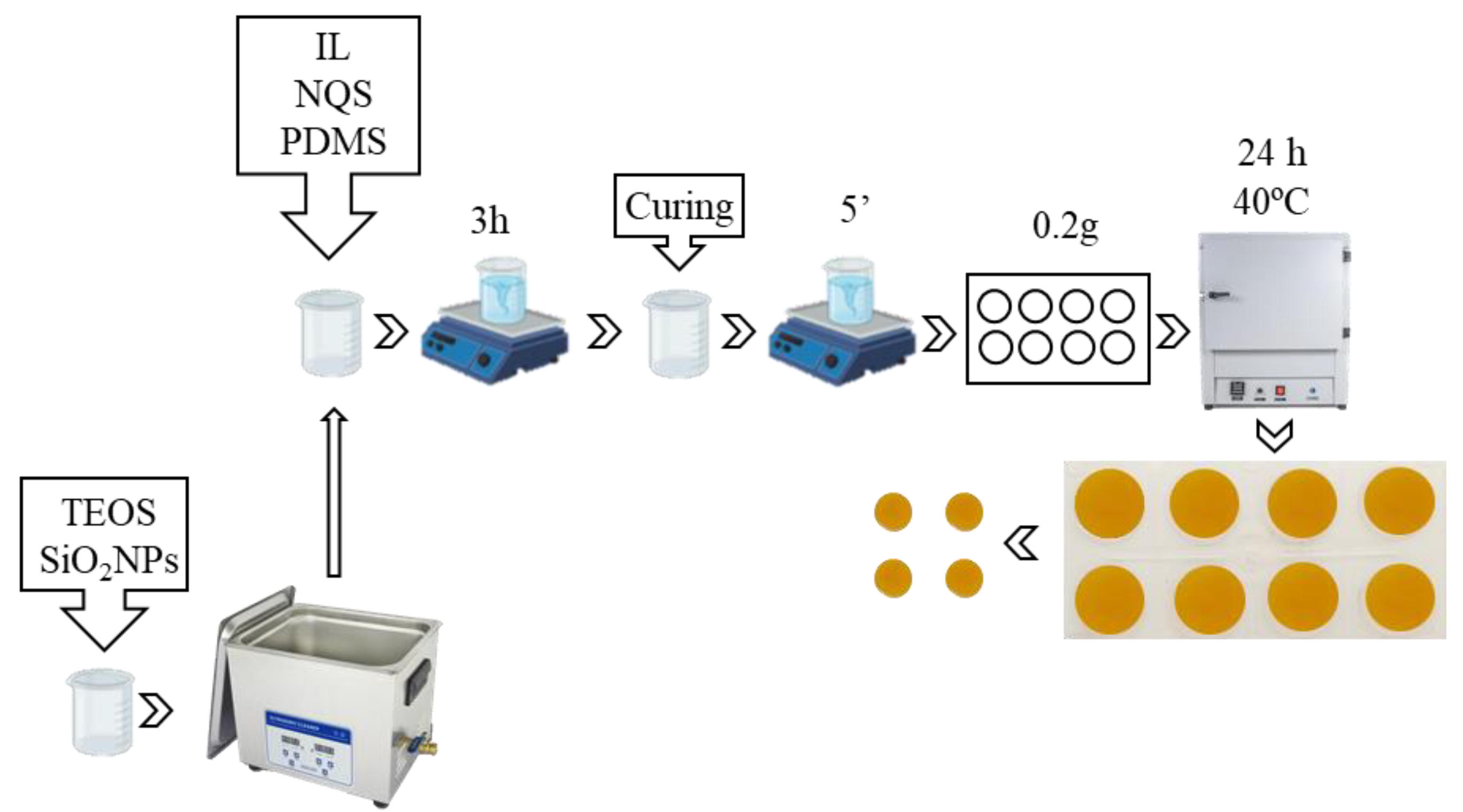
Figure 1. Steps of the sol-gel process that takes place for the sensor preparation.
Chemosensors 11 00387 g001
Figure 2. Ammonium analysis procedure in saliva as ammonia.
Chemosensors 11 00387 g002
Figure 3. Spectra of different NH4+ standard solutions. (a) Reflectance as absorbance obtained from benchtop diffuse reflectance spectrophotometer (t = 20 min) at λ = 570 nm; (b) absorbance obtained from portable fiber optic spectrometer (t = 20 min) at λ = 570 nm; the images of the three replicates for each concentration assayed and overlapped background and sensor registers obtained by the app GoSpectro as an example (c) reflectance as absorbance obtained from a benchtop diffuse reflectance spectrophotometer (t = 45 min) at λ = 570 nm, and (d) absorbance obtained from a portable fiber optic spectrometer (t = 45 min) at λ = 570 nm, and images of the three replicated for each concentration assayed. The color of each trade is related to the concentration shown in (b) for t = 20 min. and (d) for t = 45 min.
Chemosensors 11 00387 g003
Figure 4. Ammonium concentrations in 100 μL of real saliva samples (blue, one o two replicates) and fortifications (orange, one o two replicates) of these were determined by absorbance with diffuse reflectance spectrophotometry (n = 30). Assay time: 20 min. and * 45 min. Spiked concentration: 0.04 mg NH4+/mL saliva.
Chemosensors 11 00387 g004
Figure 5. Ammonium concentrations in 100 μL of real saliva samples (blue) and fortifications (orange) of these were determined by absorbance with miniaturized fiber optic spectrophotometry coupled to a smartphone with the use of the Go Spectro app (n = 30). Assay time: 20 min. and * 45 min. Spiked concentration: 0.04 mg NH4+/mL saliva.
Chemosensors 11 00387 g005
Figure 6. Ammonium concentration in 100 μL of real saliva samples (blue) and fortifications (orange) of these is determined by the intensity in color plane R (n = 30). Assay time: 20 min. and * 45 min. Spiked concentration: 0.04 mg NH4+/mL saliva.
Chemosensors 11 00387 g006
Figure 7. Designed App for calculating the analyte concentration of a sample using: (A) spectra obtained by the fiber optic minispectrometer and (B) the intensity in color planes R (red), G (green), and B (blue).
Chemosensors 11 00387 g007
Table 1. Salivary pH study (n = 12) and pHs of the saliva after basification. For more explanation, see the text.
SamplesUrinary pHpH (NaOH)pH (CO32−/HCO3)
S16.7012.2810.81
S25.7612.2010.13
S37.1611.9910.47
S46.8611.6910.53
S56.4112.0910.09
S76.7912.1310.50
S86.3711.7410.17
S95.0911.879.87
S106.3112.3410.30
S117.1812.2410.30
S126.8911.8810.19
Table 2. Figures of merit for the determination of ammonium at a measurement time of 20 min by the three measurement methods. Intraday (n = 3 and 6 points for each graph) and interday (n = 6 and 6 points for each graph). (a) concentration expressed as µg NH3/mL air atmosphere; (b) concentration expressed as mg NH4+/mL saliva. C= concentration.
Reaction
Time
Method LinearityLODLinear Interval
LOQ-Higher C
b1 ± Sb1b0 ± Sb0R2
t = 20 minDiffuse reflectanceIntraday(a) 0.366 ± 0.0190.088 ± 0.0150.980.060.20–1.5
(b) 0.70 ± 0.040.088 ± 0.0150.980.030.10–0.74
Interday(a) 0.342 ± 0.0190.092 ± 0.0140.980.060.20–1.5
(b) 0.65 ± 0.040.092 ± 0.0140.980.030.11–0.74
Go SpectroIntraday(a) 0.322 ± 0.0150.098 ± 0.0070.990.140.44–0.95
(b) 0.61 ± 0.030.098 ± 0.0070.990.080.2–0.56
Interday(a) 0.31 ± 0.090.095 ± 0.0050.990.150.45–0.95
(b) 0.58 ± 0.020.095 ± 0.0050.990.080.2–0.56
RGB (Red)Intraday(a) −77 ± 6212 ± 30.970.160.49–0.95
(b) −146 ± 12212 ± 30.970.090.26–0.56
Interday(a) −67 ± 5214 ± 20.980.190.56–0.95
(b) −127 ± 9214 ± 20.980.100.30–0.56
Table 3. Figures of merit for the determination of ammonium at a measurement time of 45 min by the three different measurement methods. Intraday (n = 3 and 6 points for each graph) and interday (n = 6 and 6 points for each graph). (a) concentration expressed as µg NH3/mL air atmosphere; (b) concentration expressed as mg NH4+/mL saliva. C = concentration.
Reaction
Time
Measurement LinearityLODLinear Interval LOQ-Higher C
b1 ± Sb1b0 ± Sb0R2
t = 45 minDiffuse reflectanceIntraday(a) 1.06 ± 0.090.041 ± 0.0130.970.020.06–0.56
(b) 2.02 ± 0.170.041 ± 0.0130.970.010.03–0.15
Interday(a) 1.04 ± 0.060.049 ± 0.0090.980.020.07–0.56
(b) 1.97 ± 0.120.049 ± 0.0090.980.010.04–0.15
Go SpectroIntraday(a) 0.92 ± 0.050.049 ± 0.0130.980.050.15–0.56
(b) 1.75 ± 0.100.049 ± 0.0130.980.030.08–0.15
Interday(a) 0.89 ± 0.070.069 ± 0.0060.990.050.16–0.56
(b) 1.69 ± 0.050.069 ± 0.0060.990.030.08–0.15
RGB (Red)Intraday(a) −152 ± 8217 ± 20.980.080.25–0.56
(b) −287 ± 15217 ± 20.980.040.13–0.26
Interday(a) −140 ± 8213 ± 20.980.090.27–0.56
(b) −265 ± 15213 ± 20.980.050.14–0.26
Table 4. Comparison of external calibration with standard addition at 45 min (n = 6 and 6 points for each graph).
Measurement Linearity (mg NH4+/mL Saliva)
b1 ± sb1b0 ± sb0R2
Diffuse reflectanceExternal calibration1.97 ± 0.110.049 ± 0.0090.98
Standard addition1.53 ± 0.100.052 ± 0.0060.99
Go SpectroExternal calibration1.69 ± 0.050.069 ± 0.0060.99
Standard addition1.174 ± 0.0140.0212 ± 0.00080.99
RGB (Red)External calibration−265 ± 15213 ± 20.98
Standard addition−239 ± 42188 ± 30.97
Disclaimer/Publisher’s Note: The statements, opinions and data contained in all publications are solely those of the individual author(s) and contributor(s) and not of MDPI and/or the editor(s). MDPI and/or the editor(s) disclaim responsibility for any injury to people or property resulting from any ideas, methods, instructions or products referred to in the content.