Article

Analysis of Solid Formulates Using UV-Visible Diffused Reflectance Spectroscopy with Multivariate Data Processing Based on Net Analyte Signal and Standard Additions Method

Department of Chemistry “Giacomo Ciamician”, University of Bologna, Via Piero Gobetti 85, 40126 Bologna, Italy
*
Author to whom correspondence should be addressed.
Chemosensors 2024, 12(11), 227; https://doi.org/10.3390/chemosensors12110227
Submission received: 16 September 2024 / Revised: 24 October 2024 / Accepted: 29 October 2024 / Published: 1 November 2024

Abstract

:
Quality control in pharmaceutical manufacturing necessitates rigorous testing and approval, adhering to Current Good Manufacturing Practices before commercialization. The production of solid drugs presents significant industrial challenges regarding uniformity, homogeneity, and consistency. Traditional quality guidelines rely on classical analytical methods such as liquid chromatography coupled with mass spectrometry. However, the emergence of Process Analytical Technology introduced non-destructive, rapid, and cost-effective methods like UV-Visible Diffuse Reflectance Spectroscopy. The present study aimed to develop a chemometric method for quantifying Active Pharmaceutical Ingredients (APIs) in Neo Nisidine®, a solid mixture drug, using spectrophotometric data. The Net Analyte Signal (NAS) method, combined with standard additions, allowed the creation of a pseudo-univariate standard addition model, overcoming some challenges in solid-phase analysis. Successful quantifications of APIs in ideal laboratory samples and real pharmaceutical tablets were obtained. NAS-based chemometric models showed high precision and reliability, whose results were validated by comparisons with HPLC ones. The study revealed that solid-phase spectrophotometric analyses can be considered a valid alternative to API analyses. Solid-phase analysis offers non-destructive, cost-effective, and environmentally friendly benefits, enabling its integration into pharmaceutical production to improve quality control.

1. Introduction

Quality control [1] is one of the main topics in the pharmaceutical industry, which necessitates ensuring the safety, efficacy, and consistency of drugs and products before these reach consumers. Among the intricate landscape of pharmaceutical manufacturing, the production of solid drugs, such as tablets, capsules, and powders, presents several analytical challenges [2]. Achieving uniformity in solid formulations, ensuring homogeneity of active pharmaceutical ingredients (APIs) [3] within each dosage unit, and maintaining consistency across batches pose significant hurdles in such pharmaceutical productions.
Traditionally, quality control has been based on established and well-known analytical methodologies for both quality assessment and quantitative analyses [2]. One of the most important methods in this field is high-performance liquid chromatography (HPLC) coupled with mass spectrometry (MS) or diode array (DAD) detectors [4]. However, HPLC analysis often demands a remarkable quantity of time and solvents, making it costly and not environmentally sustainable [5]. In 2004, the American Food and Drug Administration (FDA), followed by the European Medicines Agency (EMA), introduced the concept of Process Analytical Technology (PAT) [6]. Based on the development of innovative, non-destructive, and efficient analytical methodologies to monitor critical process parameters, PAT’s purpose is to ensure product quality throughout manufacturing and control critical process parameters, as well as raw materials. Since its early applications, analytical spectroscopy in reflectance mode has been the perfect candidate for PAT because it is non-destructive and suitable for online monitoring. Near-infrared (NIR) spectroscopy has been extensively applied for qualitative analysis, thanks to the possibility of differentiating compounds based on the vibrational information obtained from the spectra [7]. Medium-infrared (MIR) spectroscopy has also been successfully applied to quantitative analysis [8]. However, its feasibility for quantitative analysis is still a very interesting task, as well as for online monitoring. The best spectral range for quantitative spectroscopy remains UV-Vis [2]. Therefore, for pharmaceutical quality control, solid-phase spectrophotometric techniques like UV-Vis Diffuse Reflectance Spectroscopy (UV-Vis DRS) and Attenuated Total Reflectance Infrared spectroscopy (ATR-IR) have gained attention [9]. These techniques offer rapid, non-destructive, and cost-effective facilities to directly analyze solid pharmaceutical formulations, providing insights into API quantification, homogeneity, and composition; moreover, direct analysis of solid samples fulfills some requirements of green chemistry because no solvent is needed. For these reasons, spectrophotometric methods adhere to PAT guidelines [10]. In particular, in this study, we focused on UV-Vis DRS analysis.
Solid pharmaceutical formulations are generally made by a mixture of several powders, which can be roughly divided into two groups: API and excipients. API is the constituent of the medicinal product that is actively involved in the curative action. Excipients, instead, are components devoid of any pharmacological action, but useful to meet or improve other characteristics of the formulation. For example, they protect the active ingredient from external agents, increase the volume to allow the preparation of tablets of an acceptable size [11], stabilize solutions or suspensions by preventing sedimentation of the active ingredient inside the containers, facilitate the transport and absorption of the API in the body, and make the taste more pleasant.
Therefore, excipients constitute most of the bulk of pharmaceutical products, within which, generally, a lower quantity of API is mixed. This represents a major technological challenge for the pharmaceutical industries, as it is necessary to ensure perfect homogenization and distribution of API and excipients in each product batch, requiring several chemical analyses [12].
This study aimed to evaluate a chemometric method for quantifying active pharmaceutical ingredients (APIs) in solid drug mixtures by processing multidimensional spectral data. Specifically, it focused on a case study: determining the percentages of acetylsalicylic acid, caffeine, and paracetamol in a well-known commercial solid pharmaceutical formulation (Neo Nisidine® tablets) by rapid, non-destructive, and environmentally friendly analytical protocols. In particular, powder samples were analyzed using UV-Vis DRS, and the collected spectra were processed using a multivariate method based on the Net Analyte Signal (NAS) algorithm, enabling the quantification of individual components in the presence of the others. To validate the chemometric results, samples were analyzed using a standard protocol involving HPLC-DAD [13] and a conventional univariate calibration curve. Although the system composed of acetylsalicylic acid, caffeine, and paracetamol has already been used in other studies for several purposes [14], our approach aims to quantify the same APIs in the solid phase (while generally, solutions of the three APIs are studied) using a different chemometric calibration model (NAS). The solid-phase analysis represents an innovation in this context, together with the application of NAS, which constitutes a chemometric and methodological advancement rather than merely an analytical one.
By exploiting chemometric concepts such as NAS and standard addition methods [15], researchers aim to develop robust models capable of accurately quantifying APIs in complex solid mixtures [16]. Through rigorous experimentation, multivariate data analyses, and comparison with established liquid-phase methodologies, this research seeks to delineate the efficacy and potential of solid-phase spectrophotometric techniques as a viable alternative for quality assessment in pharmaceutical manufacturing. These innovations hold the promise of improving quality control processes, offering real-time monitoring capabilities, and reducing the dependency on conventional, time-consuming analyses.

2. Materials and Methods

2.1. Samples Preparation

The present study is focused on the analyses of two different samples: (i) a laboratory sample simulating Neo Nisidine, prepared to evaluate the analytical method and NAS performances; (ii) real pharmaceutical tablets, Neo Nisidine®, purchased from a pharmacy store. The real sample chosen belongs to the antipyretic and anti-inflammatory categories.

2.1.1. Laboratory Sample

Neo Nisidine® contains acetylsalicylic acid (AAS), paracetamol (PAR), and caffeine (CAF), all of which have API properties. The formulation used in these tablets contained also magnesium stearate and lactose as excipients, used to improve the pharmacokinetics and the production of the tablets. The solid solutions were in the form of capsules. In particular, our goal was to quantify the three active ingredients in the drug.
The weight of the tablets ranged from 560 to 600 mg; the composition reported on the package leaflet states 25 mg caffeine, 200 mg paracetamol, and 250 mg acetylsalicylic acid.
Laboratory samples of paracetamol (PAR), acetylsalicylic acid (AAS), and caffeine (CAF) were prepared to simulate Neo Nisidine composition. The standards of the three substances were purchased from Sigma Aldrich (Saint Louis, MI, USA). The analytical procedure was based on the standard addition method (SAM) [17], for which four added standards for each analyte were prepared.
Four standards were obtained at the following added concentrations of pure analyte: 0, 5, 10, and 15% w/w. The preparation of added standard samples was carried out by geometric dilutions [17,18], widely used to prepare solid-phase solutions to obtain a homogenous mixture and get reproducible results. The principle behind geometric dilution is to start with the pure active ingredient, mix it to an equal quantity of the excipient or sample, and then repeat the same procedure until the desired concentration is reached.
The benchmark samples were prepared as follows. The zero-added standard sample was obtained by mixing microcrystalline cellulose (as excipient) and each API to obtain a concentration of 1.5% w/w of API in 300 mg of sample. The further added standards were prepared by mixing 125 mg of the 1.5% w/w solution with different amounts of microcrystalline cellulose and pure API to reach a final mass of 300 mg to obtain 5%, 10%, and 15% w/w concentration.
Four SAM standards at 0%, 5%, 10%, and 15% w/w concentration of the added pure APIs were prepared, mixing the right amount of the three active principles and excipients to 125 mg of the 1.5 w/w solution, reaching a final mass of 300 mg. To ensure that the mixture was more likely to be homogeneous, the binary mixture was manually ground and tumbled using a Vortex model ZX3 (VELP Scientifica, Milan, Italy) for 10 min.

2.1.2. Real Samples

For the spectrophotometric quantification, the same procedure used to quantify caffeine, paracetamol, and acetylsalicylic acid in the laboratory sample was applied to the three analytes contained in Neo Nisidine®. Hence, four added standards were prepared for each of the three analytes contained in Neo Nisidine®, and the standard addition method was applied. The four added concentrations were 0%, 5%, 10%, and 15% w/w. Laboratory standards were obtained as follows: the final mass was fixed to 300 mg; for each analyte, an amount corresponding to the chosen values of added concentrations (0 mg, 15 mg, 30 mg, 45 mg, respectively) was added to 100 mg of Neo Nisidine®, and, finally, an amount of the excipients was added to obtain a final mass of 300 mg. The starting 100 mg of Neo Nisidine® was taken from a mixture of four Neo Nisidine® tablets, ground together to obtain a single sample; in this way, better homogeneity was ensured. Moreover, a total of 12 standards had to be prepared, requiring a minimum of 1.2 g of Neo Nisidine®. On average, each Neo Nisidine® tablet weighed around 0.6 g. Therefore, by mixing four tablets, the total mass of the starting sample was 2.3061 g. The excess of Neo Nisidine®, compared to the estimated 1.2 g required for the NAS procedure, was chosen for two reasons: to account for potential errors in the sample preparation process and to ensure that an aliquot would remain available for subsequent analysis using HPLC-DAD.

2.2. Instrumentation

Quantitative analyses were carried out in the solid phase, to develop a technique that could be implemented for online analysis of a production chain or industrial process. Therefore, it is essential to use nondestructive techniques, that allow many analyses to be carried out in a short time, and that can work in the solid phase. The instruments that meet these requirements are those based on spectroscopy. The spectroscopic technique used in the present study is the UV-Vis diffuse reflectance, which will be described in the following section. HPLC-DAD analyses were also performed to compare the results from spectroscopic analyses with a standard method.

2.2.1. UV-Vis Diffuse Reflectance

The UV-Vis DRS spectra were collected using a double-beam spectrophotometer Perkin Elmer Lambda 35 (Perkin Elmer, Waltham, MA, USA) equipped with an integrating sphere accessory [19] Labsphere (North Sutton, NH, USA) RSA-PE-20, with a diameter of 150 mm. This tool allowed us to enhance and detect the UV-Vis diffuse reflectance light from an opaque and structured surface [20], which depends on the physicochemical properties and color of the surface. The analysis range was between 200 and 500 nm with a step of 0.5 nm. The spectrophotometer is equipped with two lamps for visible (model WI 64604) and UV (model D2 L638) ranges and a photomultiplier detector. Samples and the reference standard (BaSO4) for background analysis were placed in a quartz cuvette, and the blank signal was removed by subtraction.

2.2.2. HPLC-DAD

The API quantification in the real sample of Neo Nisidine® was also performed with the HPLC-DAD. Liquid chromatography is a typical standard analytical method used for quantification of API in tablets. Therefore, the concentrations obtained were compared with the spectrophotometric results for method validation. Analyses were carried out with an HPLC Agilent Technologies system (Agilent, Santa Clara, CA, USA), equipped with a degasser and DAD detector. The quantification of the analytes was obtained by a calibration line for each active ingredient relating the concentration of the standards with the area subtended by the chromatographic peaks. The instrumental parameter settings, analytical procedures, and separation methods used to perform the analyses of Neo Nisidine® are described in detail in the two following papers [21,22].

2.3. Data Processing

2.3.1. Principal Components Analysis (PCA) and Dataset Pretreatment

Before performing the NAS quantitative analysis, it is important to explore the datasets through the use of PCA. PCA is a well-known chemometric method that rotates the space of original variables to calculate a new highly informative space spanned by principal components (PCs). The scores plot obtained by PCA allows us to verify the presence of outliers that are samples or observations not compliant with the others. In the present study, for outlier detection between the replicates of each added concentration sample, a one-class modeling method was applied. For this analysis, all SAM samples for each case study were involved in the same PCA computation, and a Hotelling ellipse at a 95% confidence level [23] was computed for each added concentration. If an observation fell outside the domain described by the corresponding ellipse, it was considered an outlier and removed from the dataset [24,25].
Before quantitative analysis, the datasets filtered out by the outliers were pre-processed with different methods, to maximize information and minimize instrumental background noise, which could be very high when working in the solid phase. The pre-treatments tested in the present study were Savitzky-Golay First Derivative (using a first polynomial order, i.e., linear function, and a window of 4 points in both directions), Standard Normal Variate (SNV), Multiplicative Scattering Correction (MSC), and the combination of SNV + MSC. Details about the pre-processing techniques are reported in the following paper [26].

2.3.2. Partial Least Squares (PLS)

PLS is a multivariate regression method based on PC computation (in this case called factors), which simultaneously maximizes the correlation between scores and response, parameterized by covariance [27]. For this technique, analytical data (spectra) are used as independent variables and analyte concentration (added concentration in the present study) as dependent one.
The calibration performance is assessed using Root Mean Square Error values of calibration (RMSEC) and prediction (RMSEP), both of which must be minimized, and the coefficient of determination R2, which should be as close as possible to 1. All these parameters are calculated by comparing known concentrations with the ones recalculated by the model [28]. In particular, RMSEP was calculated by leave-one-out cross-validation [29], removing one object at a time from the dataset, calculating the model with the others, and recalculating the concentration of the excluded object.
PLS is the starting point of the NAS methodology, which will be described in the following Section. The aim of using NAS for improving PLS computation is that one of the weak points of PLS is its scarce precision when making prediction in extrapolation mode, i.e., outside the range of the response values. NAS, instead, by converting the multivariate predictors into pseudo-univariate data, make it possible to convert the multivariate problem into a univariate one, increasing the precision of predictions in extrapolation as well. However, NAS results strongly depend on the choice of the PLS factor. Therefore, the criterion of minimizing RMSEP partly guided the choice of the number of factors, aiming at reducing the irrelevant information by selecting as few factors as possible [28].

2.3.3. Net Analyte Signal (NAS)

The collected spectra were elaborated with a multivariate method based on the Net Analyte Signal (NAS) concept [30,31], which allows the quantification of a single ingredient even when mixed with other ones [18]. The presence of several ingredients in a solid mixture makes the direct analysis and quantification of one of them very harsh, due to the matrix effect. In this case, the presence of several analytes and excipients produces a multivariate signal strongly affected by the matrix effect. Hence, the PLS method cannot be used to obtain a reliable calibration model [17]. Moreover, PLS is not suitable for extrapolation, which is necessary when working with the standard addition method. For these two reasons, we extended the quantitative analysis to NAS [32]. This algorithm starts from multivariate data (it is particularly effective with spectra) to calculate a pseudo-univariate regression model, minimizing the matrix effect in the final model.
The net analyte signal is that component of the multivariate signal closely linked to the chemical characteristics of the analyte, specifically its concentration [33]. Mathematically, the NAS algorithm projects the sample spectrum in a new space that is perpendicular (i.e., independent) to the space calculated from the matrix signal. In the present study, the algorithm developed by Bro has been used [32].
The NAS procedure consists of extracting from the sample signal (xi) only the portion attributable to the analyte of interest, while removing the portion due to other species. This is carried out by projecting the original signals (xi) on the NAS space by Equation (1):
x i * = H · x i
where xi* is the i-th sample NAS signals and H is a projection matrix. To compute the projection matrix, the algorithm starts from the regression coefficients (b) of PLS at the chosen optimal factor (A) using Equation (2):
H = b A b A T b A 1 b A T
The Euclidean norm of xi* signals are then used as pseudo-univariate signals, that in turn are used to calculate a standard addition calibration line. Finally, the concentration of the zero-added sample can be extrapolated.
Selecting the optimal PLS factor (A) is a critical step to obtain a correct extrapolated concentration. Thus, for each sample, the optimal PLS factor has been chosen as the one minimizing the RMSEP of PLS and, at the same time, maximizing the determination coefficient (R2) of the NAS pseudo-univariate regression line. When it was not possible to achieve both conditions simultaneously, the PLS factor was chosen based on the best compromise between these two parameters, prioritizing the highest R2 value.
To calculate the standard deviations of the extrapolated concentrations, the Jackknife method [34] was applied. It works by iterative recalculation of the NAS model after removing one observation at a time from the training set, producing a sort of cross-validation in a leave-one-out modality. In this way, a set of extrapolated concentrations is obtained for each model, each one coming from the removal of one observation, and the standard deviation calculated from this set of values is used as the standard deviation of the general extrapolated concentration.

3. Results and Discussion

3.1. Solid Standard Solutions

The UV-Vis DRS analyses of caffeine were chosen as a case study to set up the analytical and chemometric procedures to be subsequently applied to all samples. Figure 1 shows the UV-Vis DRS spectra of the pure species used for the present study: microcrystalline cellulose (the excipient used for laboratory samples preparation), caffeine, paracetamol, and acetylsalicylic acid.
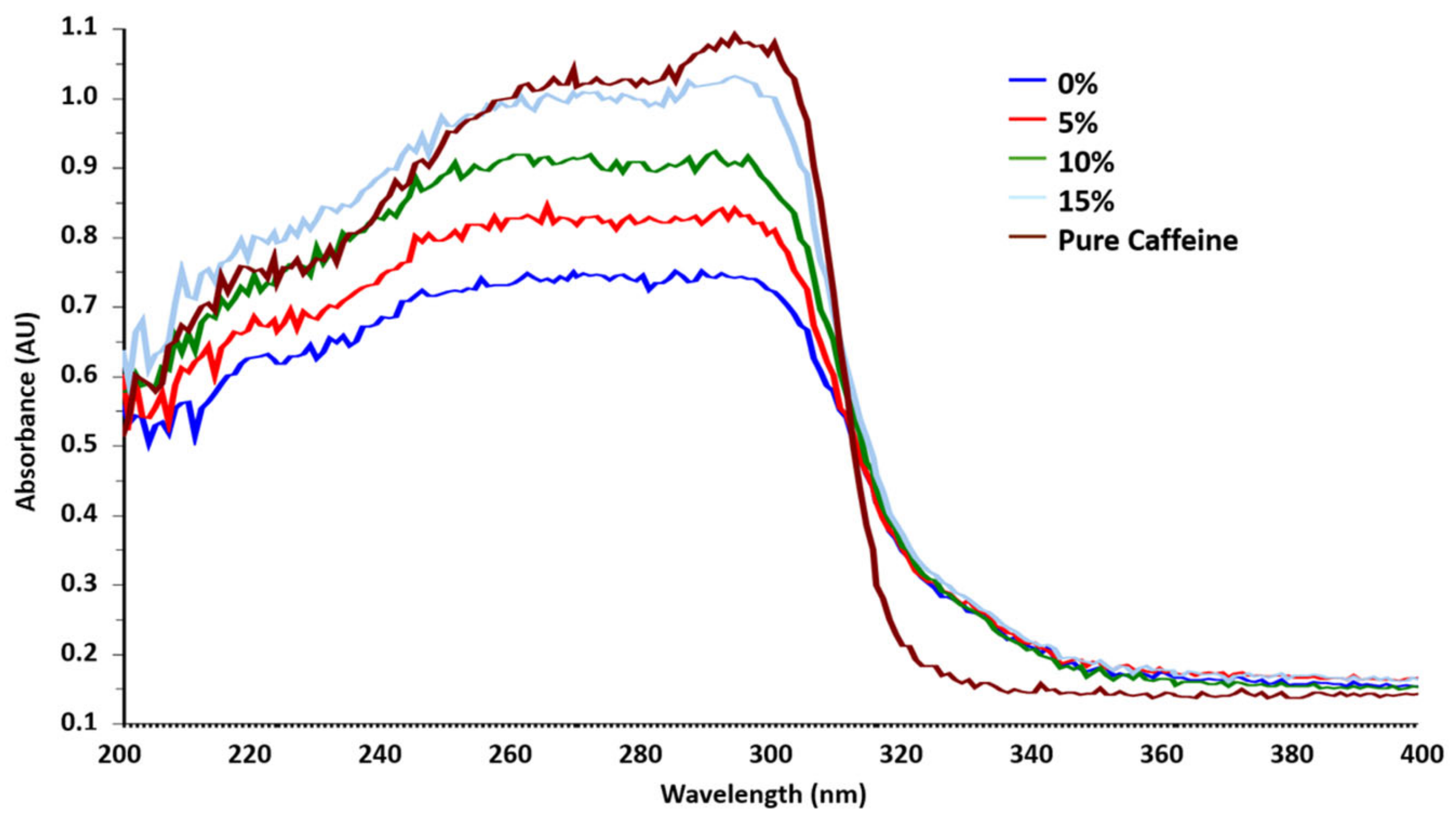
A laboratory solid solution was prepared, containing microcrystalline cellulose (excipient) and caffeine (active principle) with a caffeine concentration of 1.5% w/w. From this laboratory sample, used as the 0%-added sample, four added samples were prepared, adding pure caffeine to obtain the following added concentrations: 5%, 10%, and 15% w/w. Figure 2 shows the UV-Vis DRS spectra of standard added samples for the caffeine dataset.
The acquired UV-Vis DRS spectra used for chemometric models were organized in a matrix X, whose dimension was N × M, where N is the number of samples, and M is the number of variables (spectrophotometric wavelengths). Six replicates of each standard and of the pure caffeine (useful to evaluate the other spectra) were collected. The dataset consisted of M = 601 variables and N = 30 rows.
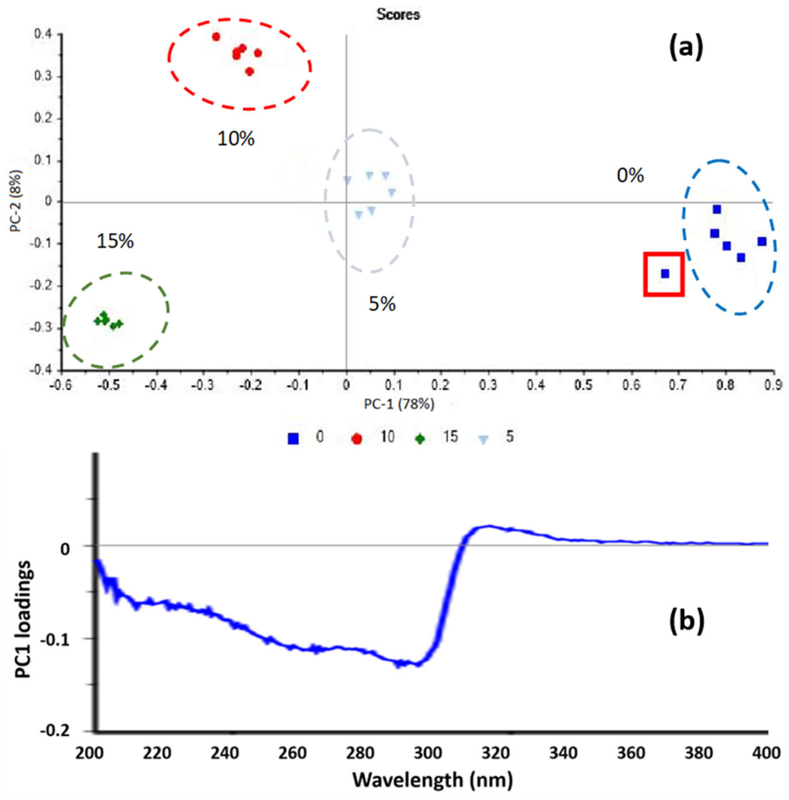
Then, a PCA model was calculated, removing the pure analyte spectra. Therefore, it was performed only on the 24 spectra of the standard samples. The scores plot is reported in Figure 3a, where PC1 explains 78% of variance, PC2 8%, and the subsequent principal components explain gradually decreasing variance. This graph was used to evaluate the repeatability of the analyses performed on the samples at the same concentration and to exclude possible outliers. Clusters were calculated for each group of samples having the same concentration by computing a Hotelling analysis (at 95% confidence) for each group. In the scores plot (Figure 3a), all points fall inside the corresponding Hotelling ellipse with the only exception of one point highlighted with a red square. This sample was considered an outlier and removed in the final model.
Moreover, the four clusters in Figure 3a, corresponding to the four different added concentrations, were well discriminated along the first principal component (PC1), which described 78% of the total explained variance. Along PC1, it was possible to see how the added samples were arranged progressively. On the left (negative PC1 values) were the samples with higher added caffeine concentration (15% w/w), while on the right (positive PC1 values) were the samples with lower added concentration of caffeine (0% w/w). Hence, PC1 proved to be anti-correlated with caffeine. This is coherent with the Loadings plot in Figure 3b, which shows that negative values of loadings are associated with the caffeine spectral range of absorption (243–302 nm). Low PC1 values were closely related to higher caffeine content, and simultaneously to lower excipient content.
The scores plots related to the datasets paracetamol and acetylsalicylic acid (Figure S1) showed no suspected outliers. Therefore, no data were removed for the computation of the NAS models.
To optimize NAS results, several pretreatment methods were applied to UV-Vis DRS spectra, and the extrapolated concentrations at the end of the NAS procedure were compared to find the best pretreatment. The following data pretreatments were tested: first-order Savitzky-Golay Derivative, SNV, MSC, and, finally, the combination of SNV + MSC.
For each model, the three control parameters, NAS-R2, PLS-RMSEP, and the extrapolated concentrations of known standards, were assessed. The extrapolated concentration of the three analytes was considered accurate when there was no significant deviation from the expected 1.5% w/w in the initially prepared reference sample. Table 1 highlights that MSC is the best pretreatment method. Specifically, the NAS model developed using the MSC-pretreated dataset yielded R2 values exceeding 0.99, and RMSEP lower than 6% of the expected value. The extrapolated concentration values were not significantly different from the expected known value of 1.5% w/w in all cases. Figure 4 shows the NAS standard addition line obtained with MSC pretreatment. The accuracy of the predicted concentration was assessed using a t-test performed with 5% significance. For each of the three analytes (Table 1, Tables S1 and S2), we obtained the best results with the MSC pretreatment.
The results from the laboratory samples confirm the feasibility of the analytical and chemometric models. Consequently, it was possible to go on with the study and apply the same methods to the two real pharmaceutical samples.

Neo Nisidine® Real Sample

This section presents the results for the real samples. In this case, the spectrophotometric results were compared with those obtained by HPLC.
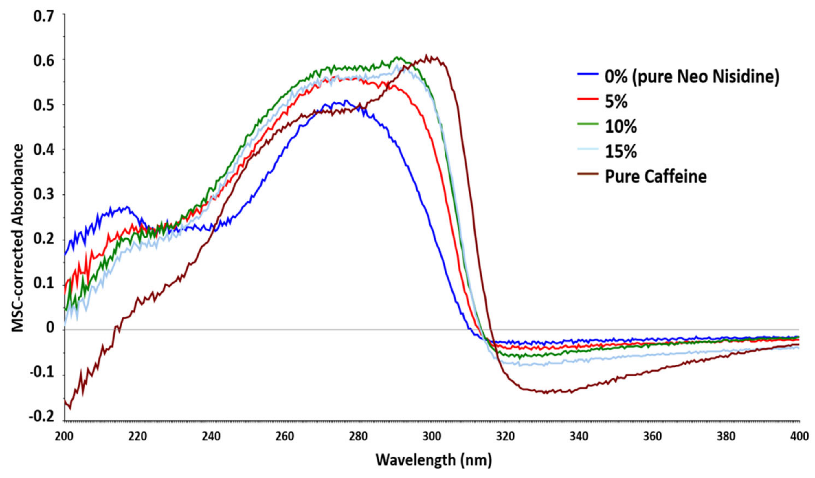
The dimensions of the real sample dataset were the same as the reference one, 30 × 601. All three datasets were preprocessed using the MSC method, which yielded the best results with the laboratory sample. Figure 5 shows the MSC-pretreated spectra of Neo Nisidine and the caffeine-added samples.
Finally, three analytes were quantified by NAS and to assess the correct quantifications obtained by UV-Vis DRS, these were compared both with the expected amounts reported in the package inserts and with the quantification performed by HPLC-DAD.
Table 2 summarizes the spectrophotometric and chromatographic quantification results and the declared values for each analyte.
The spectrophotometric-NAS results were not significantly different from the standard chromatographic technique. In addition, both results were in agreement with the theoretical concentrations stated in the package insert. The results obtained with HPLC-DAD (Figure S2) were associated with lower errors than those obtained in spectrophotometry. The higher UV-Vis errors were probably due to high inhomogeneity in the solid-phase solutions and to the several chemometric steps necessary to obtain the final results. In addition, also the preparation of added standards and a possible variability of granulometry in the solid sample may have caused an increase in errors. However, also spectrophotometric-NAS errors never overcame 10%, which is a limit generally accepted by pharmacopeia in API quantification [35].

4. Conclusions

This comprehensive study demonstrated the efficacy and potential of solid-phase spectrophotometric analysis as a valid alternative for quantifying Active Pharmaceutical Ingredients in drug formulations. The investigation began by addressing the significant challenges encountered in pharmaceutical manufacturing related to uniformity and consistency in solid drug production. Traditional quality control methods, reliant on liquid chromatography coupled with mass spectrometry or diode array detectors, have been the industry norm. However, the advent of Process Analytical Technology introduced the possibility of juxtaposing non-destructive, rapid, and cost-effective spectroscopic techniques, notably, UV-Visible Diffuse Reflectance Spectroscopy.
The primary goal achieved in this research was to create and validate a NAS-SAM analytical method. Validation was performed on reference solid mixtures created in the laboratory to quantify the initial analyte concentration present in the starting solution. Leveraging the Net Analyte Signal (NAS) concept along with the standard addition method, the study successfully formulated a NAS-SAM model. The investigation rigorously evaluated various preprocessing techniques, identifying MSC as the best one. Comparing the known analyte concentration with that predicted by the NAS model enabled excellent results to be obtained since no predicted concentration showed any significant difference from the theoretical value.
The second part of the study consisted of quantifying the percentage concentration of active ingredients present in Neo Nisidine®. The results of this study demonstrated the successful quantification of APIs in real pharmaceutical tablets. The efficacy and accuracy of the NAS-based chemometric models were underscored by their comparability with HPLC-DAD results, validating their reliability for API quantification. The UV-Vis analyses yielded results comparable with those obtained from HPLC-DAD analysis.
In conclusion, this research marks a pivotal advancement in pharmaceutical quality control as a valid alternative for API quantification. The UV-Vis DRS analysis coupled with NAS may be a suitable technique to be introduced for at-line monitoring of pharmaceutical manufacturing processes. Such at-line monitoring would involve the following procedure: samples are periodically taken from the process stream by an automatic device and brought to another automatic device that performs the standard additions, registers the spectra, and sends data to a computer, where an ad-hoc script creates the chemometric models and calculates results. By enhancing efficiency, reducing costs, and promoting environmental sustainability, this study paves the way for a new era in pharmaceutical quality control, setting a new standard for innovation and excellence in the industry.

Supplementary Materials

The following supporting information can be downloaded at: https://www.mdpi.com/article/10.3390/chemosensors12110227/s1, Figure S1: Scores plot of Paracetamol (a) and Acetylsalicylic acid (b) laboratory samples datasets; Table S1: Control parameters to evaluate the NAS regression obtained for the four pretreatments method for paracetamol laboratory samples; Table S2: Control parameters to evaluate the NAS regression obtained for the four pretreatments method for acetylsalicylic acid laboratory samples; Figure S2: HPLC chromatograms for the calibration line of paracetamol (first peak at 1.9 min), caffeine (at 2.6 min), and acetylsalicylic acid (at 8.9 min).

Author Contributions

Conceptualization, D.M.; methodology, N.K. and A.Z.; software, N.K. and M.M.; validation, A.Z.; formal analysis, N.K. and M.M.; investigation, M.M.; resources, D.M.; data curation, N.K., A.Z. and M.M.; writing—original draft preparation, N.K. and M.M.; writing—review and editing, A.Z. and D.M.; visualization, A.Z.; supervision, D.M.; project administration, D.M.; funding acquisition, D.M. All authors have read and agreed to the published version of the manuscript.

Funding

This research did not receive any specific grant from funding agencies in the public, commercial, or not-for-profit sectors.

Institutional Review Board Statement

Not applicable.

Informed Consent Statement

Not applicable.

Data Availability Statement

Dataset available on request from the authors.

Conflicts of Interest

The authors declare no competing interests.

References

  1. Mrugalska, B.; Tytyk, E. Quality Control Methods for Product Reliability and Safety. Procedia Manuf. 2015, 3, 2730–2737. [Google Scholar] [CrossRef]
  2. Macchietti, L.; Melucci, D.; Menarini, L.; Consoli, F.; Zappi, A. Analytical Comparison between Batch and Continuous Direct Compression Processes for Pharmaceutical Manufacturing Using an Innovative UV–Vis Reflectance Method and Chemometrics. Int. J. Pharm. 2024, 656, 124090. [Google Scholar] [CrossRef] [PubMed]
  3. Karpinski, P.H. Polymorphism of Active Pharmaceutical Ingredients. Chem. Eng. Technol. 2006, 29, 233–237. [Google Scholar] [CrossRef]
  4. Dogra, R.; Kumar, M.; Kumar, A.; Roverso, M.; Bogialli, S.; Pastore, P.; Mandal, U.K. Derivatization, an Applicable Asset for Conventional HPLC Systems without MS Detection in Food and Miscellaneous Analysis. Crit. Rev. Anal. Chem. 2022, 53, 1807–1827. [Google Scholar] [CrossRef] [PubMed]
  5. European Medicines Agency. Good Manufacturing Practice. Available online: https://www.ema.europa.eu/en/human-regulatory-overview/research-and-development/compliance-research-and-development/good-manufacturing-practice (accessed on 6 December 2023).
  6. Mali, A.; Jagtap, M.; Karekar, P.; Maruska, A. A Brief Review on Process Analytical Technology (PAT) Review Article. Available online: https://www.researchgate.net/publication/294053255_A_BRIEF_REVIEW_ON_PROCESS_ANALYTICAL_TECHNOLOGY_PAT_Review_Article#fullTextFileContent (accessed on 6 December 2023).
  7. Krämer, K.; Ebel, S. Application of NIR Reflectance Spectroscopy for the Identification of Pharmaceutical Excipients. Anal. Chim. Acta 2000, 420, 155–161. [Google Scholar] [CrossRef]
  8. Roggo, Y.; Chalus, P.; Maurer, L.; Lema-Martinez, C.; Edmond, A.; Jent, N. A Review of near Infrared Spectroscopy and Chemometrics in Pharmaceutical Technologies. J. Pharm. Biomed. Anal. 2007, 44, 683–700. [Google Scholar] [CrossRef]
  9. Kumar, M.; Bhatia, R.; Rawal, R.K. Applications of Various Analytical Techniques in Quality Control of Pharmaceutical Excipients. J. Pharm. Biomed. Anal. 2018, 157, 122–136. [Google Scholar] [CrossRef]
  10. Kwok, C.S.; Muntean, E.A.; Foster, W.; Mallen, C.D. Patient Pathways in Cardiology: Should Pharmaceutical and Medical Device Companies Care? Crit. Pathw. Cardiol. 2022, 21, 57–60. [Google Scholar] [CrossRef]
  11. Pereira, L.S.A.; Carneiro, M.F.; Botelho, B.G.; Sena, M.M. Calibration Transfer from Powder Mixtures to Intact Tablets: A New Use in Pharmaceutical Analysis for a Known Tool. Talanta 2016, 147, 351–357. [Google Scholar] [CrossRef]
  12. Schönbichler, S.A.; Bittner, L.K.H.; Weiss, A.K.H.; Griesser, U.J.; Pallua, J.D.; Huck, C.W. Comparison of NIR Chemical Imaging with Conventional NIR, Raman and ATR-IR Spectroscopy for Quantification of Furosemide Crystal Polymorphs in Ternary Powder Mixtures. Eur. J. Pharm. Biopharm. 2013, 84, 616–625. [Google Scholar] [CrossRef]
  13. Zappi, A.; Marassi, V.; Giordani, S.; Kassouf, N.; Roda, B.; Zattoni, A.; Reschiglian, P.; Melucci, D. Extracting Information and Enhancing the Quality of Separation Data: A Review on Chemometrics-Assisted Analysis of Volatile, Soluble and Colloidal Samples. Chemosensors 2023, 11, 45. [Google Scholar] [CrossRef]
  14. Bouhsain, Z.; Garrigues, S.; de la Guardia, M. PLS-UV Spectrophotometric method for the simultaneous determination of paracetamol, acetylsalicylic acid and caffeine in pharmaceutical formulations. Fresenius J. Anal. Chem. 1997, 357, 973–976. [Google Scholar] [CrossRef]
  15. Blanco, M.; Bautista, M.; Alcalá, M. Preparing Calibration Sets for Use in Pharmaceutical Analysis by NIR Spectroscopy. J. Pharm. Sci. 2008, 97, 1236–1245. [Google Scholar] [CrossRef] [PubMed]
  16. Sarraguça, M.C.; Lopes, J.A. The Use of Net Analyte Signal (NAS) in near Infrared Spectroscopy Pharmaceutical Applications: Interpretability and Figures of Merit. Anal. Chim. Acta 2009, 642, 179–185. [Google Scholar] [CrossRef] [PubMed]
  17. de Jong, S. SIMPLS: An Alternative Approach to Partial Least Squares Regression. Chemom. Intell. Lab. Syst. 1993, 18, 251–263. [Google Scholar] [CrossRef]
  18. Zappi, A.; Maini, L.; Galimberti, G.; Caliandro, R.; Melucci, D. Quantifying API Polymorphs in Formulations Using X-Ray Powder Diffraction and Multivariate Standard Addition Method Combined with Net Analyte Signal Analysis. Eur. J. Pharm. Sci. 2019, 130, 36–43. [Google Scholar] [CrossRef]
  19. Johnson, T.J.; Bernacki, B.E.; Redding, R.L.; Su, Y.F.; Brauer, C.S.; Myers, T.L.; Stephan, E.G. Intensity-Value Corrections for Integrating Sphere Measurements of Solid Samples Measured behind Glass. Appl. Spectrosc. 2014, 68, 1224–1234. [Google Scholar] [CrossRef]
  20. Morozzi, P.; Ballarin, B.; Arcozzi, S.; Brattich, E.; Lucarelli, F.; Nava, S.; Gómez-Cascales, P.J.; Orza, J.A.G.; Tositti, L. Ultraviolet–Visible Diffuse Reflectance Spectroscopy (UV–Vis DRS), a Rapid and Non-Destructive Analytical Tool for the Identification of Saharan Dust Events in Particulate Matter Filters. Atmos. Environ. 2021, 252, 118297. [Google Scholar] [CrossRef]
  21. Franeta, J.T.; Agbaba, D.; Eric, S.; Pavkov, S.; Aleksic, M.; Vladimirov, S. HPLC Assay of Acetylsalicylic Acid, Paracetamol, Caffeine and Phenobarbital in Tablets. Il Farm. 2002, 57, 709–713. [Google Scholar] [CrossRef]
  22. Dvořák, J.; Hájková, R.; Matysová, L.; Nováková, L.; Koupparis, M.A.; Solich, P. Simultaneous HPLC Determination of Ketoprofen and Its Degradation Products in the Presence of Preservatives in Pharmaceuticals. J. Pharm. Biomed. Anal. 2004, 36, 625–629. [Google Scholar] [CrossRef]
  23. Brereton, R.G. One-class classifiers. J. Chemom. 2011, 25, 225–246. [Google Scholar] [CrossRef]
  24. Louen, C.; Ding, S.X. Distribution Independent Threshold Setting Based on One-Class Support Vector Machine. IFAC-PapersOnLine 2020, 53, 11307–11312. [Google Scholar] [CrossRef]
  25. Seliya, N.; Abdollah Zadeh, A.; Khoshgoftaar, T.M. A Literature Review on One-Class Classification and Its Potential Applications in Big Data. J. Big Data 2021, 8, 122. [Google Scholar] [CrossRef]
  26. Rinnan, Å.; Berg, F.V.D.; Engelsen, S.B. Review of the Most Common Pre-Processing Techniques for near-Infrared Spectra. TrAC Trends Anal. Chem. 2009, 28, 1201–1222. [Google Scholar] [CrossRef]
  27. Rajalahti, T.; Kvalheim, O.M. Multivariate Data Analysis in Pharmaceutics: A Tutorial Review. Int. J. Pharm. 2011, 417, 280–290. [Google Scholar] [CrossRef]
  28. Wold, S.; Sjöström, M.; Eriksson, L. PLS-Regression: A Basic Tool of Chemometrics. Chemom. Intell. Lab. Syst. 2001, 58, 109–130. [Google Scholar] [CrossRef]
  29. Hawkins, D.M.; Basak, S.C.; Mills, D. Assessing model fit by cross-validation. J. Chem. Inf. Comput. Sci. 2003, 43, 579–586. [Google Scholar] [CrossRef]
  30. Ni, W.; Brown, S.D.; Man, R. The Relationship between Net Analyte Signal/Preprocessing and Orthogonal Signal Correction Algorithms. Chemom. Intell. Lab. Syst. 2009, 98, 97–107. [Google Scholar] [CrossRef]
  31. Ferré, J.; Faber, N.M. Net Analyte Signal Calculation for Multivariate Calibration. Chemom. Intell. Lab. Syst. 2003, 69, 123–136. [Google Scholar] [CrossRef]
  32. Bro, R.; Andersen, C.M. Theory of Net Analyte Signal Vectors in Inverse Regression. J. Chemom. 2003, 17, 646–652. [Google Scholar] [CrossRef]
  33. Lorber, A. Error Propagation and Figures of Merit for Quantification by Solving Matrix Equations. Anal. Chem. 1986, 58, 1167–1172. [Google Scholar] [CrossRef]
  34. Stute, W. The Statistical Analysis of Kaplan-Meier Integrals. IMS Lect. Notes Monogr. Ser. 1995, 27, 231–254. [Google Scholar] [CrossRef]
  35. Kupiec, T.C.; Vu, N.; Branscum, D. Quality-control analytical methods: Homogeneity of dosage forms. Int. J. Pharm. Compd. 2008, 12, 340–343. [Google Scholar]
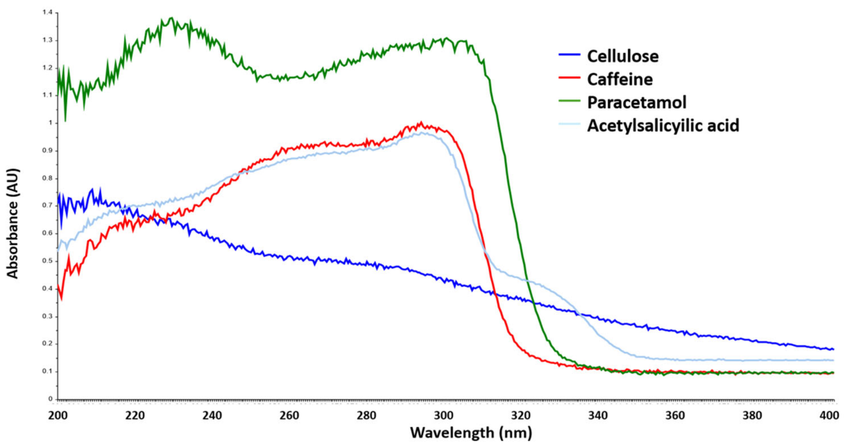
Figure 1. UV-Vis DRS spectra of the pure components used in the present study.
Chemosensors 12 00227 g001
Figure 2. UV-Vis DRS spectra of the standard added samples for the caffeine dataset.
Chemosensors 12 00227 g002
Figure 3. PCA performed on the caffeine dataset: (a) scores plot in PC1, PC2 coordinates; (b) loadings plot of PC1.
Chemosensors 12 00227 g003
Figure 4. NAS standard addition line for caffeine dataset and MSC pretreatment.
Chemosensors 12 00227 g004
Figure 5. UV-Vis DRS spectra of the dataset obtained by additions of caffeine to Neo Nisidine. All spectra were pretreated with MSC.
Chemosensors 12 00227 g005
Table 1. Control parameters to evaluate the NAS regression obtained for the four pretreatments method.
PretreatmentSNVFirst der.MSCMSC + SNV
RMSEP1.110.8390.05400.0650
R20.88630.90100.99570.9522
Number of PLS factors5966
NAS prediction (% w/w)2.323.931.541.88
Table 2. Spectrophotometric and chromatographic results for Neo Nisidine. The expected API concentration is the one declared in the information leaflet. Errors were calculated at a significance level of 5%.
Case StudyAAS (% w/w)PAR (% w/w)CAF (% w/w)
UV-Vis44 ± 434 ± 24.7 ± 0.4
HPLC-DAD43.0 ± 0.935.2 ± 0.84.2 ± 0.1
Expected43.434.74.3
Disclaimer/Publisher’s Note: The statements, opinions and data contained in all publications are solely those of the individual author(s) and contributor(s) and not of MDPI and/or the editor(s). MDPI and/or the editor(s) disclaim responsibility for any injury to people or property resulting from any ideas, methods, instructions or products referred to in the content.