Perspective

Microneedle-Based Sensors for Wearable Diagnostics

by
Hamdi Ben Halima
,
Boris Lakard
and
Nicole Jaffrezic-Renault
*
Institute of UTINAM, University of Franche-Comté, 16 Route de Gray, 25030 Besancon Cedex, France
*
Author to whom correspondence should be addressed.
Chemosensors 2025, 13(2), 68; https://doi.org/10.3390/chemosensors13020068
Submission received: 19 January 2025 / Revised: 9 February 2025 / Accepted: 13 February 2025 / Published: 15 February 2025
(This article belongs to the Section Electrochemical Devices and Sensors)

Abstract

:
Microneedles (MNs) are miniature medical devices, presented as an array of micro-scale needles, each under 1 mm in height with sharp tips. MN technology is becoming a diagnostic platform associated with several qualities: no pain and no risk of infection, offering accuracy, comfort, and usability. Monitoring biomarkers in interstitial fluid (ISF) in real time is crucial for tracking changes in metabolism and assisting in the early diagnosis of chronic illnesses. Some examples of MN sensors are summarized here: the real-time sensing of two metabolites (lactate and glucose or alcohol and glucose), the transdermal tracing of pH, Na+, K+, Ca2+, Li+, and Cl, transdermal methotrexate (MTX) monitoring, the transdermal sensing of tyrosinase enzyme as a melanoma biomarker, the integration of CRISPR technology for nucleic acid analysis, and monitoring plant sap pH in the leaves of several plants. The challenges to be addressed to realize the transition to widespread One Health solutions are analyzed.

1. Introduction

Microneedles (MNs) are miniature medical devices, presented as an array of micro-scale needles, each under 1 mm in height with sharp tips, initially developed in 1996 for painless drug delivery [1]. MN technology is becoming a diagnostic platform that is associated with several qualities: no pain and no risk of infection, offering accuracy, comfort, and usability [2,3,4]. Unlike conventional wearable devices on the surface that are prone to detachment or signal interference, MN-based systems securely anchor to the skin and penetrate only the outermost layers, bypassing the pain receptors and blood vessels in deeper dermal tissues. This minimally invasive approach enables stable, precise access to interstitial fluid (ISF), a rich reservoir of biomarkers, without the discomfort or risks associated with traditional methods. With a volume at least three times that of blood and proximity to the skin’s surface, ISF offers easier, less invasive access to biomarkers than blood collection [4].
Specific designs, such as hollow, dissolvable, solid, and hydrogel-based MNs, are optimized for targeting penetration and extracting fluid [5]. MN-based electrochemical sensors can be of different structures: a conductive solid MN array [6,7], a hollow MN array filled with a conductive paste [8,9], a hollow MN array allowing for the extraction of ISF toward miniaturized electrodes at its backside [10], or a polymeric MN array which can uptake ISF with inserted miniaturized electrodes [11]. Fluorescent MNs combine the minimally invasive nature of MN technology with the high sensitivity of fluorescence-based detection, incorporating probes or dyes for the real-time analysis of biomolecules directly from ISF. Functionalized with biorecognition elements, these MNs offer a targeted approach to capture biomarkers, enabling direct detection [12]. Colorimetric MNs combine minimally invasive technology with accessible colorimetric sensing, using biorecognition changes. This allows straightforward instrument-free diagnostics, which is ideal for POC, and integrates digital imaging tools and low-resource settings [13].
Monitoring biomarkers in ISF in real-time is crucial for tracking changes in metabolism and assisting in the early diagnosis of chronic illnesses. Nevertheless, the partitioning of analytes between blood and ISF needs to be considered. Small molecules such as glucose, ions, drugs, or small biomarkers (up to 3 kDa) easily diffuse through blood capillaries to ISF and are at a level similar to that of blood. Larger macromolecules are found at lower levels than in the blood, which have an extremely low LOD [14].
As a perspective paper, this article does not present an exhaustive review of MN sensors as in recent reviews [15,16,17]; only one example of MN sensors for the detection of metabolites, electrolytes, drugs, proteins, nucleic acid in ISF, and pH in plant leaves is presented. Then, MN sensors on the market are described and the challenges are analyzed.

2. MN Sensors Applied to Human Health

2.1. Real-Time Sensing of Two Metabolites (Lactate and Glucose or Alcohol and Glucose)

The development and validation of an MN array for the real-time detection of two metabolites (lactate and glucose or alcohol and glucose) in the isotope blood fluid (ISF) of volunteers has been described [6]. There is evidence within the clinical community that the regulation of insulin delivery in people with type 1 diabetes, based solely on glucose detection, is not accurate. Despite this high demand, to date, there is no device capable of the continuous and simultaneous measurement of glucose–lactate or alcohol–lactate in real-time.
Cr/Pt/Ag-metalized and -specifically functionalized PMMA microneedles were used as working electrodes. They were then covered with an electrodeposited film of o-phenylenediamine (oPD), and a layer of an enzyme–chitosan mixture (Gox for glucose, Lox for lactate, and AOx for alcohol) cross-linked with PEGDE (poly(ethylene glycol) diglycidyl ether). Subsequent layers of chitosan and PVC (polyvinyl chloride) containing triton X were added to the WE (working electrode) surface. The reference electrodes were chlorinated. MN sensors demonstrated accurate body performance in monitoring all tested biomarkers. Other types of MN sensors were recently proposed for the detection of glucose: a non-enzymatic MN sensor [18], a tri-compartmental MN platform to enable the easy detachment of the microneedle array from the substrate [19], and a biomimetic MN theranostic platform (MNTP) for the intelligent and precise management of diabetes [20].

2.2. Transdermal Tracing of Electrolytes

Multi-ion detection is clinically relevant as it can provide reliable assessments of electrolyte and other disorders. An MN system for the transdermal tracking of pH, Na+, K+, Ca2+, Li+, and Cl has been presented [21]. Potentiometric multi-ion measurements were performed using an MN system consisting of a silicone rubber substrate with seven stainless steel MNs (1500 μm in length and 150 μm in diameter). All WEs (ion-selective electrodes, ISEs) were based on a three-layer structure: (i) a carbon ink film to enhance conductivity, (ii) an MWCNT (multiwalled carbon nanotubes) film as an ion-electron transducer, and (iii) the ion-selective membrane (ISM). The RE (reference electrode) was coated with a layer of Ag/AgCl ink, followed by a polyvinyl butyral (PVB) membrane. An additional layer of polyurethane (PU) was finally added to the RE. Near-Nernst sensitivity, sufficient stability, and resilience to skin penetration were demonstrated by ex vivo (with rat skin pieces) and in vivo (measurements on the body of euthanized rats) tests.

2.3. Rapid Detection of Methotrexate

Methotrexate (MTX) is a widely used drug for the treatment of cancer, rheumatoid arthritis, and psoriasis. If the blood concentration of MTX is >10 μM 24 h after administration, the patient should be treated with leucovorin. Therefore, the rapid detection of MTX levels in the human body is essential for patient safety. The first MN-based electrochemical sensor for the transdermal monitoring of MTX was designed [22]. First, hollow MNs were modified with conductive pastes; the working electrode was functionalized with chitosan (CHI) cross-linked with glutaraldehyde (GA) to endow the sensor with anti-fouling and preconcentration capabilities. The analytical characterization of the MN-based sensor was performed in vitro in protein-enriched artificial interstitial fluid (AISF) and ex vivo in porcine skin using a Franz diffusion cell configuration.

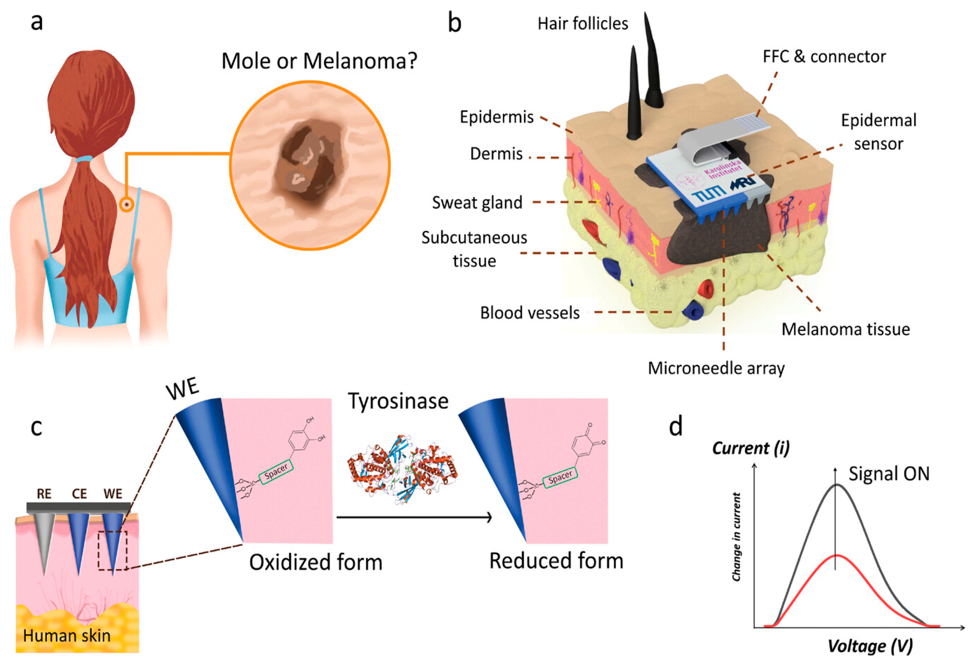
2.4. Transdermal Sensing of Tyrosinase Enzyme as Melanoma Biomarker

An epidermal bioelectronic patch for the transdermal sensing of melanoma biomarkers was developed using a microneedle array integrated with a novel target-specific chemo-responsive probe to enable the detection of tyrosinase enzyme (Tyr) through human skin tissue [23] (Figure 1).
Tyr is an important enzyme that is intricately involved in melanin production and is a critical biomarker for melanoma diagnostics. Elevated Tyr levels in melanoma cells result in aberrant pigment synthesis, making it a potential diagnostic biomarker. The fabrication of polyurethane (PU)- and poly(3,4-ethylenedioxythiophene)–poly(styrene sulfonate) (PEDOT:PSS)-based composite microneedle arrays based on molding in a silicon mold was implemented (Figure 2a), followed by the fabrication of a novel methodology to generate a Tyr-responsive bio interface through successive modification via silanization and cross-coupling methods to yield l-3,4-dihydroxyphenylalanine (L-dopa)-terminated surfaces (Figure 2b). Due to the inaccessibility of human skin samples directly from melanoma patients, we used an established model to demonstrate the measurement of topically dosed Tyr via direct injection to the epidermis. SWV (square-wave voltammetry) and CA (cyclic voltammetry) measurements were performed using transdermal microneedle sensors on Tyr--dosed and untreated skin samples every 24 h for 3 days. Transdermal microneedle sensors developed in this study could differentiate untreated and Tyr-dosed skin samples and successfully detect Tyr at physiological levels on human skin tissue without causing any skin irritation or hypoallergic reactions to microneedle application.

2.5. Real-Time cfDNA Extraction and Monitoring

The integration of CRISPR technology into wearable devices marks a significant advancement in nucleic acid analysis. Renowned for its precise gene-editing capabilities guided by programmable single guide RNA, CRISPR offers high specificity and accuracy. Yang et al. [24] exploited this potential by combining reverse iontophoresis with CRISPR-115 Cas9-enabled graphene biointerfaces on conductive MNs for real-time cfDNA extraction and monitoring. This CRISPR-based wearable device enabled the continuous monitoring of cfDNA biomarkers for conditions like sepsis and kidney transplantation, benefiting from the synergistic effects of graphene biointerfaces and CRISPR-Cas9. Tested in immunodeficient mouse models, the system demonstrated stable sensitivity for up to ten days in vivo. Focusing on sepsis intervention caused by pathogens, such as Epstein–Barr virus (EBV), Staphylococcus aureus (SA), and Pseudomonas aeruginosa (PA), the same group developed a fully integrated wireless wearable device for the long-term, stable monitoring of ultra-trace unamplified nucleic acids (cell-free DNAs and RNAs) in vivo. The system utilized Natronobacterium gregoryi Argonaute (NgAgo) and tetrahedral DNA nanostructures (TDNs) for real-time nucleic acid identification. The device featured a stretchable epidermal patch with enrichment capabilities, a customized MN biosensor, and a flexible circuit board. Researchers investigated nucleic acid identification via NgAgo/guide DNA and signal transformation within the Debye distance. In vivo tests confirmed its suitability for the real-time monitoring of cell-free DNA and RNA for up to 14 days with a sensitivity of 0.3 fM. This technology provides a reliable method for on-body nucleic acid detection and enhances opportunities for ultrasensitive and stable sepsis monitoring in intensive care settings [25].

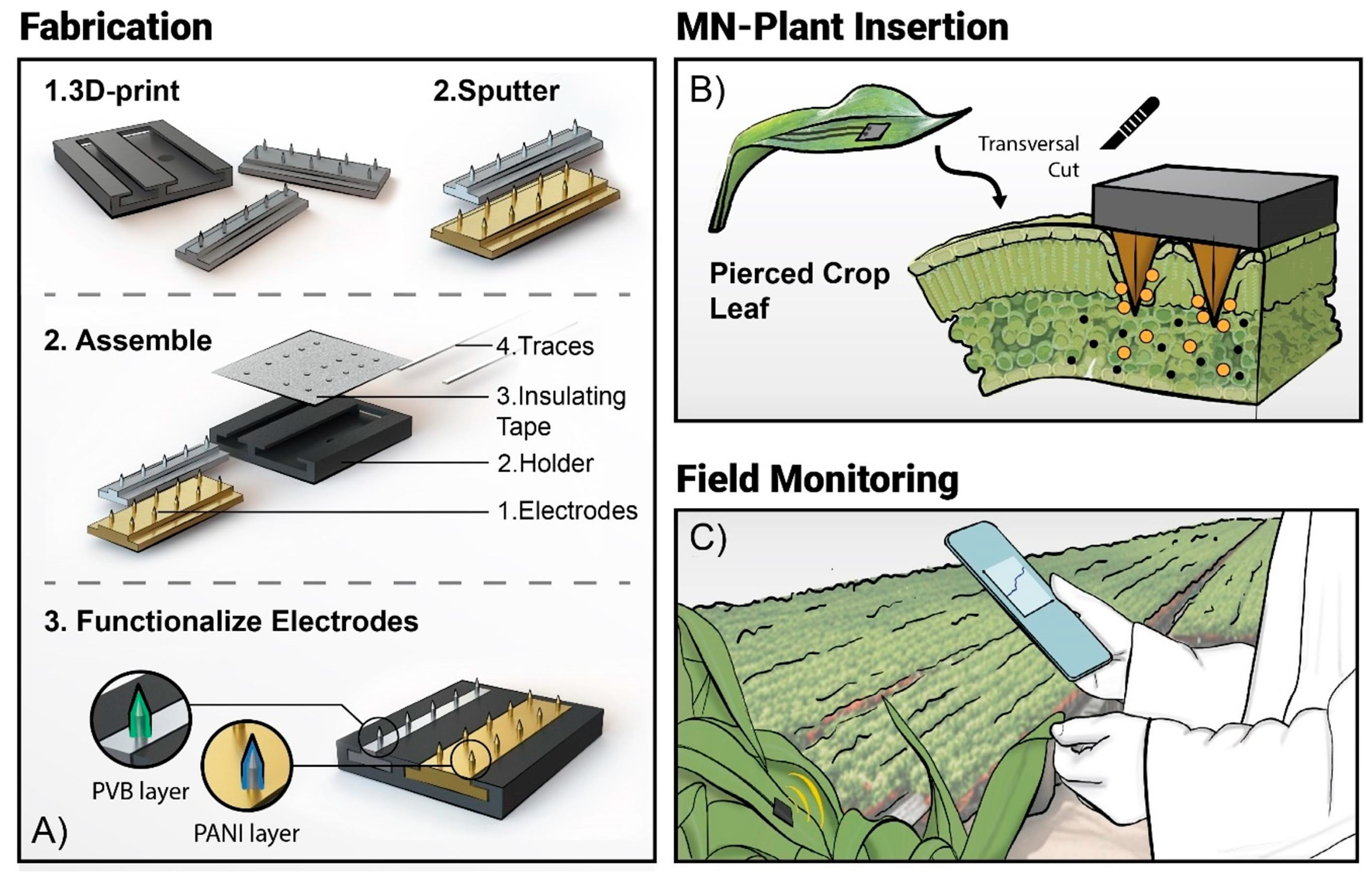
3. MN Sensors Applied to Precision Agriculture

Precision agriculture aims to optimize production under various conditions and uncertainties. Recently, precision agriculture has focused on monitoring chemical parameters in plant sap [26,27]. Wearable electrochemical sensors are promising for transdermal monitoring of biomarkers on human skin or plant leaves [28,29]. A wearable MN sensor for monitoring plant sap pH in leaves of several plants (Hydrangea macrophylla, Peperomia polybotrya, Pilea peperomioides, Coleus sp.) is described in Figure 3 [30]. First, the fabrication of a solid MN array composed of MNs of 300 μm diameter, 900 μm height, and 30 μm tip using an affordable (<500 EUR) stereolithographic 3D printer is described. The WE is functionalized with polyaniline (PANI) to impart pH sensitivity and the RE with a polyvinyl butyral (PVB) membrane to ensure potential stability. The results given by several MN pH sensors inserted into the leaves of different plants for several days showed changes in plant sap pH due to water stress and water events.

4. MN Devices on the Market

Numerous medical domains, such as anesthesia, immunity, scarring, and skin conditions, have been the subject of in-depth investigation regarding the MN platform. Although a lot of work has been focused on developing MN sensors in the lab, there are currently not many MN devices on the market that are accessible for purchase. Until now, continuous glucose monitoring systems (CGMs) based on MN sensors like the Dexcom G6, Abbott Freestyle Libre 2, and Medtronic Guardian are being commercialized. For diabetic patients, MN devices offer a conformable and real-time glucose monitoring alternative to finger-prick blood samples. The cost and short operating duration of CGM devices have hindered the general adoption of MN-based systems, even with their commercial success. More research on large-scale, low-cost MN manufacturing processes will advance the application of MN sensors in real-world settings [31].

5. Challenges

The advent of MN technology has revolutionized wearable sensors by enabling continuous, non-invasive access to ISF for health monitoring. This evolution, from basic offline sensors to sophisticated multifunctional platforms, highlights the immense potential of next-generation MN devices. These platforms promise comprehensive health monitoring, real-time treatment adjustments, and a seamless user experience. However, several key challenges must be addressed to fully realize their potential and transition to widespread MN-based healthcare solutions, including the following:

5.1. User Comfort and Longevity

A key component of the next-generation healthcare platform is user comfort. MNs are generally painless and minimally intrusive, but their comfort during long-term use is sometimes overlooked. The maximum depth of penetration for MNs is around 1000 μm. Skin thickness variability, influenced by age, gender, and body mass index, challenges the daily use of MN-based technologies. MN patches, designed for single use, lack long-term stability, making frequent replacements impractical, especially for chronic illness patients needing sustained medication delivery. While bioelectronics may remain functional, MN patches degrade or deplete, limiting reuse. Extending device longevity or developing methods to replenish functional components is essential.
Polymeric MN sensors experience considerable difficulties in terms of stability and resilience to mechanical stresses during wear, especially when used for prolonged periods. These strains can potentially impair the sensors’ dependability, thus improvements in design and materials science are required to make them more resilient.

5.2. Biofouling and Biocompatibility

An important issue that must be addressed early in the design phase is biofouling, which is the accumulation of biological materials on the sensor surface. Without proper mitigation strategies, biofouling can significantly impair sensor performance, reducing the accuracy and reliability of the data collected.
It is critical to improve MN sensors’ biocompatibility. This can be accomplished by integrating natural biomaterials. Maintaining long-term functionality and user safety requires these enhancements. Skin enzymes have the potential to break down enzyme-based MN sensors, which can eventually reduce their efficacy. To solve this problem, novel strategies for preserving the enzymes or creating more reliable substitutes are needed.

5.3. Optimized Design and Multiplexing

Exact diagnostics are made more difficult by the restricted amount of ISF that can be retrieved, especially for procedures that need larger sample volumes. This limitation highlights the necessity for extremely sensitive analytical techniques that can handle tiny ISF volumes. Different rates of component drainage from the skin might cause imbalance and inaccuracy in MN-based systems, like glucose-sensing tattoos. Furthermore, reliable monitoring and analysis can be complicated by the skewing of biomolecule concentrations caused by the ISF extraction method, especially for large molecules. This emphasizes how meticulous design and optimization of MN systems are necessary to guarantee dependable and constant functioning.
The variety of analytes is one of the practical limitations. There have been numerous reports of multiplexed MN-based sensors; however, most of these sensors are intended to identify common molecules such as lactate, glucose, and electrolytes. On the other hand, macromolecules that are clinically relevant, like DNA/RNA, proteins, and peptides, are usually overlooked. The combined analysis of many indicators would provide the MN sensors with more insightful opportunities for a thorough health assessment. It is expected that monitoring biomarkers will cover a wide variety of analytes for comprehensive healthcare strategies and will also go beyond small molecules to macromolecules.

5.4. Standardization and Regulatory Approval

Despite advancements in MN sensing materials and structures, few MN-based devices have reached the market due to challenges in standardizing production and ensuring clinical reliability. Complex, multi-step fabrication processes hinder product uniformity, requiring years to validate stability and dependability. Additionally, MN manufacturing relies on costly equipment or labor-intensive methods, failing to meet commercial demands for mass production, affordability, and simplicity. These issues significantly impede the commercialization of MN-based products.
Adhering to Good Manufacturing Practices (GMPs), as required by regulatory agencies such as the FDA (Food and Drug Administration) and EMA (European Medicines Agency), entails significant financial outlays and operational difficulties. These include the need to create facilities that comply with GMP regulations and provide workers with considerable training. Regulatory approval for medical devices is a lengthy and rigorous process requiring extensive preclinical and clinical research to ensure safety, efficacy, and reliability.

5.5. Necessary Interdisciplinarity

Additionally, many MN devices lack essential features such as flexibility, breathability, and impermeability. Advancing wearable solutions demands interdisciplinary collaboration across electronics, physics, materials science, and clinical medicine. Despite the challenges of translating lab concepts into commercial products, recent breakthroughs in MN technology show promise for delivering precise, personalized healthcare solutions suitable for routine health and wellness monitoring. The integration of cutting-edge technologies with MN patches opens transformative avenues for research and development in this field. Leveraging advanced materials such as 2D nanomaterials, liquid metals, and biodegradable compounds could revolutionize MN devices by imparting self-healing properties, enhanced flexibility, and environmental sustainability. Additionally, advancements in surface engineering and intelligent functionalization offer the potential to optimize hydrophobicity, hydrophilicity, and enable stimuli-responsive drug delivery systems. Future designs of sampling MNs must strategically balance extraction time, sample volume, and analyte detection accuracy to meet clinical and user requirements. Short extraction times that improve user compliance while ensuring sufficient sample volumes are essential for precise analyte quantification without compromise. The integration of MNs with transdermal bioelectronics, such as microfluidic chips and impedance sensors, could significantly enhance drug delivery, diagnostics, and sampling capabilities. Emerging technologies like triboelectric nanogenerators, wireless power systems, and AI algorithms present transformative opportunities for MN-based devices, enabling customized real-time therapies and diagnostics. However, substantial efforts are required to transition these innovations into practical, wearable healthcare solutions.

Author Contributions

Writing—original draft preparation, H.B.H. and B.L.; writing—review and editing, N.J.-R. All authors have read and agreed to the published version of the manuscript.

Funding

This research received no external funding.

Conflicts of Interest

The authors declare no conflicts of interest.

References

  1. Henry, S.; McAllister, D.V.; Allen, M.G.; Prausnitz, M.G. Microfabricated Microneedles: A Novel Approach to Transdermal Drug Delivery. J. Pharm. Sci. 1998, 87, 922–925. [Google Scholar] [CrossRef] [PubMed]
  2. Smith, A.A.; Li, R.; Tse, Z.T.H. Reshaping healthcare with wearable biosensors. Sci. Rep. 2023, 13, 4998. [Google Scholar] [CrossRef] [PubMed]
  3. Ometov, A.; Shubina, V.; Klus, L.; Skibińska, J.; Saafi, S.; Pascacio, P.; Flueratoru, L.; Gaibor, D.Q.; Chukhno, N.; Chukhno, O. A survey on wearable technology: History, state-of-the-art and current challenges. Comput. Netw. 2021, 193, 108074. [Google Scholar] [CrossRef]
  4. Kim, J.; Campbell, A.S.; de Ávila, B.E.F.; Wang, J. Wearable biosensors for healthcare monitoring. Nat. Biotechnol. 2019, 37, 389–406. [Google Scholar] [CrossRef] [PubMed]
  5. Dong, Y.; Mao, S.; Chen, S.; Ma, J.; Jaffrezic-Renault, N.; Guo, Z. Opportunities and challenges of microneedle electrochemical sensors for interstitial fluid detection. TrAC Trends Anal. Chem. 2024, 180, 117891. [Google Scholar] [CrossRef]
  6. Tehrani, F.; Teymourian, H.; Wuerstle, B.; Kavner, J.; Patel, R.; Furmidge, A.; Aghavali, R.; Hosseini-Toudeshki, H.; Brown, C.; Zhang, F.; et al. An Integrated Wearable Microneedle Array for the Continuous Monitoring of Multiple Biomarkers in Interstitial Fluid. Nat. Biomed. Eng. 2022, 6, 1214–1224. [Google Scholar] [CrossRef]
  7. Lin, S.; Cheng, X.; Zhu, J.; Wang, B.; Jelinek, D.; Zhao, Y.; Wu, T.-Y.; Horrillo, A.; Tan, J.; Yeung, J.; et al. Wearable Microneedle-Based Electrochemical Aptamer Biosensing for Precision Dosing of Drugs with Narrow Therapeutic Windows. Sci. Adv. 2022, 8, eabq4539. [Google Scholar] [CrossRef]
  8. Parrilla, M.; Vanhooydonck, A.; Johns, M.; Watts, R.; De Wael, K. 3D-Printed Microneedle-Based Potentiometric Sensor for PH Monitoring in Skin Interstitial Fluid. Sens. Actuators B Chem. 2023, 378, 133159. [Google Scholar] [CrossRef]
  9. Goud, K.Y.; Mahato, K.; Teymourian, H.; Longardner, K.; Litvan, I.; Wang, J. Wearable Electrochemical Microneedle Sensing Platform for Real-Time Continuous Interstitial Fluid Monitoring of Apomorphine: Toward Parkinson Management. Sens. Actuators B Chem. 2022, 354, 131234. [Google Scholar] [CrossRef]
  10. Parrilla, M.; Detamornrat, U.; Dominguez-Robles, J.; Donnelly, R.F.; De Wael, K. Wearable Hollow Microneedle Sensing Patches for the Transdermal Electrochemical Monitoring of Glucose. Talanta 2022, 249, 123695. [Google Scholar] [CrossRef]
  11. Zheng, L.; Zhu, D.; Wang, W.; Liu, J.; Thng, S.T.G.; Chen, P. A Silk-Microneedle Patch to Detect Glucose in the Interstitial Fluid of Skin or Plant Tissue. Sens. Actuators B Chem. 2022, 372, 132626. [Google Scholar] [CrossRef]
  12. Zhang, J.; Zheng, Y.; Lee, J.; Hoover, A.; King, S.A.; Chen, L.; Zhao, J.; Lin, Q.; Yu, C.; Zhu, L. Continuous glucose monitoring enabled by fluorescent nanodiamond boronic hydrogel. Adv. Sci. 2023, 10, 2203943. [Google Scholar] [CrossRef]
  13. He, R.; Liu, H.; Fang, T.; Niu, Y.; Zhang, H.; Han, F.; Gao, B.; Li, F.; Xu, F. A Colorimetric Dermal Tattoo Biosensor Fabricated by Microneedle Patch for Multiplexed Detection of Health-Related Biomarkers. Adv. Sci. 2021, 8, 2103030. [Google Scholar] [CrossRef] [PubMed]
  14. Friedel, M.; Thompson, I.A.P.; Kasting, G.; Polsky, R.; Cunningham, D.; Soh, H.T.; Heikenfeld, J. Opportunities and Challenges in the Diagnostic Utility of Dermal Interstitial Fluid. Nat. Biomed. Eng. 2023, 7, 1541–1555. [Google Scholar] [CrossRef]
  15. Vora, L.K.; Sabri, A.H.; McKenna, P.E.; Himawan, A.; Hutton, A.R.J.; Detamornrat, A.; Paredes, A.J.; Larraneta, E.; Donnelly, R.F. Microneedle-based biosensing. Nat. Rev. Bioeng. 2024, 2, 64–81. [Google Scholar] [CrossRef]
  16. Li, J.; Wei, M.; Gao, B. A review of recent advances in microneedle-based sensing within the dermal ISF that could transform medical testing. ACS Sens. 2024, 9, 1149–1161. [Google Scholar] [CrossRef] [PubMed]
  17. Poudineh, M. Microneedle Assays for Continuous Health Monitoring: Challenges and Solutions. ACS Sens. 2024, 9, 535–542. [Google Scholar] [CrossRef]
  18. Chinnadayyala, S.R.; Park, J.; Satti, A.T.; Kim, D.; Cho, S. Minimally invasive and continuous glucose monitoring sensor based on non-enzymatic porous platinum black-coated gold microneedles. Electrochim. Acta 2021, 369, 137691. [Google Scholar] [CrossRef]
  19. Ma, T.Q.; He, Q.Y.; Li, X.Y.; Zhang, B.; Li, Y.L.; Chen, L.J.; You, X.Q.; Liu, J. Wearable microneedle sensor with replaceable sensor base for multi-purpose detecting in skin interstitial fluid. Microchem. J. 2024, 203, 110933. [Google Scholar] [CrossRef]
  20. Yang, J.; Zheng, S.; Ma, D.; Zhang, T.; Huang, X.; Huang, S.; Chen, H.J.; Wang, J.; Jiang, L.; Xie, X. Masticatory system–inspired microneedle theranostic platform for intelligent and precise diabetic management. Sci. Adv. 2022, 8, eabo6900. [Google Scholar] [CrossRef]
  21. Molinero-Fernández, A.; Casanova, A.; Wang, Q.; Cuartero, M.; Crespo, G.A. In Vivo Transdermal Multi-Ion Monitoring with a Potentiometric Microneedle-Based Sensor Patch. ACS Sens. 2023, 8, 158–166. [Google Scholar] [CrossRef] [PubMed]
  22. Parrilla, M.; Detamornrat, U.; Domínguez-Robles, J.; Tunca, S.; Donnelly, R.F.; De Wael, K. Wearable microneedle-based array patches for continuous electrochemical monitoring and drug delivery: Toward a closed-loop system for methotrexate treatment. ACS Sens. 2023, 8, 4161–4170. [Google Scholar] [CrossRef] [PubMed]
  23. Poursharifi, N.; Hassanpouramiri, M.; Zink, A.; Ucuncu, M.; Parlak, O. Transdermal Sensing of Enzyme Biomarker Enabled by Chemo-Responsive Probe-Modified Epidermal Microneedle Patch in Human Skin Tissue. Adv. Mater. 2024, 36, 2403758. [Google Scholar] [CrossRef]
  24. Yang, B.; Kong, J.; Fang, X. Programmable CRISPR-Cas9 microneedle patch for long-term capture and real-time monitoring of universal cell-free DNA. Nat. Commun. 2022, 13, 3999. [Google Scholar] [CrossRef] [PubMed]
  25. Yang, B.; Wang, H.; Kong, J.; Fang, X. Long-term monitoring of ultratrace nucleic acids using tetrahedral nanostructure-based NgAgo on wearable microneedles. Nat. Commun. 2024, 15, 1936. [Google Scholar] [CrossRef]
  26. Yin, H.; Cao, Y.; Marelli, B.; Zeng, X.; Mason, A.J.; Cao, C. Soil sensors and plant wearables for smart and precision agriculture. Adv. Mater. 2021, 33, 2007764. [Google Scholar] [CrossRef]
  27. Coatsworth, P.; Gonzalez-Macia, L.; Collins, A.S.P.; Bozkurt, T.; Güder, F. Continuous monitoring of chemical signals in plants under stress. Nat. Rev. Chem. 2022, 7, 7–25. [Google Scholar] [CrossRef]
  28. Vulpe, G.; Liu, G.; Oakley, S.; Pletsas, D.; Yang, G.; Dutra, R.; Guy, O.; Liu, Y.; Waldron, M.; Neary, J.; et al. Wearable technology for one health: Charting the course of dermal biosensing. Biosens. Bioelectron. X 2024, 19, 100500. [Google Scholar] [CrossRef]
  29. Wang, B.; Lu, H.; Jiang, S.; Gao, B. Recent advances of microneedles biosensors for plants. Anal. Bioanal. Chem. 2024, 416, 55–69. [Google Scholar] [CrossRef] [PubMed]
  30. Parrilla, M.; Steijlen, A.; Kerremans, R.; Jacobs, J.; den Haan, L.; De Vreese, J.; Van Noten Geron, Y.; Clerx, P.; Watts, R.; De Wael, K. Wearable platform based on 3D-printed solid microneedle potentiometric pH sensor for plant monitoring. Chem. Eng. J. 2024, 500, 157254. [Google Scholar] [CrossRef]
  31. Hu, Y.; Chatzilakou, E.; Pan, Z.; Traverso, G.; Yetisen, A.K. Microneedle Sensors for Point-of-Care Diagnostics. Adv. Sci. 2024, 11, 2306560. [Google Scholar] [CrossRef]
Figure 1. Schematic illustration of a smart probe-modified epidermal microneedle sensor patch. Representative demonstration of cutaneous melanosis on the female patient’s back (a), schematic illustration of human skin layers and application of the epidermal microneedle array patch on the melanoma skin area (b). Working principle of the smart probe on the microneedle surface and chemical transformation upon interaction with Tyr enzyme (c), and generation of representative SWV signal with ON signal principle via an epidermal sensor device (d). (From [23]).
Chemosensors 13 00068 g001
Figure 2. (a) Schematic demonstration of the silicone mold (i), injection of the conductive polymer composite (ii), and peeling process of the microneedle array (iii). After the peeling process, the microneedle array was laser-cut to generate individual electrodes (iv), one of the 2 × 6 arrays was sputter-coated with Ag/AgCl ink to define the reference electrode (v), and finally, all the parts were connected before insertion onto the skin (vi). Schematic representation of the surface modification of the single PU-PEDOT:PSS microneedle (working electrode) by the Tyr-sensitive smart probe (b). (From [23]).
Chemosensors 13 00068 g002
Figure 3. Illustration of the concept of portable continuous pH monitoring. (A) Microneedle arrays are fabricated by low-cost 3D printing technique, sputtered with thin metal layer and arranged on a novel MN electrode configuration for functionalization with PVB and PANI. (B) MN pH sensor is inserted into the plant for metabolic assessment of plant health. (C) Vision of decentralization of MN-based sensors for digital plant health monitoring. (From [30] with permission).
Chemosensors 13 00068 g003
Disclaimer/Publisher’s Note: The statements, opinions and data contained in all publications are solely those of the individual author(s) and contributor(s) and not of MDPI and/or the editor(s). MDPI and/or the editor(s) disclaim responsibility for any injury to people or property resulting from any ideas, methods, instructions or products referred to in the content.

