Article

A Label-Free Fluorescent Sensor Based on the Formation of Poly(thymine)-Templated Copper Nanoparticles for the Sensitive and Selective Detection of MicroRNA from Cancer Cells

Department of Chemistry, National University of Singapore, Singapore 117543, Singapore
*
Author to whom correspondence should be addressed.
Chemosensors 2020, 8(3), 52; https://doi.org/10.3390/chemosensors8030052
Submission received: 8 June 2020 / Revised: 4 July 2020 / Accepted: 6 July 2020 / Published: 8 July 2020
(This article belongs to the Section Applied Chemical Sensors)

Abstract

:
In this work, a simple and label-free fluorescence “off” to “on” platform was designed for the sensitive and selective detection of microRNA (miRNA) in cancer cells. This method utilized a padlock DNA-based rolling circle amplification (P-RCA) to synthesize fluorescent poly(thymine) (PolyT) which acted as a template for the synthesis of copper nanoparticles (CuNPs) within 10 minutes under mild conditions. While the repeated PolyT sequence was used as the template for CuNP synthesis, other non-PolyT parts (single strand-DNAs without the capacity to act as the template for CuNP formation) served as “smart glues” or rigid linkers to build complex nanostructures. Under the excitation wavelength of 340 nm, the synthesized CuNPs emitted strong red fluorescence effectively at 620 nm. To demonstrate the use of this method as a universal biosensor platform, lethal-7a (let-7a) miRNA was chosen as the standard target. This sensor could achieve highly sensitive and selective detection of miRNA in the presence of other homologous analogues for the combination of P-RCA with the fluorescent copper nanoparticle. Overall, this novel label-free method holds great potential in the sensitive detection of miRNA with high specificity in real samples.

1. Introduction

MicroRNAs (miRNAs) are non-coding short RNAs present in animals, plants, and some viruses [1]. Previous evidence has shown that miRNAs are vital regulators for post-transcription, leading to the silencing of genes through target degradation or translational repression [2,3]. As well as this regulatory role in gene expression, numerous experiments have confirmed that miRNA disorder is related to certain types of diseases [4]. Thus, the highly selective and sensitive recognition of miRNAs is important for better identification and analysis of genetic functions. The in vitro polymerization of nucleotides due to their signal amplification ability has been widely used in the analysis of miRNAs, including polymerase chain reaction (PCR) [5], strand displacement amplification (SDA) [6] and rolling circle amplification (RCA) [7]. These techniques usually employ dye-labelled probes or fluorophores as signal reporters. The synthesis and attachment of traditional signal reporters generally require extensive and time-consuming operations [8]. Furthermore, the degree of nonspecific recognition caused by these signal reporters raises their environmental susceptibility and reduces sensitivity.
Owing to their distinctive linear, geometric nanosized structures, unique molecular recognition characteristics, and strong affinities for metal ions, certain types of nucleic acids have been utilized as templates for synthesizing metallic nanostructures [9,10,11,12,13]. This can be carried out firstly by binding metal ions onto the nucleic acid template, followed by the reduction of nucleic acid-complexed metal ions through chemical approaches [14,15]. This method has already been utilized for assembling metal nanoclusters with high photochemical stability and adjustable fluorescence emission that are appropriate for biochemical analysis systems [16,17], as well as for designing conductive linear nucleic acid nanowires by depositing metal atoms onto the nucleic acid template [18]. To overcome the drawbacks of traditional fluorophores and dye-labelled probes, DNA-templated nanoparticles (NPs) with intense fluorescence emission signals have been developed, which contain single-stranded or double-stranded DNA-templated NPs [19,20,21,22,23]. While silver nanoparticles are popularly employed for the detection of various analytes, their synthesis is complex and time-consuming, requiring several hours [24,25]. Hence, in this study, copper nanoparticles were utilized due to their quick and simple synthesis at room temperature. The nanoparticles exhibited comparable fluorescence intensities to the widely used silver nanoparticles, quickening the detection process. Both single and double-stranded poly(thymine) (PolyT)-templated copper nanoparticles (CuNPs) present great potential for applications in biochemical analysis, owing to their easy synthesis, admirable photo-physical properties, and excellent biocompatibility [26,27,28,29,30]. Contrary to double-stranded DNA, the single-stranded DNA (ss-DNA) template is able to maintain its linearity without hybridizing with another strand, making it a better programmable building block for nanotechnology. In addition, the sizes of fluorescent CuNPs are easily controllable by adjusting the lengths of the PolyT ss-DNA templates during synthesis [22,23].
In this work, we design a sensitive and selective primer ligation-extension rolling circle amplification strategy for simple and economic miRNA analysis by utilizing nano-dye PolyT-CuNPs synthesized in situ [8]. The detailed procedure of this assay is illustrated in Scheme 1. As reported, nucleotides have high affinities for several metal cations, and these localized metal ions could be reduced to form metallic nanomaterials along the contour of the DNA scaffold [14,31,32]. Firstly, the miRNA recognizes and hybridizes with the padlock probe to achieve the circulation of the padlock probe in the presence of DNA ligase. Secondly, the short miRNA primer triggers the RCA reaction and efficiently elongateds to an ss-DNA including a repeated PolyT sequence complementary to the polyadenylic acid (PolyA) sequence in the padlock template. Next, the formed ss-DNA served as a template for the in-situ formation of the fluorescent nano-dye CuNPs. At the same time, other ss-DNAs without the capacity to act as the template for CuNP formation (non-PolyT parts) served as “smart glues” or rigid linkers to build complex nanostructures. By adjusting the lengths of the PolyT and non-PolyT parts, alternating metallized and non-metallized parts could be obtained. Moreover, PolyT-templated CuNPs display intense fluorescence emission with large Mega-Stokes shifts [22,23], making them perfectly matched as excellent fluorescent sensors for the recognition of target miRNAs in complex biological systems since they provide high sensitivity by eliminating intense background fluorescence signals from complicated biological environments. The larger the Mega-Stokes shifts, the smaller the overlap between the absorption and emission spectra. Hence, the lowering of the fluorescence efficiency, caused by energy transfer, can be avoided.

2. Experimental

2.1. Reagents and Materials

The synthetic DNA/RNA molecules used in the experiment were acquired from Integrated DNA Technologies (Singapore), and their sequences (5′→3′) are listed in Table S1. The oligonucleotides were high-performance liquid chromatography (HPLC)-purified. Escherichia coli (E. coli) DNA ligase was purchased from AIT Biotech (Singapore) and Bacillus phage phi29 (Phi29) DNA polymerase was purchased from Thermo Fisher Scientific (South San Francisco, CA, USA). Ascorbic acid and CuSO4·5H2O were purchased from Sigma Aldrich Chemical Co., Ltd. (Singapore). The other chemicals were of analytical grade and purchased from Sigma Aldrich Chemical Co., Ltd. (Singapore) and used without any further purification. Ultrapure water (resistivity of >18.2 MΩ.cm) was used for the preparation of all buffer solutions, and was obtained through a Millipore Milli-Q water purification system.

2.2. Amplification Reaction

First, an appropriate amount of the target miRNA was mixed with 100 nM padlock DNA template in a 40 μL 1× ligation-reaction buffer solution, which was denatured at 90 °C for 10 min to promote hybridization and then slowly cooled down to room temperature. Next, 5 U E. coli DNA ligase was mixed with the ligation solution and kept at 37 °C for 1 h. Subsequently, 40 µL of RCA reaction buffer (Phi29 polymerase reaction buffer) containing 600 µM of deoxynucleoside triphosphate (dNTP) and 40 U of Phi29 DNA polymerase was added into the resulting solution. The polymerization reaction was carried out at 37 °C for 4 h. Following this, the Phi29 DNA polymerase enzyme was inactivated by maintaining the reaction solution in a water bath at 65 °C for 10 min. The final solution was slowly cooled to room temperature for further use.

2.3. Formation of PolyT-CuNPs

PolyT-templated CuNPs were synthesized according to methods reported previously [22,33,34,35,36]. The reaction was carried out in 1 mL of buffer solution consisting of 20 mM 3-(N-morpholino)propanesulfonic acid (MOPS) and 300 mM NaCl at pH 7.5. The final polymerization reaction solution contained the above buffer solution, 500 µM CuSO4, and 3 mM ascorbic acid, which were completely mixed and incubated in dark conditions for 10 min at room temperature for the formation of fluorescent CuNPs. It was noted that DNA-templated copper nanoparticles could be sensitive to thiol-containing molecules, such as dithiothreitol (DTT), in the reaction buffer for DNA ligation. Nevertheless, since DTT always existed in the RCA reaction for all the CuNP formation reactions with the same concentration, we were able to ignore the effect of DTT on the formation of CuNPs.

2.4. Cell Culture, RNA Extraction, and qPCR

The miRNAs of the lethal-7 (let-7) family are cancer-related miRNAs. Here we use lthal-7a (let-7a) as an example for the development of the method. Accumulated evidence indicates that let-7a generally functions as a tumor suppressor in several human cancers, although the role of let-7a in cancers remains unclear. It is known that let-7 miRNA family members display high sequence homology, thus there is a great challenge in distinguishing the minor differences among them. Moreover, their expression levels are tightly related to cell development and human cancer [37]. The specificity of this sensor was evaluated by comparing the fluorescence intensity of the model target miRNA let-7a to the single-base mismatched miRNAs let-7c and let-7f and the double-base mismatched miRNAs let-7b and let-7d. The cells were cultured under the following culture conditions: 5% CO2/95% air and 37 °C. The growth medium for Medical Research Council cell strain 5 (MRC-5, normal cell) was ATCC-formulated Eagle’s Minimum Essential Medium (Catalog No. 30-2003), 1% (vol/vol) penicillin-streptomycin, and 10% (vol/vol) FBS. The growth medium for HeLa cells was ATCC-formulated Eagle’s Minimum Essential Medium (Catalog No. 30-2003), 1% (vol/vol) penicillin-streptomycin, and 10% (vol/vol) fetal bovine serum (FBS). The growth media for H1299 (Lung cancer cell) was ATCC-formulated RPMI-1640 Medium (Catalog No. 30-2001) + 1% (vol/vol) penicillin-streptomycin + 10% (vol/vol) FBS. Total RNA of the cultured cells was extracted using a miRNeasy RNA extraction kit (Qiagen V.V., Hilden, Germany) according to the recommended procedure. Let-7a detection was performed on the extracted total RNA. The reference values of let-7a were obtained by qPCR.

2.5. Apparatus

UV-Vis absorption spectra were recorded using an Agilent Cary 60 UV-Vis Spectrophotometer (Agilent Technologies Singapore Pte Ltd, Singapore). Fluorescence excitation and emission spectra were recorded on an Agilent Cary Eclipse Fluorescence Spectrophotometer (Agilent Technologies Singapore Pte Ltd, Singapore). Transmission electron microscopy (TEM) was carried out on an FEI Tecnai F20 (FEI Company, Hillsboro, OR) using a 200 kV acceleration. Surface morphology examination of the samples was conducted by atomic force microscopy (AFM) using an XE-100 system from Park Systems (Suwon, South Korea). All the measurements were carried out at room temperature unless stated otherwise.

3. Results and Discussion

3.1. Principle of MiRNA Detection

Among the large number of miRNAs, the let-7 family has been identified to be responsible for the timing of cell fate determination [38,39]. The human let-7 family includes 13 members positioned on nine kinds of chromosomes, and most members have been associated with the adjustment of drug sensitivity in cancers [40,41]. Accumulated evidence indicates that let-7a generally functions as a tumor suppressor in several human cancers. However, the role of let-7a in cancers remains unclear. For our sensor, we employed let-7a as a target to demonstrate its advantages. In the presence of the padlock DNA template, dNTP, E. coli DNA ligase, and Phi29 DNA polymerase, the let-7a miRNA primer could initiate the RCA amplification process and be elongated to a long ss-DNA containing repeated PolyT sequences, which could act as the template to form CuNPs by incubation with an appropriate amount of copper (Ⅱ) ions and sodium ascorbate. As reported, nucleotides have high affinities for several metal cations, and these localized metal ions could be reduced to form metallic nanomaterials along the contour of the DNA scaffold [14,31,32]. It was noticed that the formation of CuNPs on the PolyT template was due to binding interactions between Cu2+ ions and thymine, and that the thymine-complexed Cu2+ ions were reduced to Cu0 by ascorbic acid in the shape of the PolyT template. As shown in previous results [22], random ss-DNA, PolyA, polycytidylic acid (PolyC), and polyguanylic acid (PolyG) cannot act as templates for the formation of CuNPs. Nevertheless, they can provide resistance in creating nanostructures with alternating metallized and non-metallized parts. Firstly, the target miRNA recognized and hybridized with the padlock probe to achieve the circulation of the padlock probe in the presence of DNA ligase. Secondly, the miRNA primer triggered the RCA reaction and was efficiently elongated to an ss-DNA including a repeated PolyT sequence complementary to the PolyA sequence in the padlock template. Next, the formed ss-DNA served as a template for the in situ formation of the fluorescent nano-dye CuNPs. At the same time, other ss-DNAs without the capacity to act as the template for CuNP formation (non-PolyT part) served as “smart glues” or rigid linkers to build complex nanostructures. The RCA-mediated PolyT-CuNPs display an intense absorption peak near 340 nm (Figure 1a), which is consistent with previous observations [22]. Also, under the irradiation of UV light, the two samples above showed different fluorescence images, which clearly indicates the successful in situ formation of the RCA-mediated PolyT-CuNPs. As observed with the naked eye, the sample without the addition of let-7a was colorless. In contrast, the sample with the addition of let-7a showed an intense red fluorescence signal. The characteristic fluorescence peak is illustrated in Figure 1b. In the presence of let-7a, the RCA-mediated PolyT-CuNPs exhibited an intense emission peak at around 620 nm with 340 nm excitation. Without miRNA let-7a, there was no obvious fluorescence signal detected around 620 nm.

3.2. Characterization of CuNPs

Next, transmission electron microscopy (TEM) and atomic force microscopy (AFM) experiments were carried out to further confirm the morphology of the RCA-mediated PolyT-CuNPs. As shown in the TEM images (Figure 2a–c), the PolyT-templated CuNPs were spherical in shape, and of approximately 2–4 nm in diameter. Size distribution quantification was performed by measuring 40 NP diameters and then determining the averaged diameter to be 2.8 ± 1.2 nm. The concentration of Figure 2a refers to the 1.0 nM target miRNA in the amplification process. Figure 2b,c show the TEM images for the diluted samples of PolyT-templated CuNPs. In Figure 2d, the AFM results show a long white line which could be attributed to the formation of an RCA-mediated PolyT ss-DNA strand connecting adjacent CuNPs, as deduced from Scheme 1. According to the length analysis of all the ss-DNA strands, it was noted that their lengths were multiples of the length of the padlock DNA template. The heights of the line profiles (3–3.5 nm) of the AFM image (Figure 2e,f) correspond to the diameters of the CuNPs, which were consistent with the average diameter of the CuNPs estimated from the TEM images. Hence, the above observations confirmed that the RCA amplification process could be effectively initiated by the target miRNA to yield the elongated ss-DNA containing repeated PolyT sequences. Furthermore, the formed PolyT part could function as a template for CuNPs, and the non-PolyT part could serve as the linker for the building of these linear complex nanostructures.

3.3. Optimization of Sensor Conditions

Moreover, the essential roles of the padlock DNA, E. coli DNA ligase, dNTP, and Phi29 DNA polymerase for the formation of the PolyT-CuNPs were further explored. In the absence of miRNA and E. coli DNA ligase, the noncircular padlock DNA (pDNA) could not trigger the RCA process as it was still a short single-stranded noncircular DNA template. Phi29 DNA polymerase is a kind of high-fidelity enzyme that has a strong strand-displacing capability, proofreading activity, and high processivity [42]. Additionally, this enzyme is stable enough to keep linear kinetics at room temperature overnight, eliminating the use of thermal cycling. Therefore, RCA-mediated fluorescent CuNPs could not be synthesized in the absence of any one of miRNA, the padlock DNA template, E. coli DNA ligase, Phi29 DNA polymerase, or dNTP, which was confirmed by the resulting fluorescence signals in the control experiments (Figure 3).
According to the principle of the sensing approach (Scheme 1), the fluorescence intensity of PolyT-templated CuNPs greatly determines the sensor performance of the designed method. The reduction of Cu2+ by sodium ascorbate in the presence of the PolyT template was achieved immediately after intensive mixing of all the reactants. Herein, we fixed the length of PolyT to T30, which was sufficient for the detection of fluorescence in the nanomolar range and an appropriate length for designing and inserting into the padlock template for the RCA reaction. The reaction conditions, such as the concentration of Cu2+, concentration of ascorbic acid, and incubation time, were optimized accordingly. Firstly, the concentration of sodium ascorbate was fixed while the Cu2+ concentration was adjusted. The fluorescence signal at 620 nm increased with increasing Cu2+ concentration and reached its maximum intensity at 500 µM (Figure S1). Next, when fixing the concentration of Cu2+ at 500 µM, it can be observed that with the increase in ascorbate concentration, the fluorescence intensity at 620 nm reached a plateau after 3 mM. Subsequently, the incubation time with ascorbate and Cu2+ as well as the resulting RCA solution were also explored. It is shown in Figure S3 that the fluorescence signal rose remarkably with an increase in incubation time from 0 to 10 min and then remained constant. It was noted that the fluorescence increased rapidly within the first 5 min, indicating that this designed approach was quick and sensitive in tracing the PolyT sequence. To ensure a short incubation time, an incubation time of 10 min was maintained for all further experiments [16,43]. Moreover, the CuNPs could preserve over 80% of their original fluorescence even after 2 h (Figure S3), suggesting that the prepared CuNPs exhibit high stability and hence are suitable for analytical applications. In conclusion, highly fluorescent CuNPs were synthesized after incubating the solution containing specific amounts of T30 template probe, 500 µM Cu2+, and 3 mM sodium ascorbate for 10 min.
The polymerization process determines the quantity of PolyT template formed in the system. Thus, it is important to optimize the parameters in the RCA reaction to improve the reaction efficiency. RCA reaction usually promises high sensitivity under the highly effective polymerase-Phi29 DNA polymerase. However, this amplification is limited as the background signal comes from non-specific hybridization amplification, which appears to relate to the padlock probe concentration. Therefore, it is necessary to optimize the padlock probe concentration to decrease the background signal and increase the sensitivity of the assay. It was observed that while the fluorescence intensity increased with the increment of padlock probe concentration, the background fluorescence intensity barely changed. This phenomenon implies that the ligation RCA-mediated PolyT-CuNPs approach reduces the possibility of nonspecific amplification, which helps in hindering the background noise and improving the sensitivity. To enhance the sensitivity and selectivity of this assay, the other reaction conditions such as the concentration of dNTP, E. coli DNA ligase, and Phi29 DNA polymerase and the incubation time were also investigated in the following part. The E. coli DNA ligase concentration determines the ligation of the padlock probe, which is the key factor in triggering the polymerization process. The dNTP and Phi29 DNA polymerase concentrations are crucial factors affecting PolyT DNA amplification by RCA. From Figures S4–S6, it can be observed that when the concentrations of E. coli DNA ligase, dNTP, and Phi29 DNA polymerase were 5 U, 600 μM, and 40 U, respectively, the fluorescence intensity could attain a steady value. As a result, 0.25 U/µl DNA ligase, 600 μM of each dNTP, and 0.5 U/µL of Phi29 DNA polymerase were utilized for all the following RCA reactions. In addition, the incubation time of the RCA reaction was chosen to be 4 h to increase the efficacy of the RCA reaction and by extension the sensing platform (Figure S7)

3.4. Detection of MiRNA

Under optimal conditions, the sensitivity of this assay was shown by investigating the connection between the fluorescence signal and the let-7a miRNA concentrations. As seen in Figure 4, the miRNA calibration curve shows a linear dynamic range from 100 fM to 1.0 nM with a coefficient of 0.99 (R2 = 0.98); the specific equation is log10 (RFU) = 0.501 log10 (c) − 1.002, where RFU is the intensity at the peak of 620 nm and c refers to the let-7a miRNA concentration. The peak at 620 nm was chosen as the standard to evaluate the linear relationship between the intensity of fluorescence and the concentration of miRNA let-7a. Figure 5 demonstrated that the fluorescence signal recorded when the target miRNA let-7a concentration varied from 100 fM to 1.0 nM. In the figure, 0.0 fM of the target miRNA corresponds to the background signal of the assay. This background signal is negligibly low, suggesting minimal interference. The detection limit (LOD = 3σ/S, σ and S are the standard deviation and slope of the calibration curve, respectively) was estimated to be 70.6 fM or 5.6 amol, which is lower than most of the previously published fluorescence sensors for miRNA constructed on double-stranded-DNA-templated (ds-DNA-templated) CuNPs [8,26,44]. The high magnitude of amplification efficiency from the joint action of polymerases confirms that high sensitivity is realized in the RCA method. The results suggest that the utilization of the RCA approach could effectively amplify the reporter-PolyT sequence numbers in the final detection solution for the formation of PolyT-CuNPs and build a linear nanostructure by non-PolyT sequence, thus introducing the RCA approach with great potential for ultrasensitive nucleic acid sensing. A comparison of our method with previously reported biosensor-based methods for the detection of miRNA is given in Table 1 [6,26,45,46,47,48,49,50]. Compared with the majority of these previous methods, our ss-DNA-templated CuNP-based sensor has the advantages of simplicity, robustness, high sensitivity (detection limit of 70 fM), a large detection range (from 100 fM to 1.0 nM), and a relatively short detection time (~5.5 h). Among the previous methods, an electrochemical method reported a low detection limit of 0.045 fM [47]. However, the linear range achievable was narrower compared with our method (three orders of magnitude vs. four orders of magnitude), and there would be a need to replace or clean the electrodes used for electrochemical detection. Another fluorescence-based method also reported a low detection limit (0.27 fM) [50]. However, the method required the use of several additional enzymes (including nicking endonuclease and high-fidelity polymerase). The method described in this work is more convenient for use in the analysis of clinical samples for the detection of miRNAs from cancer cells.

3.5. Specificity Study

It can be noted from Figure 6 that both the single-base mismatched and double-base mismatched miRNAs were clearly distinguished from let-7a. Moreover, the fluorescence signals from let-7f were the lowest at the same concentration with other miRNAs, although there was only one-base difference compared to let-7a. The reason could be that this single-base mismatch was located on the ligation part, which would decrease the efficiency of the ligation reaction significantly. By utilizing the ligation reaction for the RCA template, this miRNA sensor was capable of detecting single nucleotide polymorphism. The results for both let-7b and let-7d are two-base mismatches compared to let-7a. However, let-7d has a relatively higher fluorescence signal since one of the two-base mismatches of let-7d is distant from the 3′-end, which has a negligible effect on the efficiency of the 3′ elongation polymerase. Nevertheless, perfectly matched let-7a exhibited the strongest fluorescence response, and hence could be easily distinguished from other let-7 miRNAs. Therefore, this demonstrates the high specificity of the miRNA assay.
In addition, to investigate the universal practicability of the RCA-based sensor, microRNA-122 (miR-122), whose sequence is irrelevant to that of let-7 miRNAs, was randomly chosen as an example to demonstrate the universal specificity of the sensor system. The fluorescence intensity of miR-122 was below the detection level implying that miR-122 did not interfere with the detection of let-7a. These results confirm that this RCA-PolyT sensing strategy exhibited good selectivity which could discriminate between perfectly matched and mismatched sequences effectively among members of the same miRNA family, as well as unrelated miRNAs. The high selectivity ensures that this assay has great potential for applications in the detection of miRNAs in real biological systems.

3.6. Detection of MiRNA in Real Samples

To assess the practicability of the proposed assay, we detected the concentrations of miRNA let-7a in three total RNA samples extracted from cultured cells. In Table 2, the amounts of let-7a in these total RNA samples detected by the present method are summarized; they were well matched with the values obtained by the qPCR method. The relative errors of the results obtained by the present method were estimated to be less than 20%.
This sensor could achieve the highly sensitive and selective detection of miRNA in the presence of other homologous analogues for the combination of padlock DNA-based rolling circle amplification (P-RCA) with the fluorescent copper nanoparticle. The majority of nucleic acid dyes suffer from small Stokes shift and non-specific binding, which cause relatively high background interference. The red emission of DNA-templated CuNPs (λex = 340 nm, λem = 620 nm) with Mega-Stokes shifting (280 nm) could act as an excellent fluorescent indicator, resulting in lower interference in the analysis. Furthermore, primer sequences were used to enhance specificity in the selective primer ligation-extension rolling circle amplification strategy.
It is worth noting that the proposed method has significant advantages over qPCR, since isothermal amplification by the RCA process has a significant selectivity improvement through the additional use of the ligase and is much more convenient than qPCR since it does not require thermal cycling. These advantages would significantly enhance the flexibility of the sensor for straightforward miRNA determination. This sensor could be used to analyze miRNA expression levels in different cancer cells. However, it cannot be directly used for in vivo cancer diagnosis since the extraction of total RNA from cells is required for fluorescence detection.

4. Conclusions

In summary, we developed a direct and efficient method for miRNA detection by an “off” to “on” fluorescence platform, via the combination of fluorescent PolyT-CuNPs with the RCA nucleic acid signal amplification strategy. The proposed strategy was able to achieve a LOD of 70.2 fM and a wide detection range from 100 fM to 1.0 nM. The excellent performance of the proposed bioassay can be attributed to the following reasons. Firstly, RCA as a signal amplification strategy could effectively synthesize a large amount of PolyT sequences with a trace amount (70 fM) of the target primer. Secondly, the detection approach of this sensor utilized RCA-PolyT-templated fluorescent CuNPs as the signal reporter, which provided a simple and cost-effective strategy without the need for complicated synthetic reactions to obtain fluorescent silver or gold nanocluster compounds, which is much more expensive. Furthermore, the synthesized CuNPs could emit intense red fluorescence with exceptional characteristic Mega-Stokes shifts, which could minimize interferences arising from complex matrices. Hence, this method could be used for the visual determination of nucleic acids under a UV lamp. The results show that the method could achieve high sensitivity and selectivity in the analysis of miRNA in real samples. The detection method developed in this work may provide a useful tool for miRNA detection in a wide range of potential applications, such as biological analysis, fluorescence imaging, and clinical diagnostics.

Supplementary Materials

The following are available online at https://www.mdpi.com/2227-9040/8/3/52/s1, File S1: Table S1. Sequences of synthesized oligonucleotides (5′-3′) used in the article, Table S2. Sequences of miRNA let-7 family (5′→3′), Figure S1. Fluorescence spectra of PolyT-CuNPs at different concentrations of Cu2+ (1-7: 100, 200, 300, 400, 500, 600, 700 µM), Figure S2. Fluorescence spectra of polyT-CuNPs at different concentrations of sodium ascorbate (1-7: 0.5, 1.0, 1.5, 2.0, 2.5, 3.0, 3.5 mM), Figure S3. Investigation of the stability of T30 templated CuNPs, Figure S4. The relationship between the fluorescence intensity and the concentration of dNTP, Figure S5. The relationship between the fluorescence intensity and the concentration of Phi 29 DNA Polymerase, Figure S6. The relationship between the fluorescence intensity and the concentration of E. coli DNA ligase and Figure S7. Influence of polymerization time on the formation of polyT-CuNPs.

Author Contributions

Conceptualization, Q.M. and Z.G.; methodology, Q.M. and H.D.; resources: S.F.Y.L.; original draft preparation, writing—review and editing, Q.M., Z.G. and S.F.Y.L.; supervision: S.F.Y.L. All authors have read and agreed to the published version of the manuscript.

Funding

Financial support of this work by the Ministry of Education (R-143-000-B48-114) is gratefully acknowledged.

Acknowledgments

Financial support of this work by the Ministry of Education (R-143-000-B48-114) is gratefully acknowledged.

Conflicts of Interest

The authors declare that there is no conflict of interest regarding the publication of this paper.

References

  1. He, L.; Hannon, G.J. MicroRNAs: Small RNAs with a big role in gene regulation. Nat. Rev. Genet. 2004, 5, 522–531. [Google Scholar] [CrossRef] [PubMed]
  2. Bartel, B. MicroRNAs: Target recognition and regulatory functions. Cell 2009, 136, 215–233. [Google Scholar] [CrossRef] [PubMed] [Green Version]
  3. Krek, A.; Grün, D.; Poy, M.N.; Wolf, R.; Rosenberg, L.; Epstein, E.J.; MacMenamin, P.; Da Piedade, I.; Gunsalus, K.C.; Stoffel, M.; et al. Combinatorial microRNA target predictions. Nat. Genet. 2005, 37, 495–500. [Google Scholar] [CrossRef] [PubMed]
  4. Lu, J.; Getz, G.; Miska, E.A.; Alvarez-Saavedra, E.; Lamb, J.; Peck, D.; Sweet-Cordero, A.; Ebert, B.L.; Mak, R.H.; Ferrando, A.A.; et al. MicroRNA expression profiles classify human cancers. Nature 2005, 435, 834–838. [Google Scholar] [CrossRef] [PubMed]
  5. Yu, C.-Y.; Ye, B.-C.; Yin, B.-C. A universal real-time PCR assay for rapid quantification of microRNAs via the enhancement of base-stacking hybridization. Chem. Commun. 2013, 49, 8247. [Google Scholar] [CrossRef]
  6. Zhang, J.; Li, C.; Zhi, X.; Ramón, G.A.; Liu, Y.; Zhang, C.; Pan, F.; Cui, D. Hairpin DNA-Templated silver nanoclusters as novel beacons in strand displacement amplification for MicroRNA detection. Anal. Chem. 2015, 88, 1294–1302. [Google Scholar] [CrossRef] [PubMed]
  7. Deng, R.; Tang, L.; Tian, Q.; Wang, Y.; Lin, L.; Li, J. Toehold initiated rolling circle amplification for visualizing individual MicroRNAs in situ in single cells. Angew. Chem. Int. Ed. 2014, 53, 2389–2393. [Google Scholar] [CrossRef]
  8. Wang, X.-P.; Yin, B.-C.; Ye, B.-C. A novel fluorescence probe of dsDNA-Templated copper nanoclusters for quantitative detection of microRNAs. RSC Adv. 2013, 3, 8633. [Google Scholar] [CrossRef]
  9. Stobiecka, M.; Chalupa, A. DNA strand replacement mechanism in molecular beacons encoded for the detection of cancer biomarkers. J. Phys. Chem. B 2016, 120, 4782–4790. [Google Scholar] [CrossRef]
  10. Stobiecka, M.; Dworakowska, B.; Jakiela, S.; Lukasiak, A.; Chalupa, A.; Zembrzycki, K. Sensing of survivin mRNA in malignant astrocytes using graphene oxide nanocarrier-supported oligonucleotide molecular beacons. Sens. Actuators B Chem. 2016, 235, 136–145. [Google Scholar] [CrossRef]
  11. Ratajczak, K.; Krazinski, B.E.; Kowalczyk, A.; Dworakowska, B.; Jakiela, S.; Stobiecka, M. Optical biosensing system for the detection of survivin mRNA in colorectal cancer cells using a graphene oxide carrier-bound oligonucleotide molecular beacon. Nanomaterials 2018, 8, 510. [Google Scholar] [CrossRef] [PubMed] [Green Version]
  12. Li, J.; Deng, T.; Chu, X.; Tan, W.; Jiang, J.-H.; Shen, G.; Yu, R. Rolling circle amplification combined with gold nanoparticle aggregates for highly sensitive identification of single-nucleotide polymorphisms. Anal. Chem. 2010, 82, 2811–2816. [Google Scholar] [CrossRef] [PubMed]
  13. Stobiecka, M.; Molinero, A.A.; Chałupa, A.; Hepel, M. Mercury/Homocysteine ligation-induced ON/OFF-switching of a T–T mismatch-based oligonucleotide molecular beacon. Anal. Chem. 2012, 84, 4970–4978. [Google Scholar] [CrossRef] [PubMed]
  14. Becerril, H.A.; Woolley, A.T. DNA-Templated nanofabrication. Chem. Soc. Rev. 2009, 38, 329–337. [Google Scholar] [CrossRef] [PubMed]
  15. Houlton, A.; Pike, A.R.; Galindo, M.A.; Horrocks, B.R. DNA-based routes to semiconducting nanomaterials. Chem. Commun. 2009, 1797–1806. [Google Scholar] [CrossRef]
  16. Thomas, A. Blue emitting gold nanoclusters templated by poly-cytosine DNA at low pH and poly-adenine DNA at neutral pH. Chem. Commun. 2012, 48, 6845–6847. [Google Scholar]
  17. Petty, J.T.; Zheng, J.; Hud, N.V.; Dickson, R.M. DNA-Templated Ag Nanocluster formation. J. Am. Chem. Soc. 2004, 126, 5207–5212. [Google Scholar] [CrossRef]
  18. Richards, C.I.; Choi, S.; Hsiang, J.-C.; Antoku, Y.; Vosch, T.; Bongiorno, A.; Tzeng, Y.-L.; Dickson, R.M. Oligonucleotide-stabilized Ag Nanocluster fluorophores. J. Am. Chem. Soc. 2008, 130, 5038–5039. [Google Scholar] [CrossRef] [Green Version]
  19. Jia, X.; Li, J.; Han, L.; Ren, J.; Yang, X.; Wang, E. DNA-hosted copper Nanoclusters for fluorescent identification of single nucleotide polymorphisms. ACS Nano 2012, 6, 3311–3317. [Google Scholar] [CrossRef]
  20. Rotaru, A.; Dutta, S.; Jentzsch, E.; Gothelf, K.; Mokhir, A. Selective dsDNA-Templated formation of copper nanoparticles in solution. Angew. Chem. Int. Ed. 2010, 49, 5665–5667. [Google Scholar] [CrossRef]
  21. Song, Q.; Shi, Y.; He, D.-C.; Xu, S.; Ouyang, J. Sequence-dependent dsDNA-Templated formation of fluorescent copper nanoparticles. Chem. A Eur. J. 2014, 21, 2417–2422. [Google Scholar] [CrossRef]
  22. Qing, Z.; He, X.; He, D.; Wang, K.; Xu, F.; Qing, T.; Yang, X. Poly (thymine) Templated selective formation of fluorescent copper nanoparticles. Angew. Chem. Int. Ed. 2013, 52, 9719–9722. [Google Scholar] [CrossRef]
  23. Liu, G.; Shao, Y.; Peng, J.; Dai, W.; Liu, L.; Xu, S.; Wu, F.; Wu, X. Highly thymine-dependent formation of fluorescent copper nanoparticles templated by ss-DNA. Nanotechnology 2013, 24, 345502. [Google Scholar] [CrossRef]
  24. Chen, W.-Y.; Lan, G.-Y.; Chang, H.-T. Use of fluorescent DNA-Templated Gold/Silver Nanoclusters for the detection of sulfide ions. Anal. Chem. 2011, 83, 9450–9455. [Google Scholar] [CrossRef] [PubMed]
  25. Zhang, Y.; Zhu, C.-F.; Zhang, L.; Tan, C.; Yang, J.; Chen, B.; Wang, L.; Zhang, H. DNA-Templated silver Nanoclusters for multiplexed fluorescent DNA detection. Small 2014, 11, 1385–1389. [Google Scholar] [CrossRef]
  26. Xu, F.; Shi, H.; He, X.; Wang, K.; He, D.; Guo, Q.; Qing, Z.; Yan, L.; Ye, X.; Li, D.; et al. Concatemeric dsDNA-Templated copper nanoparticles strategy with improved sensitivity and stability based on rolling circle replication and its application in MicroRNA detection. Anal. Chem. 2014, 86, 6976–6982. [Google Scholar] [CrossRef] [PubMed]
  27. Ma, N.; Sargent, E.H.; Kelley, S.O. One-step DNA-programmed growth of luminescent and biofunctionalized nanocrystals. Nat. Nanotechnol. 2008, 4, 121–125. [Google Scholar] [CrossRef] [PubMed] [Green Version]
  28. Chen, J.; Liu, J.; Fang, Z.; Zeng, L. Random dsDNA-templated formation of copper nanoparticles as novel fluorescence probes for label-free lead ions detection. Chem. Commun. 2012, 48, 1057–1059. [Google Scholar] [CrossRef]
  29. Zhang, L.; Zhao, J.; Duan, M.; Zhang, H.; Jiang, J.-H.; Yu, R. Inhibition of dsDNA-Templated copper nanoparticles by pyrophosphate as a label-free fluorescent strategy for alkaline phosphatase assay. Anal. Chem. 2013, 85, 3797–3801. [Google Scholar] [CrossRef]
  30. Qing, Z.; Zhu, L.; Yang, S.; Cao, Z.; He, X.; Wang, K.; Tan, W. In situ formation of fluorescent copper nanoparticles for ultrafast zero-background Cu2+ detection and its toxicides screening. Biosens. Bioelectron. 2016, 78, 471–476. [Google Scholar] [CrossRef]
  31. Yin, J.; He, X.; Wang, K.; Qing, Z.; Wu, X.; Shi, H.; Yang, X. One-step engineering of silver nanoclusters–aptamer assemblies as luminescent labels to target tumor cells. Nanoscale 2012, 4, 110–112. [Google Scholar] [CrossRef] [PubMed]
  32. Monson, C.; Woolley, A.T. DNA-Templated construction of copper nanowires. Nano Lett. 2003, 3, 359–363. [Google Scholar] [CrossRef]
  33. Qing, Z.; He, X.; Qing, T.; Wang, K.; Shi, H.; He, D.; Zou, Z.; Yan, L.; Xu, F.; Ye, X.; et al. Poly(Thymine)-Templated fluorescent copper nanoparticles for ultrasensitive label-free nuclease assay and its inhibitors screening. Anal. Chem. 2013, 85, 12138–12143. [Google Scholar] [CrossRef] [PubMed]
  34. Zhang, L.; Cai, Q.; Li, J.; Ge, J.; Wang, J.-Y.; Dong, Z.-Z.; Li, Z. A label-free method for detecting biothiols based on poly(thymine)-templated copper nanoparticles. Biosens. Bioelectron. 2015, 69, 77–82. [Google Scholar] [CrossRef]
  35. Wang, H.-B.; Zhang, H.-D.; Chen, Y.; Liu, Y. A fluorescent biosensor for protein detection based on poly(thymine)-templated copper nanoparticles and terminal protection of small molecule-linked DNA. Biosens. Bioelectron. 2015, 74, 581–586. [Google Scholar] [CrossRef]
  36. Ou, L.; Li, X.; Liu, H.; Li, L.; Chu, X. Poly(thymine)-templated fluorescent copper nanoparticles for ultrasensitive label-free detection of Pb2+ ion. Anal. Sci. 2014, 30, 723–727. [Google Scholar] [CrossRef] [Green Version]
  37. Johnson, S.M.; Großhans, H.; Shingara, J.; Byrom, M.; Jarvis, R.; Cheng, A.; Labourier, E.; Reinert, K.L.; Brown, D.; Slack, F.J. RAS is regulated by the let-7 MicroRNA family. Cell 2005, 120, 635–647. [Google Scholar] [CrossRef] [Green Version]
  38. Pasquinelli, A.; Reinhart, B.; Slack, F.; Maller, B.; Ruvkun, G. Conservation across animal phylogeny of the sequence and temporal regulation of the 21 nucleotide C. elegans let-7 heterochronic regulatory RNA. Nature 2000, 408, 86–89. [Google Scholar] [CrossRef]
  39. Reinhart, B.J.; Slack, F.J.; Basson, M.; Pasquinelli, A.E.; Bettinger, J.C.; Rougvie, A.E.; Horvitz, H.R.; Ruvkun, G. The 21-nucleotide let-7 RNA regulates developmental timing in Caenorhabditis elegans. Nature 2000, 403, 901–906. [Google Scholar] [CrossRef]
  40. Takamizawa, J.; Chamoto, K.; Tsuji, T.; Funamoto, H.; Kosaka, A.; Matsuzaki, J.; Sato, T.; Konishi, H.; Fujio, K.; Yamamoto, K.; et al. Reduced expression of the let-7 MicroRNAs in human lung cancers in association with shortened postoperative survival. Cancer Res. 2004, 64, 3753–3756. [Google Scholar] [CrossRef] [Green Version]
  41. Calin, G.A.; Sevignani, C.; Dumitru, C.D.; Hyslop, T.; Noch, E.; Yendamuri, S.; Shimizu, M.; Rattan, S.; Bullrich, F.; Negrini, M. Human microRNA genes are frequently located at fragile sites and genomic regions involved in cancers. Proc. Natl. Acad. Sci. USA 2004, 101, 2999–3004. [Google Scholar] [CrossRef] [Green Version]
  42. Broyde, S.; Wang, L.; Rechkoblit, O.; Geacintov, N.E.; Patel, D.J. Lesion processing: High-fidelity versus lesion-bypass DNA polymerases. Trends Biochem. Sci. 2008, 33, 209–219. [Google Scholar] [CrossRef] [Green Version]
  43. Yeh, H.-C.; Sharma, J.; Han, J.J.; Martinez, J.S.; Werner, J.H. A DNA−silver nanocluster probe that fluoresces upon hybridization. Nano Lett. 2010, 10, 3106–3110. [Google Scholar] [CrossRef]
  44. Zhang, Y.; Chen, Z.; Tao, Y.; Wang, Z.; Ren, J.; Qu, X. Hybridization chain reaction engineered dsDNA for Cu metallization: An enzyme-free platform for amplified detection of cancer cells and microRNAs. Chem. Commun. 2015, 51, 11496–11499. [Google Scholar] [CrossRef]
  45. Ye, T.; Chen, J.; Liu, Y.; Ji, X.; Zhou, G.; He, Z. Periodic fluorescent silver clusters assembled by rolling circle amplification and their sensor application. ACS Appl. Mater. Interfaces 2014, 6, 16091–16096. [Google Scholar] [CrossRef]
  46. Qiu, X.; Wang, P.; Cao, Z. Hybridization chain reaction modulated DNA-hosted silver nanoclusters for fluorescent identification of single nucleotide polymorphisms in the let-7 miRNA family. Biosens. Bioelectron. 2014, 60, 351–357. [Google Scholar] [CrossRef] [PubMed]
  47. Chi, B.-Z.; Liang, R.-P.; Qiu, W.-B.; Yuan, Y.-H.; Qiu, J.-D. Direct fluorescence detection of microRNA based on enzymatically engineered primer extension poly-thymine (EPEPT) reaction using copper nanoparticles as nano-dye. Biosens. Bioelectron. 2017, 87, 216–221. [Google Scholar] [CrossRef] [PubMed]
  48. Miao, P.; Zhang, T.; Xu, J.; Tang, Y. Electrochemical detection of miRNA combining T7 exonuclease-assisted cascade signal amplification and DNA-Templated copper nanoparticles. Anal. Chem. 2018, 90, 11154–11160. [Google Scholar] [CrossRef] [PubMed]
  49. Zhu, Y.; Wang, H.; Wang, L.; Zhu, J.; Jiang, W. Cascade signal amplification based on copper nanoparticle-reported rolling circle amplification for ultrasensitive electrochemical detection of the prostate cancer biomarker. ACS Appl. Mater. Interfaces 2016, 8, 2573–2581. [Google Scholar] [CrossRef]
  50. Park, K.W.; Batule, B.S.; Kang, K.S.; Park, K.S.; Park, H.G. Rapid and ultrasensitive detection of microRNA by target-assisted isothermal exponential amplification coupled with poly (thymine)-templated fluorescent copper nanoparticles. Nanotechnology 2016, 27, 425502. [Google Scholar] [CrossRef] [PubMed]
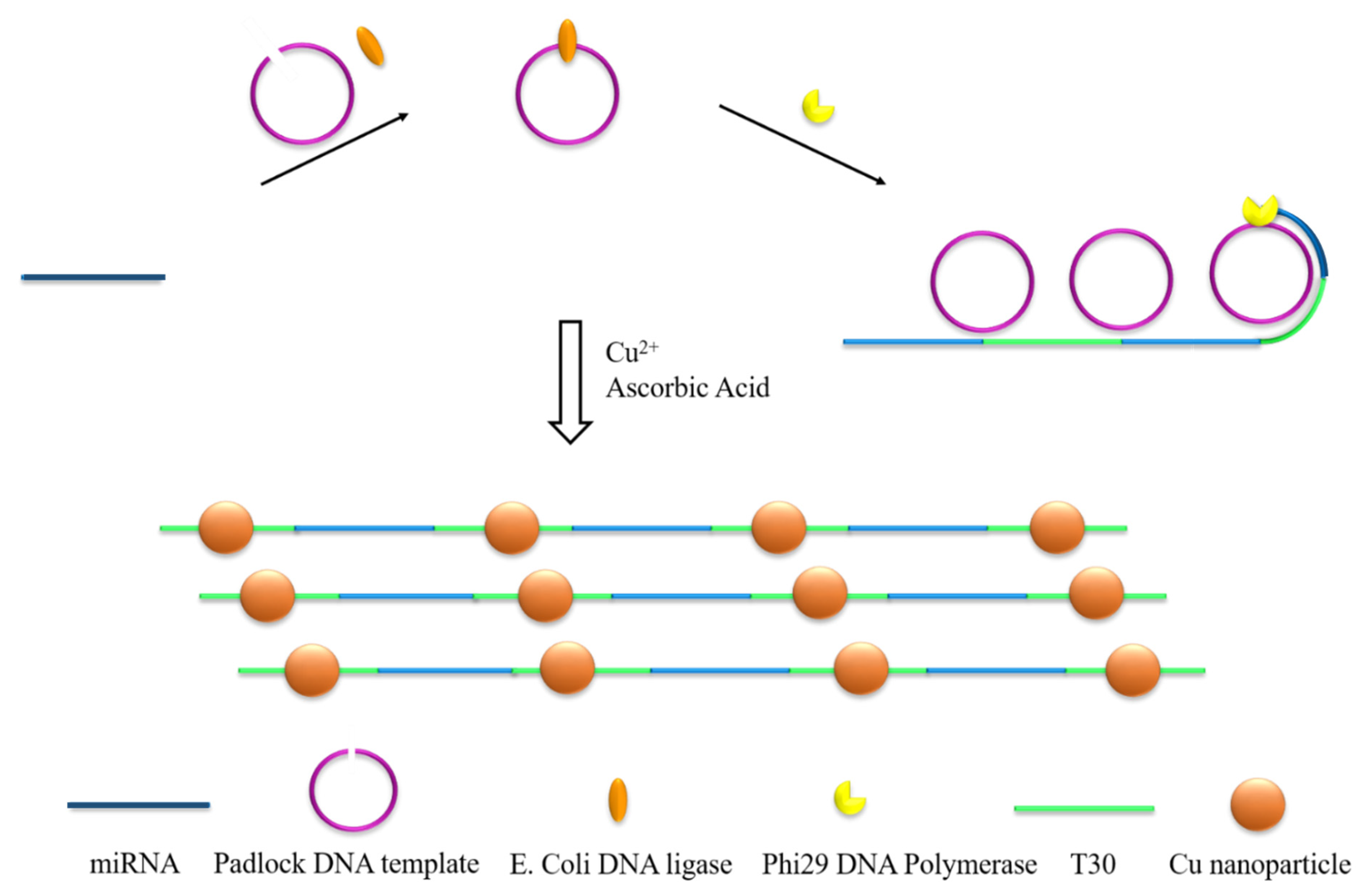
Scheme 1. Illustration of the principle of the label-free microRNA (miRNA) assay. The dark blue lines represent miRNA and the light blue lines represent the corresponding single-stranded DNA (ss-DNA).
Chemosensors 08 00052 sch001
Figure 1. UV-Vis spectra (a) of the poly(thymine) PolyT-templated copper nanoparticles (CuNPs) with 1.0 nM target miRNA (line 2). Fluorescence emission spectra (b) of the rolling circle amplification (RCA)-PolyT-templated CuNPs. CuNPs were formed in dark conditions for 10 min at room temperature.
Chemosensors 08 00052 g001
Figure 2. TEM images (ac) of the PolyT-templated CuNPs (concentrations: [b] = [c] = [a]/10, a: [target miRNA] = 1.0 nM). Atomic force microscopy (AFM) images of the PolyT-templated CuNPs (d,e) and AFM line profiles (f) of red, green, and blue lines in e. CuNPs were formed in dark conditions for 10 min at room temperature.
Chemosensors 08 00052 g002
Figure 3. Fluorescence emission spectra under different conditions: miRNA (1.0 nM) + pDNA + E. coli DNA ligase + Phi29 DNA polymerase + dNTP (line 1); miRNA + pDNA + Phi29 DNA polymerase + dNTP (line 2); miRNA + pDNA + E. coli DNA ligase + dNTP (line 3); miRNA + E. coli DNA ligase + Phi29 DNA polymerase + dNTP (line 4); pDNA + E. coli DNA ligase + Phi29 DNA polymerase + dNTP (line 5). The padlock DNA-based rolling circle amplification (P-RCA) reaction was at 37 ℃ for 5 h.
Chemosensors 08 00052 g003
Figure 4. The performance of the RCA-based nano-biosensor with increasing let-7a miRNA (pM) concentration at the peak of 620 nm; from 1 to 8: (1) 0, (2) 0.1, (3) 1.0, (4) 10, (5) 50, (6) 100, (7) 500, (8) 1000. The padlock DNA-based rolling circle amplification (P-RCA) reaction was at 37 ℃ for 5 h.
Chemosensors 08 00052 g004
Figure 5. The calibration curve of let-7a under optimized reaction conditions. The padlock DNA-based rolling circle amplification (P-RCA) reaction was at 37 ℃ for 5 h, and replicated three times.
Chemosensors 08 00052 g005
Figure 6. Specificity of the PolyT-RCA biosensor with different miRNAs (10 pM). The padlock DNA-based rolling circle amplification (P-RCA) reaction was at 37 ℃ for 5 h.
Chemosensors 08 00052 g006
Table 1. Comparison of reported methods for the detection of miRNA based on fluorescent nanomaterial.
Description Linear RangeDetection LimitDetection MethodTotal Analysis TimeRef.
DNA-templated silver
nanoclusters/hairpin-DNA (AgNCs/HpDNA) and strand-displacement amplification
0 to 0.05 µM0.05 µMFluorescence~6.0 h[6]
ds-DNA-templated copper nanoparticles (CuNPs) and rolling circle amplification (RCA)10 to 400 pM10 pMFluorescence~16.5 h[26]
Silver cluster and rolling circle amplification6 to 300 pM0.84 pMFluorescence~8.5 h[45]
Hybridization chain reaction and DNA-hosted silver nanoclusters3.12 to 50 nM0.78 nMFluorescence~6.5 h[46]
Poly(thymine)-templated CuNPs and enzymatically engineered primer extension1 pM to 1nM100 fMFluorescence~3.5 h[47]
T7 exonuclease-assisted cascade signal amplification and DNA-templated copper nanoparticles0.1 fM to 0.1 pM0.045 fMElectrochemical ~5.0 h[48]
Cascade signal amplification based on CuNPs and reported rolling circle amplification Prostate cancer biomarker
0.05–500 fg/mL
0.020 ± 0.001 fg/mL prostate specific antigen (PSA)Electrochemical ~16.0 h[49]
Target-assisted isothermal exponential amplification (TAIEA) and CuNPs70 fM to 700 nM0.27 fMFluorescence~1.0 h[50]
ss-DNA-templated CuNP strand and rolling circle amplification 100 fM to 1.0 nM70.6 fMFluorescence~5.5 hThis work
Table 2. Summary of miRNA let-7a concentrations in real samples.
Lung Cancer Cells
(106 copies μg−1)
HeLa Cells
(106 copies μg−1)
Normal Cells
(106 copies μg−1)
This assay1.7 ± 0.323.7 ± 0.685.4 ± 0.91
qPCR method1.8 ± 0.303.3 ± 0.805.6 ± 0.80