Article

An Aptamer-Array-Based Sample-to-Answer Biosensor for Ochratoxin A Detection via Fluorescence Resonance Energy Transfer

1
Department of Pharmacy, Fujian Health College, Fuzhou 350101, China
2
Department of Biomedical Engineering, University of Houston, Houston, TX 77204, USA
3
Department of Medical Technology, Fujian Health College, Fuzhou 350101, China
*
Author to whom correspondence should be addressed.
Chemosensors 2021, 9(11), 309; https://doi.org/10.3390/chemosensors9110309
Submission received: 16 September 2021 / Revised: 16 October 2021 / Accepted: 28 October 2021 / Published: 30 October 2021

Abstract

:
Food toxins are a hidden threat that can cause cancer and tremendously impact human health. Therefore, the detection of food toxins in a timely manner with high sensitivity is of paramount importance for public health and food safety. However, the current detection methods are relatively time-consuming and not practical for field tests. In the present work, we developed a novel aptamer-chip-based sample-to-answer biosensor (ACSB) for ochratoxin A (OTA) detection via fluorescence resonance energy transfer (FRET). In this system, a cyanine 3 (Cy3)-labeled OTA-specific biotinylated aptamer was immobilized on an epoxy-coated chip via streptavidin-biotin binding. A complementary DNA strand to OTA aptamer at the 3′-end was labeled with a black hole quencher 2 (BHQ2) to quench Cy3 fluorescence when in proximity. In the presence of OTA, the Cy3-labeled OTA aptamer bound specifically to OTA and led to the physical separation of Cy3 and BHQ2, which resulted in an increase of fluorescence signal. The limit of detection (LOD) of this ACSB for OTA was 0.005 ng/mL with a linearity range of 0.01–10 ng/mL. The cross-reactivity of ACSB against other mycotoxins, ochratoxin B (OTB), aflatoxin B1 (AFB1), zearalenone (ZEA), or deoxynilvalenol (DON), was less than 0.01%. In addition, this system could accurately detect OTA in rice samples spiked with OTA, and the mean recovery rate of the spiked-in OTA reached 91%, with a coefficient of variation (CV) of 8.57–9.89%. Collectively, the ACSB may represent a rapid, accurate, and easy-to-use platform for OTA detection with high sensitivity and specificity.

1. Introduction

Mycotoxins are secondary metabolites of fungi and can cause severe health problems [1]. There are growing concerns of food safety issues due to the contamination of mycotoxins. Ochratoxin A (OTA) is a mycotoxin produced by several fungal species including Aspergillus ochraceus, A. carbonarius, A. niger and Penicillium verrucosum [2]. OTA occurs in a wide variety of commonly contaminated agricultural commodities, such as grains, nuts, spices, coffee beans, olives, grapes, dried fruits, wine, and meat products [3,4]. OTA is notorious for its teratogenicity, embryotoxicity, genotoxicity, neurotoxicity, carcinogenicity, nephrotoxicity, and immunosuppressive effects [5]. OTA can restrain protein synthesis, affect saccharide and calcium metabolisms, increase lipid peroxidation, and disrupt mitochondrial functions. As reviewed by others, OTA exposure could lead to adverse health effects in different populations, such as Balkan endemic nephropathy, chronic interstitial nephropathy, and other renal diseases [2]. The EU commission postulated maximal residue levels (MRLs) for OTA in different food samples were reviewed previously [6] and new standards of 3 µg/kg for cereals and cereal products, 0.2–1 µg/kg for wine, 0.2 µg/kg for beer, 8 µg/kg for green coffee beans, and 4 µg/kg for roasted coffee beans and coffee products as well as a much lower acceptable daily intake (ADI) of 1.5 ng OTA/kg body weight were suggested [6]. Therefore, attention has been focused on the development of a rapid, sensitive, and robust detection system for OTA in contaminated food.
Thus far, several analytical methods have been used for the determination of OTA, such as thin-layer chromatography (TLC) [7], high-performance liquid chromatography (HPLC) [8,9], mass spectrometry (MS) [10,11], and enzyme-linked immunosorbent assay (ELISA) [12]. These traditional detection techniques have been instrumental in the measurement of OTA in laboratory settings. In recent years, several economical and rapid immunoassays such as label-free electrochemical techniques have exhibited reasonable sensitivity and accuracy in the determination of OTA [13,14,15]. Despite these promising attempts, the quantitative capability, reproducibility, sensitivity, ease of use, and robustness remain a challenge, particularly for the purpose of field applications.
Recently there has been great interest in aptamers, artificial antibodies made of specific synthetic DNA or RNA oligonucleotides that can bind to a particular target molecule, such as proteins, peptides, or nucleotides. A technique called sequential evolution of ligands by exponential enrichment (SELEX) screens for highly selective aptamers that bind biomolecules such as antibiotics [16], mycotoxins [17], proteins [18], and even whole cells [19]. There have been many emerging aptamer-based techniques, such as biosensors and assays for the detection of mycotoxins, in the past decade. Aptamers have been used in the determination of mycotoxins such as fumonisin B-1 [20], OTA [21,22], aflatoxin B1 (AFB1) [23], and zearalenone (ZEA) [24]. Due to heterogeneous distribution of mycotoxins in staple crops, the analytical methods needs to have low limit of detection (LOD) and high specificity with low cost to allow for extensive sampling and testing [25]. Microarray-based analytical format is advantageous as it has high throughput, high sensitivity, high specificity, and low reagent consumptions; hence, this technology holds great potential for disease marker detection, toxicological response profiling, and pharmaceutical target screening [26]. Several electrochemical immunoassay-based microarrays have been used for mycotoxin detection [27].
In the present work, a novel aptamer-chip-based sample-to-answer biosensor (ACSB) for OTA detection via fluorescence resonance energy transfer (FRET) was developed. This is a simple, fast, and sensitive OTA assay built on a microarray chip. A biotinylated, Cy3-labeled, OTA-specific aptamer complex 5′-Cy3-aptamer-biotin-3′ and a BHQ2-labeled oligonucleotide complementary to the OTA-specific aptamer sequence were used to construct the ACSB, where FRET will occur or be disrupted on the basis of the absence or presence or OTA, respectively. The ACSB system is a highly sensitive and robust technology that may have great potential for field test of food toxins.

2. Materials and Methods

2.1. Chemicals, Reagents, and Instruments

Streptavidin, OTA, AFB1, ZEA, and deoxynilvalenol (DON) were purchased from Sigma-Aldrich (St. Louis, MO, USA). Ochratoxin B (OTB) was purchased from Santa Cruz Biotechnology Inc. (Dallas, TX, USA). Super G™ Blocking Buffer was purchased from GRACE BIO-LABS Inc. (Bend, OR, USA). OTA aptamer 5′-biotin-GATCGGGTGTGGGTGGCGTAAAGGGAGCATCGGACA-Cy3-3′ and the complementary strand 5′-BHQ2-TGTCCGATGC-3′ [28] were synthesized in bio basic Inc. (Amherst, NY, USA). Streptavidin was diluted with PBS buffer, and then 500 picoliters of the sample was printed onto an epoxy-coated slide with a microarray printing robot (sciFLEXARRAYER S3; Scienion) at 25 °C in an atmosphere of 60% humidity, as described previously [29]. The array detection procedure was according to our previous work [30,31,32,33]. The fluorescence signal on the chip was scanned and analyzed with a GenePix 4000B scanner and a Genepix Pro 7 software (Molecular Devices, San Jose, CA, USA).

2.2. Development of Array Sensor and Streptavidin Coating

An epoxy-modified polymer slide (STRATEC Consumables GmbH) was used for substrate immobilization and subsequent detection as described in our previous work [30,31,32,33]. In this work, streptavidin was used as the first layer on the slide to immobilize biotin-labeled aptamers. Streptavidin was diluted to final concentrations of 1, 2, 3.9, 7.8, 15.6, 31.2, 62.5, 125, 250, 500, 750, and 1000 ng/mL with PBS buffer and printed on an epoxy-coated chip. The chip was incubated for 12 h at 4 °C. Then the chip was blocked with Super G blocking buffer at room temperature for 2 h. The chip was then washed three times with PBS-Tween 20 and then washed with ddH2O. The Cy3-labeled biotinylated OTA aptamer was diluted to 1000 nmol/L with Tris-HCl buffer and 20 μL was added to each well of the chip. After incubation at 25 °C for 1 h, the chip was washed again and air-dried. The fluorescence intensity of the spots on the chip was analyzed with GenePix Pro 7. The optimal streptavidin concentration was determined on the basis of the quality of the spots as indicated by the fluorescence intensity.

2.3. Immobilization and Optimization of OTA Aptamer Concentrations on the Chip

OTA aptamer was diluted to final concentrations of 1, 2, 3.9, 7.8, 15.6, 31.2, 62.5, 125, 250, 500, and 1000 nmol/L with Tris-HCl buffer (pH = 7.5; 120 mmol/L NaCl, 40 mmol/L MgCl2, 20 mmol/L CaCl2) and added into the chamber on the chip where optimal concentration of streptavidin was printed. The chip was incubated at 25 °C on the shaker for 1 h, followed by washing and air-drying of the chip. The fluorescence intensity of the spots on the chip were analyzed to determine the optimal concentration of OTA aptamer.

2.4. Optimization of the Complementary Strand Concentration

The complementary DNA strand of the OTA aptamer was diluted to final concentrations of 1, 2, 3.9, 7.8, 15.6, 31.2, 62.5, 125, 250, 500, and 1000 nmol/L with Tris-HCl buffer (pH = 7.5; 120 mmol/L NaCl, 40 mmol/L MgCl2, 20 mmol/L CaCl2) and added to each well on the chip where optimal concentrations of OTA aptamer was printed. The slide was incubated at 37 °C on the shaker for 1 h, and then washed and air-dried. The slide was then scanned to examine if the Cy3 fluorescence was quenched by BHQ2.

2.5. Standard Curve for Determination of OTA

The OTA standard was dissolved with methanol and diluted with reaction buffer 10 mmol/L HEPES (pH 7.0; 120 mmol/L NaCl, 5 mmol/L KCI, 20 mmol/L MgCl2, 20 mmol/L CaCl2). The OTA standard was diluted to the following concentrations: 0.001 ng/mL, 0.005 ng/mL, 0.01 ng/mL, 0.05 ng/mL, 0.1 ng/mL, 0.5 ng/mL, 1 ng/mL, 5 ng/mL, 10 ng/mL, 50 ng/mL, and 100 ng/mL. Each concentration of OTA standard was added onto the wells on the chip where Cy3 fluorescence was quenched by BHQ2. The slide was kept at 45 °C on the shaker for 30 min. After the reaction, the slide was washed and air-dried for fluorescence scanning with a GenePix 4000B scanner (Molecular Devices, San Jose, CA, USA). The fluorescence signal response to various concentrations of OTA standards was plotted using Graphpad Prism 7.

2.6. Specificity Test

To determine the specificity of this on-chip OTA assay, we used four mycotoxins including OTB, AFB1, ZEA, and DON as controls to determine if they could also be recognized by the OTA-specific ACSB. Four mycotoxins were dissolved in methanol and diluted to 0.1 ng/mL, 1 ng/mL, and 10 ng/mL with reaction buffer, and added into the well of the OTA assay chip where Cy3-labeled OTA aptamer was reacted with BHQ2 probe as described above.

2.7. Recovery Test of OTA in Artificially Contaminated Rice Samples

Rice was selected as a matrix for testing the performance of the on-chip OTA assay. Rice was milled into particles less than 1 mm. Analytical grade OTA (Sigma-Aldrich, St. Louis, MO, USA) was spiked into an aliquot of 1 g rice sample to give final OTA concentrations of 1, 10, and 100 ng/g. Each of these samples was mixed with 10 mL of methanol, and the entire mixture was vortexed for 5 min, sonicated for 30 min, and then centrifuged at 10,000× g for 10 min. The supernatant was collected and filtrated with a 0.45 μm filter membrane. Blank controls were also prepared in the same way as above except that methanol only was used instead of the OTA. The OTA in the extract was diluted with HEPES buffer prior to the assay. OTA levels in these samples were also measured using an OTA ELISA KIT purchased from REAGEN Inc. (Haddon Township, NJ, USA) according to the user’s manual. A SpectraMax® M3 Multi-Mode Microplate Reader (Molecular Devices, San Jose, CA, USA) was used to detect the signals of ELISA.

3. Results

To construct the ACSB system, as illustrated in Figure 1, we first biotinylated the OTA-specific aptamer for effective immobilization of the aptamer on the chip, maintaining the natural structure of the aptamer without comprising its ability for OTA binding. Streptavidin was printed on the chip and then was bound by 5′-Cy3-aptamer-biotin-3′. At this step, the Cy3 could fluoresce. Upon the addition of a BHQ2-labeled oligonucleotide, which is partially complementary to the OTA aptamer, the fluorescence of Cy3 was quenched by BHQ2 due to the proximity of the two labels through aptamer-DNA probe binding. However, in the presence of OTA, Cy3-labeled OTA aptamer could bind specifically to OTA, leading to the structural change of the aptamer and the subsequent separation of the BHQ2 from Cy3, which results in the recovery of the fluorescence signal of Cy3.

3.1. Streptavidin Coating and Optimization of Streptavidin Concentration

According to the design of our ACSB system, the OTA-specific aptamer was immobilized onto the chip via biotin-streptavidin binding. Biotin was conjugated onto the aptamer, and streptavidin was printed on the chip. To determine the optimal concentration of streptavidin needed to immobilize sufficient OTA-specific aptamer on the chip, we prepared various concentrations of streptavidin and printed them on the chip to determine which concentration of streptavidin could give the best fluorescence signal. Excessive 5′-Cy3-aptamer-biotin-3′ was added onto the chip to react with various concentrations of streptavidin. As shown in Figure 2, the mean fluorescence intensity was increased with the increase of the streptavidin concentration printed on the chip. The mean fluorescence intensity could reach a maximum of 60,000 arbitrary units (a.u.) when the streptavidin concentration was around 500 ng/mL. The mean fluorescence intensity did not change much when streptavidin concentration was higher than 500 ng/mL. This suggests that the optimal streptavidin concentration is 500 ng/mL in order to immobilize sufficient amount of the OTA-specific aptamer in the development of the ACSB.

3.2. Immobilization of Cy3-Labled OTA Aptamer on the Chip and Optimization of OTA Aptamer Concentration

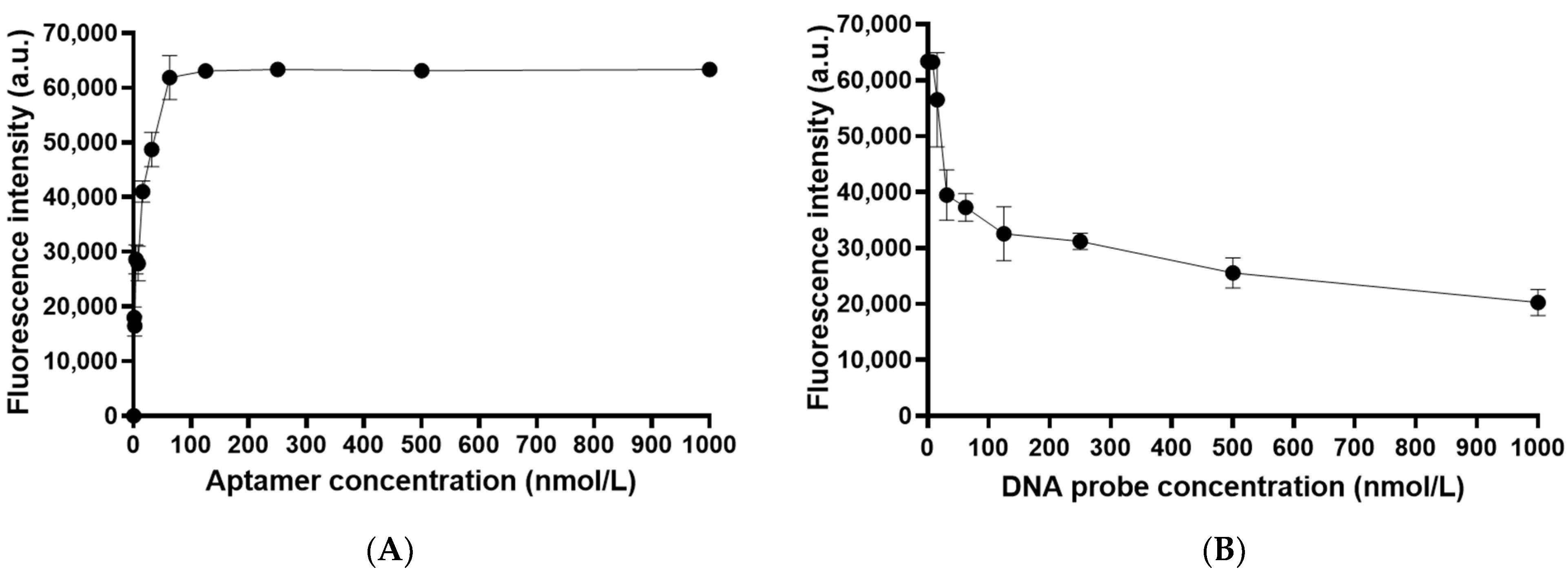
Next, the response of the fluorescence signal of this system to various concentrations of Cy3-labeled OTA-specific aptamer was tested to determine the optimal aptamer concentration that gives the best signal. The OTA-specific aptamer was diluted into different concentrations and added to different subarray wells on the chip for incubation at 25 °C for 1 h. After the scanning of the chip, the fluorescence signal was quantified using Genepix Pro 7 software (Molecular Devices, LLC, San Jose, CA, USA). As shown in Figure 3A, the mean fluorescence values were increased with the increase of the OTA aptamer concentration. At the aptamer concentration of 150 nM, the mean fluorescence intensity reached a plateau. Hence, in order to achieve the best signal in the development of the ACSB, the optimal aptamer concentration was found to be 150 nM.

3.3. Optimization of the Concentration of BHQ2-Labeled DNA Probe

In this study, the ACSB was designed for one-step detection of OTA. On the chip, a pre-coated Cy3-labeled OTA-specific aptamer is bound by a BHQ2-labeled DNA probe via complementary binding, which leads to the quenching of Cy3 fluorescence via FRET, and this will establish a low fluorescence baseline F0. F0 is then used as a starting point for the one-step assay, where an increased fluorescence Fa will be generated upon OTA sample addition as assay readout. Therefore, the concentration of the DNA probe is critical in establishing the baseline of the ACSB assay. The DNA probe was diluted into 1, 2, 3.9, 7.8, 15.6, 31.2, 62.5, 125, 250, 500, and 1000 nM with Tris-HCl buffer (pH = 7.5; 120 mM NaCl, 40 mM MgCl2, 20 mM CaCl2). A total of 20 μL of each dilution was added into each well on the ACSB chip. After FRET reaction, the fluorescence intensity of each spot on the chip was analyzed with GenePix Pro 7 software. As shown in Figure 3B, the mean fluorescence intensity was decreased with the increase of the concentration of DNA probe. The mean fluorescence intensity was lower than 25,000 a.u. when the DNA probe concentration was higher than 500 nM. This indicates that the fluorescence of the Cy3-labeld aptamer was quenched in the presence of 500 nM DNA probe, which was labeled with BHQ2. Only a minor decrease of the mean fluorescence intensity was observed when the DNA probe was further increased. This suggests that the optimal concentration of the DNA probe is around 500 nM.

3.4. Detectability and Sensitivity of ACSB for Ochratoxin A (OTA)

Next, the capability of the ACSB to detect OTA quantitatively via fluorescence resonance energy transfer (FRET) was examined. The entire experiments were executed via a signal “on–off–on” process as shown in Figure 4A. First, 9 × 4 = 36 spot replicates of Cy3-labeled biotinylated aptamer were printed on the streptavidin-coated chip where the fluorescence signal was “on”. Upon addition of the quencher BHQ2-labeled DNA probe on the chip, the fluorescence was quenched and the signal was “off”; however, upon further addition of OTA (the analyte), the OTA–aptamer binding induced the separation between Cy3 and BHQ2, which led to the signal “on” again. The fluorescence signals from the above “on–off–on” process were quantified using Genepix Pro7 (Figure 4B). OTA dose-dependent response curve was established using various concentrations of OTA (X-axis) and fluorescence increase (Y-axis) upon the addition of OTA (Figure 4C). To prepare various concentrations of OTA, we diluted the OTA standard to 0.001 ng/mL, 0.005 ng/mL, 0.01 ng/mL, 0.05 ng/mL, 0.1 ng/mL, 0.5 ng/mL, 1 ng/mL, 5 ng/mL, 10 ng/mL, 50 ng/mL, and 100 ng/mL. A total of 20 μL of each concentration was added into each well on the chip to start the FRET reaction. As shown on the Figure 4C, the mean fluorescence intensity was increased with the increase of the OTA concentration. The trend was reflected in a dose-dependent manner in the recovery of fluorescence signals. There was a good linearity ranging from 0.01 to 10 ng/mL with a LOD of 0.005 ng/mL for OTA.

3.5. Specificity of ACSB in Detecting OTA

To investigate the specificity of the ACSB, we used four mycotoxins, namely, OTB, AFB1, ZEA, and DON determine if there was any cross-reactivity with the sensor. All mycotoxins were dissolved with methanol and diluted to 0.1 ng/mL, 1 ng/mL, or 10 ng/mL with reaction buffer, and 20 μL of each mycotoxin standard was added into the well of the chip. The measurement steps were the same as described above. The detection and analysis results are shown in Figure 5.
As shown in Figure 5, the mean fluorescence intensity was lower than 1000 a.u. when the concentration of AFB1, ZEA, or DON was at 10 ng/mL or lower. The mean fluorescence intensity was lower than 2000 a.u. when OTB was at 10 ng/mL or lower, which was far below the mean fluorescence intensity of 40,000 a.u. when OTA was at 0.1~10 ng/mL. These results indicate that there was barely any cross-reactivity of the OTA sensor in detecting other mycotoxins including OTB.

3.6. Recovery Test of OTA in Artificially Contaminated Rice Samples

Rice was selected as a matrix for testing the performance of ACSB in detecting OTA. Rice samples were milled and artificially contaminated with OTA (dissolved in methanol) to give final concentrations of 1, 10, or 100 ng/g. The recovery of OTA from artificially contaminated rice is shown in Table 1.
As shown in Table 1, the ACSB exhibited high levels of recovery of OTA spiked into rice samples. Mean recoveries were 91%, 90%, and 85% for OTA spiked-in with 1, 10, and 100 ng/g, respectively. The systematic error was relatively low with a coefficient of variation (CV) ranging from 8.57 to 9.89%. The same three sets of samples (each with three replicates) were also analyzed with a commercial ELISA kit to compare with the results obtained using ACSB. The correlation between ELISA data and ACSB data was analyzed using Pearson test, and the correlation coefficient r = 0.99 and p-value < 0.0001 were found. This suggests that the results obtained using ACSB method are highly consistent with the conventional ELISA measurement.

4. Discussion

In the present study, an aptamer-based sensor technology for OTA detection on a microarray/chip platform was developed. The microarray/chip-based technology provides superior sensitivity; minimum sample/reagent requirement; and, most importantly, the preprinted target-capture module and ready-to-use interface for reaction. Microarrays have been widely applied in basic biological research and clinical diagnosis, such as protein interaction studies [34], immune profiling [35], vaccine development [36], biomarker discovery [37], disease diagnostics [38], and drug screening and discovery [39], among others. Microarray technologies have been used for mycotoxin detection due to their sensitivity [40]. Electrochemical immunoassay-based microarrays have also been used for mycotoxin detection with promising sensitivity, selectivity, and simplicity in use [27].
In a surface plasmonic resonance-based multiplex microimmunoassay for the detection of DON and ZEA via a competitive inhibition immunoassay format, LODs of 84 and 68 μg/kg for DON and 64 and 40 μg/kg for ZEA in maize and wheat samples were achieved, respectively [41]. In a microarray for screening OTA in green coffee extract on a fully automated flow-through device via over 20 assay-regeneration cycles, a reduced assay time (within 12 min) with a limit of quantitation (LOQ) of OTA of 0.3 μg/L was achieved [42]. In an indirect competitive immunoassay on regenerable, reusable glass microchips for the parallel determination of aflatoxins, ochratoxin A, deoxynivalenol, and fumonisin B1 in oat extracts, a fully automated flow-through device with chemiluminescence readout was achieved within 19 min [43,44]. In a surface-enhanced Raman scattering (SERS)-based immunosensor for the detection of three mycotoxins (AFB1, ZEA, OTA) in food on a single gold chip, a LOD of 0.061–0.066 μg/kg for AFB1, 0.53–0.57 μg/kg for ZEA, and 0.26–0.29 μg/kg for OTA in foodstuff was established [45].
However, immunoassays for mycotoxins require artificial mycotoxin antigens and antibodies that need complicated production processes and relatively stringent storage conditions. Moreover, different batches of antibodies exhibited different titers in antigen binding that may affect the performance of the sensor and the accuracy of detection. In comparison to antibodies, aptamers are more chemically stable under most environmental conditions with a longer shelf life and can be reversibly denatured without loss of specificity [25]. By the virtues of simplicity, rapidness, and low-cost in assays, aptamer-based biosensors are more promising in detection of various mycotoxins with high sensitivity and selectivity compared to antibody-based methods [17].
Thus far, a few aptamer-based microarray methods have been reported in the detection of mycotoxins. In an aptamer microarray on the TiO2-porous silicon (PSi) surface for simultaneous screening of multiple mycotoxins, a dynamic linear detection range of 0.1–10 ng/mL for OTA, 0.01–10 ng/mL for AFB1, and 0.001–10 ng/mL for FB1, and a LOD of 15.4, 1.48, and 0.21 pg/mL for OTA, AFB1, and FB1, respectively, was achieved [46]. In a high-throughput photonic crystal microsphere (PHCM) suspension array for multiplexing mycotoxins detection in cereal samples, a dynamic linear detection range of 0.1 pg/mL to 0.1 ng/mL for AFB1/OTA and 0.1–10 ng/mL for FB1, was achieved [47]. Given the relatively tedious fabrication procedures of these assays, apparently there is a long way to go before developing them into easy-to-repeat and test-friendly devices [48,49].
The novelty of this work lies in the following: (1) the simple and straightforward fabrication process of the aptamer chip. Any commercial microarray slide can be directly used, and the printing of substrates and aptamer takes under 10 min. (2) High reproducibility. As shown in Figure 4, each test site can be integrated with multiple replicates for high reproducibility. (3) Capability of sample-to-answer detection. With a preprinted chip, the entire assay can be performed in one step. Therefore, this biosensor allows for simple, fast, and sensitive detection of OTA through FRET mechanism. Although this is only a proof-of-concept study where the signal “on–off–on” process was demonstrated, the sensor indeed is capable of detecting OTA in one step, given the fact that the Cy3-labeled OTA aptamer can be preprinted and the BHQ2-labeled DNA probe can also be preprinted or added on top of the aptamer layer, prior to detection. In other words, this sensor can be premade and is ready to use upon the addition of a “real” sample for OTA detection in one step. Therefore, this device is indeed a sample-to-answer device by design. In laboratory settings, this sensor allows for establishing a standard curve and performing sample detection in a quantitative manner; however, in field settings, this sensor allows for a fast, easy, sample-to-answer assay of food toxin in a qualitative way by reporting “yes” or “no” toxin in a contaminated food sample. This ACSB exhibited comparable sensitivity and specificity in OTA detection compared to most sensors as discussed above. Compared to label-free approaches such as SERS or electrochemical sensors, ACSB may have greater potentials for repeated measurements in improving accuracy or towards the development of one-shot, high-throughput, and multiplexing assays. The advantage of FRET lies in the fact that it establishes a clear baseline of readout at each step so that the assay is more robust. Future work will focus on the improvement of the affinity of target-specific aptamer as well as the sensitivity of the FRET components and testing of the multiplexing capability of this sensor.

5. Conclusions

In conclusion, a simple, sensitive, and highly selective on-chip biosensor was developed with great potential in sample-to-answer detection of OTA in food samples. This array format enables further development of high-throughput and multiplexing biosensors that allow for simultaneous detection of multiple mycotoxins or biomarkers in multiple samples.

Author Contributions

T.W. and Y.L. (Yongning Li) conceived and designed this study; Y.L. (Yongning Li), Y.L. (Yaxi Li), M.X., G.T., W.W., Y.W., M.F., S.Z., C.T. and B.Y. performed the experiments, collected the data, and analyzed the data; Y.L. (Yongning Li), Z.P. and T.W. wrote and revised the manuscript. All authors have read and agreed to the published version of the manuscript.

Funding

This research received no external funding.

Institutional Review Board Statement

Not applicable.

Informed Consent Statement

Not applicable.

Data Availability Statement

Not applicable.

Acknowledgments

Y.N.L. was partly supported by a visiting scholar program of Outstanding Professional Leader of Colleges and Universities, Fujian.

Conflicts of Interest

The authors declare no conflict of interest.

References

  1. Bennett, J.W.; Klich, M. Mycotoxins. Clin. Microbiol. Rev. 2003, 16, 497–516. [Google Scholar] [CrossRef] [PubMed] [Green Version]
  2. Bui-Klimke, T.R.; Wu, F. Ochratoxin A and human health risk: A review of the evidence. Crit. Rev. Food Sci. Nutr. 2015, 55, 1860–1869. [Google Scholar] [CrossRef] [Green Version]
  3. Duarte, S.C.; Pena, A.; Lino, C.M. A review on ochratoxin A occurrence and effects of processing of cereal and cereal derived food products. Food. Microbiol. 2010, 27, 187–198. [Google Scholar] [CrossRef]
  4. Covarelli, L.; Beccari, G.; Marini, A.; Tosi, L. A review on the occurrence and control of ochratoxigenic fungal species and ochratoxin A in dehydrated grapes, non-fortified dessert wines and dried vine fruit in the Mediterranean area. Food Control 2012, 26, 347–356. [Google Scholar] [CrossRef]
  5. JECFA. Safety Evaluation f Certain Mycotoxins in Food—Prepared by the 56th Meeting of the JECFA (Joint FAO/WHO Expert Committee on Food Additives)—FAO Food and Nutrition Paper 74/WHO Foods Additives Series 47. 2001. Available online: https://inchem.org/documents/jecfa/jecmono/v47je01.htm (accessed on 16 September 2021).
  6. Petzinger, E.; Ziegler, K. Ochratoxin A from a toxicological perspective. J. Vet. Pharmacol. Ther. 2000, 23, 91–98. [Google Scholar] [CrossRef]
  7. Santos, E.A.; Vargas, E.A. Immunoaffinity column clean-up and thin layer chromatography for determination of ochratoxin A in green coffee. Food Addit. Contam. 2002, 19, 447–458. [Google Scholar] [CrossRef]
  8. Degelmann, P.; Becker, M.; Herderich, M.; Humpf, H.U. Determination of ochratoxin A in beer by high-performance liquid chromatography. Chromatographia 1999, 49, 543–546. [Google Scholar] [CrossRef]
  9. Tessini, C.; Mardones, C.; von Baer, D.; Vega, M.; Herlitz, E.; Saelzer, R.; Silva, J.; Torres, O. Alternatives for sample pre-treatment and HPLC determination of Ochratoxin A in red wine using fluorescence detection. Anal. Chim. Acta 2010, 660, 119–126. [Google Scholar] [CrossRef] [PubMed]
  10. Reinsch, M.; Topfer, A.; Lehmann, A.; Nehls, I.; Panne, U. Determination of ochratoxin A in beer by LC-MS/MS ion trap detection. Food Chem. 2007, 100, 312–317. [Google Scholar] [CrossRef]
  11. Olsson, J.; Borjesson, T.; Lundstedt, T.; Schnurer, J. Detection and quantification of ochratoxin A and deoxynivalenol in barley grains by GC-MS and electronic nose. Int. J. Food Microbiol. 2002, 72, 203–214. [Google Scholar] [CrossRef]
  12. Yu, F.Y.; Chi, T.F.; Liu, B.H.; Su, C.C. Development of a sensitive enzyme-linked immunosorbent assay for the determination of ochratoxin A. J. Agric. Food Chem. 2005, 53, 6947–6953. [Google Scholar] [CrossRef]
  13. Wei, M.; Wang, C.L.; Xu, E.S.; Chen, J.; Xu, X.L.; Wei, W.; Liu, S.Q. A simple and sensitive electrochemiluminescence aptasensor for determination of ochratoxin A based on a nicking endonuclease-powered DNA walking machine. Food Chem. 2019, 282, 141–146. [Google Scholar] [CrossRef] [PubMed]
  14. Kaur, N.; Bharti, A.; Batra, S.; Rana, S.; Rana, S.; Bhalla, A.; Prabhakar, N. An electrochemical aptasensor based on graphene doped chitosan nanocomposites for determination of Ochratoxin A. Microchem. J. 2019, 144, 102–109. [Google Scholar] [CrossRef]
  15. Wei, M.; Zhang, W.Y. The determination of Ochratoxin A based on the electrochemical aptasensor by carbon aerogels and methylene blue assisted signal amplification. Chem. Cent. J. 2018, 12, 45. [Google Scholar] [CrossRef] [Green Version]
  16. Zhou, L.; Gan, N.; Zhou, Y.; Li, T.; Cao, Y.; Chen, Y. A label-free and universal platform for antibiotics detection based on microchip electrophoresis using aptamer probes. Talanta 2017, 167, 544–549. [Google Scholar] [CrossRef]
  17. Guo, X.; Wen, F.; Zheng, N.; Saive, M.; Fauconnier, M.L.; Wang, J. Aptamer-Based Biosensor for Detection of Mycotoxins. Front. Chem. 2020, 8, 195. [Google Scholar] [CrossRef] [PubMed] [Green Version]
  18. Jung, Y.; Lee, C.Y.; Park, K.S.; Park, H.G. Sensitive and specific detection of proteins based on target-responsive DNA polymerase activity. Anal. Chim. Acta 2019, 1059, 80–85. [Google Scholar] [CrossRef] [PubMed]
  19. Chen, M.; Yu, Y.; Jiang, F.; Zhou, J.; Li, Y.; Liang, C.; Dang, L.; Lu, A.; Zhang, G. Development of Cell-SELEX Technology and Its Application in Cancer Diagnosis and Therapy. Int. J. Mol. Sci. 2016, 17, 2079. [Google Scholar] [CrossRef] [Green Version]
  20. Chen, X.J.; Bai, X.J.; Li, H.Y.; Zhang, B.L. Aptamer-based microcantilever array biosensor for detection of fumonisin B-1. RSC Adv. 2015, 5, 35448–35452. [Google Scholar] [CrossRef]
  21. Costantini, F.; Sberna, C.; Petrucci, G.; Reverberi, M.; Domenici, F.; Fanelli, C.; Manetti, C.; de Cesare, G.; DeRosa, M.; Nascetti, A.; et al. Aptamer-based sandwich assay for on chip detection of Ochratoxin A by an array of amorphous silicon photosensors. Sens. Actuators B Chem. 2016, 230, 31–39. [Google Scholar] [CrossRef]
  22. Cruz-Aguado, J.A.; Penner, G. Determination of Ochratoxin A with a DNA Aptamer. J. Agric. Food Chem. 2008, 56, 10456–10461. [Google Scholar] [CrossRef]
  23. Chen, L.; Wen, F.; Li, M.; Guo, X.; Li, S.; Zheng, N.; Wang, J. A simple aptamer-based fluorescent assay for the detection of Aflatoxin B1 in infant rice cereal. Food Chem. 2017, 215, 377–382. [Google Scholar] [CrossRef] [PubMed]
  24. Goud, K.Y.; Hayat, A.; Satyanarayana, M.; Kumar, V.S.; Catanante, G.; Gobi, K.V.; Marty, J.L. Aptamer-based zearalenone assay based on the use of a fluorescein label and a functional graphene oxide as a quencher. Microchim. Acta 2017, 184, 4401–4408. [Google Scholar] [CrossRef]
  25. Ruscito, A.; Smith, M.; Goudreau, D.N.; DeRosa, M.C. Current Status and Future Prospects for Aptamer-Based Mycotoxin Detection. J. AOAC Int. 2016, 99, 865–877. [Google Scholar] [CrossRef]
  26. Bertone, P.; Snyder, M. Advances in functional protein microarray technology. FEBS J. 2005, 272, 5400–5411. [Google Scholar] [CrossRef] [PubMed]
  27. Zhang, Z.; Li, P.; Hu, X.; Zhang, Q.; Ding, X.; Zhang, W. Microarray technology for major chemical contaminants analysis in food: Current status and prospects. Sensors 2012, 12, 9234–9252. [Google Scholar] [CrossRef] [Green Version]
  28. Zhao, H.; Xiang, X.Y.; Chen, M.J.; Ma, C.B. Aptamer-Based Fluorometric Ochratoxin A Assay Based on Photoinduced Electron Transfer. Toxins 2019, 11, 65. [Google Scholar] [CrossRef] [PubMed] [Green Version]
  29. Yuan, Y.; Wang, H.; Lin, Z.-T.; Hong, X.; Heon, M.; Wu, T. Protein arrays II: Antigen arrays. In Functional Genomics; Springer: Berlin/Heidelberg, Germany, 2017; pp. 271–277. [Google Scholar]
  30. Wu, T.; Du, Y.; Han, J.; Singh, S.; Xie, C.; Guo, Y.; Zhou, X.J.; Ahn, C.; Saxena, R.; Mohan, C. Urinary angiostatin—A novel putative marker of renal pathology chronicity in lupus nephritis. Mol. Cell. Proteom. 2013, 12, 1170–1179. [Google Scholar] [CrossRef] [Green Version]
  31. Yuan, Y.; Qiu, J.; Lin, Z.T.; Li, W.; Haley, C.; Mui, U.N.; Ning, J.; Tyring, S.K.; Wu, T. Identification of Novel Autoantibodies Associated with Psoriatic Arthritis. Arthritis Rheumatol. 2019, 71, 941–951. [Google Scholar] [CrossRef]
  32. Qiu, J.; Yuan, Y.; Li, Y.; Haley, C.; Mui, U.N.; Swali, R.; Mohan, C.; Tyring, S.K.; Wu, T. Discovery of IgG4 Anti-Gliadin Autoantibody as a Potential Biomarker of Psoriasis Using an Autoantigen Array. Proteom. Clin. Appl. 2019, e1800114. [Google Scholar] [CrossRef] [PubMed]
  33. Yuan, Y.; Lin, Z.-T.; Wang, H.; Hong, X.; Heon, M.; Wu, T. Protein arrays I: Antibody arrays. In Functional Genomics; Springer: Berlin/Heidelberg, Germany, 2017; pp. 261–269. [Google Scholar]
  34. Bulyk, M.L. DNA microarray technologies for measuring protein-DNA interactions. Curr. Opin. Biotechnol. 2006, 17, 422–430. [Google Scholar] [CrossRef] [Green Version]
  35. Liang, L.; Juarez, S.; Nga, T.V.; Dunstan, S.; Nakajima-Sasaki, R.; Davies, D.H.; McSorley, S.; Baker, S.; Felgner, P.L. Immune profiling with a Salmonella Typhi antigen microarray identifies new diagnostic biomarkers of human typhoid. Sci. Rep. 2013, 3, 1043. [Google Scholar] [CrossRef]
  36. Monsurro, V.; Marincola, F.M. Microarray analysis for a comprehensive immunological-status evaluation during cancer vaccine immune monitoring. J. Biomed. Biotechnol. 2011, 2011, 307297. [Google Scholar] [CrossRef] [PubMed]
  37. Dang, K.; Zhang, W.; Jiang, S.; Lin, X.; Qian, A. Application of Lectin Microarrays for Biomarker Discovery. ChemistryOpen 2020, 9, 285–300. [Google Scholar] [CrossRef]
  38. Balashanmugam, M.V.; Shivanandappa, T.B.; Nagarethinam, S.; Vastrad, B.; Vastrad, C. Analysis of Differentially Expressed Genes in Coronary Artery Disease by Integrated Microarray Analysis. Biomolecules 2019, 10, 35. [Google Scholar] [CrossRef] [Green Version]
  39. Schumacher, S.; Muekusch, S.; Seitz, H. Up-to-Date Applications of Microarrays and Their Way to Commercialization. Microarrays 2015, 4, 196–213. [Google Scholar] [CrossRef] [PubMed] [Green Version]
  40. Logrieco, A.; Arrigan, D.W.; Brengel-Pesce, K.; Siciliano, P.; Tothill, I. DNA arrays, electronic noses and tongues, biosensors and receptors for rapid detection of toxigenic fungi and mycotoxins: A review. Food Addit. Contam. 2005, 22, 335–344. [Google Scholar] [CrossRef]
  41. Dorokhin, D.; Haasnoot, W.; Franssen, M.C.; Zuilhof, H.; Nielen, M.W. Imaging surface plasmon resonance for multiplex microassay sensing of mycotoxins. Anal. Bioanal. Chem. 2011, 400, 3005–3011. [Google Scholar] [CrossRef] [Green Version]
  42. Sauceda-Friebe, J.C.; Karsunke, X.Y.; Vazac, S.; Biselli, S.; Niessner, R.; Knopp, D. Regenerable immuno-biochip for screening ochratoxin A in green coffee extract using an automated microarray chip reader with chemiluminescence detection. Anal. Chim. Acta 2011, 689, 234–242. [Google Scholar] [CrossRef]
  43. Oswald, S.; Karsunke, X.Y.; Dietrich, R.; Martlbauer, E.; Niessner, R.; Knopp, D. Automated regenerable microarray-based immunoassay for rapid parallel quantification of mycotoxins in cereals. Anal. Bioanal. Chem. 2013, 405, 6405–6415. [Google Scholar] [CrossRef]
  44. Oswald, S.; Dietrich, R.; Martlbauer, E.; Niessner, R.; Knopp, D. Microarray-Based Immunoassay for Parallel Quantification of Multiple Mycotoxins in Oat. Methods Mol. Biol. 2017, 1536, 143–156. [Google Scholar] [CrossRef] [PubMed]
  45. Li, Y.; Chen, Q.; Xu, X.; Jin, Y.; Wang, Y.; Zhang, L.; Yang, W.; He, L.; Feng, X.; Chen, Y. Microarray surface enhanced Raman scattering based immunosensor for multiplexing detection of mycotoxin in foodstuff. Sens. Actuators B Chem. 2018, 266, 115–123. [Google Scholar] [CrossRef]
  46. Liu, R.; Li, W.; Cai, T.; Deng, Y.; Ding, Z.; Liu, Y.; Zhu, X.; Wang, X.; Liu, J.; Liang, B.; et al. TiO2 Nanolayer-Enhanced Fluorescence for Simultaneous Multiplex Mycotoxin Detection by Aptamer Microarrays on a Porous Silicon Surface. ACS Appl. Mater. Interfaces 2018, 10, 14447–14453. [Google Scholar] [CrossRef] [PubMed]
  47. Yang, Y.; Li, W.; Shen, P.; Liu, R.; Li, Y.; Xu, J.; Zheng, Q.; Zhang, Y.; Li, J.; Zheng, T. Aptamer fluorescence signal recovery screening for multiplex mycotoxins in cereal samples based on photonic crystal microsphere suspension array. Sens. Actuators B Chem. 2017, 248, 351–358. [Google Scholar] [CrossRef]
  48. Rasooly, A.; Herold, K.E. Food microbial pathogen detection and analysis using DNA microarray technologies. Foodborne Pathog. Dis. 2008, 5, 531–550. [Google Scholar] [CrossRef] [PubMed] [Green Version]
  49. Arora, P.; Sindhu, A.; Dilbaghi, N.; Chaudhury, A. Biosensors as innovative tools for the detection of food borne pathogens. Biosens. Bioelectron. 2011, 28, 1–12. [Google Scholar] [CrossRef] [PubMed]
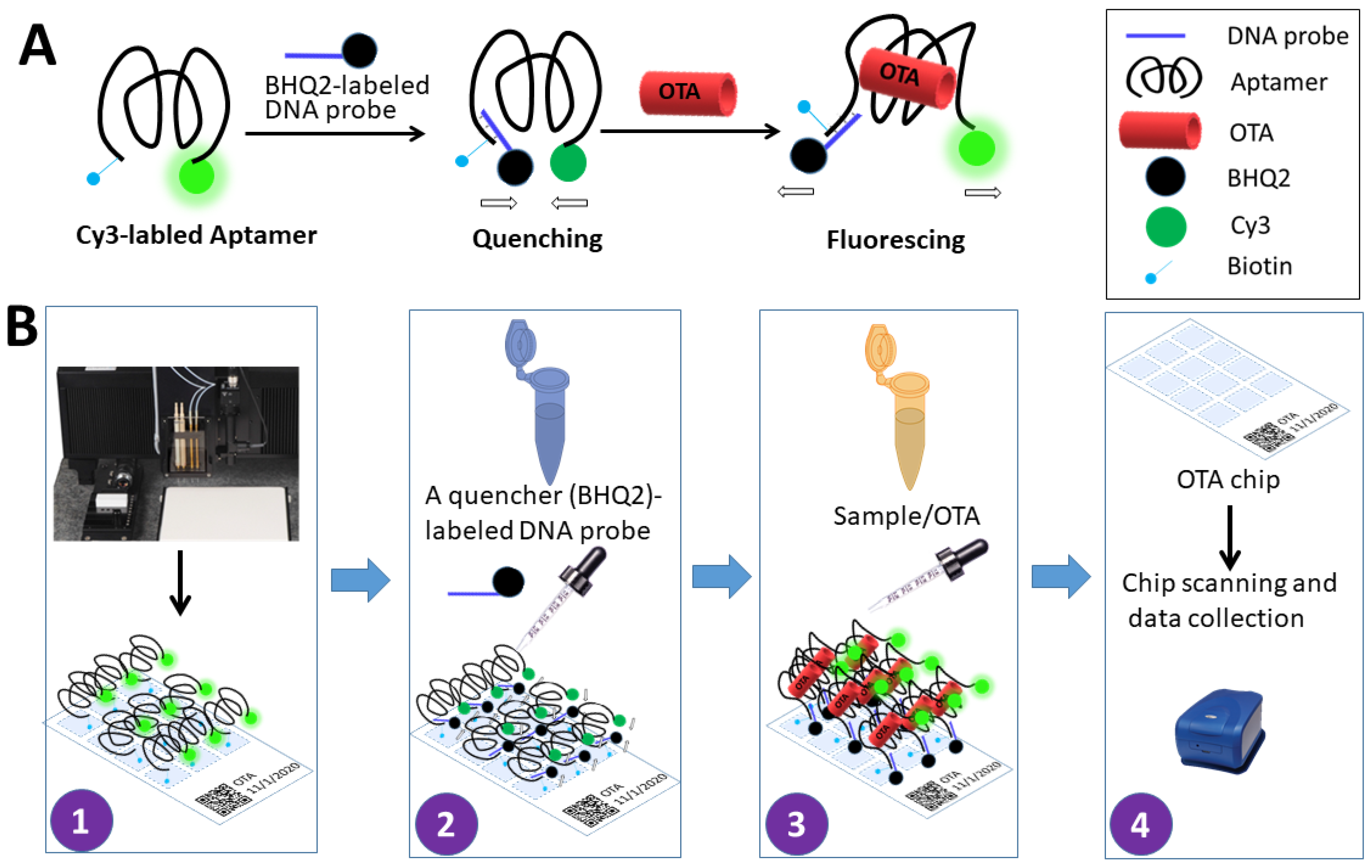
Figure 1. Detection of ochratoxin A on an aptamer-based chip via fluorescence resonance energy transfer. When bound by a BHQ2-labeled oligonucleotide complementary to the OTA-specific aptamer sequence, the fluorescence of the Cy3-labeled OTA aptamer was quenched via fluorescence resonance energy transfer (FRET). The subsequent OTA binding to the aptamer caused the separation of the fluorophore Cy3 from the quencher BHQ2, which led to the generation of fluorescence signals (A). This detection system could be built on a chip and the signal can be analyzed using a fluorescence scanner (B). (1) Printing Cy3-labeled OTA aptamer on the chip; (2) Adding a quencher (BHQ2)-labled DNA probe onto the chip; (3) Adding OTA samples on the chip; (4) Chip scanning and data collection.
Chemosensors 09 00309 g001
Figure 2. The response of fluorescence intensity to various concentrations of streptavidin in a dose-dependent manner. Various concentrations of streptavidin were printed on the chip and incubated with Cy3-labeled OTA-specific aptamers for the detection of fluorescence signal.
Chemosensors 09 00309 g002
Figure 3. Optimization of the Cy3-labeled OTA-specific aptamer concentration in the development of ACSB (A). Fluorescence signal response was analyzed at various concentrations of OTA-specific aptamer. Optimization of the concentration of the BHQ2-labeled DNA probe for the construction of ACSB (B). Fluorescence signal response was analyzed at various concentrations of DNA probe used to achieve FRET-induced fluorescence quenching of the Cy3 labeled-OTA aptamer.
Chemosensors 09 00309 g003
Figure 4. Aptamer-chip-based sample-to-answer biosensor (ACSB) for ochratoxin A (OTA) detection via fluorescence resonance energy transfer (FRET). Signal on–off–on process is shown in (A): Cy3-labeled biotinylated aptamer was printed on the streptavidin-coated chip (in 9 × 4 = 36 spot replicates) where the fluorescence signal was on. Upon addition of the quencher BHQ2-labeled DNA probe on the chip, the fluorescence was quenched and the signal was off; however, upon further addition of OTA (the analyte), the OTA–aptamer binding induced the separation between Cy3 and BHQ2, which led to the signal-on again. The fluorescence signals from the above on–off–on process were quantified using Genepix Pro7 (B). OTA dose-dependent response curve was established using various concentrations of OTA (X-axis, log10 scale) and fluorescence increase (Y-axis) upon the addition of OTA (C).
Chemosensors 09 00309 g004
Figure 5. The specificity of the OTA aptamer-based ACSB system in detecting OTA. Four mycotoxins, namely, OTB, AFB1, ZEA, and DON, at 0.1 ng/mL, 1 ng/mL, or 10 ng/mL were used to determine if there was cross-reactivity with the OTA aptamer-based ACSB.
Chemosensors 09 00309 g005
Table 1. Application of biosensor for OTA detection in artificially contaminated samples.
OTA Spiked-In
(ng/g)
OTA Detected (ng/g)Mean Recovery
ACSB (%)
Coefficient of
Variation, ACSB (%)
ELISAACSBPearson Correlation
10.83 ± 0.070.91 ± 0.09r = 0.99
p < 0.0001
919.89
109.21 ± 0.419.03 ± 0.87909.63
10088.56 ± 5.3785.26 ± 7.31858.57
Values represent the mean of three replicates ± SD in both ELISA and ACSB experiments. The correlation between ELISA data and ACSB data was analyzed using Pearson test, and the correlation coefficient r and p-value were calculated.
Publisher’s Note: MDPI stays neutral with regard to jurisdictional claims in published maps and institutional affiliations.