Case Report

Dental Phenotype with Minor Ectodermal Symptoms Suggestive of WNT10A Deficiency

by
Victoria-Eugenia García-Martínez
1,
Ximo Galiana-Vallés
2,
Otilia Zomeño-Alcalá
2,
Raquel Rodríguez-López
2,
Carmen Llena
3,4,*,
María del Carmen Martínez-Romero
5,6,7,8 and
Encarna Guillén-Navarro
6,7,9
1
Alaquas Health Center, Departament General University Hospital, 46070 Valencia, Spain
2
Laboratory of Molecular Genetics, Clinical Analysis Service, Consortium General University Hospital, 46014 Valencia, Spain
3
Primary Care Dentistry, Departament General University Hospital, 46070 Valencia, Spain
4
Departament of Stomatology, Universitat de Valencia, 46010 Valencia, Spain
5
Molecular Genetics Section, Biochemistry and Clinical Genetics Center, University Clinical Hospital Virgen de la Arrixaca, Health Sciences PhD Program-UCAM, 30109 Murcia, Spain
6
IMIB-Pascual Parrilla, 30007 Murcia, Spain
7
CIBERER-ISCIII, 28029 Madrid, Spain
8
Faculty of Medicine and Health Sciences, UCAM Catholic University of Murcia, 30109 Murcia, Spain
9
Medical Genetics Section, Pediatrics Department, University Clinical Hospital Virgen de la Arrixaca, University of Murcia (UMU), 30120 Murcia, Spain
*
Author to whom correspondence should be addressed.
Children 2023, 10(2), 356; https://doi.org/10.3390/children10020356
Submission received: 15 January 2023 / Revised: 7 February 2023 / Accepted: 8 February 2023 / Published: 10 February 2023
(This article belongs to the Special Issue Current Advances in Pediatric Dental Diseases and Their Treatment)

Abstract

:
Ectodermal dysplasias (EDs) represent a heterogeneous group of genetic disorders characterized by the abnormal development of ectodermal-derived tissues. They include the involvement of the hair, nails, skin, sweat glands, and teeth. Pathogenic variants in EDA1 (Xq12–13.1; OMIM*300451), EDAR (2q11-q13; OMIM*604095), EDARADD (1q42-q43, OMIM*606603), and WNT10A (2q35; OMIM*606268) genes are responsible for most EDs. Bi-allelic pathogenic variants of WNT10A have been associated with autosomal recessive forms of ED, as well as non-syndromic tooth agenesis (NSTA). The potential phenotypic impact of associated modifier mutations in other ectodysplasin pathway genes has also been pointed out. We present on an 11-year-old Chinese boy with oligodontia, with conical-shaped teeth as the main phenotype, and other very mild ED signs. The genetic study identified the pathogenic variants WNT10A (NM_025216.3): c.310C > T; p. (Arg104Cys) and c.742C > T; p. (Arg248Ter) in compound heterozygosis, confirmed by parental segregation. In addition, the patient had the polymorphism EDAR (NM_022336.4): c.1109T > C, p. (Val370Ala) in homozygosis, named EDAR370. A prominent dental phenotype with minor ectodermal symptoms is very suggestive of WNT10A mutations. In this case, the EDAR370A allele might also attenuate the severity of other ED signs.

1. Introduction

The ectoderm is one of the three germ layers of the embryo. Around the third week of development, it differentiates to form the central and peripheral nervous system, skin, oral mucosa, tooth enamel, mucosa of the nostrils, sweat glands, hair, and nails, among other structures.
The specific differentiation of cells of ectodermal origin is regulated by very specific signaling pathways, such as WNT, BMP “bone morphogenic protein”, and FGF “fibroblast growth factor” pathways [1]. Structures of ectodermal origin (e.g., hair, teeth, nails) arise from cross-interactions between the ectodermal epithelium and the mesenchyme [2].
Ectodermal dysplasias (EDs) represent a heterogeneous group of genetic disorders characterized by the abnormal development of ectodermal-derived tissues, although most of them are also associated with abnormal development of mesoderm-derived structures and, sometimes, intellectual disability [3]. They are considered rare diseases, with a prevalence of 1:10,000 to 1:100,000. They can show any of the possible Mendelian inheritance patterns, and although clinical features are common to many of them, some syndromes have specific clinical findings. At present, about 100 separate EDs have been described [3]. Pathogenic variants in the EDA1, EDAR, EDARADD, and WNT10A genes are responsible for the majority of EDs [4].
Based on current genetic knowledge, it is possible to approach these rare pathologies from a molecular perspective [5]. The first genetic alteration identified as a cause of ED was the loss of the EDA gene [6]. Subsequent studies identified the EDA receptor defect (EDAR), the adaptor protein EDARADD “EDAR-associated death domain”, and TRAF6 “TNF receptor-associated actor 6˝ genes [7,8].
The genetic basis of almost 50% of the conditions historically classified as EDs and the underlying causative genetic alterations in most of the most prevalent ED conditions are now known. In addition, it is now clear that many of the genes are affected in ED functions in common molecular pathways in the development of ectodermal derivatives. The categorization of EDs is complex, and different classification systems have succeeded each other by combining clinical and genetic data [3,9,10,11,12,13]. In the present proposed classification system by Wright, conditions are grouped based on the molecular pathway, the genotype, and the phenotype. The main groups are related to the EDA/NF-KappaB pathway, the WNT (wingless-type) pathway, the TP63 (tumor protein p63) pathway, and structural proteins. The rest of the EDs are included in a full list of almost 100 different conditions, which will require additional changes in the future due to the identification of new genes.
We present the clinical case of a boy in whom the first finding was the presence of conical teeth and oligodontia. These signs led to the diagnosis of mild ED, which was associated with WNT10A pathogenic variants and EDAR polymorphism.

2. Clinical Case

All procedures performed in this study were in accordance with the ethical standards of the institutional and/or national research committee and with the 1964 Helsinki Declaration and its later amendments or comparable ethical standards. Written informed consent was obtained from the parents for publication and for the presentation of clinical and radiographical images (Figure 1, Figure 2, Figure 3 and Figure 4).
We present the case of an 11-year-old Chinese boy who was referred to the dentist at the age of 23 months. In the first visit, it was noted that the boy was missing the left maxillary lateral incisor (6.2) and the maxillary and mandibular second molars (5.5, 6.5, 7.5, 8.5) and that he had conical-shaped anterior teeth (5.2, 7.2, 7.1, 8.1, 8.2) (Figure 1). The clinical examination results of his hair, eyes, eyebrows, nails, fingers, and skin were normal, except for dryness of the skin. His weight and height were at the 10th percentile. According to the parents, dental eruption was age-appropriate, at around 6 months of age. Psychomotor development was normal, and there were no previous episodes of fever or other significant pathologies. He was born at term after an uncomplicated pregnancy. The parents were nonconsanguineous. The paternal grandparents came from Daxue, China, and the maternal grandparents from Yuhu Village, China. The paternal grandparents came from Daxue, China, and the maternal grandparents from Yuhu Village, China.
In a new dental visit, at the age of 6 years, an orthopantomography was performed, where multiple agenesis was observed in both the primary and permanent dentition. Given that more than six permanent teeth were missing, it was labelled as oligodontia [14] (Figure 2). He also had a comprehensive phenotypic evaluation, and a genetic study was performed.
The child was followed up by the dentist. At present, the patient is 11 years old with normal psychomotor development, and his dental phenotype showed two conical maxillary central incisors and the first permanent molars. There were still conical-shaped anterior primary teeth and first primary molars. The germs of the second permanent molars were visible (Figure 3). The patient was then monitored by the dentist for treatment planning to address the growth and development of the jaws and subsequent rehabilitative treatment.

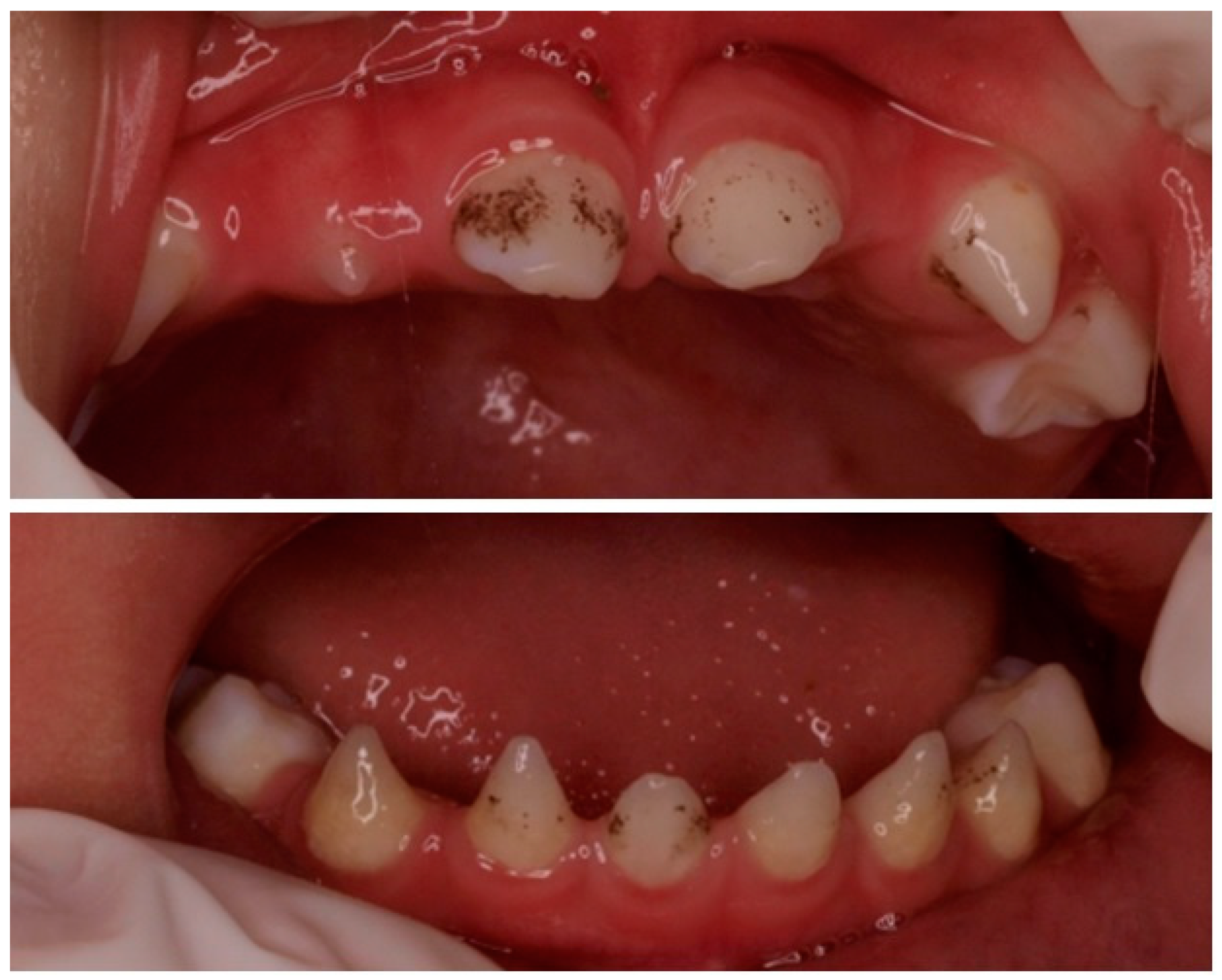
2.1. Phenotype Study

The phenotype was described using Human Phenotype Ontology (HPO) terms [15], which provide a standardized vocabulary of phenotypic abnormalities encountered in human disease. Abnormality of the primary teeth (HP:0006481) which had a conical-shaped and abnormality of the primary molar morphology (HP:0006344) that had an irregular coronal morphology and highly divergent roots were observed. Oligodontia (HP:0000677), tooth agenesis (HP:0009804), dental malocclusion (HP:0000689), abnormality of dental morphology (HP:0006482), the agenesis of mandibular premolars (HP:0011053), and a smooth tongue (HP:0010298) were also present. Abnormality of the skin (HP:0000951) was noted, being slightly atrophic with the loss of fingerprints on the thumbs of the hands. Mild keratosis pilaris (HP:0032152) was found on the cheeks and multiple hyperpigmented lentiginous macules <5 mm on the upper back and the buttocks. Likewise, erythema (HP:0010783) and some fissures were observed in the soles of the feet, suggestive of atopic pulpitis. The scalp hair was apparently normal, whose microscopic study showed a normal morphology and preserved birefringence. Sparse eyebrows (HP:0045075), palmoplantar keratoderma (HP:0000982) (Figure 4), and facial telangiectases (HP:0007380) were noted. No alterations in sweating, including hypohidrosis or hyperhidrosis, were present. See the HPO phenotypic description of the child in Table 1.
In the family dermatological evaluation, keratosis pilaris (HP:0032152) was observed in the father; plantar hyperkeratosis (HP:0007556) was observed in the mother; and dry skin (HP:0000958) was observed in the father, mother, and youngest son. To rule out possible anomalies in the size or number of teeth, the parents were comprehensively evaluated clinically and radiologically by a dentist. No significant alterations were found.

2.2. Genetic Study

The methodology used was the massive parallel sequencing (next-generation sequencing or NGS) of all the coding and splicing regions of a total of 96 genes involved in the different types of ED. The test was performed by capture enrichment with specific probes (SureSelect XT® Agilent) and subsequent sequencing in Illumina equipment (Miseq). Bioinformatic analysis was performed using Illumina Studio 3.0 Database Software to annotate variants: db SNP, 1000 genomes, ExAC, and Variant Server. Reference assembly (CRCh37/hg19). The minimum depth of filtered coverage in this analysis was 100X.
The genes included in the panel were: AXIN2, BRAF, CDH3, COL11A1, CTSC, CTSK, CYLN2, DKC1, DLX3, DSP, ED1, EDAR, EDAR2, EDARADD, EEC1 (ECE1), ELN, RCC2, ERCC3, EVC, EVC2, FGFR10, FGFR2, FGFR3, FLNA, GATA3, GAJ1, GJB2, GJB6, GTF2I, GTF2IRD1, GTF2IRD2, HRAS, IFT122, IFT43, INSR, KCTD1, KRAS2, KREMEN1, KRT14, KRT16, KRT17, KRT6A, KRT6B, KRTHB1, KRTHB3, KRTHB5, KRTHB6, LIMK1, LRP6, MBTS2, MEK1 = MAP2K1, MEK2 = MAP2K2, MSX1, NEMO = IKBKG, IKK1, IKK2, NFKB1, NFKB2, NOLA3 = NOP10, OFD1, PAX9, PIGL, PKP1, POC1A, PORCN, PVRL1, PVRL4, RECQL4, RFC2, RIPK4, RMRP, ROGDI, SETBP1, SHH, TBX3, TERC, TERT, TGF2H5, TINF2, TP63, TRAF6, TRPS1, TTDN1, TWIST2, UBR1, WDR19, WDR35, WHN y, and WNT10A.
Compound heterozygous variants, confirmed by parental segregation, were found in the WNT10A (NM_025216.2): c.310C > T; p. (Arg104Cys) (Clin Var: 532827, dbSNP: 764658964) (ƒ = 0.0000517, gnomAD exomes v. 2.1.1) of maternal origin and the c.742C > T; p. (Arg248Ter) (ClinVar: 265293; dbSNP: rs886039453) (ƒ = 0.00000843, gnomAD Exomes v. 2.1.1) of paternal origin in the patient’s germline DNA, in exons 2 and 3, respectively.
This result was confirmed by Sanger sequencing. Both variants were registered as pathogenic in public databases such as the Human Gene Mutation Database (HGMD®). In addition, the patient carried the EDAR (NM_022336.4): c.1109T > C; p. (Val370Ala) (ClinVar: 5858, dbSNP: rs3827760) (ƒ = 0.154, gnomAD Exomes v. 2.1.1) benign variant in homozygosis, as did both parents [16].

3. Discussion

Dental agenesis is one of the most common craniofacial anomalies. Depending on the number of missing teeth, it is considered hypodontia when less than six teeth are missing (excluding the third molars), oligodontia when more than six teeth are missing, or anodontia when all the teeth are missing [14]. Dental anomalies may be isolated [17] or syndromic. They can also be familial or occur sporadically.
According to the literature, pathogenic variants in the WNT101A gene lead to a wide clinical spectrum of ectodermal disorders. This wide genetic allelic heterogeneity involves at least three WNT101A-related phenotypes: odonto–onycho–dermal dysplasia (OODD, OMIM#257980), autosomal recessive (AR), Schöpf–Schulz–Passarge syndrome (SSPS, OMIM#224750), autosomal recessive AR and non-syndromic tooth agenesis (NSTA), or selective tooth agenesis type 4, (STHAG4, OMIM#150400) with an autosomal recessive or autosomal dominant hereditary pattern. One of the syndromic forms in which oligodontia or anodontia occurs is ED, which is also associated with dry skin, fine hair, and sweating problems. The most commonly associated ED genes are WNT10A [18] (Wnt family member 10 A), EDA (ectodysplasin A), EDAR (ectodysplasin A receptor) [19], and EDARADD (EDAR-associated death domain) [20,21]), which are also candidate genes for non-syndromic tooth agenesis (NSTA). In 2017 and 2019, keratinocyte differentiation factor l (KDF1) was also shown to result in ED [22,23]. Patients with mutations in KDF1 present with abnormal skin, nails, and hair; a complete absence of permanent dental germs; and other abnormal ectodermal-derived tissues and organs.
Our patient presented with a prominent dental phenotype and minor ectodermal signs (mild skin anomalies, sparse eyebrows, and brittle nails), which were only identified under comprehensive dermatological evaluation. This phenotype was associated with pathogenic variants previously described, p. (Arg104Cys) [4] of maternal origin and p. (Arg248Ter) [24] of paternal origin observed in compound heterozygosis in the WNT10A gene. The frequency of the pathogenic variant inherited from the father, WNT10A: c.742C > T; p. (Arg248Ter), is precisely described in East Asian populations, with an overall frequency of 0.00084%.
The father comes from a region with a high potential for geographic isolation in eastern China, Daxue, a mountainous area of Tibet. The maternally inherited pathogenic variant, WNT10A: c.310C > T; p. (Arg104Cys), generates a less impactful amino acid change in the protein it encodes; its validated frequency is 0.065% in East Asian populations and it has not been identified in South Asia.
Both parents, as carriers, confirm the recessive inheritance pattern attributed in this case to the WNT10A gene. Neither of them presented with oligodontia or a complete ED phenotype, except dry skin (HP:0000958), keratosis pilaris (HP:0032152) in the father, and plantar hyperkeratosis (HP:0007556) in the mother. It should be noted that the paternal WNT10A pathogenic variant p. (Arg248Ter) is a nonsense mutation that leads to the end of protein synthesis, resulting in a smaller nonfunctional protein.
Pathogenic variants in the WNT10A gene have been associated with variable phenotypes, ranging from asymptomatic to a severe ED phenotype. Heterozygous pathogenic variants can lead to tooth agenesis. Homozygous or compound heterozygous WNT10A variants, as in the case of our patient, may lead to a wide phenotypic spectrum, from STHAG4 to odonto–onycho–dermal dysplasia and Schöpf–Schulz–Passarge syndrome [25]. The latter is a rare autosomal recessive ED characterized by palmoplantar keratoderma, hypotrichosis, hypodontia, nail dystrophy, and multiple apocrine hydrocystomas in the eyelids that develop with age [26], as well as adnexal skin tumors [27].
It has been observed that the number of missing teeth in the permanent dentition strongly depends on whether the affected individual is a homozygous or heterozygous carrier of mutations in the WNT10A gene. It is very likely that the phenotype depends on the characteristics and location of the genetic changes, and, consequently, on the deficiency generated in the Wnt10 protein. In general, patients carrying biallelic nonsense pathogenic variants in WNT10A have a much more severe dental agenesis, whereas heterozygous individuals carrying a nonsense or a missense pathogenic variant are often unaffected or have a mild phenotype. Heterozygous compound patients were missing up to 6 permanent teeth, whereas homozygotes were usually missing 6 to 26 teeth, mostly around 16 [28]. This is consistent with our case, where the patient carried compound heterozygous variants in the WNT10A gene and showed severe dental involvement, while the parents did not present any dental involvement. Recently, WNT10A-linked oligo/hypodontia phenotypes have been described to be related with minor ectodermal manifestations, such as mild hair and nail anomalies, as described in our patient [29].
According to the literature, the compound heterozygous genotype in the WNT10A gene and its resulting phenotype that we report have not been previously described. Our patient could share a clinical condition with STHAG4 or odonto–onico–dermal dysplasia. A broad clinical spectrum has been defined among homozygous carriers of these variants separately, as a WNT10A homozygote genotype for c.310C > T; p. (Arg104Cys) delineated in ED-affected Turkish children [30] or an adult patient of Asian origin affected with SSPS [31]. Similarly, the entire clinical range has been observed in patients with the WNT10A: c. 742C > T; p. (Arg248Ter) homozygous variant associated with NSTA [32], OODD [33], or SSPS in an elderly patient [34]. Potentially, our patient could even evolve to this last condition. On the other hand, it has also been published that heterozygous carriers of these variants do not show dental loss, as it has also been observed in the parents of this patient [24,35]. Table 2 presents the clinical features described in cases with referred variants in homozygous or compound heterozygous.
Regarding the variant in the EDAR gene: c. 1109T > C; p. (Val370Ala), it has been suggested that it could act as a modifying variant of the ED phenotype in patients carrying causal variants in the EDA gene, one of the main susceptibility genes for ED [36]. It is difficult to attribute the very mild ectodermal manifestations in our patient to the protective effect of the homozygous EDAR370A allele, since there are not specific analyzed cohorts to establish such an association with WNT10A gene mutation carriers. However, we cannot exclude it, given the relationship among the different molecular pathways involved in ectodermal derivatives development. It is difficult to attribute to the fact that the patient is a carrier of such a homozygous polymorphism, the few symptoms that he presents at the dermatological level or in other structures, since there are no series analyzed to establish such an association with mutation carriers in the WNT10A gene.
According to the functional prediction software for genetic variants Alamut Software, version 1.5.1, the physical–chemical characteristics of the T/C nucleotide change that generates the polymorphism could be important for the Edar protein that it encodes.
The fact that both parents were homozygous for the EDAR370A allele, and therefore the patient, is not surprising. The frequency of this variant in the population from which the patient’s ancestors came from has reversed to become the consensus allele, with 92.1% described in East Asian populations. The allelic frequencies described for the C allele in European, African, and Caucasian populations have remained between 1 and 2%. Its genetic drift shows great contrasts between different populations and ethnic groups. Some authors have suggested that its homozygous genotype could confer some advantage. Its evolutionary conservation is evident in the population of origin of the patient’s family [37].

4. Conclusions

Our patient presented a prominent dental phenotype and minor ectodermal signs carrying biallelic WNT10A pathogenic variants in compound heterozygosis. This supports previous reports and highlights the importance of searching skin and nail anomalies in patients consulting for apparently isolated dental anomalies to establish an accurate diagnosis during childhood. The association of the EDAR370A homozygous allele might also attenuate the severity of ED signs in this patient, although more related data are needed.

Author Contributions

V.-E.G.-M. and C.L. identification of the patient, clinical and radiological explorations, follow-up of the case and drafting of the manuscript; O.Z.-A., acquisition of samples; M.d.C.M.-R. and E.G.-N., genetic analysis, and interpretation of the data; X.G.-V., interpretation of the data and drafting of the manuscript; and R.R.-L. and E.G.-N., critical revision and final version. All authors have read and agreed to the published version of the manuscript.

Funding

The genetic study was carried out in the context of a research project funded by the Carlos III Health Institute (ISCIII), Madrid (Spain). (PI14/01259, PI17/00796 y, PI21/01082), co-supported with the European Regional Development Fund (ERDF). Encarna Guillén-Navarro, University Clinical Hospital Virgen de la Arrixaca, IMIB, UMU, Murcia (Spain).

Institutional Review Board Statement

Not applicable.

Informed Consent Statement

All procedures performed in this study were in accordance with the ethical standards of the institutional and/or national research committee and with the 1964 Helsinki Declaration and its later amendments or comparable ethical standards. Written informed consent was obtained from the parents for publication and for the presentation of clinical and radiographical images (Figure 1, Figure 2, Figure 3 and Figure 4).

Data Availability Statement

The data provided are clinical and analytical data derived from the genetic study, which are described in the body of the document. There are no other data to be provided.

Acknowledgments

Esteve of the Dermatology Department of the General University Hospital, Valencia (Spain). The Genetics Laboratory of the General University Hospital, Valencia (Spain).

Conflicts of Interest

The authors declare no conflict of interest.

References

  1. Patthey, C.; Gunhaga, L. Signaling pathways regulating ectodermal cell fate choices. Exp. Cell Res. 2014, 321, 11–16. [Google Scholar] [CrossRef]
  2. Naveau, A.; Seidel, K.; Klein, O.D. Tooth, hair and claw: Comparing epithelial stem cell niches of ectodermal appendages. Exp. Cell Res. 2014, 325, 96–103. [Google Scholar] [CrossRef]
  3. Wright, J.T.; Fete, M.; Schneider, H.; Zinser, M.; Koster, M.I.; Clarke, A.J.; Hadj-Rabia, S.; Tadini, G.; Pagnan, N.; Visinoni, A.F.; et al. Ectodermal dysplasiasClassification and organization by phenotype, genotype and molecular pathway. Am. J. Med. Genet. A 2019, 179, 442–447. [Google Scholar] [CrossRef]
  4. Cluzeau, C.; Hadj-Rabia, S.; Jambou, M.; Mansour, S.; Guigue, P.; Masmoudi, S.; Bal, E.; Chassaing, N.; Vincent, M.C.; Viot, G.; et al. Only four genes (EDA1, EDAR, EDARADD and WNTA10A) account for 90% of hypohidrotic/anhidrotic ectodermal dysplasia cases. Hum. Mutat. 2011, 32, 70–72. [Google Scholar] [CrossRef]
  5. Itin, P.H. Etiology and pathogenesis of ectodermal dysplasias. Am. J. Med. Genet. A 2014, 164, 2472–2477. [Google Scholar] [CrossRef]
  6. Kere, J.; Srivastava, A.K.; Montonen, O.; Zonana, J.; Thomas, N.; Ferguson, B.; Munoz, F.; Morgan, D.; Clarke, A.; Baybayan, P.; et al. X-linked anhidrotic (hypohidrotic) ectodermal dysplasia is caused by mutation in a novel transmembrane protein. Nat. Genet. 1996, 13, 409–416. [Google Scholar] [CrossRef]
  7. Headon, D.J.; Emmal, S.A.; Ferguson, B.M.; Tucker, A.S.; Justice, M.-J.; Sharpe, P.T.; Zonana, J.; Overbeek, P.A. Gene defect in ectodermal dysplasia implicates a death domain adapter in development. Nature 2001, 414, 913–916. [Google Scholar] [CrossRef]
  8. Chassaing, N.; Bourthoumieu, S.; Cossee, M.; Calvas, P.; Vincent, M.C. Mutations in EDAR account for one-quarter of non-ED1-related hypohidrotic ectodermal dysplasia. Hum. Mutat. 2006, 27, 255–259. [Google Scholar] [CrossRef]
  9. Pinheiro, M.; Freire-Maia, N. Ectodermal dysplasias: A clinical classification and a causal review. Am. J. Med. Genet. A 1994, 53, 153–162. [Google Scholar] [CrossRef]
  10. Priolo, M.; Silengo, M.; Lerone, M.; Ravazzolo, R. Ectodermal dysplasias: Not only «skin» deep. Clin. Genet. 2000, 58, 415–430. [Google Scholar] [CrossRef]
  11. Lamartine, J. Towards a new classification of ectodermal dysplasia. Clin. Exp. Dermatol. 2003, 28, 351–355. [Google Scholar] [CrossRef]
  12. Irvine, A.D. Towards a unified classification of the ectodermal dysplasias: Opportunities outweigh challenges. Am. J. Med. Genet. A 2009, 149, 1970–1972. [Google Scholar] [CrossRef]
  13. Di Giovanna, J.J.; Priolo, M.; Itin, P. Approach towards a new classification for ectodermal dysplasias: Integration of the clinical and molecular knowledge. Am. J. Med. Genet. A 2009, 149, 2068–2070. [Google Scholar] [CrossRef]
  14. de Smalen, A.; van Nunen, D.P.F.; Hermus, R.R.; Ongkosuwito, E.M.; van Wijk, A.J.; Don Griot, J.P.W.; Breugem, C.C.; Kramer, G.J.C. Permanent tooth agenesis in non-syndromic Robin sequence and cleft palate: Prevalence and patterns. Clin. Oral Investig. 2017, 21, 2273–2281. [Google Scholar] [CrossRef]
  15. Köhler, S.; Gargano, M.; Matentzoglu, N.; Carmody, L.C.; Lewis-Smith, D.; Vasilevsky, N.A.; Danis, D.; Balagura, G.; Baynam, G.; Brower, A.M.; et al. The Human Phenotype Ontology in 2021. Nucleic Acids Res. 2021, 49, D1207–D1217. [Google Scholar] [CrossRef]
  16. rs3827760 (SNP)—Citations—Homo_Sapiens—Ensembl Genome Browser 108 [Internet]. Ensembl.org. Available online: https://oct2022.archive.ensembl.org/Homo_sapiens/Variation/Citations?db=core;r=2:108896645-108897645;v=rs3827760;vdb=variation;vf=183839455 (accessed on 25 October 2022).
  17. Fauzi, N.H.; Ardini, Y.D.; Zainuddin, Z.; Lestari, W. A review on non-syndromic tooth agenesis associated with PAX9 mutations. Jpn. Dent. Sci. Rev. 2018, 54, 30–36. [Google Scholar] [CrossRef]
  18. Doolan, B.J.; Onoufriadis, A.; Kantaputra, P.; McGrath, J.A. WNT I0 A, dermatology and dentistry. Brit. J. Dermatol. 2021, 185, 1105–1111. [Google Scholar] [CrossRef]
  19. Han, Y.; Wang, X.; Zheng, L.; Zhu, T.; Li, Y.; Hong, J.; Xu, C.; Wang, P.; Gao, M. Pathogenic EDA Mutations in Chinese Han Families With Hypohidrotic Ectodermal Dysplasia and Genotype-Phenotype: A Correlation Analysis. Front. Genet. 2020, 11, 21. [Google Scholar] [CrossRef]
  20. Okita, T.; Asano, N.; Yasuno, S.; Shimomura, Y. Functional studies for a dominant mutation in the EDAR gene responsible for hypohidrotic ectodermal dysplasia. J. Dermatol. 2019, 46, 710–715. [Google Scholar] [CrossRef]
  21. Chassaing, N.; Cluzeau, C.; Bal, E.; Guigue, P.; Vincent, M.C.; Viot, G.; Ginisty, D.; Munnich, A.A.; Calvas, P. Mutations in EDARADD account for a small proportion of hypohidrotic ectodermal dysplasia cases. Brit. J. Dermatol. 2010, 162, 1044–1048. [Google Scholar] [CrossRef]
  22. Shamseldin, H.E.; Khalifa, O.; Binamer, Y.M.; Almutawa, A.; Arold, S.T.; Zaidan, H.; Alkuraya, F.S. KDF1, encoding keratinocyte differentiation factor 1, is mutated in a multigenerational family with ectodermal dysplasia. Hum. Genet. 2017, 136, 99–105. [Google Scholar] [CrossRef]
  23. Manaspon, C.; Thaweesapphithak, S.; Osathanon, T.; Suphapeetiporn, K.; Porntaveetus, T.; Shotelersuk, V. A novel de novo mutation substantiates KDF1 as a gene causing ectodermal dysplasia. Br. J. Dermatol. 2019, 18, 419–420. [Google Scholar] [CrossRef]
  24. Yang, J.; Wang, S.K.; Choi, M.; Reid, B.M.; Hu, Y.; Lee, Y.L.; Herzog, C.R.; Kim-Berman, H.; Lee, M.; Benke, P.J.; et al. Taurodontism, variations in tooth number, and misshapened crowns in WNT10A null mice and human kindreds. Mol. Genet. Genomic. Med. 2015, 3, 40–58. [Google Scholar] [CrossRef]
  25. Martínez-Romero, M.C.; Ballesta-Martínez, M.J.; López-González, V.; GIEDE (Spanish multidisciplinary research group for ectodermal dysplasia). EDA, EDAR, EDARADD and WNT10A allelic variants in patients with ectodermal derivative impairment in the Spanish population. Orphanet. J. Rare Dis. 2019, 14, 281. [Google Scholar] [CrossRef]
  26. Tziotzios, C.; Petrof, G.; Liu, L.; Verma, A.; Wedgeworth, E.K.; Mellerio, J.E.; McGrath, J.A. Clinical features and WNT10A mutations in seven unrelated cases of Schopf-Schulz-Passarge syndrome. Br. J. Dermatol. 2014, 171, 1211–1214. [Google Scholar] [CrossRef]
  27. Riera-Monroig, J.; Martinez-Romero, M.C.; Alos, L.; Guillen-Navarro, E.; Mascaro, J.M., Jr. Eccrine Syringofibroadenoma as a clue for the diagnosis of Schopf-Schulz-Passarge syndrome in acquired palmoplantar keratoderma. J. Cutan. Pathol. 2020, 47, 987–989. [Google Scholar] [CrossRef]
  28. Mues, G.; Bonds, J.; Xiang, L.; Vieira, A.R.; Seymen, F.; Klein, O.; D’Souza, R.N. The WNT10A gene in ectodermal dysplasias and selective tooth agenesis. Am. J. Med. Genet. A 2014, 164, 2455–2460. [Google Scholar] [CrossRef]
  29. Pagnan, N.A.; Visinoni, A.F. Update on ectodermal dysplasia clinical classification. Am. J. Med. Genet. A 2014, 164, 2415–2423. [Google Scholar] [CrossRef]
  30. Güven, Y.; Bal, E.; Altunoglu, U.; Yücel, E.; Hadj-Rabia, S.; Koruyucu, M.; Tuna, E.B.; Çıldır, S.; Aktören, O.; Bodemer, C.; et al. Turkish Ectodermal Dysplasia Cohort: From Phenotype to Genotype in 17 Families. Cytogenet. Genome Res. 2019, 157, 189–196. [Google Scholar] [CrossRef]
  31. Hsu, T.C.; Lee, J.Y.; Hsu, M.M.; Chao, S.C. Case report of Schopf-Schulz-Passarge syndrome resulting from a missense mutation, p.Arg104Cys, in WNT10A. J. Dermatol. 2018, 45, 475–478. [Google Scholar] [CrossRef]
  32. Zeng, B.; Zhao, Q.; Li, S.; Lu, H.; Lu, J.; Ma, L.; Zhao, W.; Yu, D. Novel EDA or EDAR Mutations Identified in Patients with X-Linked Hypohidrotic Ectodermal Dysplasia or Non-Syndromic Tooth Agenesis. Genes 2017, 8, 259. [Google Scholar] [CrossRef] [PubMed]
  33. Yu, M.; Wong, S.W.; Han, D.; Cai, T. Genetic analysis: Wnt and other pathways in nonsyndromic tooth agenesis. Oral Dis. 2019, 25, 646–651. [Google Scholar] [CrossRef] [PubMed]
  34. Zimmermann, C.E.; Soufi, M.; Ruppert, V.; Schaefer, J.R.; von Domarus, H. Schopf-Schulz-Passarge Syndrome: Previously Unreported WNT10A Genotype and Phenotypes in 9 Family Members. Acta Derm.-Venereol. 2019, 99, 113–114. [Google Scholar] [CrossRef]
  35. Capalbo, A.; Alonso Valero, R.; Jimenez-Almazan, J.; Mir Pardo, P.; Fabiani, M.; Jiménez, D.; Simon, C.; Martin-Rodriguez, J. Optimizing clinical exome design and parallel gene-testing for recessive genetic conditions in preconception carrier screening: Translational research genomic data from 14,125 exomes. PLoS Genet. 2019, 15, e1008409. [Google Scholar] [CrossRef]
  36. Cluzeau, C.; Hadj-Rabia, S.; Bal, E.; Clauss, F.; Munnich, A.; Bodemer, C.; Headon, D.; Smahi, A. The EDAR370A allele attenuates the severity of hypohidrotic ectodermal dysplasia caused by EDA gene mutation. Br. J. Dermatol. 2012, 166, 678–681. [Google Scholar] [CrossRef]
  37. Chang, S.H.; Jobling, S.; Brennan, K.; Headon, D.J. Enhanced Edar signalling has pleiotropic effects on craniofacial and cutaneous glands. PLoS ONE 2009, 4, e7591. [Google Scholar] [CrossRef]
Figure 1. Presence of conical anterior temporary teeth. Absence of the left lateral maxillary incisor and the maxillary and mandibular second molars.
Children 10 00356 g001
Figure 2. Clinical and radiographical findings at 6 years old.
Children 10 00356 g002
Figure 3. Clinical and radiographical findings at 11 years old.
Children 10 00356 g003
Figure 4. Erythema and some fissures on the soles of the feet suggestive of atopic pulpitis.
Children 10 00356 g004
Table 1. Comprehensive phenotypic description of the patient based on the WNT10A gene using Human Phenotype Ontology (HPO).
HPO_TERM_IDHPO_TERM_NAMEPatientDescription
HP:0006344Abnormality of primary molar morphologyYESIrregular coronal morphology and highly divergent roots.
HP:0006481Abnormality of primary teethYESFront teeth are conical-shaped.
HP:0011056Agenesis of first permanent molar toothNO
HP:0001798AnonychiaNO
HP:0006482Abnormality of dental morphologyYES
HP:0001792Small nailsNO
HP:0002209Sparse scalp hairNO
HP:0000478Abnormality of the eyeNO
HP:0010298Smooth tongueYES
HP:0011053Agenesis of mandibular premolarYESAlso agenesis of the maxillary premolars.
HP:0008388Abnormal toenail morphologyNO
HP:0000668HypodontiaNO
HP:0001806OnycholysisNO
HP:0031405PoromaNO
HP:0000975HyperhidrosisNO
HP:0002671Basal cell carcinomaNO
HP:0008391Dystrophic fingernailsNO
HP:0005216Impaired masticationNO
HP:0000007Autosomal recessive inheritance
HP:0000613PhotophobiaNO
HP:0001231Abnormal fingernail morphologyNO
HP:0010764Short eyelashesNO
HP:0006342Peg-shaped maxillary lateral incisorsYES
HP:0006289Agenesis of central incisorNO
HP:0009804Tooth agenesisYES1.4,1.5, 2.2, 2.4, 2.5, 3.1, 3.2, 3.3, 3.4, 3.5, 4.1, 4.2, 4.3, 4.4, 4.5, 5.5, 6.2, 6.5,7.5, 8.5.
HP:0001596AlopeciaNO
HP:0001810Dystrophic toenailsNO
HP:0000951Abnormality of the skinYESMultiple hyperpigmented lentiginous macules <5 mm on the upper back and buttocks. Appearance was slightly atrophic with loss of fingerprints on the thumbs of the hands.
HP:0031454Apocrine hidrocystomaNO
HP:0000684Delayed eruption of teethNO
HP:0000320Bird-like faciesNO
HP:0002231Sparse body hairNO
HP:0011219Short faceNO
HP:0000202Oral cleftNO
HP:0000685Hypoplasia of teethNO
HP:0000958Dry skinYES
HP:0002860Squamous cell carcinomaNO
HP:0012472EclabionNO
HP:0000968Ectodermal dysplasiaYES
HP:0006297Enamel hypoplasiaNO
HP:0002164Nail dysplasiaYESBrittle toenails.
HP:0001595Abnormal hair morphologyNO
HP:0011313Narrow nailsNO
HP:0008070Sparse hairNO
HP:0001807Ridged nailsNO
HP:0006323Premature loss of primary teethNO
HP:0007380Facial telangiectasiaNO
HP:0000677OligodontiaYES
HP:0000689Dental malocclusionYES
HP:0000691MicrodontiaNO
HP:0000696Delayed eruption of permanent teethNO
HP:0100840Aplasia/hypoplasia of the eyebrowNO
HP:0002213Fine hairNO
HP:0007410Palmoplantar hyperhidrosisNO
HP:0010783ErythemaYESErythema and some fissures were observed in the balls of the feet, suggestive of atopic pulpitis.
HP:0007556Plantar hyperkeratosisNO
HP:0011359Dry hairNO
HP:0000679TaurodontiaNO
HP:0000687Widely spaced teethYES
HP:0000690Agenesis of maxillary lateral incisorsYESAgenesis of a maxillary lateral incisor, temporary and permanent.
HP:0032152Keratosis pilarisYESMild keratosis pilaris on the cheeks.
HP:0001816Thin nailsNO
HP:0025114HypergranulosisNO
HP:0000982Palmoplantar keratodermaNO
HP:0025092Epidermal acanthosisNO
HP:0006336Short dental rootNO
HP:0100615Ovarian neoplasmNO
HP:0040162OrthokeratosisNO
HP:0045075Sparse eyebrowYESSparse hair in eyebrow tail.
HP:0000966HypohidrosisNO
HP:0006349Agenesis of permanent teethYES
HP:0011051Agenesis of premolarYES
HP:0011078Abnormality of canineYES
Table 2. Clinical features described in cases with referred variants in homozygous or compound heterozygous.
Cases ReportedGüven et al., 2019 [29]Hsu et al., 2018 [30]Yang et al., 2015 [34]Yu et al., 2019 [32]Zimmermann et al., 2017 [33]Novel Case
SexF MMF MM
Age at diagnosis (years)DNA (child)548.5145311
Tooth agenesis
HP:0006482Abnormality of dental morphologyYESYESYESYESYESYES
HP:0006349 Agenesis of permanent teethYESYESYESYESYESYES
HP:0000677 OligodontiaYESYESYESYESYESYES
Sweating
HP:0000966HipohidrosisNONONOYESNONO
HP:0007410Palmoplantar hyperhidrosisYESNONONOYESNO
Skin
HP:0000958Dry skinYESYESNOYESYESYES
HP:0000982Palmoplantar keratodermaNOYESNONOYESNO
HP:0031454Apocrine hidrocystomaNOYESNONOYESNO
Hair
HP:0002209Sparse scalp hairYESYESNONOYESNO
HP:0002231Sparse body hairNDYESNONOYESYES
Nails
HP:0002164Nail dysplasiaYESYESNOYESYESYES
Clinical
Diagnosis
EDSSPSSTHAGOODDSSPSSTHAG with mild ED
Variants in
WNT10A (NM_025216.3)
c. 310 C > Ac. 310 C > Ac. 310 C > A/c. 637 T > Ac. 742 C > Tc. 742 C > T/c. 321 C > Ac. 310 C > A/c. 742 C > T
Protein changep. (Arg104Cys)p. (Arg104Cys)p. (Arg104Cys)/p. (Gly213Ser)p. (Arg248Ter)p. (Arg248Ter)/p. (Cys107Ter)p. (Arg104Cys)/p. (Arg248Ter)
ZigosityHomozygoteHomozygoteCompound heterozygousHomozygoteCompound heterozygousCompound heterozygous
F, female; M, male; DNA, data not available; ED, ectodermal dysplasia; SSPS, Schöpf–Schulz–Passarge syndrome; STHAG, selective tooth agenesis type 4; OODD, odonto–onycho–dermal dysplasia.
Disclaimer/Publisher’s Note: The statements, opinions and data contained in all publications are solely those of the individual author(s) and contributor(s) and not of MDPI and/or the editor(s). MDPI and/or the editor(s) disclaim responsibility for any injury to people or property resulting from any ideas, methods, instructions or products referred to in the content.

