Review

Exploring the Genetic Landscape of Childhood Glaucoma

by
Yang Pan
and
Takeshi Iwata
*
National Institute of Sensory Organs, NHO Tokyo Medical Center, Tokyo 152-8902, Japan
*
Author to whom correspondence should be addressed.
Children 2024, 11(4), 454; https://doi.org/10.3390/children11040454
Submission received: 6 March 2024 / Revised: 3 April 2024 / Accepted: 8 April 2024 / Published: 9 April 2024
(This article belongs to the Special Issue Pediatric Eye Disease: Screening, Causes and Treatment)

Abstract

:
Childhood glaucoma, a significant cause of global blindness, represents a heterogeneous group of disorders categorized into primary or secondary forms. Primary childhood glaucoma stands as the most prevalent subtype, comprising primary congenital glaucoma (PCG) and juvenile open-angle glaucoma (JOAG). Presently, multiple genes are implicated in inherited forms of primary childhood glaucoma. This comprehensive review delves into genetic investigations into primary childhood glaucoma, with a focus on identifying causative genes, understanding their inheritance patterns, exploring essential biological pathways in disease pathogenesis, and utilizing animal models to study these mechanisms. Specifically, attention is directed towards genes such as CYP1B1 (cytochrome P450 family 1 subfamily B member 1), LTBP2 (latent transforming growth factor beta binding protein 2), TEK (TEK receptor tyrosine kinase), ANGPT1 (angiopoietin 1), and FOXC1 (forkhead box C1), all associated with PCG; and MYOC (myocilin), associated with JOAG. Through exploring these genetic factors, this review aims to deepen our understanding of the intricate pathogenesis of primary childhood glaucoma, thereby facilitating the development of enhanced diagnostic and therapeutic strategies.

Graphical Abstract

1. Introduction

Glaucoma constitutes a group of chronic and progressive optic neuropathies distinguished by progressive visual field loss and optic disc damage, standing as the leading cause of irreversible blindness globally [1]. It can occur at all ages, with early-onset cases (before 40 years) typically displaying Mendelian inheritance patterns, while adult-onset forms (after 40 years) exhibit inheritance as complex traits [2]. Generally, mutations in genes causing early-onset glaucoma are rare but have significant biological effects, whereas variants contributing to adult-onset forms are more common with minor effects [3]. Childhood glaucoma, denoted by the onset occurring before 18 years of age, represents a subset within the early-onset category [4].
Various definitions and classification systems have been used to describe different subtypes of childhood glaucoma, lacking consensus until recently. To address this situation, the Childhood Glaucoma Research Network (CGRN) has developed a classification system, adopted by the World Glaucoma Association and the American Board of Ophthalmology [5]. Diagnosis of childhood glaucoma requires at least two of the following criteria: (1) intraocular pressure (IOP) of 21 mmHg or higher; (2) glaucomatous optic nerve damage, such as increased cupping, focal notching, or cup-to-disc asymmetry of 0.2 or more between both eyes; (3) corneal changes, such as an increased corneal diameter or Haab striae; or (4) visual field defects consistent with glaucomatous optic nerve damage. According to the CGRN system, childhood glaucoma is categorized as primary or secondary, with primary glaucoma being further subdivided into primary congenital glaucoma (PCG) and juvenile open-angle glaucoma (JOAG) [5]. PCG typically presents with characteristic signs in neonates and infants, while JOAG is often asymptomatic and may be detected incidentally or through family screening [6].
The incidence of childhood glaucoma varies among different populations and subtypes. Six published reports have consistently identified PCG as the prevalent common subtype, with studies from Egypt (55.07% from 207 samples [7]; 68.25% from 652 samples [8]), the USA (19.2% from 205 samples [9]), Brazil (43.95% from 496 samples [10]), India (35.27% from 275 samples [11]), and Australia/New Zealand (57.59% from 290 samples [4]). In contrast, two studies from Thailand and Boston have identified PCG (20.8% in Thailand; 29% in Boston) as the second most common subtype [12]. These findings underscore the variability in prevalence among different geographic populations and suggest potential genetic influences.
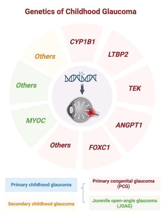
Primary childhood glaucoma is typically caused by genetic variants following a Mendelian pattern of inheritance [4]. The important genes implicated in this condition include CYP1B1 (cytochrome P450 family 1 subfamily B member 1), LTBP2 (latent transforming growth factor beta binding protein 2), FOXC1 (forkhead box C1), TEK (TEK receptor tyrosine kinase, also known as TIE2), and ANGPT1 (angiopoietin 1) for PCG; and MYOC (myocilin) for JOAG. These gene variants typically exhibit strong penetrance but variable expressivity, contributing to a diverse phenotypic spectrum and overlap between clinical entities. This review aims to explore recent advancements and new insights into the genetics underlying Mendelian forms of primary childhood glaucoma. While the precise functions of these genes and the effects of their variants on health and disease remain incompletely understood, these discoveries offer valuable insights for enhancing clinical diagnoses in childhood glaucoma, despite the challenges in accurately establishing such diagnoses.

2. Primary Congenital Glaucoma (PCG)

PCG represents a severe manifestation of the disease, characterized by infant or early-childhood (before three years old) ocular hypertension, enlarged eye globes, and optic neuropathy, often leading to vision loss and blindness despite treatment [13]. Notably, PCG constitutes 18% of children enrolled in institutions for blind people globally [14]. Although PCG occurs across all ethnic groups, its incidence varies based on ethnic background, with rates from 1:1250 in endogamous populations to 1:30,000 in ethnically diverse populations [15,16]. Typically inherited as an autosomal recessive trait, PCG is more prevalent in regions and cultures that practice consanguinity. Linkage analysis studies have identified five loci (GLC3A (2p22–p21) [17], GLC3B (1p36.2–p36.1) [18], GLC3C (14q24.3) [19], GLC3D (14q24) [20], and GLC3E (9p21) [21]), and three genes (CYP1B1, LTBP2, and TEK) located at these loci [14,22,23,24]. Additionally, genomic analyses, including whole-exome and direct Sanger sequencing analyses, have demonstrated that mutations in the ANGPT1 and FOXC1 genes also contribute to PCG [25,26].

2.1. CYP1B1

2.1.1. CYP1B1 Mutations in PCG

The CYP1B1 gene is located on chromosome 2p22–21 and consists of only three exons, including a non-coding exon 1 [27]. It was the first gene to be genetically linked to PCG, with its mutations being the most frequently identified genetic defect causing autosomal recessive PCG globally. To date, over 150 distinct mutations of CYP1B1 have been identified in PCG cases worldwide. The prevalence of CYP1B1 mutations in PCG varies significantly across different populations. Specifically, CYP1B1 mutations have been reported in approximately 80–100% of Saudi Arabians and Slovakian Rom populations [28], as well as in 70% of Iran [29], 50% of Brazil [30], and 44% of India [31]. Conversely, the incidence of PCG caused by CYP1B1 mutations is lower in Indonesian, Japanese, and Han Chinese populations, standing at 33.3%, 20%, and 17.2%, respectively [32]. Consanguineous marriages, known for their increased risk of developing autosomal recessive disorders [33], contribute to the prevalence of CYP1B1 mutations causing autosomal recessive PCG. This practice is particularly prevalent in Muslim-majority countries in North and sub-Saharan Africa, the Middle East, and parts of West, Central, and South Asia [34,35]. Therefore, the variation in the incidence of CYP1B1 mutations in PCG can be largely attributed to the prevalence of consanguineous marriage within these populations.
Moreover, the most prevalent CYP1B1 mutation varies among different populations (Figure 1). The point mutation CYP1B1 p. Gly61Glu (G61E) is the predominant mutation in Saudi Arabia, Portugal, Vietnam, Morocco, and Iran [36]. Conversely, the second point mutation, p. Glu387Lys (E387K), is the most common mutation observed in Slovakian Rom and Hungarian populations [36]. In Han Chinese and Pakistani populations, p.Arg390His (R390H) stands as the most frequent mutation, causing a severe form of PCG that is resistant to surgical interventions and medications [37]. Additionally, certain mutations serve as the most common mutation within specific populations but not in others. For instance, CYP1B1 p.Arg444Gln (p.R444Q) predominates in Japanese populations [38], c.970_971dupAT (p.T325SfsX104) is prevalent in Koreans [39], g.8214_8215delAG (p.Val460fs) is common in Brazilians [40], and p.R368H is notable among Indians [40]. A mixed European study identified 31 CYP1B1 mutations in 56 PCG patients (34.7%), with p.E387K being the most prevalent mutation [41]. However, the exact impact of CYP1B1 disruption in various European populations remains unclear due to limited samples, necessitating further research.

2.1.2. CYP1B1 Protein Function in PCG

The CYP1B1 protein belongs to the cytochrome P450 (CYP) enzyme family, which plays a crucial role in catalyzing oxidation reactions of various organic substrates [14]. It is expressed in numerous adult and fetal extrahepatic tissues, including brain, kidney, prostate, breast, and ocular tissues [42]. Although the precise function of CYP1B1 in the eye remains elusive, several pathways regulated by this protein have been implicated in PCG.
First, CYP1B1 is integral to properly developing trabecular meshwork (TM), a critical tissue for controlling IOP in relation to PCG pathogenesis. The strong expression of CYP1B1 in ocular tissues underscores its importance in normal eye development and function in both mice and humans [43,44]. Mice lacking Cyp1b1 exhibit anomalies such as small or absent Schlemm’s canal (SC), TM fibers, and angle closure [45]. The precise mechanism by which CYP1B1 contributes to TM development remains under investigation; however, some studies show its involvement in modulating oxidative stress and extracellular matrix proteins [46].
Secondly, the role of CYP1B1 in angiogenesis is noteworthy. Its expression in neurovascular structures, including astrocytes, suggests an involvement in postnatal retinal vascular development and pathological neovascularization [47,48,49]. Some studies indicate that CYP1B1 expression modulates oxidative stress, activates nuclear factor-κB (NF-κB), and upregulates thrombospondin-2 (TSP2) in retinal vascular cells, influencing angiogenesis [48]. However, its role in retinal astrocytes, another major component of retinal vasculature, remains poorly understood.
Lastly, CYP1B1 is implicated in metabolic pathways relevant to glaucoma. It participates in the metabolism of vitamin A, crucial for embryonic eye development, and melatonin, a hormone with diverse ocular effects [42]. Specifically, CYP1B1 catalyzes retinol to retinal, the major forms of vitamin A, and further transforms the catalyzed retinal into retinoic acid [50]. Furthermore, CYP1B1 contributes to melatonin signaling, which has significant metabolic functions [51]. Melatonin synthesis in the eye, acting as a CYP1B1 inhibitor, and its modulation of retinal functions, such as dopamine synthesis, photoreceptor activity, and IOP regulation, suggest a potential link between CYP1B1-mediated retinoid metabolism, melatonin signaling, and glaucoma [52,53]. Moreover, CYP1B1 generates 12-(R)-HETE from arachidonic acid, serving as an endogenous corneal arachidonate metabolite [54,55]. Research by Masferrer et al. has shown that 12-(R)-HETE can effectively lower IOP in rabbits [55]. Additionally, lipidomic analyses have identified differences in endogenous metabolism within the eye between glaucoma patients and controls, indicating the potential involvement of metabolic pathways in the development of glaucoma [56]. These findings collectively suggest a potential link between CYP1B1-mediated metabolic pathways and the pathogenesis of glaucoma.
In summary, CYP1B1 plays multifaceted roles in PCG pathogenesis, influencing TM development, angiogenesis, and metabolic pathways crucial for ocular homeostasis. Further research is warranted to elucidate the precise mechanisms underlying its involvement in glaucoma and to explore its potential as a therapeutic target.

2.1.3. CYP1B1 Animal Models

The expression pattern of Cyp1b1 in mice exhibits tissue-specific characteristics throughout development [57]. During early embryonic stages, Cyp1b1 mRNA is detectable in various regions, including the eye, hindbrain, brachial arches, forelimb bud, ligaments supporting the primordial liver, and kidney [58]. Subsequently, its expression becomes confined to the eye region and forelimb bud, with discernible alterations being observed in the neural retina as development progresses. Immunohistochemical studies in adult mice reveal the presence of Cyp1b1 in the eye, particularly in the ciliary and corneal epithelia, retinal cells, and ganglion cells [46]. Notably, developmental changes in Cyp1b1 protein levels are also observed in the lens epithelium. Studies on Cyp1b1−/− mice, homozygous Cyp1b1-knockout (KO) mice, underscore the crucial role of constitutive Cyp1b1 expression in TM cells for their development and function [45]. Overall, the dynamic expression of cyp1b1 during both prenatal and postnatal development suggests its importance in guiding proper tissue development and function, particularly in the eye.
In 1999, a Cyp1b1-KO mouse line on the 129/Sv background was generated to elucidate the role of CYP1B1 in the metabolic activation of 7,12-dimethylbenz[α]anthracene [59]. Similarly, the role of CYP1B1 in PCG was investigated using Cyp1b1-null mice on a mixed 129 × 1/SvJ × C57BL/6J background, revealing defects reminiscent of those observed in PCG patients [60]. Notably, the severity of these defects was heightened in albino mice deficient in tyrosinase, an enzyme crucial for the production of melanin and l-dihydroxyphenylalanine (l-Dopa) [60]. Additionally, the symptoms were alleviated by administering l-Dopa, potentially through reducing oxidative stress. More recently, Cyp1b1-KO mice on a pure C57BL/6J background were used to explore the effects of Cyp1b1 on hypertension, revealing decreased TM collagen, elevated TM endothelial cell counts, and elevated collagen lesion scores compared to controls; these scores were observed to increase progressively with age [61].
Moreover, studies by Amirmokhtari et al. demonstrate that naive Cyp1b1-KO mice develop an anatomically intact retinal projection without overt signs of glaucomatous pathology [62]. However, following pressure elevation, accelerated degradation of axonal transport from the retina to the superior colliculus and altered morphology of the nodes of Ranvier and adjacent paranodes in the optic nerves were observed [62]. Their results suggest that the absence of Cyp1b1 expression alone may not be sufficient to induce glaucomatous pathology in mice. However, it may render retinal axons more susceptible to IOP elevation. Using a Cyp1b1-null mouse model, researchers have discovered that the tyrosinase gene (Tyr) acts as a modifier of the drainage structure phenotype, and the severe dysgenesis in eyes lacking both Cyp1b1 and Tyr can be alleviated via dihydroxyphenylalanine [60]. Additionally, in zebrafish, CYP1B1 indirectly influences neural crest migration by contributing to the closure of the ocular fissure [63]. Thus, selective upregulation of CYP1B1 expression in the eye holds promise for novel therapeutic strategies in PCG treatment.

2.2. LTBP2

2.2.1. LTBP2 Mutations in PCG

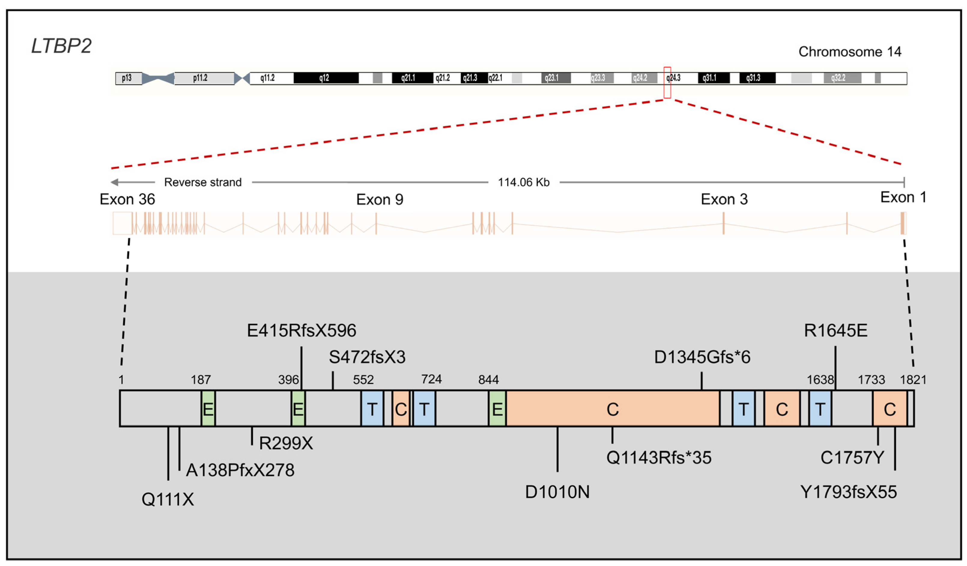
LTBP2, located on chromosome 14q24 within the GLC3D locus, is composed of 36 exons and encodes a matrix protein containing 1821 amino acids (aa) (Figure 2) [16]. In 2009, Ail et al. identified four homozygous null mutations in LTBP2, including p.Ala138fs*278 (p.A138PfsX278), p.Arg299*(p.R299X), p.Glu415fs*596 (p.E415RfsX596), and p.Gln111*(p.Q111X), as causative mutations of PCG in consanguineous families from Pakistan and individuals of Gypsy ethnicity [23]. Among these mutations, the truncating LTBP2 p.Arg299* (p.R299X) mutation is the most prevalent Gypsy PCG mutation, accounting for approximately one-third of disease alleles (33.8%, from 34 families) [64]. In the same year, Narooie-Nejad et al. performed whole-genome autozygosity mapping in Iranian PCG families and identified two loss-of-function (LoF) mutations in LTBP2 (p.S472fsX3 and p.Y1793fsX55) [24]. Subsequently, in 2016, Micheal et al. reported two novel mutations (p.R1645E and p.D1345Gfs*6) from consanguineous Pakistani PCG families using whole-exome sequencing [65]. Three novel LTBP2 mutations (p.D1010N, p.Q1143Rfs*35, and p.C1757Y) were recently discovered in three consanguineous Pakistani PCG families [66].

2.2.2. LTBP2 Protein Function in PCG

The LTBP2 protein plays a crucial role in tissue repair and cell adhesion, with ocular studies revealing its expression in both TM and ciliary processes [15]. Structurally, LTBP2 belongs to a superfamily of fibrillin and LTBP proteins, characterized by tandem arrays of epidermal growth factor-like motifs and interspersed TGFβ-binding protein-like motifs [67]. Unlike other isoforms, LTBP2 does not bind to latent TGFβ, suggesting functions independent of latent TGFβ storage and activation [68]. However, Hirani et al. reported an interaction between the C-terminal region of LTBP-2 and the N-terminal region of fibrillin-1, suggesting a role in regulating TGFβ activation by releasing LTBP-1 from microfibrils [69]. Considering the association of TGFβ with IOP [70], these results suggest the contribution of pathogenic LTBP2 mutations to PCG through TGFβ-related mechanisms.
Localization studies by Ail et al. confirmed the expression of LTBP2 in the anterior segment of the eye, particularly the ciliary process, indicating its importance in the normal development of the anterior chamber by maintaining ciliary muscle tone [23]. Moreover, these findings suggest that LTBP2 deficiencies may disrupt the elasticity of ciliary body structures, affecting the support provided to adjacent tissues such as Schlemm’s canal or the scleral spur [71]. Alternatively, alterations in the elasticity of the scleral spur could adversely affect the architecture of the TM, considering the anatomical connections between components of the ciliary body and the scleral spur [72]. Additionally, LTBP2’s involvement in tissue repair and cell adhesion suggests potential roles in maintaining microfibril and elastin fiber functions [24,73], as evidenced by its interactions with fibrillin-1 and fibulin 5 [74,75]. Despite these insights, the precise function of LTBP2 in PCG is yet to be fully elucidated.

2.2.3. LTBP2 Mouse Models

Investigating the in vivo functions of LTBP2 has posed a challenge, as the initial report of Ltbp2-null mice indicated a halt in development beyond embryonic day 6.5, indicating an essential role for LTBP2 in early embryogenesis [76]. Interestingly, PCG patients with homozygous nonsense LTBP2 mutations do not exhibit lethal birth defects or early developmental disorders, which presents a discrepancy between human and mouse models. However, this contradiction was recently addressed with the introduction of new Ltbp2-null mice, wherein exon 1 was deleted in the presence of Cre recombinase [75]. Remarkably, these mice were viable and fertile without any noticeable abnormalities. Detailed examinations revealed fragmented microfibrils in their ciliary zonules and dislocated lenses. Additionally, it was demonstrated that LTBP2 binds to fibrillin-1, and the addition of recombinant LTBP2 promotes the assembly of microfibril bundles in cultured cells and organ-cultured eyeballs. These findings underscore the significance of LTBP2 in eye development. However, further investigations are warranted to elucidate its role in PCG.

2.3. TEK and ANGPT1

TEK, located on chromosome 14q24 within the GLC3E locus [21], is an integral component of the angiopoietin–TIE (tunica interna endothelial cell kinase) signaling pathway, which includes three ligands (ANGPT1, ANGPT2, and ANGPT4) and two receptors (TIE1 and TEK, also known as TIE2) [77]. Known as an endothelial growth factor pathway, the ANGPT–TIE system extends its involvement to processes such as inflammation, metastasis, and lymphangiogenesis [78]. Notably, heterozygous LoF variants in TEK (Figure 3) or its primary ligand ANGPT1 (Figure 4) have been associated with PCG [14,26].

2.3.1. TEK and ANGPT1 Mutations in PCG

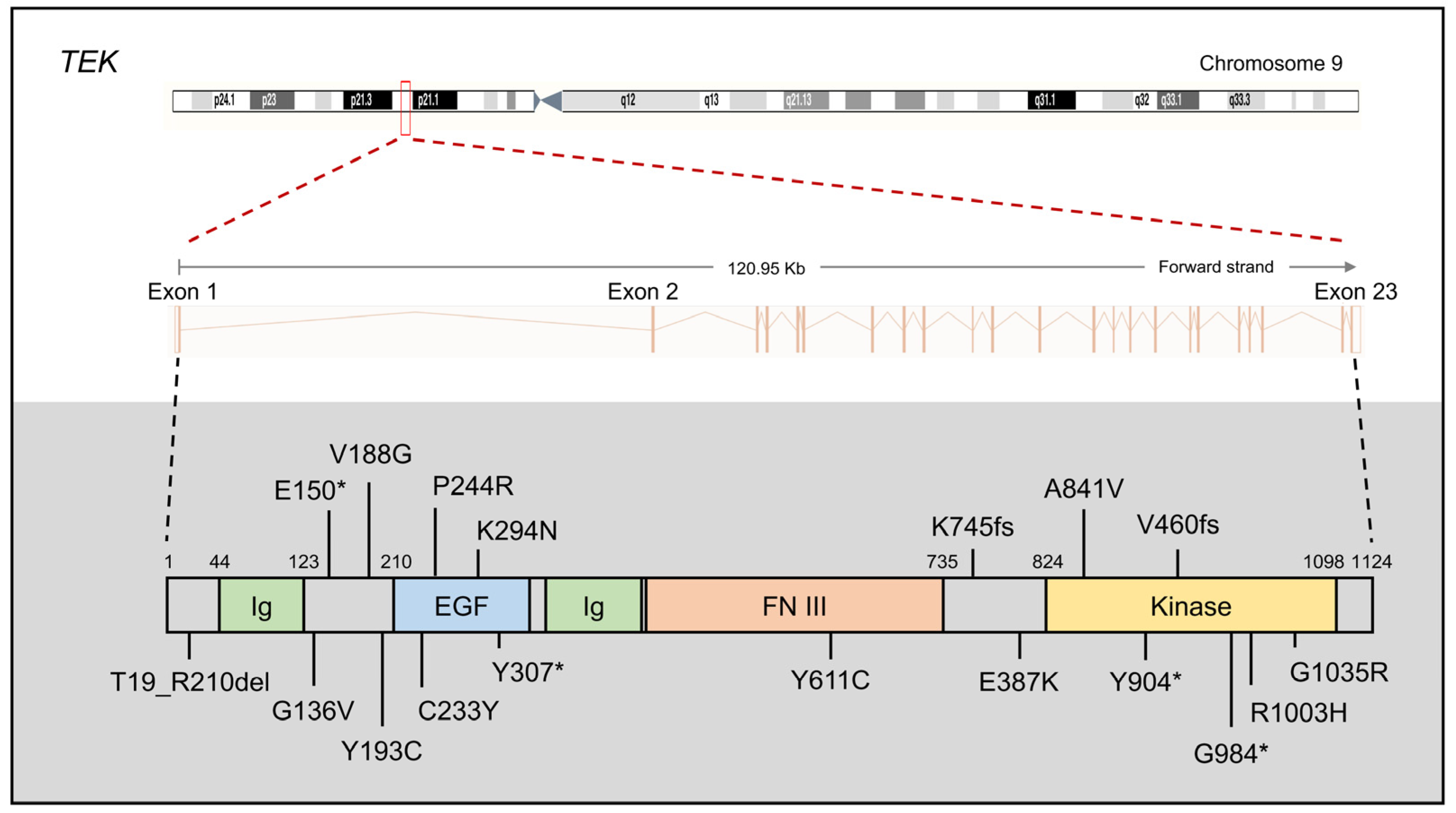
The TEK gene comprises 23 coding exons encoding a principal product of 1124 aa. In 2016, Souma et al. first identified 10 heterozygous novel/rare TEK variants (p.T19_R210del, p.E150*, p.C233Y, p.K294N, p.Y307*, p.Y611C, p.K745fs, p.G984*, c.760+2T>C, and c.3300+2delT) as disease-causing mutations following dominant inheritance [14]. Seven of these mutations lead to abnormal LoF, including the absence of intact protein production, increased proteasomal degradation, impaired phosphorylation of critical tyrosine residue, changes in subcellular localization and diminished responsiveness to ligands, and protein aggregation. The remaining three mutations are also likely to be disease-associated. TEK could be implicated in the etiology of PCG, supported by their findings of the PCG-like phenotype in Angpt1/Angpt2 and Tek conditional KO mice [79]. In 2020, Young et al. identified eight TEK variants in families affected by PCG (p.G136V, p.Y904*, p.V188G, p.Y193C, p.P244R, p.A841V, p.G1035R, and c.1624 +5G>A), further supporting TEK’s role as a causative factor in PCG [80]. A subsequent study in China demonstrated that TEK mutations, particularly p.R1003H located in the protein kinase domain, may act as pathogenic mutation and contribute to PCG [81]. Taken together, these findings underscore the fact that defects in the ANGPT–TIE pathway caused by reduced TEK constitute a novel mechanism of the PCG disease. It is likely that investigating additional genes involved in forming and maintaining the ANGPT–TIE pathway could provide further insights.
Given the similarity in PCG-like phenotypes observed in Angpt1/Angpt2 and Tek conditional KO mice, it has been hypothesized that other genes within the ANGPT–TIE system, such as ANGPT1, ANGPT2, and ANGPT4, might also play a role in PCG. In 2017, Thomson et al. identified heterozygosity for two novel nonsense variants (p.Q236*, p.R494*) and one rare missense variant (p.K249R) in ANGPT1 in three PCG families without other known PCG-causing genes from a cohort of 284 PCG families [26]. The ANGPT1 p.Q236* mutation introduces a premature stop codon within exon 4, resulting in a truncated protein lacking the C-terminal receptor-binding domain required for interactions with TEK [26]. The truncated ANGPT1 protein may have a dominant-negative effect on ANGPT1 signaling if it escapes the nonsense-mediated decay pathway. Additionally, Knight et al. identified an ANGPT1 mutation in a cohort of 290 Australian PCG cases [4]. Furthermore, common variants in ANGPT1 have been implicated as risk alleles for POAG in GWAS studies of 4986 cases and 58,426 controls from four racial/ethnic groups (non-Hispanic whites, Hispanics/Latinos, East Asians, and African Americans) [82].

2.3.2. The Protein Function of TEK and ANGPT1

The TEK protein, identified as the ANGPT receptor discovered in 1992, form a small subfamily of growth factor receptor tyrosine kinases (RTKs) [77]. Its expression is primarily confined to the endothelium, including the endothelia of SC and collector channels, crucial components of the aqueous humor (AH) outflow pathway [83]. ANGPT1 is a paracrine ligand expressed by mesenchymal cells and acts as a potent TEK agonist, supporting endothelial cell survival, vessel stability, and endothelial barrier function [84]. The activation of TEK by ANGPT1 triggers downstream signaling through the protein kinase AKT (also known as protein kinase B), leading to the inhibition of transcription factor forkhead box protein O1 (FOXO1) and the suppression of FOXO1′s target genes, including ANGPT2 [77].
Elevated IOP, a well-recognized key risk factor in PCG, is attributed to defects in the AH outflow pathway rather than increased AH production [14]. AH, produced by the ciliary body, drains mainly through SC and the uveoscleral pathways, and any disruption in fluid homeostasis can lead to elevated IOP [85,86]. The significant expression of the TEK receptor in the SC endothelium and the critical role of ANGPT–TIE signaling in SC development leads to significant outcomes when Tek or both major ANGPT ligands are deleted in mice after embryonic day 16.5. These outcomes include the developmental loss of SC, a notable increase in IOP, and a rapid and complete loss of retinal ganglion cells, all of which ultimately contribute to the development of glaucoma [79]. Thus, pathogenetic mutations in TEK and ANGPT1 may contribute to elevated IOP and the development of PCG.

2.3.3. TEK and ANGPT1 Mouse Models

Since 2002, several mouse models of ocular disease related to the ANGPT–TIE system have been generated, including KO models for Angpt1 or Angpt2, conditional deletion of Angpt1, Angpt2, or Tek, as well as transgenic mouse models overexpressing retinal/ubiquitous ANGPT1 or ANGPT2 [77]. These models have provided insights into the role of the ANGPT–TIE system in retinal and choroidal neovascularization, particularly relevant to diseases affecting the outer retina and Bruch’s membrane, such as neovascular age-related macular degeneration (nAMD) [87].
Conditional deletion of Angpt1, or both Angpt1 and Angpt2, resulted in mice rapidly developing a severe glaucoma phenotype, characterized by the complete absence of SC, buphthalmos (enlargement of the eyeball), markedly elevated IOP, and severe damage to the neural retina [79]. The phenotype observed in mice lacking both Angpt1 and Angpt2 was more severe than that in mice lacking Angpt1 alone, with higher IOP levels of 35.58 ± 2.01 mmHg compared to 23.53 ± 1.50 mmHg at 8 weeks of age, suggesting compensatory mechanisms between ANGPT ligands within SC [26]. This severe phenotype was also replicated in mice lacking the Angpt receptor TEK. Conditional KO of Tek from embryonic day 17.5 resulted in the complete absence of SC and significantly elevated IOP, while heterozygous deletion of Tek led to a severely hypomorphic SC and increased IOP [14]. This dose–effect relationship clearly demonstrates the essential role of signaling through the classical ANGPT–TIE axis in SC formation [14,26].

2.4. FOXC1

The FOXC1 gene, located on chromosome 6p25 and containing a single exon, encodes the FOXC1 protein, a member of the forkhead transcription factor family, which shares a highly conserved forkhead DNA-binding domain and plays essential roles in regulating various biological processes both during development and throughout adult life [88]. FOXC1 is essential for the development of multiple organs, including the eyes, skull, teeth, cardiovascular system, kidneys, and hematopoietic stem cells [88].
Mutations in FOXC1 have been associated with anterior segment dysgenesis (ASD), particularly in Axenfeld–Rieger syndrome (ARS), globally [25]. ARS describes patients with Axenfeld–Rieger anomalies and additional systemic features, such as maxillary hypoplasia, teeth abnormalities, redundant umbilical skin, and congenital heart defects [89]. Diagnosis can be challenging in children, especially without a thorough ophthalmic examination under anesthesia [89]. Approximately 50% of individuals with ARS develop glaucoma, highlighting the significance of FOXC1 in glaucoma pathogenesis [90]. Previous investigations have identified FOXC1 mutations in PCG patients, suggesting the potential role of FOXC1 in PCG pathogenesis, particularly in cases where patients carry both FOXC1 and CYP1B1 alleles [25]. Recent findings have further supported this notion with the identification of rare pathogenic FOXC1 variants in PCG cases without known CYP1B1 mutations, indicating that FOXC1 sequencing could aid in PCG diagnosis, particularly when ARS is suspected [90]. In addition, some studies have implicated FOXC1 in early-onset glaucoma associated with ASD and other ocular or non-ocular diseases [25].
One study using homozygous and heterozygous Foxc1-KO mice has demonstrated malformations in the anterior segment, leading to abnormalities in ocular drainage structures [91]. Additionally, a zebrafish model has shown that aberrant regulation of retinal ganglion cell (RGC) numbers may contribute to developing PCG and early-onset glaucoma due to FOXC1 mutations [92].

3. Juvenile Open-Angle Glaucoma (JOAG)

Open-angle glaucoma, characterized by normal anatomic structures in the eye, typically develops in adults over 50, referred to as POAG. However, there are rare instances where this condition occurs in children or young adults before the age of 35, known as JOAG [3]. JOAG is inherited as an autosomal dominant trait and is characterized by extremely elevated IOP, often exceeding 50 mmHg, necessitating surgical treatment [16]. Several genetic loci have been associated with JOAG, including GLC1A (1q24.3–q25.2) [93], GLC1J (9q22) [94], GLC1K (20p12) [94], GLC1M (5q22.1–q32) [95], and GLC1N (15q22–q24) [96], with the MYOC gene identified within the GLC1A locus [97].

3.1. MYOC

3.1.1. MYOC Mutations in JOAG

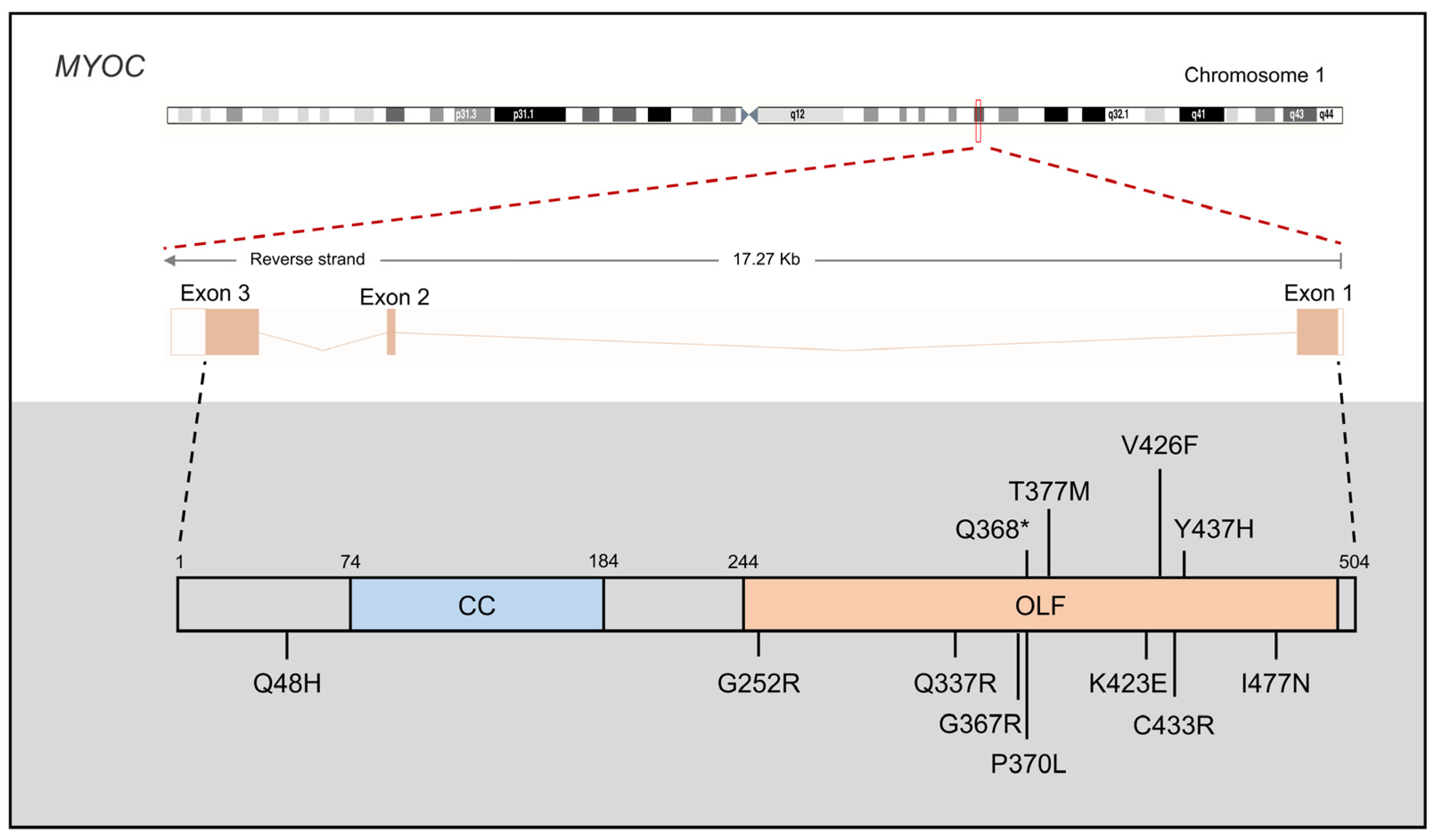
The MYOC gene, also known as the TM-inducible glucocorticoid response (TIGR) gene, is located on chromosome 1q24 and contains three exons. In 1997, Stone et al. initially linked MYOC mutations to JOAG [97]. While MYOC mutations can lead to both JOAG and adult-onset POAG, the majority of these mutations result in JOAG, often manifesting as a familial disease [2]. Currently, over 250 MYOC mutations have been reported, of which 37.7% are considered pathogenic [98]. Notably, approximately 97% of disease-causing MYOC mutations localize to exon 3, which encodes the olfactomedin homology domain (myocilin allele-specific phenotype database: myocilin.com, accessed on 24 January 2024). Although most pathogenic MYOC mutations are missense alleles (84%), the most common MYOC mutation identified in JOAG is a nonsense mutation known as p.Gln368stop, causing protein aggregation [98]. In addition to being a prevalent JOAG-causing mutation, MYOC p.Pro370Leu is associated with severe pathology [99], and MYOC p.Tyr437His is correlated with the form of glaucoma characterized by an earlier age of onset and significantly elevated IOP levels [100]. Moreover, nine additional MYOC mutations have been identified by multiple independent research groups (Figure 5). Furthermore, certain MYOC mutations, including p.N57D [101], p.C245Y [102], p.P274R [103], p.I345M [104], p.T377R [105], p.D384H [106], p.E385K [107], p.Y453Mfs*11 [104], and p.I499S [101], have been exclusively reported by single research teams.

3.1.2. MYOC Protein Function in PCG

MYOC, a secreted glycoprotein, is expressed in various eye structures, including the retina, ciliary body, and TM, with the highest mRNA level detected in TM tissue [108,109,110,111]. As well as the expression of MYOC is also demonstrated in skeletal muscles, heart, brain, and testes [112]. Despite its widespread expression, MYOC does not appear essential for ocular health, as evidenced by both KO animal models and individuals with homozygous, likely null mutations who do not exhibit glaucoma phenotypes [113,114]. Notably, the expression of MYOC is influenced by several molecules, including steroids, TGF-ꞵ1, and the protein optineurin, particularly within cultured TM cells [115].
Molecular and biochemical studies have elucidated that mutated MYOC tends to accumulate in the endoplasmic reticulum (ER) rather than proper release, leading to activation of the unfolded protein response (UPR) and subsequent ER stress [112,116]. Under normal circumstances, the UPR triggers reduced translation and increased chaperone levels to aid in proper protein folding and secretion [117]. However, in cases of overwhelming misfolding, cell apoptosis may ensue [117]. TM cells, particularly sensitive to prolonged ER stress, may eventually perish, thereby contributing to elevated IOP and the development of glaucoma in individuals with MYOC mutations [112].

3.1.3. MYOC Mouse Models

Various research groups have utilized mouse genetics to investigate the role of MYOC in both normal and pathological retinal functions. In both 2001 and 2004, Kim et al. and Gould et al., respectively, demonstrated that MYOC mutations are gain-of-function mutations, as neither KO nor transgenic Myoc mouse model did not lead to a glaucoma phenotype or elevated IOP [114,118]. Consequently, subsequent investigations focused on examining the disease-specific MYOC p.Y437H mutation in mice, responsible for a severe form of JOAG [100]. Initially, expression of human MYOC p.Y437H and the corresponding mouse Myoc mutation in mouse lens did not lead to elevated IOP or RGC loss [3]. Until 2007, Shepard et al. crucially observed elevated IOP upon adenovirus-mediated expression of human MYOC p.Y437H in the iridocorneal angle [119]. Notably, human MYOC contains a peroxisome targeting signal absent in mouse Myoc, which is critical for mutant MYOC induced toxicity in TM cells; thus, the corresponding mouse Myoc did not develop the glaucoma phenotype. Subsequent studies from independent groups confirmed IOP elevation using various strategies to express human MYOC p.Y437H in mouse eyes, suggesting alternative pathogenic mechanisms for other MYOC mutations [120].

4. Discussion

Currently, six genes are known to be implicated in childhood primary glaucoma, with mutation carriers often exhibiting variable phenotypes. These genes display diverse inheritance patterns: CYP1B1 and LTBP2 follow an autosomal recessive pattern, while TEK and ANGPT1 adhere to autosomal dominant inheritance. FOXC1 and MYOC mutations are detected in both autosomal dominant cases and sporadic PCG instances. The prevalence of CYP1B1 mutations in PCG varies significantly across populations, with higher frequencies seen in regions characterized by high consanguinity [121]. Similarly, most LTBP2 mutations leading to PCG are concentrated in areas with pronounced consanguinity, notably Pakistan and Iran. Numerous genetic and molecular studies have provided insights into the biological processes underlying ophthalmic disorders. This review focused on these gene-associated mutations in childhood primary glaucoma, their protein function, and relevant mouse models.
Childhood glaucoma forms typically manifest with elevated IOP. Nevertheless, familial instances of normal-tension glaucoma also exist [122]. OPTN, TBK1, and METTL23 are responsible for early onset normal-tension glaucoma with autosomal dominant inheritance, characterized by significant optic atrophy despite normal IOP [70,123]. OPTN and TBK1 are pivotal in critical cellular processes, notably autophagy and NF-kB signaling, with the encoded proteins known to interact [123]. In addition, METTL23 catalyzes H3R17 dimethylation in the retina, which is critical for RGC homeostasis by negatively regulating NF-κB–mediated TNF-α and IL-1β feedback [70]. These findings suggest that genes encoding NTG-related proteins or those involved in autophagy and NF-kB signaling may also contribute to childhood glaucoma.
Furthermore, PCG is classified under ASD, characterized by congenital improper development of anterior ocular tissues [120]. ASD, in any form, elevates the risk of childhood glaucoma by 50% [124]. Subtle anterior segment abnormalities in early childhood significantly contribute to later-in-life IOP elevation, independent of clinical severity [125]. The multifactorial etiology involves genetic variants, which remain incompletely understood at the cellular and molecular levels despite advances in sequencing technology. Continued research is crucial for identifying gene functions in normal development and understanding how mutations contribute to ASD, enabling the development of diagnostic and screening tests to identify at risk individuals before irreversible optic nerve damage occurs.
Moreover, primary childhood glaucoma patients may have pathogenetic mutations in one or more causing genes, with potential regulatory or interactional relationships among them. CYP1B1 may act as a modifier gene for MYOC expression, and a digenic mode of inheritance involving CYP1B1 and MYOC, as well as CYP1B1 and TEK, has been considered [126]. Currently, genetic testing yields a 40% chance of identifying a genetic cause [127]. Further exploration into the intricate genetic mechanisms underlying PCG is imperative for enhancing diagnostic accuracy and developing targeted therapies to manage the condition and improve patient outcomes effectively.

Author Contributions

Conceptualization, T.I.; writing—original draft preparation, Y.P.; writing—review and editing, T.I. and Y.P.; visualization, Y.P.; supervision, T.I.; funding acquisition, T.I. and Y.P. All authors have read and agreed to the published version of the manuscript.

Funding

This research was funded by the Japan Agency for Medical Research and Development (AMED, 15ek0109072h0003, 16ek0109072h0003, 17ek0109282s0001, 18ek0109282h0002, 19ek0109282h0003); the Japanese Ministry of Health, Labour and Welfare (H22-Kankaku-Ippan-002); and the National Hospital Organization (R2-NHO(Kankaku)-02). This work was also supported by grants to Y.P. from the Japan Society for the Promotion of Science (JSPS KAKENHI, Grant Numbers 20K18366 and 23K15923).

Institutional Review Board Statement

Not applicable.

Informed Consent Statement

Not applicable.

Data Availability Statement

Not applicable.

Conflicts of Interest

The authors declare no conflict of interest.

References

  1. Minegishi, Y.; Nakayama, M.; Iejima, D.; Kawase, K.; Iwata, T. Significance of Optineurin Mutations in Glaucoma and Other Diseases. Prog. Retin. Eye Res. 2016, 55, 149–181. [Google Scholar] [CrossRef] [PubMed]
  2. Wiggs, J.L.; Pasquale, L.R. Genetics of Glaucoma. Hum. Mol. Genet. 2017, 26, R21–R27. [Google Scholar] [CrossRef] [PubMed]
  3. Pan, Y.; Iwata, T. Molecular Genetics of Inherited Normal Tension Glaucoma. Indian J. Ophthalmol. 2024. [Google Scholar] [CrossRef] [PubMed]
  4. Knight, L.S.W.; Ruddle, J.B.; Taranath, D.A.; Goldberg, I.; Smith, J.E.H.; Gole, G.; Chiang, M.Y.; Willett, F.; D’Mellow, G.; Breen, J.; et al. Childhood and Early Onset Glaucoma Classification and Genetic Profile in a Large Australasian Disease Registry. Ophthalmology 2021, 128, 1549–1560. [Google Scholar] [CrossRef] [PubMed]
  5. Weinreb, R.N.; World Glaucoma Association (Eds.) Childhood Glaucoma: The 9th Consensus Report of the World Glaucoma Association; [...Vancouver, July, 17–20, 2013]; Consensus Series/World Glaucoma Association; Kugler: Amsterdam, The Netherlands, 2013; ISBN 978-90-6299-239-3. [Google Scholar]
  6. Karaconji, T.; Zagora, S.; Grigg, J.R. Approach to Childhood Glaucoma: A Review. Clin. Exp. Ophthalmol. 2022, 50, 232–246. [Google Scholar] [CrossRef] [PubMed]
  7. Mokbel, T.H.; El Hefney, E.M.; Hagras, S.M.; ALNagdy, A.A.; Badawi, A.E.; Kasem, M.A.; El Shaer, S.M. Childhood Glaucoma Profile in Dakahelia, Egypt: A Retrospective Study. Int. J. Ophthalmol. 2018, 11, 674–680. [Google Scholar] [CrossRef] [PubMed]
  8. El Sayed, Y.M.; Elhusseiny, A.M.; Gawdat, G.I.; Esmael, A.F.; Elhilali, H.M. Childhood Glaucoma Profile in a Tertiary Centre in Egypt According to the Childhood Glaucoma Research Network Classification. PLoS ONE 2023, 18, e0279874. [Google Scholar] [CrossRef]
  9. Hoguet, A.; Grajewski, A.; Hodapp, E.; Chang, T.P. A Retrospective Survey of Childhood Glaucoma Prevalence According to Childhood Glaucoma Research Network Classification. Indian J. Ophthalmol. 2016, 64, 118. [Google Scholar] [CrossRef] [PubMed]
  10. Lopes, N.L.; Gracitelli, C.P.B.; Rolim-de-Moura, C. Childhood Glaucoma Profile in a Brazilian Tertiary Care Center Using Childhood Glaucoma Research Network Classification. J. Glaucoma 2021, 30, 129–133. [Google Scholar] [CrossRef]
  11. Senthil, S.; Badakere, S.; Ganesh, J.; Krishnamurthy, R.; Dikshit, S.; Choudhari, N.; Garudadri, C.; Mandal, A. Profile of Childhood Glaucoma at a Tertiary Center in South India. Indian J. Ophthalmol. 2019, 67, 358. [Google Scholar] [CrossRef]
  12. Shen, R.; Li, V.S.W.; Wong, M.O.M.; Chan, P.P.M. Pediatric Glaucoma—From Screening, Early Detection to Management. Children 2023, 10, 181. [Google Scholar] [CrossRef] [PubMed]
  13. Lewis, C.J.; Hedberg-Buenz, A.; DeLuca, A.P.; Stone, E.M.; Alward, W.L.M.; Fingert, J.H. Primary Congenital and Developmental Glaucomas. Hum. Mol. Genet. 2017, 26, R28–R36. [Google Scholar] [CrossRef] [PubMed]
  14. Souma, T.; Tompson, S.W.; Thomson, B.R.; Siggs, O.M.; Kizhatil, K.; Yamaguchi, S.; Feng, L.; Limviphuvadh, V.; Whisenhunt, K.N.; Maurer-Stroh, S.; et al. Angiopoietin Receptor TEK Mutations Underlie Primary Congenital Glaucoma with Variable Expressivity. J. Clin. Investig. 2016, 126, 2575–2587. [Google Scholar] [CrossRef]
  15. Lim, S.-H.; Tran-Viet, K.-N.; Yanovitch, T.L.; Freedman, S.F.; Klemm, T.; Call, W.; Powell, C.; Ravichandran, A.; Metlapally, R.; Nading, E.B.; et al. CYP1B1, MYOC, and LTBP2 Mutations in Primary Congenital Glaucoma Patients in the United States. Am. J. Ophthalmol. 2013, 155, 508–517.e5. [Google Scholar] [CrossRef] [PubMed]
  16. Fan, B.J.; Wiggs, J.L. Glaucoma: Genes, Phenotypes, and New Directions for Therapy. J. Clin. Investig. 2010, 120, 3064–3072. [Google Scholar] [CrossRef]
  17. Sarfarazi, M.; Akarsu, N.A.; Hossain, A.; Turacli, E.M.; Aktan, G.S.; Barsoum-Homsy, M.; Chevrette, L.; Sayli, S.B. Assignment of a Locus (GLC3A) for Primary Congenital Glaucoma (Buphthalmos) to 2p21 and Evidence for Genetic Heterogeneity. Genomics 1995, 30, 171–177. [Google Scholar] [CrossRef]
  18. Akarsu, A. A Second Locus (GLC3B) for Primary Congenital Glaucoma (Buphthalmos) Maps to the 1p36 Region. Hum. Mol. Genet. 1996, 5, 1199–1203. [Google Scholar] [CrossRef] [PubMed]
  19. Stoilov, I.; Sarfarazi, M. The Third Genetic Locus (GLC3C) for Primary Congenital Glaucoma (PCG) Maps to Chromosome 14q24.3. Investig. Ophthalmol. Vis. Sci. 2002, 43, 3015. [Google Scholar]
  20. Firasat, S.; Riazuddin, S.A.; Hejtmancik, J.F.; Riazuddin, S. Primary Congenital Glaucoma Localizes to Chromosome 14q24.2-24.3 in Two Consanguineous Pakistani Families. Mol. Vis. 2008, 14, 1659–1665. [Google Scholar]
  21. Shue, A.; Wong, M.O.; Freedman, S.F. Primary Congenital Glaucoma. In Albert and Jakobiec’s Principles and Practice of Ophthalmology; Albert, D., Miller, J., Azar, D., Young, L.H., Eds.; Springer International Publishing: Cham, Switzerland, 2020; pp. 1–40. ISBN 978-3-319-90495-5. [Google Scholar]
  22. Stoilov, I. Identification of Three Different Truncating Mutations in Cytochrome P4501B1 (CYP1B1) as the Principal Cause of Primary Congenital Glaucoma (Buphthalmos) in Families Linked to the GLC3A Locus on Chromosome 2p21. Hum. Mol. Genet. 1997, 6, 641–647. [Google Scholar] [CrossRef]
  23. Ali, M.; McKibbin, M.; Booth, A.; Parry, D.A.; Jain, P.; Riazuddin, S.A.; Hejtmancik, J.F.; Khan, S.N.; Firasat, S.; Shires, M.; et al. Null Mutations in LTBP2 Cause Primary Congenital Glaucoma. Am. J. Hum. Genet. 2009, 84, 664–671. [Google Scholar] [CrossRef] [PubMed]
  24. Narooie-Nejad, M.; Paylakhi, S.H.; Shojaee, S.; Fazlali, Z.; Rezaei Kanavi, M.; Nilforushan, N.; Yazdani, S.; Babrzadeh, F.; Suri, F.; Ronaghi, M.; et al. Loss of Function Mutations in the Gene Encoding Latent Transforming Growth Factor Beta Binding Protein 2, LTBP2, Cause Primary Congenital Glaucoma. Hum. Mol. Genet. 2009, 18, 3969–3977. [Google Scholar] [CrossRef] [PubMed]
  25. Chakrabarti, S.; Kaur, K.; Rao, K.N.; Mandal, A.K.; Kaur, I.; Parikh, R.S.; Thomas, R. The Transcription Factor Gene FOXC1 Exhibits a Limited Role in Primary Congenital Glaucoma. Investig. Ophthalmol. Vis. Sci. 2009, 50, 75. [Google Scholar] [CrossRef] [PubMed]
  26. Thomson, B.R.; Souma, T.; Tompson, S.W.; Onay, T.; Kizhatil, K.; Siggs, O.M.; Feng, L.; Whisenhunt, K.N.; Yanovitch, T.L.; Kalaydjieva, L.; et al. Angiopoietin-1 Is Required for Schlemm’s Canal Development in Mice and Humans. J. Clin. Investig. 2017, 127, 4421–4436. [Google Scholar] [CrossRef] [PubMed]
  27. Tang, Y.M.; Wo, Y.-Y.P.; Stewart, J.; Hawkins, A.L.; Griffin, C.A.; Sutter, T.R.; Greenlee, W.F. Isolation and Characterization of the Human Cytochrome P450 CYP1B1 Gene. J. Biol. Chem. 1996, 271, 28324–28330. [Google Scholar] [CrossRef]
  28. Bejjani, B.A. Multiple CYP1B1 Mutations and Incomplete Penetrance in an Inbred Population Segregating Primary Congenital Glaucoma Suggest Frequent de Novo Events and a Dominant Modifier Locus. Hum. Mol. Genet. 2000, 9, 367–374. [Google Scholar] [CrossRef] [PubMed]
  29. Chitsazian, F.; Tusi, B.K.; Elahi, E.; Saroei, H.A.; Sanati, M.H.; Yazdani, S.; Pakravan, M.; Nilforooshan, N.; Eslami, Y.; Mehrjerdi, M.A.Z.; et al. CYP1B1 Mutation Profile of Iranian Primary Congenital Glaucoma Patients and Associated Haplotypes. J. Mol. Diagn. 2007, 9, 382–393. [Google Scholar] [CrossRef]
  30. Stoilov, I.R.; Costa, V.P.; Vasconcellos, J.P.C.; Melo, M.B.; Betinjane, A.J.; Carani, J.C.E.; Oltrogge, E.V.; Sarfarazi, M. Molecular Genetics of Primary Congenital Glaucoma in Brazil. Investig. Ophthalmol. Vis. Sci. 2002, 43, 1820–1827. [Google Scholar]
  31. Chakrabarti, S.; Kaur, K.; Mandal, A. Primary Congenital Glaucoma and the Involvement of CYP1B1. Middle East. Afr. J. Ophthalmol. 2011, 18, 7. [Google Scholar] [CrossRef]
  32. Cascella, R.; Strafella, C.; Germani, C.; Novelli, G.; Ricci, F.; Zampatti, S.; Giardina, E. The Genetics and the Genomics of Primary Congenital Glaucoma. BioMed Res. Int. 2015, 2015, 321291. [Google Scholar] [CrossRef]
  33. Ben-Omran, T.; Al Ghanim, K.; Yavarna, T.; El Akoum, M.; Samara, M.; Chandra, P.; Al-Dewik, N. Effects of Consanguinity in a Cohort of Subjects with Certain Genetic Disorders in Qatar. Mol. Genet. Genom. Med. 2020, 8, e1051. [Google Scholar] [CrossRef]
  34. Jalal Abbasi-Shavazi, M.; McDonald, P.; Hosseini-Chavoshi, M. Modernization or Cultural Maintenance: The Practice of Consanguineous Marriage in Iran. J. Biosoc. Sci. 2008, 40, 911–933. [Google Scholar] [CrossRef]
  35. Bittles, A.H.; Black, M.L. Consanguinity, Human Evolution, and Complex Diseases. Proc. Natl. Acad. Sci. USA 2010, 107, 1779–1786. [Google Scholar] [CrossRef] [PubMed]
  36. Shah, M.; Bouhenni, R.; Benmerzouga, I. Geographical Variability in CYP1B1 Mutations in Primary Congenital Glaucoma. JCM 2022, 11, 2048. [Google Scholar] [CrossRef] [PubMed]
  37. Ou, Z.; Liu, G.; Liu, W.; Deng, Y.; Zheng, L.; Zhang, S.; Feng, G. Bioinformatics Analysis of CYP1B1 Mutation Hotspots in Chinese Primary Congenital Glaucoma Patients. Biosci. Rep. 2018, 38, BSR20180056. [Google Scholar] [CrossRef]
  38. Mashima, Y.; Suzuki, Y.; Sergeev, Y.; Ohtake, Y.; Tanino, T.; Kimura, I.; Miyata, H.; Aihara, M.; Tanihara, H.; Inatani, M.; et al. Novel Cytochrome P4501B1 (CYP1B1) Gene Mutations in Japanese Patients with Primary Congenital Glaucoma. Investig. Ophthalmol. Vis. Sci. 2001, 42, 2211–2216. [Google Scholar]
  39. Kim, H.-J.; Suh, W.; Park, S.C.; Kim, C.Y.; Park, K.H.; Kook, M.S.; Kim, Y.Y.; Kim, C.-S.; Park, C.K.; Ki, C.-S.; et al. Mutation Spectrum of CYP1B1 and MYOC Genes in Korean Patients with Primary Congenital Glaucoma. Mol. Vis. 2011, 17, 2093–2101. [Google Scholar]
  40. De Melo, M.B.; Mandal, A.K.; Tavares, I.M.; Ali, M.H.; Kabra, M.; De Vasconcellos, J.P.C.; Senthil, S.; Sallum, J.M.F.; Kaur, I.; Betinjane, A.J.; et al. Genotype-Phenotype Correlations in CYP1B1-Associated Primary Congenital Glaucoma Patients Representing Two Large Cohorts from India and Brazil. PLoS ONE 2015, 10, e0127147. [Google Scholar] [CrossRef] [PubMed]
  41. López-Garrido, M.-P.; Medina-Trillo, C.; Morales-Fernandez, L.; Garcia-Feijoo, J.; Martínez-de-la-Casa, J.-M.; García-Antón, M.; Escribano, J. Null CYP1B1 Genotypes in Primary Congenital and Nondominant Juvenile Glaucoma. Ophthalmology 2013, 120, 716–723. [Google Scholar] [CrossRef]
  42. Li, F.; Zhu, W.; Gonzalez, F.J. Potential Role of CYP1B1 in the Development and Treatment of Metabolic Diseases. Pharmacol. Ther. 2017, 178, 18–30. [Google Scholar] [CrossRef]
  43. Choudhary, D.; Jansson, I.; Sarfarazi, M.; Schenkman, J.B. Physiological Significance and Expression of P450s in the Developing Eye. Drug Metab. Rev. 2006, 38, 337–352. [Google Scholar] [CrossRef] [PubMed]
  44. Doshi, M.; Marcus, C.; Bejjani, B.A.; Edward, D.P. Immunolocalization of CYP1B1 in Normal, Human, Fetal and Adult Eyes. Exp. Eye Res. 2006, 82, 24–32. [Google Scholar] [CrossRef] [PubMed]
  45. Zhao, Y.; Wang, S.; Sorenson, C.M.; Teixeira, L.; Dubielzig, R.R.; Peters, D.M.; Conway, S.J.; Jefcoate, C.R.; Sheibani, N. Cyp1b1 Mediates Periostin Regulation of Trabecular Meshwork Development by Suppression of Oxidative Stress. Mol. Cell. Biol. 2013, 33, 4225–4240. [Google Scholar] [CrossRef] [PubMed]
  46. Falero-Perez, J.; Song, Y.-S.; Sorenson, C.M.; Sheibani, N. CYP1B1: A Key Regulator of Redox Homeostasis. Trends Cell Mol. Biol. 2018, 13, 27–45. [Google Scholar]
  47. Palenski, T.L.; Sorenson, C.M.; Jefcoate, C.R.; Sheibani, N. Lack of Cyp1b1 Promotes the Proliferative and Migratory Phenotype of Perivascular Supporting Cells. Lab. Investig. 2013, 93, 646–662. [Google Scholar] [CrossRef]
  48. Tang, Y.; Scheef, E.A.; Wang, S.; Sorenson, C.M.; Marcus, C.B.; Jefcoate, C.R.; Sheibani, N. CYP1B1 Expression Promotes the Proangiogenic Phenotype of Endothelium through Decreased Intracellular Oxidative Stress and Thrombospondin-2 Expression. Blood 2009, 113, 744–754. [Google Scholar] [CrossRef] [PubMed]
  49. Rieder, C.R.; Ramsden, D.B.; Williams, A.C. Cytochrome P450 1B1 mRNA in the Human Central Nervous System. Mol. Pathol. 1998, 51, 138–142. [Google Scholar] [CrossRef] [PubMed]
  50. Choudhary, D.; Jansson, I.; Stoilov, I.; Sarfarazi, M.; Schenkman, J.B. Metabolism of Retinoids and Arachidonic Acid by Human and Mouse Cytochrome P450 1b1. Drug Metab. Dispos. 2004, 32, 840–847. [Google Scholar] [CrossRef]
  51. Korkmaz, A.; Topal, T.; Tan, D.-X.; Reiter, R.J. Role of Melatonin in Metabolic Regulation. Rev. Endocr. Metab. Disord. 2009, 10, 261–270. [Google Scholar] [CrossRef]
  52. Alkozi, H.A.; Navarro, G.; Franco, R.; Pintor, J. Melatonin and the Control of Intraocular Pressure. Prog. Retin. Eye Res. 2020, 75, 100798. [Google Scholar] [CrossRef]
  53. Chang, T.K.H.; Chen, J.; Yang, G.; Yeung, E.Y.H. Inhibition of Procarcinogen-bioactivating Human CYP1A1, CYP1A2 and CYP1B1 Enzymes by Melatonin. J. Pineal Res. 2010, 48, 55–64. [Google Scholar] [CrossRef]
  54. Malik, K.U.; Jennings, B.L.; Yaghini, F.A.; Sahan-Firat, S.; Song, C.Y.; Estes, A.M.; Fang, X.R. Contribution of Cytochrome P450 1B1 to Hypertension and Associated Pathophysiology: A Novel Target for Antihypertensive Agents. Prostaglandins Other Lipid Mediat. 2012, 98, 69–74. [Google Scholar] [CrossRef] [PubMed]
  55. Masferrer, J.L.; Dunn, M.W.; Schwartzman, M.L. 12(R)-Hydroxyeicosatetraenoic Acid, an Endogenous Corneal Arachidonate Metabolite, Lowers Intraocular Pressure in Rabbits. Investig. Ophthalmol. Vis. Sci. 1990, 31, 535–539. [Google Scholar]
  56. Aribindi, K.; Guerra, Y.; Lee, R.K.; Bhattacharya, S.K. Comparative Phospholipid Profiles of Control and Glaucomatous Human Trabecular Meshwork. Investig. Ophthalmol. Vis. Sci. 2013, 54, 3037. [Google Scholar] [CrossRef] [PubMed]
  57. Stoilov, I.; Rezaie, T.; Jansson, I.; Schenkman, J.B.; Sarfarazi, M. Expression of Cytochrome P4501b1 (Cyp1b1) during Early Murine Development. Mol. Vis. 2004, 10, 629–636. [Google Scholar] [PubMed]
  58. Sheibani, N.; Zhao, Y.; Sorenson, C. Cytochrome P450 1B1 and Primary Congenital Glaucoma. J. Ophthalmic Vis. Res. 2015, 10, 60. [Google Scholar] [CrossRef] [PubMed]
  59. Buters, J.T.M.; Sakai, S.; Richter, T.; Pineau, T.; Alexander, D.L.; Savas, U.; Doehmer, J.; Ward, J.M.; Jefcoate, C.R.; Gonzalez, F.J. Cytochrome P450 CYP1B1 Determines Susceptibility to 7,12-Dimethylbenz[a]Anthracene-Induced Lymphomas. Proc. Natl. Acad. Sci. USA 1999, 96, 1977–1982. [Google Scholar] [CrossRef] [PubMed]
  60. Libby, R.T.; Smith, R.S.; Savinova, O.V.; Zabaleta, A.; Martin, J.E.; Gonzalez, F.J.; John, S.W.M. Modification of Ocular Defects in Mouse Developmental Glaucoma Models by Tyrosinase. Science 2003, 299, 1578–1581. [Google Scholar] [CrossRef] [PubMed]
  61. Teixeira, L.B.C.; Zhao, Y.; Dubielzig, R.R.; Sorenson, C.M.; Sheibani, N. Ultrastructural Abnormalities of the Trabecular Meshwork Extracellular Matrix in Cyp1b1-Deficient Mice. Vet. Pathol. 2015, 52, 397–403. [Google Scholar] [CrossRef]
  62. Amirmokhtari, N.; Foresi, B.D.; Dewan, S.S.; Bouhenni, R.A.; Smith, M.A. Absence of Cytochrome P450-1b1 Increases Susceptibility of Pressure-Induced Axonopathy in the Murine Retinal Projection. Front. Cell Dev. Biol. 2021, 9, 636321. [Google Scholar] [CrossRef]
  63. Williams, A.L.; Eason, J.; Chawla, B.; Bohnsack, B.L. Cyp1b1 Regulates Ocular Fissure Closure Through a Retinoic Acid–Independent Pathway. Investig. Ophthalmol. Vis. Sci. 2017, 58, 1084. [Google Scholar] [CrossRef] [PubMed]
  64. Azmanov, D.N.; Dimitrova, S.; Florez, L.; Cherninkova, S.; Draganov, D.; Morar, B.; Saat, R.; Juan, M.; Arostegui, J.I.; Ganguly, S.; et al. LTBP2 and CYP1B1 Mutations and Associated Ocular Phenotypes in the Roma/Gypsy Founder Population. Eur. J. Hum. Genet. 2011, 19, 326–333. [Google Scholar] [CrossRef]
  65. Micheal, S.; Siddiqui, S.N.; Zafar, S.N.; Iqbal, A.; Khan, M.I.; Den Hollander, A.I. Identification of Novel Variants in LTBP2 and PXDN Using Whole-Exome Sequencing in Developmental and Congenital Glaucoma. PLoS ONE 2016, 11, e0159259. [Google Scholar] [CrossRef] [PubMed]
  66. Rauf, B.; Irum, B.; Khan, S.Y.; Kabir, F.; Naeem, M.A.; Riazuddin, S.; Ayyagari, R.; Riazuddin, S.A. Novel Mutations in LTBP2 Identified in Familial Cases of Primary Congenital Glaucoma. Mol. Vis. 2020, 26, 14–25. [Google Scholar] [PubMed]
  67. Saharinen, J.; Hyytiäinen, M.; Taipale, J.; Keski-Oja, J. Latent Transforming Growth Factor-β Binding Proteins (LTBPs)—Structural Extracellular Matrix Proteins for Targeting TGF-β Action. Cytokine Growth Factor Rev. 1999, 10, 99–117. [Google Scholar] [CrossRef] [PubMed]
  68. Robertson, I.B.; Horiguchi, M.; Zilberberg, L.; Dabovic, B.; Hadjiolova, K.; Rifkin, D.B. Latent TGF-β-Binding Proteins. Matrix Biol. 2015, 47, 44–53. [Google Scholar] [CrossRef] [PubMed]
  69. Hirani, R.; Hanssen, E.; Gibson, M.A. LTBP-2 Specifically Interacts with the Amino-Terminal Region of Fibrillin-1 and Competes with LTBP-1 for Binding to This Microfibrillar Protein. Matrix Biol. 2007, 26, 213–223. [Google Scholar] [CrossRef] [PubMed]
  70. Pan, Y.; Suga, A.; Kimura, I.; Kimura, C.; Minegishi, Y.; Nakayama, M.; Yoshitake, K.; Iejima, D.; Minematsu, N.; Yamamoto, M.; et al. METTL23 Mutation Alters Histone H3R17 Methylation in Normal-Tension Glaucoma. J. Clin. Investig. 2022, 132, e153589. [Google Scholar] [CrossRef] [PubMed]
  71. Tandon, P.N.; Autar, R. Flow of Aqueous Humor in the Canal of Schlemm. Math. Biosci. 1989, 93, 53–78. [Google Scholar] [CrossRef]
  72. Carreon, T.; Van Der Merwe, E.; Fellman, R.L.; Johnstone, M.; Bhattacharya, S.K. Aqueous Outflow—A Continuum from Trabecular Meshwork to Episcleral Veins. Prog. Retin. Eye Res. 2017, 57, 108–133. [Google Scholar] [CrossRef]
  73. Mahmud, N.; Eisner, C.; Purushothaman, S.; Storer, M.A.; Kaplan, D.R.; Miller, F.D. Nail-Associated Mesenchymal Cells Contribute to and Are Essential for Dorsal Digit Tip Regeneration. Cell Rep. 2022, 41, 111853. [Google Scholar] [CrossRef] [PubMed]
  74. Désir, J.; Sznajer, Y.; Depasse, F.; Roulez, F.; Schrooyen, M.; Meire, F.; Abramowicz, M. LTBP2 Null Mutations in an Autosomal Recessive Ocular Syndrome with Megalocornea, Spherophakia, and Secondary Glaucoma. Eur. J. Hum. Genet. 2010, 18, 761–767. [Google Scholar] [CrossRef] [PubMed]
  75. Inoue, T.; Ohbayashi, T.; Fujikawa, Y.; Yoshida, H.; Akama, T.O.; Noda, K.; Horiguchi, M.; Kameyama, K.; Hata, Y.; Takahashi, K.; et al. Latent TGF-β Binding Protein-2 Is Essential for the Development of Ciliary Zonule Microfibrils. Hum. Mol. Genet. 2014, 23, 5672–5682. [Google Scholar] [CrossRef] [PubMed]
  76. Shipley, J.M.; Mecham, R.P.; Maus, E.; Bonadio, J.; Rosenbloom, J.; McCarthy, R.T.; Baumann, M.L.; Frankfater, C.; Segade, F.; Shapiro, S.D. Developmental Expression of Latent Transforming Growth Factor β Binding Protein 2 and Its Requirement Early in Mouse Development. Mol. Cell. Biol. 2000, 20, 4879–4887. [Google Scholar] [CrossRef] [PubMed]
  77. Saharinen, P.; Eklund, L.; Alitalo, K. Therapeutic Targeting of the Angiopoietin–TIE Pathway. Nat. Rev. Drug Discov. 2017, 16, 635–661. [Google Scholar] [CrossRef]
  78. Kizhatil, K.; Ryan, M.; Marchant, J.K.; Henrich, S.; John, S.W.M. Schlemm’s Canal Is a Unique Vessel with a Combination of Blood Vascular and Lymphatic Phenotypes That Forms by a Novel Developmental Process. PLoS Biol. 2014, 12, e1001912. [Google Scholar] [CrossRef] [PubMed]
  79. Thomson, B.R.; Heinen, S.; Jeansson, M.; Ghosh, A.K.; Fatima, A.; Sung, H.-K.; Onay, T.; Chen, H.; Yamaguchi, S.; Economides, A.N.; et al. A Lymphatic Defect Causes Ocular Hypertension and Glaucoma in Mice. J. Clin. Investig. 2014, 124, 4320–4324. [Google Scholar] [CrossRef] [PubMed]
  80. Young, T.L.; Whisenhunt, K.N.; Jin, J.; LaMartina, S.M.; Martin, S.M.; Souma, T.; Limviphuvadh, V.; Suri, F.; Souzeau, E.; Zhang, X.; et al. SVEP1 as a Genetic Modifier of TEK-Related Primary Congenital Glaucoma. Investig. Ophthalmol. Vis. Sci. 2020, 61, 6. [Google Scholar] [CrossRef]
  81. Qiao, Y.; Chen, Y.; Tan, C.; Sun, X.; Chen, X.; Chen, J. Screening and Functional Analysis of TEK Mutations in Chinese Children With Primary Congenital Glaucoma. Front. Genet. 2021, 12, 764509. [Google Scholar] [CrossRef]
  82. Choquet, H.; Paylakhi, S.; Kneeland, S.C.; Thai, K.K.; Hoffmann, T.J.; Yin, J.; Kvale, M.N.; Banda, Y.; Tolman, N.G.; Williams, P.A.; et al. A Multiethnic Genome-Wide Association Study of Primary Open-Angle Glaucoma Identifies Novel Risk Loci. Nat. Commun. 2018, 9, 2278. [Google Scholar] [CrossRef]
  83. Van Zyl, T.; Yan, W.; McAdams, A.; Peng, Y.-R.; Shekhar, K.; Regev, A.; Juric, D.; Sanes, J.R. Cell Atlas of Aqueous Humor Outflow Pathways in Eyes of Humans and Four Model Species Provides Insight into Glaucoma Pathogenesis. Proc. Natl. Acad. Sci. USA 2020, 117, 10339–10349. [Google Scholar] [CrossRef] [PubMed]
  84. Koh, G.Y. Orchestral Actions of Angiopoietin-1 in Vascular Regeneration. Trends Mol. Med. 2013, 19, 31–39. [Google Scholar] [CrossRef] [PubMed]
  85. Toris, C.B.; Yablonski, M.E.; Wang, Y.-L.; Camras, C.B. Aqueous Humor Dynamics in the Aging Human Eye. Am. J. Ophthalmol. 1999, 127, 407–412. [Google Scholar] [CrossRef] [PubMed]
  86. Alm, A.; Nilsson, S.F.E. Uveoscleral Outflow—A Review. Exp. Eye Res. 2009, 88, 760–768. [Google Scholar] [CrossRef] [PubMed]
  87. Pan, Y.; Fu, Y.; Baird, P.N.; Guymer, R.H.; Das, T.; Iwata, T. Exploring the Contribution of ARMS2 and HTRA1 Genetic Risk Factors in Age-Related Macular Degeneration. Prog. Retin. Eye Res. 2023, 97, 101159. [Google Scholar] [CrossRef] [PubMed]
  88. Golson, M.L.; Kaestner, K.H. Fox Transcription Factors: From Development to Disease. Development 2016, 143, 4558–4570. [Google Scholar] [CrossRef] [PubMed]
  89. Kaushik, S.; Dubey, S.; Choudhary, S.; Ratna, R.; Pandav, S.; Khan, A. Anterior Segment Dysgenesis: Insights into the Genetics and Pathogenesis. Indian J. Ophthalmol. 2022, 70, 2293. [Google Scholar] [CrossRef] [PubMed]
  90. Siggs, O.M.; Souzeau, E.; Pasutto, F.; Dubowsky, A.; Smith, J.E.H.; Taranath, D.; Pater, J.; Rait, J.L.; Narita, A.; Mauri, L.; et al. Prevalence of FOXC1 Variants in Individuals With a Suspected Diagnosis of Primary Congenital Glaucoma. JAMA Ophthalmol. 2019, 137, 348. [Google Scholar] [CrossRef] [PubMed]
  91. Smith, R.S. Haploinsufficiency of the Transcription Factors FOXC1 and FOXC2 Results in Aberrant Ocular Development. Hum. Mol. Genet. 2000, 9, 1021–1032. [Google Scholar] [CrossRef]
  92. Umali, J.; Hawkey-Noble, A.; French, C.R. Loss of Foxc1 in Zebrafish Reduces Optic Nerve Size and Cell Number in the Retinal Ganglion Cell Layer. Vis. Res. 2019, 156, 66–72. [Google Scholar] [CrossRef]
  93. Sheffield, V.C.; Stone, E.M.; Alward, W.L.M.; Drack, A.V.; Johnson, A.T.; Streb, L.M.; Nichols, B.E. Genetic Linkage of Familial Open Angle Glaucoma to Chromosome 1q21–Q31. Nat. Genet. 1993, 4, 47–50. [Google Scholar] [CrossRef] [PubMed]
  94. Wiggs, J.L.; Lynch, S.; Ynagi, G.; Maselli, M.; Auguste, J.; Del Bono, E.A.; Olson, L.M.; Haines, J.L. A Genomewide Scan Identifies Novel Early-Onset Primary Open-Angle Glaucoma Loci on 9q22 and 20p12. Am. J. Hum. Genet. 2004, 74, 1314–1320. [Google Scholar] [CrossRef] [PubMed]
  95. Pang, C.P.; Fan, B.J.; Canlas, O.; Wang, D.Y.; Dubois, S.; Tam, P.O.S.; Lam, D.S.C.; Raymond, V.; Ritch, R. A Genome-Wide Scan Maps a Novel Juvenile-Onset Primary Open Angle Glaucoma Locus to Chromosome 5q. Mol. Vis. 2006, 12, 85–92. [Google Scholar] [PubMed]
  96. Wang, D.Y.; Fan, B.J.; Chua, J.K.H.; Tam, P.O.S.; Leung, C.K.S.; Lam, D.S.C.; Pang, C.P. A Genome-Wide Scan Maps a Novel Juvenile-Onset Primary Open-Angle Glaucoma Locus to 15q. Investig. Ophthalmol. Vis. Sci. 2006, 47, 5315. [Google Scholar] [CrossRef] [PubMed]
  97. Stone, E.M.; Fingert, J.H.; Alward, W.L.M.; Nguyen, T.D.; Polansky, J.R.; Sunden, S.L.F.; Nishimura, D.; Clark, A.F.; Nystuen, A.; Nichols, B.E.; et al. Identification of a Gene That Causes Primary Open Angle Glaucoma. Science 1997, 275, 668–670. [Google Scholar] [CrossRef] [PubMed]
  98. Wang, H.; Li, M.; Zhang, Z.; Xue, H.; Chen, X.; Ji, Y. Physiological Function of Myocilin and Its Role in the Pathogenesis of Glaucoma in the Trabecular Meshwork (Review). Int. J. Mol. Med. 2018, 43, 671–681. [Google Scholar] [CrossRef]
  99. Gupta, V.; Somarajan, B.I.; Gupta, S.; Chaurasia, A.K.; Kumar, S.; Dutta, P.; Gupta, V.; Sharma, A.; Tayo, B.O.; Nischal, K. The Inheritance of Juvenile Onset Primary Open Angle Glaucoma. Clin. Genet. 2017, 92, 134–142. [Google Scholar] [CrossRef] [PubMed]
  100. Alward, W.L.M.; Fingert, J.H.; Coote, M.A.; Johnson, A.T.; Lerner, S.F.; Junqua, D.; Durcan, F.J.; McCartney, P.J.; Mackey, D.A.; Sheffield, V.C.; et al. Clinical Features Associated with Mutations in the Chromosome 1 Open-Angle Glaucoma Gene (GLC1A). N. Engl. J. Med. 1998, 338, 1022–1027. [Google Scholar] [CrossRef] [PubMed]
  101. Shimizu, S.; Lichter, P.R.; Johnson, A.T.; Zhou, Z.; Higashi, M.; Gottfredsdottir, M.; Othman, M.; Moroi, S.E.; Rozsa, F.W.; Schertzer, R.M.; et al. Age-Dependent Prevalence of Mutations at the GLC1A Locus in Primary Open-Angle Glaucoma. Am. J. Ophthalmol. 2000, 130, 165–177. [Google Scholar] [CrossRef]
  102. Fan, B.J.; Leung, D.Y.L.; Wang, D.Y.; Gobeil, S.; Raymond, V.; Tam, P.O.S.; Lam, D.S.C.; Pang, C.P. Novel Myocilin Mutation in a Chinese Family with Juvenile-Onset Open-Angle Glaucoma. Arch. Ophthalmol. 2006, 124, 102–106. [Google Scholar] [CrossRef]
  103. Markandaya, M.; Ramesh, T.K.; Selvaraju, V.; Dorairaj, S.K.; Prakash, R.; Shetty, J.; Kumar, A. Genetic Analysis of an Indian Family with Members Affected with Juvenile-Onset Primary Open-Angle Glaucoma. Ophthalmic Genet. 2004, 25, 11–23. [Google Scholar] [CrossRef] [PubMed]
  104. Svidnicki, P.V.; Braghini, C.A.; Costa, V.P.; Schimiti, R.B.; de Vasconcellos, J.P.C.; de Melo, M.B. Occurrence of MYOC and CYP1B1 Variants in Juvenile Open Angle Glaucoma Brazilian Patients. Ophthalmic Genet. 2018, 39, 717–724. [Google Scholar] [CrossRef] [PubMed]
  105. Waryah, A.M.; Narsani, A.K.; Sheikh, S.A.; Shaikh, H.; Shahani, M.Y. The Novel Heterozygous Thr377Arg MYOC Mutation Causes Severe Juvenile Open Angle Glaucoma in a Large Pakistani Family. Gene 2013, 528, 356–359. [Google Scholar] [CrossRef] [PubMed]
  106. Huang, C.; Xie, L.; Wu, Z.; Cao, Y.; Zheng, Y.; Pang, C.-P.; Zhang, M. Detection of Mutations in MYOC, OPTN, NTF4, WDR36 and CYP1B1 in Chinese Juvenile Onset Open-Angle Glaucoma Using Exome Sequencing. Sci. Rep. 2018, 8, 4498. [Google Scholar] [CrossRef]
  107. Criscione, J.; Ji, W.; Jeffries, L.; McGrath, J.M.; Soloway, S.; Pusztai, L.; Lakhani, S. Identification of a Novel MYOC Variant in a Hispanic Family with Early-Onset Primary Open-Angle Glaucoma with Elevated Intraocular Pressure. Cold Spring Harb. Mol. Case Stud. 2019, 5, a004374. [Google Scholar] [CrossRef] [PubMed]
  108. Takahashi, H.; Noda, S.; Imamura, Y.; Nagasawa, A.; Kubota, R.; Mashima, Y.; Kudoh, J.; Oguchi, Y.; Shimizu, N. Mouse Myocilin (Myoc) Gene Expression in Ocular Tissues. Biochem. Biophys. Res. Commun. 1998, 248, 104–109. [Google Scholar] [CrossRef] [PubMed]
  109. Ortego, J.; Escribano, J.; Coca-Prados, M. Cloning and Characterization of Subtracted cDNAs from a Human Ciliary Body Library Encoding TIGR, a Protein Involved in Juvenile Open Angle Glaucoma with Homology to Myosin and Olfactomedin. FEBS Lett. 1997, 413, 349–353. [Google Scholar] [CrossRef]
  110. Karali, A.; Russell, P.; Stefani, F.H.; Tamm, E.R. Localization of Myocilin/Trabecular Meshwork--Inducible Glucocorticoid Response Protein in the Human Eye. Investig. Ophthalmol. Vis. Sci. 2000, 41, 729–740. [Google Scholar]
  111. Kubota, R.; Noda, S.; Wang, Y.; Minoshima, S.; Asakawa, S.; Kudoh, J.; Mashima, Y.; Oguchi, Y.; Shimizu, N. A Novel Myosin-like Protein (Myocilin) Expressed in the Connecting Cilium of the Photoreceptor: Molecular Cloning, Tissue Expression, and Chromosomal Mapping. Genomics 1997, 41, 360–369. [Google Scholar] [CrossRef]
  112. Jain, A.; Zode, G.; Kasetti, R.B.; Ran, F.A.; Yan, W.; Sharma, T.P.; Bugge, K.; Searby, C.C.; Fingert, J.H.; Zhang, F.; et al. CRISPR-Cas9–Based Treatment of Myocilin-Associated Glaucoma. Proc. Natl. Acad. Sci. USA 2017, 114, 11199–11204. [Google Scholar] [CrossRef]
  113. Wiggs, J.L. Molecular and Clinical Evaluation of a Patient Hemizygous for TIGR/MYOC. Arch. Ophthalmol. 2001, 119, 1674. [Google Scholar] [CrossRef] [PubMed]
  114. Kim, B.S.; Savinova, O.V.; Reedy, M.V.; Martin, J.; Lun, Y.; Gan, L.; Smith, R.S.; Tomarev, S.I.; John, S.W.M.; Johnson, R.L. Targeted Disruption of the Myocilin Gene (Myoc) Suggests That Human Glaucoma-Causing Mutations Are Gain of Function. Mol. Cell. Biol. 2001, 21, 7707–7713. [Google Scholar] [CrossRef]
  115. Tamm, E.R.; Russell, P.; Epstein, D.L.; Johnson, D.H.; Piatigorsky, J. Modulation of Myocilin/TIGR Expression in Human Trabecular Meshwork. Investig. Ophthalmol. Vis. Sci. 1999, 40, 2577–2582. [Google Scholar]
  116. Carbone, M.A.; Ayroles, J.F.; Yamamoto, A.; Morozova, T.V.; West, S.A.; Magwire, M.M.; Mackay, T.F.C.; Anholt, R.R.H. Overexpression of Myocilin in the Drosophila Eye Activates the Unfolded Protein Response: Implications for Glaucoma. PLoS ONE 2009, 4, e4216. [Google Scholar] [CrossRef] [PubMed]
  117. Bravo, R.; Parra, V.; Gatica, D.; Rodriguez, A.E.; Torrealba, N.; Paredes, F.; Wang, Z.V.; Zorzano, A.; Hill, J.A.; Jaimovich, E.; et al. Endoplasmic Reticulum and the Unfolded Protein Response. In International Review of Cell and Molecular Biology; Elsevier: Amsterdam, The Netherlands, 2013; Volume 301, pp. 215–290. ISBN 978-0-12-407704-1. [Google Scholar]
  118. Gould, D.B.; Miceli-Libby, L.; Savinova, O.V.; Torrado, M.; Tomarev, S.I.; Smith, R.S.; John, S.W.M. Genetically Increasing Myoc Expression Supports a Necessary Pathologic Role of Abnormal Proteins in Glaucoma. Mol. Cell. Biol. 2004, 24, 9019–9025. [Google Scholar] [CrossRef] [PubMed]
  119. Shepard, A.R.; Jacobson, N.; Millar, J.C.; Pang, I.-H.; Steely, H.T.; Searby, C.C.; Sheffield, V.C.; Stone, E.M.; Clark, A.F. Glaucoma-Causing Myocilin Mutants Require the Peroxisomal Targeting Signal-1 Receptor (PTS1R) to Elevate Intraocular Pressure. Hum. Mol. Genet. 2007, 16, 609–617. [Google Scholar] [CrossRef] [PubMed]
  120. Fernandes, K.A.; Harder, J.M.; Williams, P.A.; Rausch, R.L.; Kiernan, A.E.; Nair, K.S.; Anderson, M.G.; John, S.W.M.; Howell, G.R.; Libby, R.T. Using Genetic Mouse Models to Gain Insight into Glaucoma: Past Results and Future Possibilities. Exp. Eye Res. 2015, 141, 42–56. [Google Scholar] [CrossRef] [PubMed]
  121. Ling, C.; Zhang, D.; Zhang, J.; Sun, H.; Du, Q.; Li, X. Updates on the Molecular Genetics of Primary Congenital Glaucoma (Review). Exp. Ther. Med. 2020, 20, 968–977. [Google Scholar] [CrossRef]
  122. Gauthier, A.C.; Wiggs, J.L. Childhood Glaucoma Genes and Phenotypes: Focus on FOXC1 Mutations Causing Anterior Segment Dysgenesis and Hearing Loss. Exp. Eye Res. 2020, 190, 107893. [Google Scholar] [CrossRef]
  123. Fox, A.R.; Fingert, J.H. Familial Normal Tension Glaucoma Genetics. Prog. Retin. Eye Res. 2023, 96, 101191. [Google Scholar] [CrossRef]
  124. Sowden, J.C. Molecular and Developmental Mechanisms of Anterior Segment Dysgenesis. Eye 2007, 21, 1310–1318. [Google Scholar] [CrossRef] [PubMed]
  125. Ito, Y.A.; Walter, M.A. Genomics and Anterior Segment Dysgenesis: A Review. Clin. Exp. Ophthalmol. 2014, 42, 13–24. [Google Scholar] [CrossRef] [PubMed]
  126. Mookherjee, S.; Acharya, M.; Banerjee, D.; Bhattacharjee, A.; Ray, K. Molecular Basis for Involvement of CYP1B1 in MYOC Upregulation and Its Potential Implication in Glaucoma Pathogenesis. PLoS ONE 2012, 7, e45077. [Google Scholar] [CrossRef] [PubMed]
  127. Yu-Wai-Man, C.; Arno, G.; Brookes, J.; Garcia-Feijoo, J.; Khaw, P.T.; Moosajee, M. Primary Congenital Glaucoma Including Next-Generation Sequencing-Based Approaches: Clinical Utility Gene Card. Eur. J. Hum. Genet. 2018, 26, 1713–1718. [Google Scholar] [CrossRef] [PubMed]
Figure 1. Gene structure of CYP1B1 and the locations of the most prevalent mutations associated with primary congenital glaucoma (PCG). The most common mutations reported across different populations are indicated in red, while mutations specific to certain populations are shown in black.
Children 11 00454 g001
Figure 2. Gene structure of LTBP2 and variants associated with primary congenital glaucoma (PCG). The LTBP2 gene comprises 36 exons and codes a protein of 1821 amino acids, characterized by EGF-like (E), TB (T), and calcium-binding EGF-like (C) domains.
Children 11 00454 g002
Figure 3. Gene structure of TEK and identified primary congenital glaucoma (PCG) variants. The TEK gene consists of 23 exons and codes a protein of 1124 amino acids, featuring Ig-like (Ig), EGF-like (EGF), fibronectin type III (FN III), and protein kinase (Kinase) domains. Variants reported in PCG patients are depicted, excluding those located in intronic regions: c.1624+5G>A, c.760+2T>C, and c.3300+2delT. Asterisk (*): premature stop codon.
Children 11 00454 g003
Figure 4. Schematic representation of the ANGPT1 gene structure, protein domains, and three reported primary congenital glaucoma (PCG) mutations. The ANGPT1 gene comprises nine exons and codes for a protein of 498 amino acids, characterized by two coiled-coil (CC) domains and one fibrinogen C-terminal domain. Asterisk (*): premature stop codon.
Children 11 00454 g004
Figure 5. Gene structure of MYOC and reported 12 primary congenital glaucoma (PCG) variants by multiple independent research groups. The MYOC gene consists of 3 exons and encodes a protein of 504 amino acids, including one coiled-coil (CC) and one olfactomedin-like (OLF) domain. Asterisk (*): premature stop codon.
Children 11 00454 g005
Disclaimer/Publisher’s Note: The statements, opinions and data contained in all publications are solely those of the individual author(s) and contributor(s) and not of MDPI and/or the editor(s). MDPI and/or the editor(s) disclaim responsibility for any injury to people or property resulting from any ideas, methods, instructions or products referred to in the content.