
Large Numbers for Small Children—Up to What Age Do Infants Benefit from a Longer Echo Time in Cerebral T2 MRI Sequences?

 , ,
, ,
Abstract
:1. Introduction
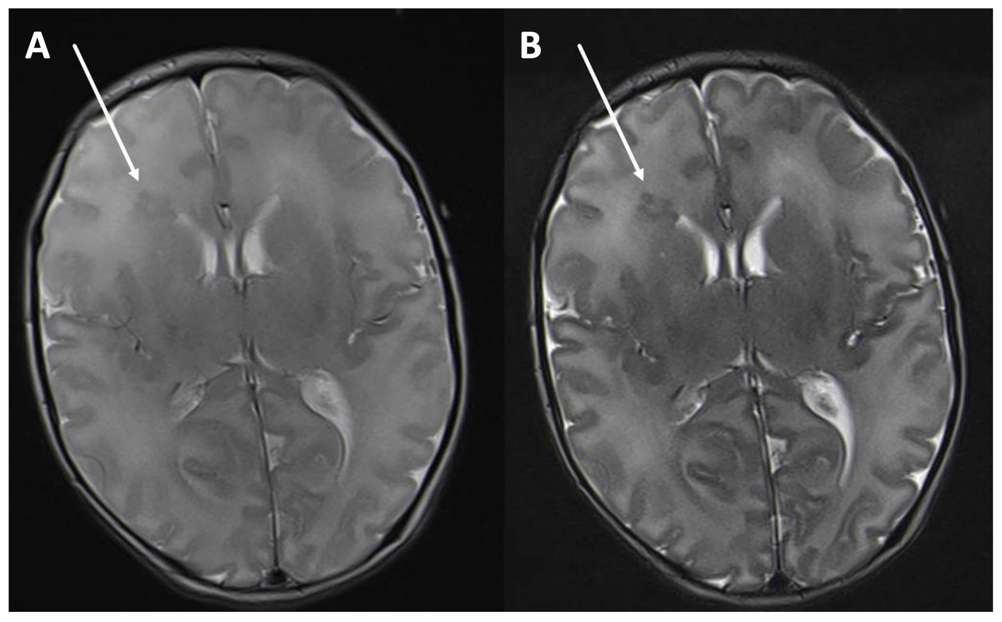
2. Materials and Methods
2.1. Patients
2.2. MRI Protocol
2.3. Quantitative MRI Analysis
3. Results
3.1. Cohort
3.2. Michelson Contrast
4. Discussion
Author Contributions
Funding
Institutional Review Board Statement
Informed Consent Statement
Data Availability Statement
Conflicts of Interest
References
- Dobbing, J.; Sands, J. Quantitative growth and development of human brain. Arch. Dis. Child. 1973, 48, 757–767. [Google Scholar] [CrossRef] [PubMed]
- Rutherford, M.A. MRI of the Neonatal Brain; Gulf Professional Publishing: Houston, TX, USA, 2002. [Google Scholar]
- Barkovich, M.J.; Barkovich, A.J. MR Imaging of Normal Brain Development. Neuroimaging Clin. N. Am. 2019, 29, 325–337. [Google Scholar] [CrossRef]
- Alonso-Ortiz, E.; Levessque, I.R.; Pike, G.B. MRI-Based Myelin Water Imaging: A Technical Review. Magn. Reson. Med. 2015, 73, 70–81. [Google Scholar] [CrossRef]
- Goksan, S.; Hartley, C.; Hurley, S.A.; Winkler, A.M.; Duff, E.P.; Jenkinson, M.; Rogers, R.; Clare, S.; Slater, R. Optimal echo time for functional MRI of the infant brain identified in response to noxious stimulation. Magn. Reson. Med. 2017, 78, 625–631. [Google Scholar] [CrossRef] [PubMed]
- Heath, F.; Hurley, S.A.; Johansen-Berg, H.; Sampaio-Baptista, C. Advances in noninvasive myelin imaging. Dev. Neurobiol. 2018, 78, 136–151. [Google Scholar] [CrossRef] [PubMed]
- Holshouser, B.A.; Ashwal, S.; Shu, S.; Hinshaw, D.B., Jr. Proton MR spectroscopy in children with acute brain injury: Comparison of short and long echo time acquisitions. J. Magn. Reson. Imaging 2000, 11, 9–19. [Google Scholar] [CrossRef]
- Li, T.; Mirowitz, S.A. Fast T2-weighted MR imaging: Impact of variation in pulse sequence parameters on image quality and artifacts. Magn. Reson. Imaging 2003, 21, 745–753. [Google Scholar] [CrossRef] [PubMed]
- Branson, H.M. Normal myelination: A practical pictorial review. Neuroimaging Clin. N. Am. 2013, 23, 183–195. [Google Scholar] [CrossRef] [PubMed]
- Righart, R.; Biberacher, V.; Jonkman, L.E.; Klaver, R.; Schmidt, P.; Buck, D.; Berthele, A.; Kirschke, J.S.; Zimmer, C.; Hemmer, B.; et al. Cortical Pathology in Multiple Sclerosis Detected by the T1/T2-Weighted Ratio from Routine Magnetic Resonance Imaging. Ann. Neurol. 2017, 82, 519–529. [Google Scholar] [CrossRef] [PubMed]
- Brown, R.W.; Cheng, Y.N.; Haacke, E.M.; Thompson, M.R. Magnetic Resonance Imaging: Physical Principles and Sequence Design, 2nd ed.; John Wiley & Sons, Inc.: Hoboken, NJ, USA, 2014. [Google Scholar]
- Soun, J.E.; Liu, M.Z.; Cauley, K.A.; Grinband, J. Evaluation of Neonatal Brain Myelination Using the T1- and T2-Weighted MRI Ratio. J. Magn. Reson. Imaging 2017, 46, 690–696. [Google Scholar] [CrossRef] [PubMed]
- Sowell, E.R.; Thompson, P.M.; Holmes, C.J.; Jernigan, T.L.; Toga, A.W. Invivoevidence for post-adolescentbrain maturation in frontal and striatal regions. Nat. Neurosci. 1999, 2, 859–861. [Google Scholar] [CrossRef] [PubMed]
- Jones, R.A.; Palasis, S.; Grattan-Smith, J.D. MRI of the neonatal brain: Optimization of spin-echo parameters. Am. J. Roentgenol. 2004, 182, 367–372. [Google Scholar] [CrossRef]
- Volpe, J.J. Neurology of the Newborn, 5th ed.; Elsevier: Philadelphia, PA, USA, 2008. [Google Scholar]
- Wiesinger, F.; Ho, M.-L. Zero-TE MRI: Principles and applications in the head and neck. Br. J. Radiol. 2022, 95, 20220059. [Google Scholar] [CrossRef] [PubMed]
- Clare, S.; Francis, S.; Morris, P.G.; Bowtell, R. single-shot T2* measurement to establish optimum echo time for fMRI: Studies of the visual, motor, and auditory cortices at 3.0 T. Magn. Reson. Med. 2001, 45, 930–933. [Google Scholar] [CrossRef] [PubMed]
- Welker, K.M.; Patton, A. Assessment of Normal Myelination with Magnetic Resonance Imaging. Semin. Neurol. 2012, 32, 015–028. [Google Scholar] [CrossRef]
- Ferrazzi, G.; Nunes, R.G.; Arichi, T.; Gaspar, A.S.; Barone, G.; Allievi, A.; Vasylechko, S.; Abaei, M.; Hughes, E.; Rueckert, D.; et al. An exploration of task based fMRI in neonates using echo-shifting to allow acquisition at longer TE without loss of temporal efficiency. Neuroimage 2016, 127, 298–306. [Google Scholar] [CrossRef] [PubMed]
- Rivkin, M.; Wolraich, D.; Als, H.; McAnulty, G.; Butler, S.; Conneman, N.; Fischer, C.; Vajapeyam, S.; Robertson, R.; Mulkern, R. Prolonged T2* values in newborn versus adult brain: Implications for fMRI studies of newborns. Magn. Reson. Med. 2004, 51, 1287–1291. [Google Scholar] [CrossRef] [PubMed]


| Patient Characteristics | |
|---|---|
| Mean Age (months) Minimum Maximum | 7.8 6 days 18 months |
| Sex Male Female | n = 54 n = 46 |
| Birth on schedule Preterm | n = 94 n = 7 |
Reason of scanning
| n = 38 n = 19 n = 19 n = 17 n = 15 n = 14 n = 12 n = 4 n = 4 n = 2 |
| Normal findings in MRI | n = 51 |
| Scanning Parameters | Dual TE (n = 71) | Single TE (n = 30) |
|---|---|---|
| TE—Echo time (ms) | 100/200 | 113/203 |
| TR—Repetition time (ms) | 6950 | 5500 |
| Echo-train length | 7 | 12 |
| Acquisition matrix | 384 × 237 | 320 × 320 |
| Number of excitations | 2 | 1 |
| Bandwidth (Hz/px) | 221 | 363 |
| Field of view | 180 × 146 | 180 × 180 |
| Slice thickness (mm) | 3 | 3 |
| Number of slices | 40 | 40 |
| Flip angle (°) | 150 | 90 |
| Scanning time (m:ss) | 3:30 | 2:17/2:25 |
| Age (Months) | Sample Size | Medium TE (MC) | Long TE (MC) | 95% CI | p-Value | |
|---|---|---|---|---|---|---|
| Lower | Upper | |||||
| 0–6 | 47 | 0.11 | 0.18 | −0.10 | −0.07 | <0.001 (*) |
| 6–10 | 18 | −0.02 | −0.01 | −0.05 | 0.00 | 0.24 |
| 10–18 | 34 | −0.10 | −0.04 | 0.03 | 0.05 | <0.001 (*) |
Disclaimer/Publisher’s Note: The statements, opinions and data contained in all publications are solely those of the individual author(s) and contributor(s) and not of MDPI and/or the editor(s). MDPI and/or the editor(s) disclaim responsibility for any injury to people or property resulting from any ideas, methods, instructions or products referred to in the content. |
© 2024 by the authors. Licensee MDPI, Basel, Switzerland. This article is an open access article distributed under the terms and conditions of the Creative Commons Attribution (CC BY) license (https://creativecommons.org/licenses/by/4.0/).
Share and Cite
Beeskow, A.B.; Hirsch, F.W.; Denecke, T.; Sorge, I.; Gräfe, D. Large Numbers for Small Children—Up to What Age Do Infants Benefit from a Longer Echo Time in Cerebral T2 MRI Sequences? Children 2024, 11, 511. https://doi.org/10.3390/children11050511
Beeskow AB, Hirsch FW, Denecke T, Sorge I, Gräfe D. Large Numbers for Small Children—Up to What Age Do Infants Benefit from a Longer Echo Time in Cerebral T2 MRI Sequences? Children. 2024; 11(5):511. https://doi.org/10.3390/children11050511
Chicago/Turabian StyleBeeskow, Anne Bettina, Franz Wolfgang Hirsch, Timm Denecke, Ina Sorge, and Daniel Gräfe. 2024. "Large Numbers for Small Children—Up to What Age Do Infants Benefit from a Longer Echo Time in Cerebral T2 MRI Sequences?" Children 11, no. 5: 511. https://doi.org/10.3390/children11050511
APA StyleBeeskow, A. B., Hirsch, F. W., Denecke, T., Sorge, I., & Gräfe, D. (2024). Large Numbers for Small Children—Up to What Age Do Infants Benefit from a Longer Echo Time in Cerebral T2 MRI Sequences? Children, 11(5), 511. https://doi.org/10.3390/children11050511