Case Report

Mesenchymal Stem Cell Therapy for Hutchinson–Gilford Progeria: Improvements in Arterial Stiffness and Bone Mineral Density in a Single Case

1
Department of Pediatrics, Inha University College of Medicine, Incheon 22212, Republic of Korea
2
Gyeonggi-Incheon Regional Specialized Rare Disease Institute, Inha University Hospital, Incheon 22332, Republic of Korea
3
Advanced Regenerative Medicine Clinical Trial Center, Inha University Hospital, Incheon 22332, Republic of Korea
4
Department of Dermatology, Inha University College of Medicine, Incheon 22212, Republic of Korea
*
Authors to whom correspondence should be addressed.
These authors contributed equally to this work.
Children 2025, 12(4), 523; https://doi.org/10.3390/children12040523
Submission received: 12 February 2025 / Revised: 24 March 2025 / Accepted: 2 April 2025 / Published: 18 April 2025
(This article belongs to the Section Translational Pediatrics)

Abstract

:
Background/Objectives: Hutchinson–Gilford progeria syndrome (HGPS) is a rare genetic disorder that cause premature aging due to LMNA mutations and progerin accumulation. Although lonafarnib, an FDA-approved farnesyltransferase inhibitor, offers modest extension of life, the disease remains progressive. As progeria is associated with stem cell depletion and mesenchymal stem cell (MSC) therapy has shown efficacy in treating atherosclerosis, we aimed to evaluate its efficacy and safety in HGPS. Methods: A 7-year-old male with classic HGPS and preexisting severe cerebrovascular disease received four intravenous infusion of bone marrow-derived MSCs (2.5 × 10⁵ cells/kg) over 8 months. Growth, metabolic, cardiovascular, musculoskeletal, auditory, and inflammatory cytokines were monitored throughout the study. Prophylactic enoxaparin was administered to prevent vascular complications. Results: MSC therapy was associated with improved lean body mass (11.5%), bone mineral density (L-spine z-score: 0.55 → 2.03), reduced arterial stiffness (9.98% reductionin pulse wave velocity), joint range of motion, dentition, and decreased sICAM-1 levels. However, Cardiovascular deterioration continued, and the patient passed away 10 months after the fourth dose, likely due to progression of the underlying vascular disease. No severe adverse effects were attributed to MSC therapy. Conclusions: MSC therapy may offer short-term benefits in arterial stiffness, bone health and inflammation in HGPS without notable safety concerns. Further studies are warranted to validate these findings, explore earlier intervention, and determine long-term efficacy and optimal dosing strategies.

1. Introduction

Classic Hutchinson–Gilford progeria syndrome (HGPS) is a rare premature aging disorder that occurs in approximately 1 in 4 million individuals. It is caused by a mutation in the LMNA gene on Chr.1q22 (c.1824C>T, p.G608G) [1,2], leading to progerin accumulation. Progerin disrupts nuclear envelope stability, impairs DNA repair, and accelerates aging. Progeria patients look similar to normal babies at birth. However, severe growth failure and sclerotic skin changes begin in infancy. Over time, distinctive features, including hair loss, prominent eyes, micrognathia, a beaked nose, lipodystrophy, and joint stiffness, become evident [3,4,5]. After the age of 2, the mean growth rate is only 3.58 cm/year, and weight gain is limited to 0.44 kg/year [6,7]. Progerin promotes rapid atherosclerosis through vascular smooth muscle cell depletion and endothelial dysfunction, leading to the formation of unstable plaques [8,9]. The mean life expectancy is about 14.5 years, with cardiovascular complications being the primary cause of death [10,11].
Lonafarnib (Zokinvy®), a farnesyltransferase inhibitor, is the only FDA approved drug for progeria, extending life expectancy by an average of 2.5 years [12,13]. It also provide positive effects on weight gain, cardiovascular stiffness, bone density, and hearing [14]. However, high cost, frequent gastrointestinal side effects, and limited accessibility in some countries pose barriers to its use. Morever, lonafarnib alone does not halt disease progression, leading to studies on combination therapies, such as zoledronic acid, pravastatin, or everolimus, to improve its efficacy [15,16].
Stem cell exhaustion is characteristic of HGPS. Increased nuclear fragility, increased mitochondrial oxidative stress, and chronic inflammation accelerate high cell turnover, telomere shortening, leading to depletes progenitor cells. [17,18,19]. Depletion of endothelial progenitor cell loss may exacerbate atherosclerosis [19] Progerin further impairs mesenchymal stem cell (MSC) function, contributing to premature aging features [20]. Stem cell depletion is also linked to key clinical signs, such as lipodystrophy, hair loss, joint stiffness, and delayed dentition [19].
Owing to their immunomodulatory, regenerative effects, MSCs therapy hasshown therapeutic potential in treating aging [21,22], atherosclerosis [23,24], and stroke [25] through paracrine and regenerative mechanisms. Commercially available MSC products enhance clinical applicability. Given their potential to address the multifaceted pathology of HGPS, we investigated the efficacy and safety of intravenous (IV) allogeneic bone marrow-derived clonal MSC therapy in a patient with classic r HGPS.

2. Case Report

2.1. Case Description

The patient was a male aged 7 years and 9 months with classic HGPS at the start of the study. This study protocol was approved by the Korean Ministry of Food and Drug Safety, Advanced Regenerative Medicine (R-3-0005), and informed consent was obtained from the patient and his parents.
The patient was born full term with a birth weight of 2.8 kg (11.8 percentile). However, severe growth failure due to poor feeding and abdominal sclerodermoid changes were observed within one month of birth. By 5 months of age, his weight (5.3 kg) and height (61 cm) had fallen below the 1st percentile. At age 5, the patient was diagnosed with progeria due to an LMNA mutation (c.1824C>T). A comprehensive evaluation was conducted at the time of diagnosis. He exhibited hair loss with prominent scalp veins, and impaired nail growth, including joint contractures, particularly in the hand and foot. Brain magnetic resonance angiography (MRA) revealed mild to moderate atherosclerosis in the bilateral internal carotid arteries and severe segmental stenosis of the bilateral vertebral arteries with collateral vessels, although the patient had no preceding neurological symptoms. Atorvastatin and clopidogrel were started on the basis of the vascular state. Echocardiography revealed normal ventricular function with trivial tricuspid regurgitation and pulmonary regurgitation. At age 6 years and 9 months, the patient experienced his first transient ischemic attack (TIA), with symptoms of severe headache, dizziness, and lower extremity weakness, all of which resolved spontaneously.

2.2. Study Design

The aim of this study was to evaluate the efficacy and safety of allogenic bone marrow-derived clonal MSCs in a patient with HGPS. This study was designed after patients visited our hospital. The study was planned for 24 months and included screening, treatment, interim evaluation, and follow-up. Five IV administrations of MSCs (2.5 × 10⁵ cells/kg each, SCM-AGH, SCM Life Sciences) were planned. The first three doses were administered at one-month intervals. An interim evaluation was conducted at three months following the third injection, after which two additional doses were administered at six-month intervals.
The inclusion criteria were as follows: (1) a typical phenotype of HGPS, (2) confirmation of HGPS by genetic testing and (3) AST/ALT < 5 times the upper limit of the normal range for age. The exclusion criteria included: (1) severe renal impairment (glomerular filtration rate [GFR] < 30 mL/min/1.73 m2), (2) received any other clinical trial treatment 90 days prior to the start of this study, and (3) severe adverse drug reactions during stem cell therapy.
Comprehensive baseline assessments were performed, including growth (anthropometric, IGF-1, IGFBP3, and age adjusted z -score), metabolic (AST, ALT, insulin, c-peptide, Hba1c, and cholesterol), proinflammatory and atherosclerosis-associated cytokines (TNF-α, IL-1β, IL-8, and IL-18; monocyte chemoattractant protein-1 [MCP-1], and soluble intercellular adhesion molecule-1 [sICAM-1]), brain imaging (MRA), cardiac (chest X-ray, electrocardiography, echocardiography, brachial-ankle pulse wave velocity [baPWV] testing, carotid intima-media thickness [cIMT] measurement), ophthalmologic and auditory (impedance, pure tone audiogram)test, and musculoskeletal (bone marrow density [BMD] and range of motion [ROM]) measurements. Blood tests were conducted during intravenous line placement for MSC infusion, minimizing additional vascular access burden. To prevent occlusive complications following MSC administration, such as pulmonary embolism, enoxaparin (0.5 mg/kg) was administered subcutaneously before and after each infusion. The study was terminated prematurely due to death 10 months after initiation, 2 months after the 4th dose of MSCs. Treatment efficacy was evaluated by comparing three periods: the two years prior to treatment, the treatment period, and the posttreatment period, encompassing the natural progression of progeria. Adverse reactions were assessed via the WHO-UMC causality assessment system and the common terminology of criteria for adverse events (CTCAE). This study was registered with the Clinical Research Information Service (CRIS number: KCT0008336).

3. Results

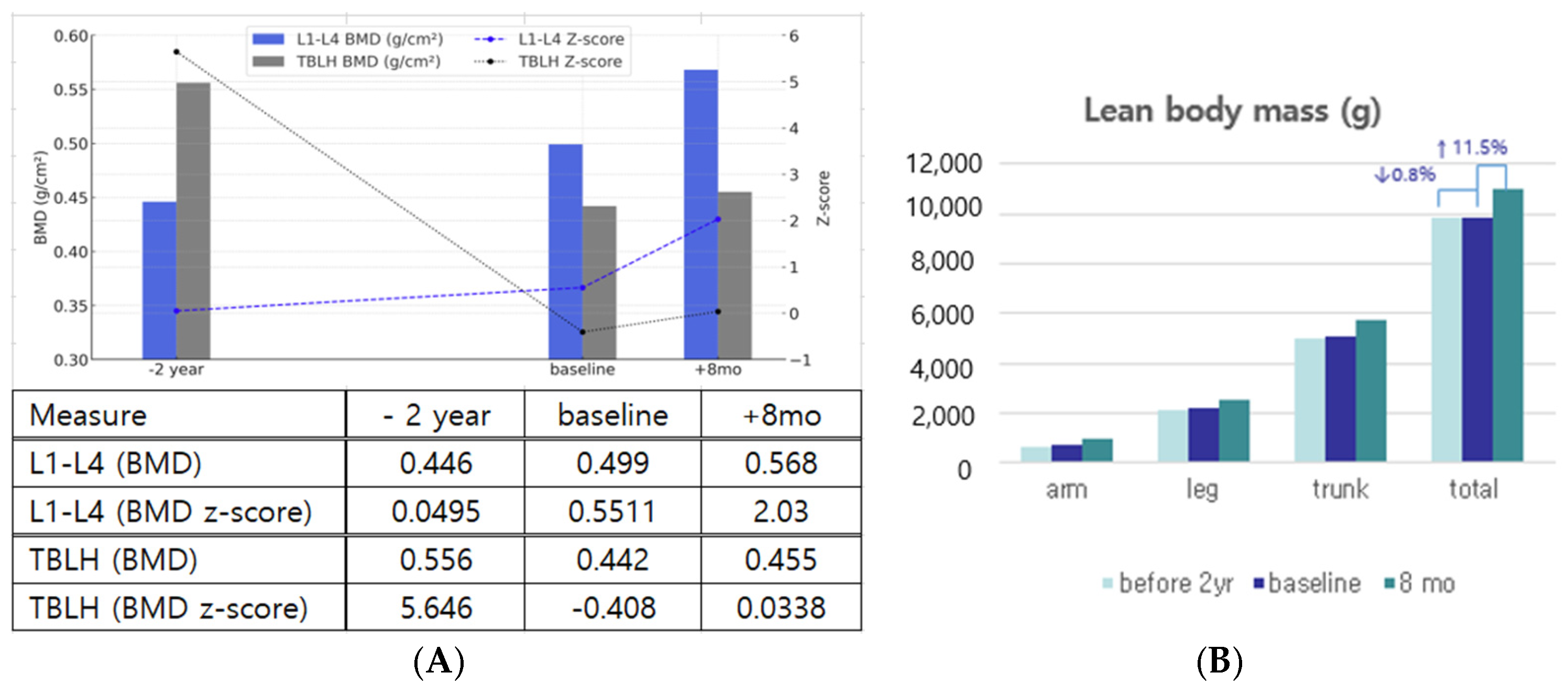
3.1. Improvements in Body Composition, Bone Mineral Density, and Dentition

Following treatment, notable improvements in bone mineral density (BMD), lean body mass, and dentition status were observed. Body composition and bone mineral density were assessed using dual-energy X-ray absorptiometry (DXA). During the pretreatment period, a slight increase in L-spine BMD was observed, whereas TBLH BMD decreased. In contrast, both measures showed significant improvement after MSC therapy. When calculated as annual rates, L1–L4 BMD increased by 5.94% per year during the pretreatment period and accelerated to 20.74% per year after MSC therapy. In contrast, TBLH BMD showed a −10.25% annual decline before treatment but improved to a 4.41% annual increase following therapy. The L1-L4 BMD z-score also improved markedly from 0.0495 to 2.03, shifting from near average to well above the mean for height age-matched children, suggesting a positive effect of MSC therapy on bone metabolism (Figure 1A). Lean body mass also significantly increased by treatment. The annual rate of total body mass change shifted from −0.4% per year (−0.8% over 2 years) before treatment to +17.25% per year (+11.5% over 8 months) after MSC therapy, suggesting a potential anabolic effect (Figure 1B). Detailed data of changes in body composition and fat mass are provided in the Supplementary Materials.
All the primary teeth were retained before treatment. After MSC administration, the previously retained deciduous teeth exhibited mobility, rapid eruption of permanent teeth, and exfoliation of primary teeth following the first (#51, #52, #71), second (#81), and fourth (#61) MSC administrations.

3.2. Amelioration of Stiffness: Joints, Tympanic Membrane, and Arterial Flexibility

Treatment led to improvements in the stiffness of joint mobility, tympanic membrane, and arterial stiffness. The ROMs of the hip, knee, shoulder, and elbow joints increased slightly. However, the ROMs of the wrist and fingers did not improve (Figure S3). A decrease in tympanic membrane stiffness was observed via tympanometry, leading to improved hearing, as confirmed by pure tone audiometry (Figure S4A,B). BaPWV, an indicator of arterial stiffness, decreased by an average of 9.98% with treatment. Specifically, the velocity decreased from 1113 cm/s to 1011 cm/s on the right side and from 1228 cm/s to 1097 cm/s on the left side (Table 1). These findings indicate that the treatment effectively improved stiffness and overall physiological function.

3.3. Short-Term Improvement in Growth and Metabolic Aspects

Weight gain improved with MSC treatment, and after two months of treatment, the greatest improvement in all indicators except height (weight, IGF-1, IGFBP3, and HbA1c) was observed. A weight gain of 1 kg was observed during the 8-month treatment period, whereas only 0.5 kg was observed over the two-year pretreatment period. The change in the z-score during the post-treatment period was +0.75, whereas that during the pretreatment period was −1.13. The increase in the growth rate is less clear, but the rate of z-score deterioration has slowed. The IGF-1 level rose from 173.1 ng/mL (z score: 0.03) to 235.6 ng/mL (z score: 1.32), and the IGFBP3 level rose from 1786.5 ng/mL (z score: −1.6) to 2664.7 ng/mL (z score: 0.37) after 2 months of treatment. In terms of metabolism, glycemic control improved initially (glycated hemoglobin [HbA1c]: 5.8% → 5.5% in 2 months). However, by 6 months after the third MSC administration, these gains had diminished (Table 1). Metabolic abnormalities associated with steatotic liver disease (MASLD) were observed with abdominal ultrasound at baseline and did not progress during treatment (Table 1).

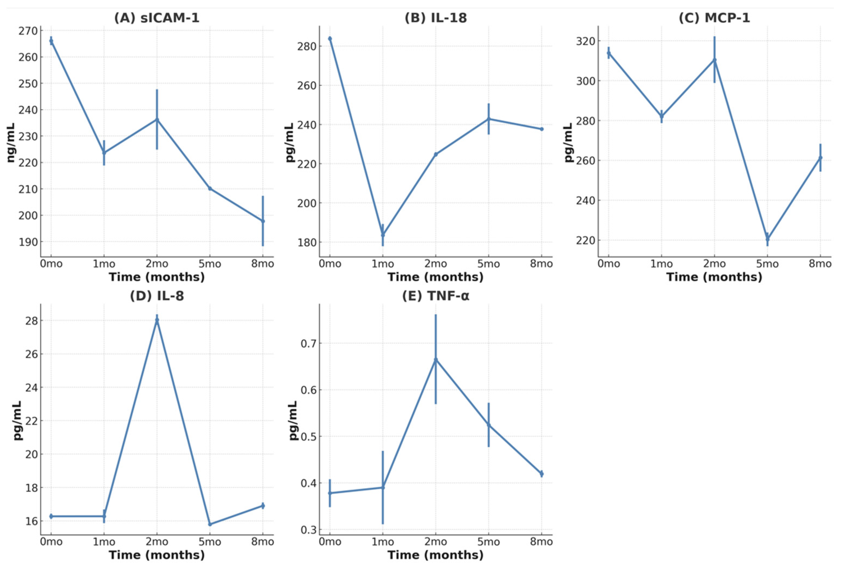
3.4. Reduction in Inflammatory Cytokines

We evaluated the cytokines IL-18, MCP-1, sICAM-1, IL-8, TNF- α, and IL-1β. IL-1β was undetectable throughout the study. Cytokine changes in this study were interpreted based on intra-patient trends (Figure 2A–E). Systemic cytokine analysis showed a sustained decrease in sICAM-1 levels with repeated MSC administration, suggesting reduced vascular inflammation. IL-18, MCP-1, and sICAM-1 levels also decreased after the first dose, indicating an anti-inflammatory effect of MSC therapy. Increased IL-8 levels with repeated MSC treatment improve endothelial cell function by increasing endothelial nitric oxide synthase (eNOS) activity and reducing oxidative stress [26]. However, in the context of cytokine changes, we assume that the efficacy of MSCs decreases over time.

3.5. Lower Efficacy for Atherosclerosis and Cardiovascular Aspects

Despite treatment, atherosclerosis and cardiovascular deterioration did not stop or reverse. Cerebrovascular imaging demonstrated progressive changes over time. Two years before treatment, severe segmental stenosis of the bilateral vertebral arteries and mild stenosis of the bilateral ICA with collateral vessels were observed (Figure 3A). At baseline, vertebral artery stenosis had progressed, and hypoplastic changes with segmental total occlusion of the left ICA were identified (Figure 3B). Follow-up imaging showed some progression, especially in the left vertebral artery, but no new major vascular events or additional occlusions were observed (Figure 3C). These changes may reflect the natural course of cerebrovascular disease in HGPS.
On the other hand, atherosclerosis and cardiac dysfunction progression were evident. The atherosclerotic marker cIMT increased during treatment. At 5 months, the right mean cIMT increased from 0.47 mm to 0.61, exceeding the normal range (0.365–0.58 mm) [27]. Additionally, right maximum cIMT also increased from 0.60 to 0.80 mm. Progression of mild diastolic dysfunction was observed through tissue Doppler imaging (early diastolic velocity 7→5.89 cm/s normal > 8 cm/s, E/e 16.1→12.28 normal < 8). Systolic function, as measured by the ejection fraction and fractional shortening, was within the normal range but declined over time. No calcification was detected on TTE during treatment (Table 1).

3.6. Safety of MSC Treatment

Unfortunately, MSC therapy did not result in an extension of the patient’s lifespan. The study was terminated due to the patient’s death 10 months after treatment initiation at age 8 years and 7 months. The presumed cause of death was post-exercise arrhythmia, considered unlikely to be related to MSC treatment. The patient was dead on arrival with asystole on ECG, and no further evaluation was possible. An autopsy was not performed in accordance with the guardians’ wishes. Following a thorough investigation, both the Korea National Institute of Health (KNIH) and the Regenerative Medicine Acceleration Foundation reviewed the termination report and concluded that the death was not related to MSC therapy.
During MSC administration, the only adverse events that occurred within 24 h of administration were nausea, vomiting, and dizziness with the first dose. At the first dose, lorazepam was administered concurrently to relieve anxiety. Without lorazepam, the side effects do not appear repeatedly, so it looks less relevant to stem cell therapy. Hand weakness lasting for an hour and a half without headache at 6 days after the first dose might be associated with his basal vascular state, which is less likely with MSC therapy. Frequent epistaxis was thought to be due to the use of clopidogrel. Thirty-six days after the 4th MSC treatment, the patient reported intermittent, stress-induced chest pain lasting 1–2 min, which resolved completely. An immediate attempt to conduct additional evaluation was made, but scheduling delays due to patient refusal because of various circumstances resulted in unexpected death during daily activities. Other mild adverse reactions associated with infection were noted (Table S2).

4. Discussion

This study was systematically designed and is, to our knowledge, the first to administer a precise cell count of MSCs to a patient with progeria. Safety was ensured through precise quantification and repeated administration of MSCs, along with comprehensive monitoring.
Like lonafarnib [13], this study confirmed that MSC therapy also has positive effects on bone structure, vascular and joint stiffness, and low-frequency sensorineural hearing. Additionally, MSC therapy improved dentition and had anti-inflammatory effects, with short-term positive effects on growth and metabolism. The Lumbar spine, predominantly composed of metabolically active trabecular bone, showed significant improvement not only in BMD but also in the BMD z-score (0.0495 → 2.03), supporting the therapeutic effect of MSCs on bone metabolism. While the TBLH region, predominantly cortical bone with lower metabolic activity, showed smaller changes (BMD: 0.442 → 0.455 g/cm2, BMD z-score: −0.408 → 0.0338), it shifted from a declining trend to stabilization. These results suggest that MSC therapy may positively influence bone metabolism in HGPS, which is consistent with the known mechanism of impaired osteoblast differentiation and low bone turnover in this disease [28].
There were two prior cases of stem cell therapy in HGPS. A single use of adipose tissue-derived stromal vascular fractions containing MSCs improved the growth rate, weight gain, and IGF-1 levels [29]. Another patient treated with cord blood cells exhibited decreased PWV, increased height, weight gain, and increased ROM of the proximal joint [30]. These results were consistent with our study. However, unlike these studies or lonafarnib trial, our case did not show improvement in cIMT. This is likely due to the patient’s severe baseline vascular disease.
The patient rapidly progressed to vascular changes. He had bilateral vertebral artery stenosis at diagnosis, and additional ICA stenosis had developed over two years. Murtada et al. reported that the aortic phenotype worsened rapidly as the disease progressed toward the terminal stage in mouse models [31]. Despite active LDL cholesterol management using lipid-lowering agents to prevent stroke, atherosclerosis progression was evident on cIMT. This aligns with clinical trial data showing the combination therapy (lonafarnib + pravastatin + zoledronate) did not outperform lonafarnib alone [15]. The failure of lipid-lowering drugs to improve arteriosclerosis is thought to be attributed to a distinct mechanism specific to progeria. Progressive diastolic dysfunction, a characteristic finding in patients with progeria [32] was also observed in TTE. While the patient’s vascular deterioration progressed rapidly, it is unlikely that MSC therapy contributed to this outcome. This is supported by well-established cardioprotective properties of bone marrow-derived MSCs (BM-MSCs), including promotion of angiogenesis, reduction in fibrosis, and preservation of cardiac function [33]. Furthermore, MSCs have demonstrated therapeutic potential in acute myocardial infarction [34]. Considering that cerebrovascular deterioration begins at early age, we believe similar changes may have also occurred in the coronary arteries.
The clinically proven drug for cardiovascular manifestations in progeria is lonafarnib, the efficacy of which has been demonstrated in a mouse model through the use of progerinin [35] and antisense oligonucleotides (ASOs) [36]. These drugs effectively lower progerin levels. Unfortunately, progerin levels were not assessed in this study. Additionally, therapeutic approaches targeting endothelial cell dysfunction, such as Angiopoietin-2 inhibition and Verteporfin (a YAP/TAZ pathway inhibitor), have also been reported to improve vascular function. This study suggests that MSC therapy may improve vascular function, as evidenced by changes in baPWV and cytokine levels. Baseline measurements revealed an elevated baPWV in the patient, with a mean value of 1170 cm/s, which exceeds the 95th percentile for a healthy 12-year-old boy (1039 cm/s) [37]. Given that the PWV naturally increases with age and height, this value indicates a significant degree of arterial stiffness. After MSC treatment, the patient demonstrated a reduction in baPWV, suggesting an improvement in arterial stiffness and a positive therapeutic response. Additionally, serial reductions in sICAM-1, an endothelial activation and inflammation marker [38,39], suggest reduced vascular inflammation and potential atherosclerosis risk mitigation.
Due to rarity of HGPS, this study included a single subject, limiting the generalizability and making statistical analysis infeasible. Nonetheless, the findings provide valuable insights. Brain MRA follow-ups suggested that MSC therapy may alleviate deterioration but not halt or reverse structural changes. A low dose of MSCs (2.5 ×10⁵ cells/kg) was chosen to prioritize vascular safety considering the patient’s compromised vascular status, and history of TIA. Some effects of MSCs are short-lived and relatively insufficient. As safety was ensured throughout this study, dose escalation and earlier intervention-prior to irreversible changes- should be considered to maximize therapeutic benefits I future study. Additionally, progerin levels could not be measured in this study, which limits the ability to directly assess biological impact of MSC therapy on disease mechanism. To place these findings in context, we next review recent advances in stem cell–based therapies for HGPS, including clinical and experimental studies.

5. Literature Review

Recent stem cell research in HGPS spans mechanistic studies, disease modeling, and in vivo and clinical investigations, providing a foundation for novel therapeutic strategies (Table 2). Scaffidi and Misteli (2008) first demonstrated that progerin impairs MSC function by activating premature aging pathway [40]. Similarly, Rosengardten et al. (2011) reported the depletion of endogenous stem cell pools in HGPS, underscoring the importance of stem cell maintenance in disease pathology [19]. These findings suggest stem cell-based approaches, including replacement strategies.
The advent of induced pluripotent stem cells(iPSCs) technology has significantly advanced HGPS research. Differentiation of iPSCs derived from HGPS fibroblasts into vascular smooth muscle cells and MSCs, offering valuable models of studying pathogenesis and paving the way for targeted therapies [41,42,43]. In an in vivo study, transplantation of muscle-derived stem/progenitor cells into progeroid mice notably extended both their healthspan and lifespan [44]. Clinical studies have also explored the efficacy of stem cell based therapies. Administration of allogeneic cord blood cells or adipose tissue derived stromal vascular fraction containing MSCs have shown improvements in growth and clinical parameters. However, these studies remain limted in duration and scope.
Overall, recent advances in stem cell research including iPSC-based modeling, transplantation of stem cells, have deepened our understanding of HGPS and underscored the potential of regenerative medicine as a therapeutic avenue. A growing trend toward combination therapies that target multiple aspects of disease pathogenesis has emereged [45]. In particular, integrating stem cell therapy with agents that reduce progerin production, such as lonafarnib, may offer synergistic effects. Therefore, further studies should clarify underlying mechanisms, and optimize combination approaches to improve clinical outcomes.
Table 2. Stem cell research on HGPS. hMSC- human Mesenchymal stem cells; HGPS—Hutchinson–Gilford progeria syndrome; SVF—stromal vascular fraction; iPSCs—induced pluripotent stem cells; MDSPCs—muscle-derived stem/progenitor cells.
Year
Authors
SubjectsMethodologyKey FindingsMechanismClinical
Implications
Limitations/Future Directions
2008 [40]
Scaffidi P et al.
hMSC Expression of progerin in hMSCs; analysis of stem cell functionDiscovered misregulation leading to premature agingProgerin interferes with the function of hMSCsProgerin activates Notch signaling pathway in hMSCsLimited to in vitro study: in vivo confirmation needed
2011 [41]
Zhang J, et al.
iPSC-derived cellsiPSC differentiation and analysisVascular smooth muscle and mesenchymal stem cell defects identifiediPSCs reveal specific cellular defects in HGPSNew targets for therapeutic interventionValidation in patient samples required
2011 [42]
Liu et al.
HGPS patient fibroblastsGeneration of iPSCs from HGPS fibroblasts; Differentiation of iPSCsiPSCs from HGPS patients lack progerin expression but resume upon differentiationReprogramming suppresses progerin expression: differentiation resumes aging-associated phenotypesiPSCs as a model for studying HGPS and drug screeningLimited to in vitro model: in vivo validation needed
2011 [19]
Rosengardten Y et al.
HGPS mouse modelAnalysis of stem cell populations and wound healing capacityHGPS mutation causes adult stem cell depletion and impaired wound healingProgerin accumulation leads to stem cell exhaustionStem cell therapies may be beneficial for HGPS patientsLimited to mouse model: human studies needed
2012 [44]
Lavasani M et al.
Progeroid miceIntraperitoneal injection of young wild-type MDSPCsExtended lifespan and healthspan of progeroid miceSecretion of factors by MDSPCs that improve tissue functionStem cell transplantation as potential therapy for progeriaLimited to animal model: human studies needed
2015 [43]
Lo Cicero A, Nissan X
iPSCsiPSC modeling of HGPSImproved understanding of disease mechanismsiPSCs recreate HGPS cellular environment for studyImproved drug screening platformTranslation to in vivo models needed
2020 [29]
Park J et al.
HGPS patientadipose SVF containing MSCIncreased height, weight and IGF-1anti-inflammatory effects via paracrine signalingProposal for the potential treatment of inflammaging-related diseasesSingle case study: larger trials needed
2021 [30]
Suh YS, et al.
HGPS patientCord blood stem cell infusion
+sirolimus
Improved growth, reduced PWV, slowed IMT progressionCord blood stem cells may provide trophic support and replace damaged cellsPotential noninvasive treatment for HGPSSingle case study: larger trials needed

6. Conclusions

Herein, we report the case of a patient with HGPS who received MSC therapy. Our findings suggest that MSC therapy for HGPS is safe and may have potential benefits, including increased bone mineral density, reduced joint stiffness and arterial stiffness, and anti-inflammatory effects. Given that a favorable safety profile was observed in this case, further studies with optimized dosing strategies and earlier intervention before significant vascular deterioration are warranted. Additionally, a personalized protocol with long-term monitoring should be developed to optimize therapeutic outcomes. Since MSC therapy has limited effects on cardiovascular disease, it may be more effective as an adjuvant therapy to lonafarnib than as monotherapy. Furthermore, beyond HGPS, stem cell therapy warrants further investigation for its potential role in aging related conditions, cardiovascular disease prevention, and the treatment of rare diseases.

Supplementary Materials

The following supporting information can be downloaded at: https://www.mdpi.com/article/10.3390/children12040523/s1. Figure S1 Changes in body composition; Figure S2. Transthoracic echocardiography (TTE) image of patient; Figure S3. Change of range of motion from baseline; Figure S4. Change of Audiological Evaluation; Table S1. Changes in body composition; Table S2. Lists of adverse reactions.

Author Contributions

Conceptualization, E.-Y.J., J.-S.P., S.-J.K., H.-T.S., J.-E.L. and G.-S.C.; methodology, E.-Y.J., J.-S.P., S.-J.K., H.-T.S., J.-E.L. and G.-S.C.; software, IBM SPSS Statistics version 26 (IBM Corp., Armonk, NY, USA); validation, E.-Y.J., J.-E.L. and G.-S.C.; formal analysis, E.-Y.J.; investigation, E.-Y.J., J.-S.P., M.Y., J.-E.L., H.-T.S., J.-E.L. and G.-S.C.; resources, J.-E.L., H.-T.S., J.-E.L. and G.-S.C.; data curation, E.-Y.J. and M.Y.; writing—original draft preparation, E.-Y.J. and J.-S.P.; writing—review and editing, E.-Y.J., J.-S.P. and J.-E.L.; visualization, E.-Y.J.; supervision, J.-E.L. and G.-S.C.; project administration, E.-Y.J., M.Y., J.-E.L. and G.-S.C.; funding acquisition, J.-E.L. and G.-S.C. All authors have read and agreed to the published version of the manuscript.

Funding

This research was supported by a grant from the Advanced Regenerative Medicine Clinical Research Program, funded by the Regenerative Medicine Acceleration Foundation under the Ministry of Health and Welfare, Republic of Korea (grant number: R–3-0005).

Institutional Review Board Statement

The study was conducted in accordance with the principles of the Declaration of Helsinki and approved by the Ethics Committee of Inha university hospital (2023-04-038, 19 June 2023).

Informed Consent Statement

Informed consent was obtained from the patients and their parents in this study.

Data Availability Statement

The original contributions presented in the study are included in the article/Supplementary Materials, further inquiries can be directed to the corresponding authors.

Acknowledgments

The authors would like to acknowledge SCM Science for providing in vitro research support and conducting in vivo safety evaluations for MSC therapy. We are deeply grateful to the patient and their family for their participation and cooperation in this study. We also appreciate the expert consultation and support from Eun Jung Shin (Department of Pediatric Cardiology) for the cardiovascular evaluation and Hee-Kwon Park (Department of Neurology) for the neurological assessment.

Conflicts of Interest

The authors have received research funding from the Regenerative Medicine Acceleration Foundation. The funders had no role in the design of the study; in the collection, analyses, or interpretation of data; in the writing of the manuscript; or in the decision to publish the results.

References

  1. De Sandre-Giovannoli, A.; Bernard, R.; Cau, P.; Navarro, C.; Amiel, J.; Boccaccio, I.; Lyonnet, S.; Stewart, C.L.; Munnich, A.; Le Merrer, M.; et al. Lamin a truncation in Hutchinson–Gilford progeria. Science 2003, 300, 2055. [Google Scholar] [CrossRef] [PubMed]
  2. Eriksson, M.; Brown, W.T.; Gordon, L.B.; Glynn, M.W.; Singer, J.; Scott, L.; Erdos, M.R.; Robbins, C.M.; Moses, T.Y.; Berglund, P. Recurrent de novo point mutations in lamin A cause Hutchinson–Gilford progeria syndrome. Nature 2003, 423, 293–298. [Google Scholar] [CrossRef] [PubMed]
  3. Jansen, T.; Romiti, R. Progeria infantum (Hutchinson–Gilford syndrome) associated with scleroderma-like lesions and acro-osteolysis: A case report and brief review of the literature. Pediatr. Dermatol. 2000, 17, 282–285. [Google Scholar] [CrossRef] [PubMed]
  4. Rork, J.F.; Huang, J.T.; Gordon, L.B.; Kleinman, M.; Kieran, M.W.; Liang, M.G. Initial cutaneous manifestations of Hutchinson–Gilford progeria syndrome. Pediatr. Dermatol. 2014, 31, 196–202. [Google Scholar] [CrossRef]
  5. Hennekam, R.C. Hutchinson–Gilford progeria syndrome: Review of the phenotype. Am. J. Med. Genet. A 2006, 140, 2603–2624. [Google Scholar] [CrossRef]
  6. Merideth, M.A.; Gordon, L.B.; Clauss, S.; Sachdev, V.; Smith, A.C.; Perry, M.B.; Brewer, C.C.; Zalewski, C.; Kim, H.J.; Solomon, B.; et al. Phenotype and course of Hutchinson–Gilford progeria syndrome. N. Engl. J. Med. 2008, 358, 592–604. [Google Scholar] [CrossRef]
  7. Gordon, L.B.; McCarten, K.M.; Giobbie-Hurder, A.; Machan, J.T.; Campbell, S.E.; Berns, S.D.; Kieran, M.W. Disease progression in Hutchinson–Gilford progeria syndrome: Impact on growth and development. Pediatrics 2007, 120, 824–833. [Google Scholar] [CrossRef]
  8. Nevado, R.M.; Hamczyk, M.R.; Gonzalo, P.; Andres-Manzano, M.J.; Andres, V. Premature Vascular Aging with Features of Plaque Vulnerability in an Atheroprone Mouse Model of Hutchinson–Gilford Progeria Syndrome with Ldlr Deficiency. Cells 2020, 9, 2252. [Google Scholar] [CrossRef]
  9. Xu, Q.; Mojiri, A.; Boulahouache, L.; Morales, E.; Walther, B.K.; Cooke, J.P. Vascular senescence in progeria: Role of endothelial dysfunction. Eur. Heart J. Open 2022, 2, oeac047. [Google Scholar] [CrossRef]
  10. Gordon, L.B.; Massaro, J.; D’Agostino, R.B., Sr.; Campbell, S.E.; Brazier, J.; Brown, W.T.; Kleinman, M.E.; Kieran, M.W.; Progeria Clinical Trials, C. Impact of farnesylation inhibitors on survival in Hutchinson–Gilford progeria syndrome. Circulation 2014, 130, 27–34. [Google Scholar] [CrossRef]
  11. Prakash, A.; Gordon, L.B.; Kleinman, M.E.; Gurary, E.B.; Massaro, J.; D’Agostino, R., Sr.; Kieran, M.W.; Gerhard-Herman, M.; Smoot, L. Cardiac Abnormalities in Patients With Hutchinson–Gilford Progeria Syndrome. JAMA Cardiol. 2018, 3, 326–334. [Google Scholar] [CrossRef] [PubMed]
  12. Gordon, L.B.; Shappell, H.; Massaro, J.; D’Agostino, R.B., Sr.; Brazier, J.; Campbell, S.E.; Kleinman, M.E.; Kieran, M.W. Association of Lonafarnib Treatment vs No Treatment With Mortality Rate in Patients With Hutchinson–Gilford Progeria Syndrome. JAMA 2018, 319, 1687–1695. [Google Scholar] [CrossRef] [PubMed]
  13. Suzuki, M.; Jeng, L.J.B.; Chefo, S.; Wang, Y.; Price, D.; Li, X.; Wang, J.; Li, R.J.; Ma, L.; Yang, Y.; et al. FDA approval summary for lonafarnib (Zokinvy) for the treatment of Hutchinson–Gilford progeria syndrome and processing-deficient progeroid laminopathies. Genet Med. 2023, 25, 100335. [Google Scholar] [CrossRef]
  14. Gordon, L.B.; Kleinman, M.E.; Miller, D.T.; Neuberg, D.S.; Giobbie-Hurder, A.; Gerhard-Herman, M.; Smoot, L.B.; Gordon, C.M.; Cleveland, R.; Snyder, B.D.; et al. Clinical trial of a farnesyltransferase inhibitor in children with Hutchinson–Gilford progeria syndrome. Proc. Natl. Acad. Sci. USA 2012, 109, 16666–16671. [Google Scholar] [CrossRef]
  15. Gordon, L.B.; Kleinman, M.E.; Massaro, J.; D’Agostino, R.B., Sr.; Shappell, H.; Gerhard-Herman, M.; Smoot, L.B.; Gordon, C.M.; Cleveland, R.H.; Nazarian, A.; et al. Clinical Trial of the Protein Farnesylation Inhibitors Lonafarnib, Pravastatin, and Zoledronic Acid in Children With Hutchinson–Gilford Progeria Syndrome. Circulation 2016, 134, 114–125. [Google Scholar] [CrossRef]
  16. Abutaleb, N.O.; Atchison, L.; Choi, L.; Bedapudi, A.; Shores, K.; Gete, Y.; Cao, K.; Truskey, G.A. Lonafarnib and everolimus reduce pathology in iPSC-derived tissue engineered blood vessel model of Hutchinson–Gilford Progeria Syndrome. Sci. Rep. 2023, 13, 5032. [Google Scholar] [CrossRef]
  17. Halaschek-Wiener, J.; Brooks-Wilson, A. Progeria of stem cells: Stem cell exhaustion in Hutchinson–Gilford progeria syndrome. J. Gerontol. A Biol. Sci. Med. Sci. 2007, 62, 3–8. [Google Scholar] [CrossRef]
  18. Bridger, J.M.; Kill, I.R. Aging of Hutchinson–Gilford progeria syndrome fibroblasts is characterised by hyperproliferation and increased apoptosis. Exp. Gerontol. 2004, 39, 717–724. [Google Scholar] [CrossRef]
  19. Rosengardten, Y.; McKenna, T.; Grochova, D.; Eriksson, M. Stem cell depletion in Hutchinson–Gilford progeria syndrome. Aging Cell 2011, 10, 1011–1020. [Google Scholar] [CrossRef]
  20. Prolla, T.A. Multiple roads to the aging phenotype: Insights from the molecular dissection of progerias through DNA microarray analysis. Mech. Ageing Dev. 2005, 126, 461–465. [Google Scholar] [CrossRef]
  21. Dabrowska, S.; Andrzejewska, A.; Janowski, M.; Lukomska, B. Immunomodulatory and Regenerative Effects of Mesenchymal Stem Cells and Extracellular Vesicles: Therapeutic Outlook for Inflammatory and Degenerative Diseases. Front. Immunol. 2020, 11, 591065. [Google Scholar] [CrossRef] [PubMed]
  22. Zhu, Y.; Ge, J.; Huang, C.; Liu, H.; Jiang, H. Application of mesenchymal stem cell therapy for aging frailty: From mechanisms to therapeutics. Theranostics 2021, 11, 5675–5685. [Google Scholar] [CrossRef] [PubMed]
  23. Ogay, V.; Sekenova, A.; Li, Y.; Issabekova, A.; Saparov, A. The Therapeutic Potential of Mesenchymal Stem Cells in the Treatment of Atherosclerosis. Curr. Stem Cell Res. Ther. 2021, 16, 897–913. [Google Scholar] [CrossRef]
  24. Kirwin, T.; Gomes, A.; Amin, R.; Sufi, A.; Goswami, S.; Wang, B. Mechanisms underlying the therapeutic potential of mesenchymal stem cells in atherosclerosis. RegenMed 2021, 16, 669–682. [Google Scholar] [CrossRef]
  25. Zhang, Y.; Dong, N.; Hong, H.; Qi, J.; Zhang, S.; Wang, J. Mesenchymal Stem Cells: Therapeutic Mechanisms for Stroke. Int. J. Mol. Sci. 2022, 23, 2550. [Google Scholar] [CrossRef]
  26. Lin, Y.L.; Yet, S.F.; Hsu, Y.T.; Wang, G.J.; Hung, S.C. Mesenchymal Stem Cells Ameliorate Atherosclerotic Lesions via Restoring Endothelial Function. Stem Cells Transl. Med. 2015, 4, 44–55. [Google Scholar] [CrossRef]
  27. Drole Torkar, A.; Plesnik, E.; Groselj, U.; Battelino, T.; Kotnik, P. Carotid Intima-Media Thickness in Healthy Children and Adolescents: Normative Data and Systematic Literature Review. Front. Cardiovasc. Med. 2020, 7, 597768. [Google Scholar] [CrossRef]
  28. Cabral, W.A.; Stephan, C.; Terajima, M.; Thaivalappil, A.A.; Blanchard, O.; Tavarez, U.L.; Narisu, N.; Yan, T.; Wincovitch, S.M.; Taga, Y.; et al. Bone dysplasia in Hutchinson–Gilford progeria syndrome is associated with dysregulated differentiation and function of bone cell populations. Aging Cell 2023, 22, e13903. [Google Scholar] [CrossRef]
  29. Pak, J.; Lee, J.H.; Jeon, J.H.; Kim, Y.B.; Jeong, B.C.; Lee, S.H. Potential Benefits of Allogeneic Haploidentical Adipose Tissue-Derived Stromal Vascular Fraction in a Hutchinson–Gilford Progeria Syndrome Patient. Front. Bioeng. Biotechnol. 2020, 8, 574010. [Google Scholar] [CrossRef]
  30. Suh, M.R.; Lim, I.; Kim, J.; Yang, P.S.; Choung, J.S.; Sim, H.R.; Ha, S.C.; Kim, M. Efficacy of Cord Blood Cell Therapy for Hutchinson–Gilford Progeria Syndrome-A Case Report. Int. J. Mol. Sci. 2021, 22, 12316. [Google Scholar] [CrossRef]
  31. Murtada, S.I.; Mikush, N.; Wang, M.; Ren, P.; Kawamura, Y.; Ramachandra, A.B.; Li, D.S.; Braddock, D.T.; Tellides, G.; Gordon, L.B.; et al. Lonafarnib improves cardiovascular function and survival in a mouse model of Hutchinson–Gilford progeria syndrome. Elife 2023, 12, e82728. [Google Scholar] [CrossRef] [PubMed]
  32. Olsen, F.J.; Gordon, L.B.; Smoot, L.; Kleinman, M.E.; Gerhard-Herman, M.; Hegde, S.M.; Mukundan, S.; Mahoney, T.; Massaro, J.; Ha, S.; et al. Progression of Cardiac Abnormalities in Hutchinson–Gilford Progeria Syndrome: A Prospective Longitudinal Study. Circulation 2023, 147, 1782–1784. [Google Scholar] [CrossRef] [PubMed]
  33. Gill, J.K.; Rehsia, S.K.; Verma, E.; Sareen, N.; Dhingra, S. Stem cell therapy for cardiac regeneration: Past, present, and future. Can. J. Physiol. Pharmacol. 2024, 102, 161–179. [Google Scholar] [CrossRef]
  34. Clavellina, D.; Balkan, W.; Hare, J.M. Stem cell therapy for acute myocardial infarction: Mesenchymal Stem Cells and induced Pluripotent Stem Cells. Expert Opin. Biol. Ther. 2023, 23, 951–967. [Google Scholar] [CrossRef]
  35. Kang, S.M.; Seo, S.; Song, E.J.; Kweon, O.; Jo, A.H.; Park, S.; Woo, T.G.; Kim, B.H.; Oh, G.T.; Park, B.J. Progerinin, an Inhibitor of Progerin, Alleviates Cardiac Abnormalities in a Model Mouse of Hutchinson–Gilford Progeria Syndrome. Cells 2023, 12, 1232. [Google Scholar] [CrossRef]
  36. Erdos, M.R.; Cabral, W.A.; Tavarez, U.L.; Cao, K.; Gvozdenovic-Jeremic, J.; Narisu, N.; Zerfas, P.M.; Crumley, S.; Boku, Y.; Hanson, G.; et al. A targeted antisense therapeutic approach for Hutchinson–Gilford progeria syndrome. Nat. Med. 2021, 27, 536–545. [Google Scholar] [CrossRef]
  37. Sougawa, Y.; Miyai, N.; Utsumi, M.; Miyashita, K.; Takeda, S.; Arita, M. Brachial-ankle pulse wave velocity in healthy Japanese adolescents: Reference values for the assessment of arterial stiffness and cardiovascular risk profiles. Hypertens. Res. 2020, 43, 331–341. [Google Scholar] [CrossRef]
  38. Singh, V.; Kaur, R.; Kumari, P.; Pasricha, C.; Singh, R. ICAM-1 and VCAM-1: Gatekeepers in various inflammatory and cardiovascular disorders. Clin. Chim. Acta 2023, 548, 117487. [Google Scholar] [CrossRef]
  39. Bui, T.M.; Wiesolek, H.L.; Sumagin, R. ICAM-1: A master regulator of cellular responses in inflammation, injury resolution, and tumorigenesis. J. Leukoc. Biol. 2020, 108, 787–799. [Google Scholar] [CrossRef]
  40. Scaffidi, P.; Misteli, T. Lamin A-dependent misregulation of adult stem cells associated with accelerated ageing. Nat. Cell Biol. 2008, 10, 452–459. [Google Scholar] [CrossRef]
  41. Zhang, J.; Lian, Q.; Zhu, G.; Zhou, F.; Sui, L.; Tan, C.; Mutalif, R.A.; Navasankari, R.; Zhang, Y.; Tse, H.F.; et al. A human iPSC model of Hutchinson Gilford Progeria reveals vascular smooth muscle and mesenchymal stem cell defects. Cell Stem. Cell 2011, 8, 31–45. [Google Scholar] [CrossRef] [PubMed]
  42. Liu, G.H.; Barkho, B.Z.; Ruiz, S.; Diep, D.; Qu, J.; Yang, S.L.; Panopoulos, A.D.; Suzuki, K.; Kurian, L.; Walsh, C.; et al. Recapitulation of premature ageing with iPSCs from Hutchinson–Gilford progeria syndrome. Nature 2011, 472, 221–225. [Google Scholar] [CrossRef] [PubMed]
  43. Lo Cicero, A.; Nissan, X. Pluripotent stem cells to model Hutchinson–Gilford progeria syndrome (HGPS): Current trends and future perspectives for drug discovery. Ageing Res. Rev. 2015, 24, 343–348. [Google Scholar] [CrossRef] [PubMed]
  44. Lavasani, M.; Robinson, A.R.; Lu, A.; Song, M.; Feduska, J.M.; Ahani, B.; Tilstra, J.S.; Feldman, C.H.; Robbins, P.D.; Niedernhofer, L.J.; et al. Muscle-derived stem/progenitor cell dysfunction limits healthspan and lifespan in a murine progeria model. Nat. Commun. 2012, 3, 608. [Google Scholar] [CrossRef]
  45. Joo, E.; Lee, J. Current Therapeutic Approaches for Hutchinson–Gilford Progeria Syndrome. J. Interdiscip. Genomics 2025, 7, 1–8. [Google Scholar]
Figure 1. (A) Changes in bone mineral density and (B) lean body mass bone mineral density. TBLH—total body less head (z-score was assessed by age); BMD—bone mineral density; L1–L4—lumbar vertebrae 1–4 (z-score was adjusted by height-matched age). Lean body mass decreased (↓) 0.8% during pretreatment over 2 years, increased (↑) 11.5% after 8 months of treatment.
Children 12 00523 g001
Figure 2. Changes in the levels of the following serum cytokines: (A) sICAM-1, (B) IL-18, (C) MCP-1, (D) IL-8, (E) TNF-α. sICAM-1—soluble intercellular adhesion molecule-1; IL-18—interleukin-18; MCP-1—monocyte chemoattractant protein-1; IL-8—interleukin-8; TNF-α—tumor necrosis factor-α; IL-1β—interleukin.
Children 12 00523 g002
Figure 3. Changes in the cerebrovascular state before and after treatment were detected via brain magnetic resonance angiography (MRA). At diagnosis, brain MRA revealed vertebral artery (VA) occlusion and a tortuous internal carotid artery (ICA) with collateral vessels. (A) At baseline, diffuse hypoplastic changes with segmental total occlusion developed in the left ICA for two years before treatment, with sustained VA stenosis. (B) At 5 months after treatment, treatment did not completely halt vascular deterioration, and no significant progression was observed (C).
Children 12 00523 g003
Table 1. Changes in growth, metabolic, and cardiac indicators over time.
−2 YearsBaseline2 Months5 Months8 Months
Before Treatment1 Month After
2nd MSC
3 Months After
3rd MSC
6 Months After
3rd MSC
GrowthHeight (cm)
z-score for height
93.2
−4.65
101.2
−5.41
102.1
−5.41
102.2
−5.46
103
−5.73
Weight (kg)
z-score for weight
11.6
−5.56
12.1
−6.89
12.6
−6.48
12.7
−6.3
13
−6.14
ALP (U/L) (ref: 120–344)178207218166207
IGF-1 (ng/mL) (ref: 90.6–268.8)145.5173.1235.6118104.4
z-score for IGF-1−0.150.031.32−1.24−1.49
IGFBP3 (ng/mL) (ref: 1620–3490)14461786266426082147
z-score for IGFBP3−1.96−1.60.37−0.04−0.96
MetabolicHbA1c (%) 5.85.55.95.8
AST/ALT (U/L)31/2234/3538/3831/4134/45
Cholesterol (mg/dL)219180165149115
CardiacCK-MB (normal 0–5 ng/mL)4.23.92.71.93.2
D-dimer (normal 0–0.5 μg/mL) 1.670.400.370.28
BP (systolic/diastolic) 99/69100/47104/51100/52117/60
BaPWV (cm/s) rt/lt 1113/1228 1011/1097
cIMT mean (mm) rt/lt0.43/0.360.47/0.46 0.61/0.51
max (mm) rt/lt0.60/0.520.60/0.68 0.80/0.68
TTE-TDI e’ (cm/s (z-score))
E/e (normal < 8)
7 (−4.04)
8 (−3.53)
16.1
5.89 (−4.6)
12.28
EF (normal 50–70%)64.3670 62.756.2
FS (normal 25–45%)34.3840 33.1828.48
Weight, height, IGF-1, and IGFBP3 increased in the short term and Hba1c decreased in the short term. IGF-1—insulin-like growth factor-1; IGFBP3—insulin-like growth factor-binding protein. Hba1c—glycated hemoglobin; BaPWV—brachial ankle pulse wave velocity; cIMT—carotid intima media thickness; TTE—transthoracic echocardiogram; TDI—tissue Doppler imaging; e’—early diastolic velocity; E/e—mitral inflow E-wave-to-early diastolic velocity ratio. EF—ejection fraction; FS—fractional shortening. Bold text indicates deterioration or out of the normal range.
Disclaimer/Publisher’s Note: The statements, opinions and data contained in all publications are solely those of the individual author(s) and contributor(s) and not of MDPI and/or the editor(s). MDPI and/or the editor(s) disclaim responsibility for any injury to people or property resulting from any ideas, methods, instructions or products referred to in the content.

