
Endoloop-Assisted Polypectomy for a Symptomatic Giant Colonic Polyp in a Pediatric Patient

Abstract
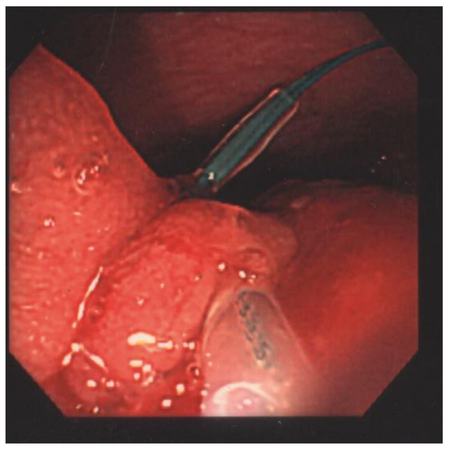
:1. Case Presentation
2. Laboratory Results and Treatment
3. Discussion
Author Contributions
Funding
Institutional Review Board Statement
Informed Consent Statement
Conflicts of Interest
References
- Teach, S.J.; Fleisher, G.R. Rectal bleeding in the pediatric emergency department. Ann. Emerg. Med. 1994, 23, 1252–1258. [Google Scholar] [CrossRef]
- Cucchiara, S.; Guandalini, S.; Staiano, A.; Devizia, B.; Capano, G.; Romaniello, G.; Poggi, V.; Tamburrini, O.; Settimi, A.; de Ritis, G. Sigmoidoscopy, colonoscopy, and radiology in the evaluation of children with rectal bleeding. J. Pediatr. Gastroenterol. Nutr. 1983, 2, 667–671. [Google Scholar] [CrossRef] [PubMed]
- Holgersen, L.O.; Mossberg, S.M.; Miller, R.E. Colonoscopy for rectal bleeding in childhood. J. Pediatr. Surg. 1978, 13, 83–85. [Google Scholar] [CrossRef]
- Katsinelos, P.; Kountouras, J.; Paroutoglou, G.; Beltsis, A.; Chatzimavroudis, G.; Zavos, C.; Vasiliadis, I.; Katsinelos, T.; Papaziogas, B. Endoloop-assisted polypectomy for large pedunculated colorectal polyps. Surg. Endosc. 2006, 20, 1257–1261. [Google Scholar] [CrossRef] [PubMed]
- Hachisu, T. A new detachable snare for hemostasis in the removal of large polyps or other elevated lesions. Surg. Endosc. 1991, 5, 70–74. [Google Scholar] [CrossRef] [PubMed]
- Luigiano, C.; Ferrara, F.; Ghersi, S.; Fabbri, C.; Cennamo, V.; Landi, P.; Polifemo, A.M.; Billi, P.; Bassi, M.; Consolo, P.; et al. Endoclip-assisted resection of large pedunculated colorectal polyps: Technical aspects and outcome. Dig. Dis. Sci. 2010, 55, 1726–1731. [Google Scholar] [CrossRef] [PubMed]
- Di Giorgio, P.; De Luca, L.; Calcagno, G.; Rivellini, G.; Mandato, M.; De Luca, B. Detachable snare versus epinephrine injection in the prevention of postpolypectomy bleeding: A randomized and controlled study. Endoscopy 2004, 36, 860–863. [Google Scholar] [CrossRef] [PubMed]
- Paspatis, G.A.; Paraskeva, K.; Theodoropoulou, A.; Mathou, N.; Vardas, E.; Oustamanolakis, P.; Chlouverakis, G.; Karagiannis, I. A prospective, randomized comparison of adrenaline injection in combination with detachable snare versus adrenaline injection alone in the prevention of postpolypectomy bleeding in large colonic polyps. Am. J. Gastroenterol. 2006, 101, 2805. [Google Scholar] [CrossRef] [PubMed]
- Kouklakis, G.; Mpoumponaris, A.; Gatopoulou, A.; Efraimidou, E.; Manolas, K.; Lirantzopoulos, N. Endoscopic resection of large pedunculated colonic polyps and risk of postpolypectomy bleeding with adrenaline injection versus endoloop and hemoclip: A prospective, randomized study. Surg. Endosc. 2009, 23, 2732–2737. [Google Scholar] [CrossRef] [PubMed]
- Binmoeller, K.F.; Thonke, F.; Soehendra, N. Endoscopic hemoclip treatment for gastrointestinal bleeding. Endoscopy 1993, 25, 167–170. [Google Scholar] [CrossRef] [PubMed]


Publisher’s Note: MDPI stays neutral with regard to jurisdictional claims in published maps and institutional affiliations. |
© 2022 by the authors. Licensee MDPI, Basel, Switzerland. This article is an open access article distributed under the terms and conditions of the Creative Commons Attribution (CC BY) license (https://creativecommons.org/licenses/by/4.0/).
Share and Cite
Lin, Y.-C.; Chou, J.-W.; Chen, A.-C.; Wu, S.-F.; Peng, C.-T.; Chen, W.; Lin, C.-H. Endoloop-Assisted Polypectomy for a Symptomatic Giant Colonic Polyp in a Pediatric Patient. Children 2022, 9, 222. https://doi.org/10.3390/children9020222
Lin Y-C, Chou J-W, Chen A-C, Wu S-F, Peng C-T, Chen W, Lin C-H. Endoloop-Assisted Polypectomy for a Symptomatic Giant Colonic Polyp in a Pediatric Patient. Children. 2022; 9(2):222. https://doi.org/10.3390/children9020222
Chicago/Turabian StyleLin, Yen-Chung, Jen-Wei Chou, An-Chyi Chen, Shu-Fen Wu, Ching-Tien Peng, Walter Chen, and Chien-Heng Lin. 2022. "Endoloop-Assisted Polypectomy for a Symptomatic Giant Colonic Polyp in a Pediatric Patient" Children 9, no. 2: 222. https://doi.org/10.3390/children9020222
APA StyleLin, Y.-C., Chou, J.-W., Chen, A.-C., Wu, S.-F., Peng, C.-T., Chen, W., & Lin, C.-H. (2022). Endoloop-Assisted Polypectomy for a Symptomatic Giant Colonic Polyp in a Pediatric Patient. Children, 9(2), 222. https://doi.org/10.3390/children9020222