The Influence of Different Rope Jumping Methods on Adolescents’ Lower Limb Biomechanics during the Ground-Contact Phase


Abstract
:1. Introduction
2. Materials and Methods
2.1. Participants
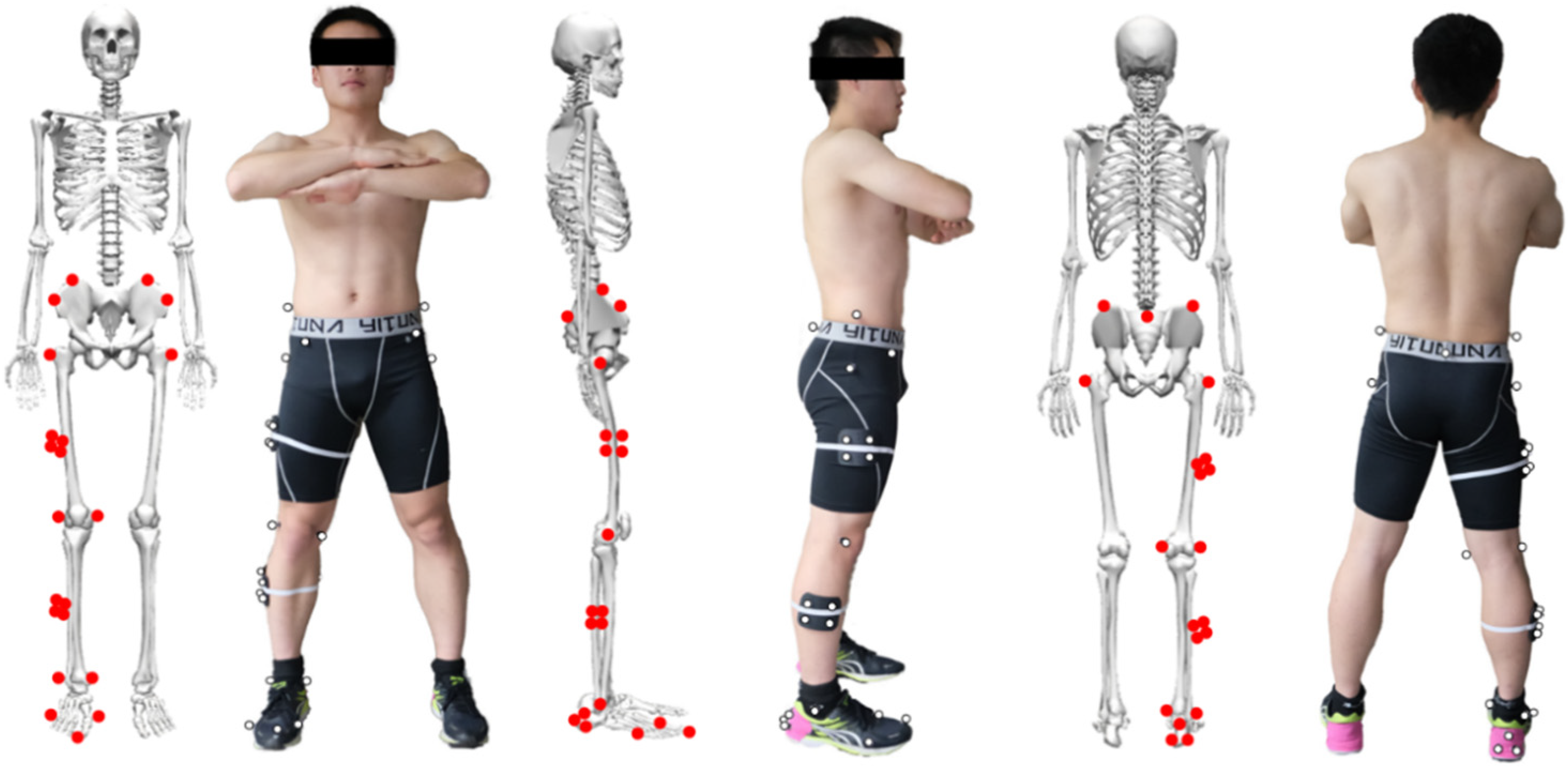
2.2. Experimental Apparatus
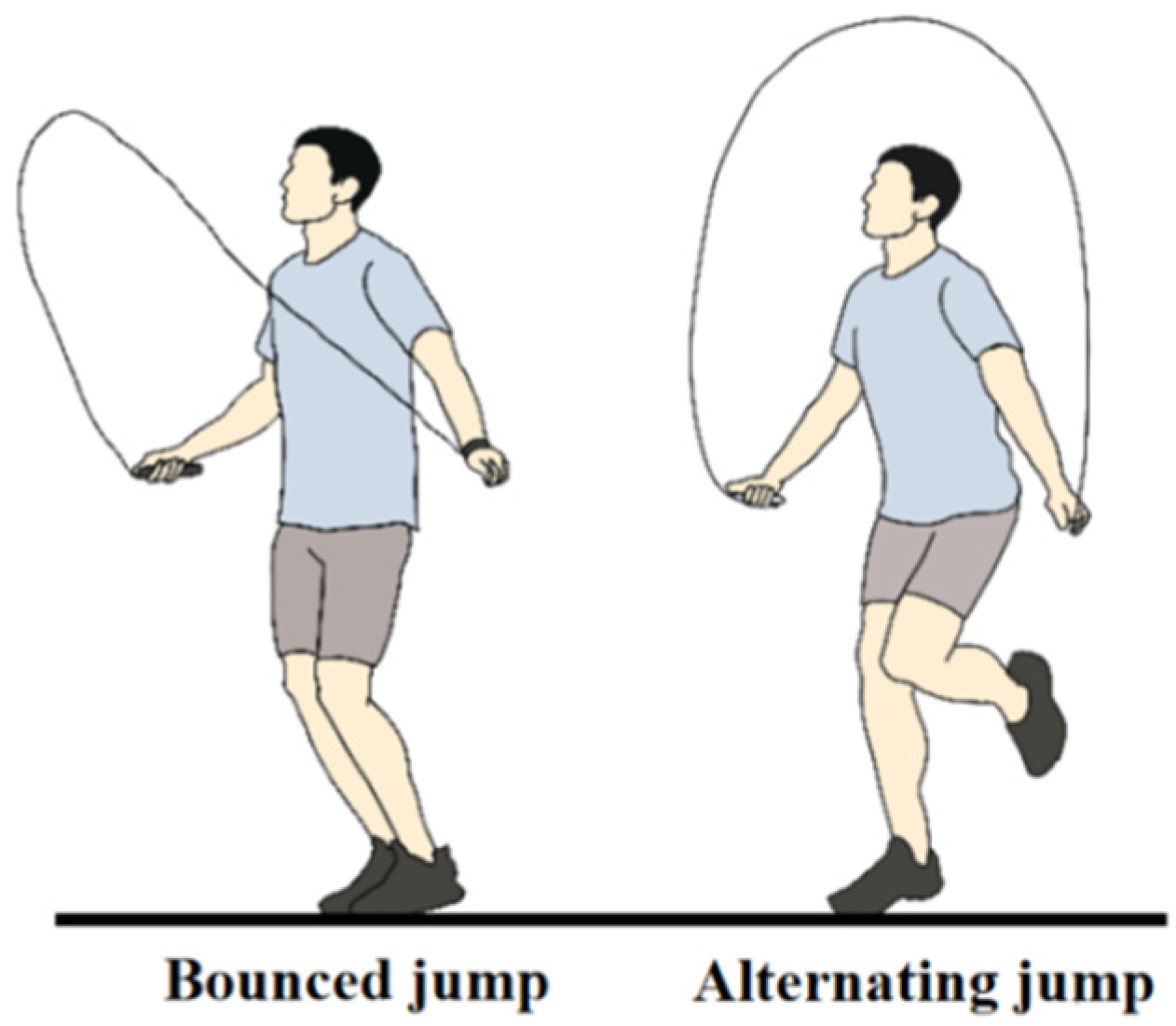
2.3. Experimental Protocol and Procedures
2.4. Data Collection and Processing
2.5. Statistical Analysis
3. Results
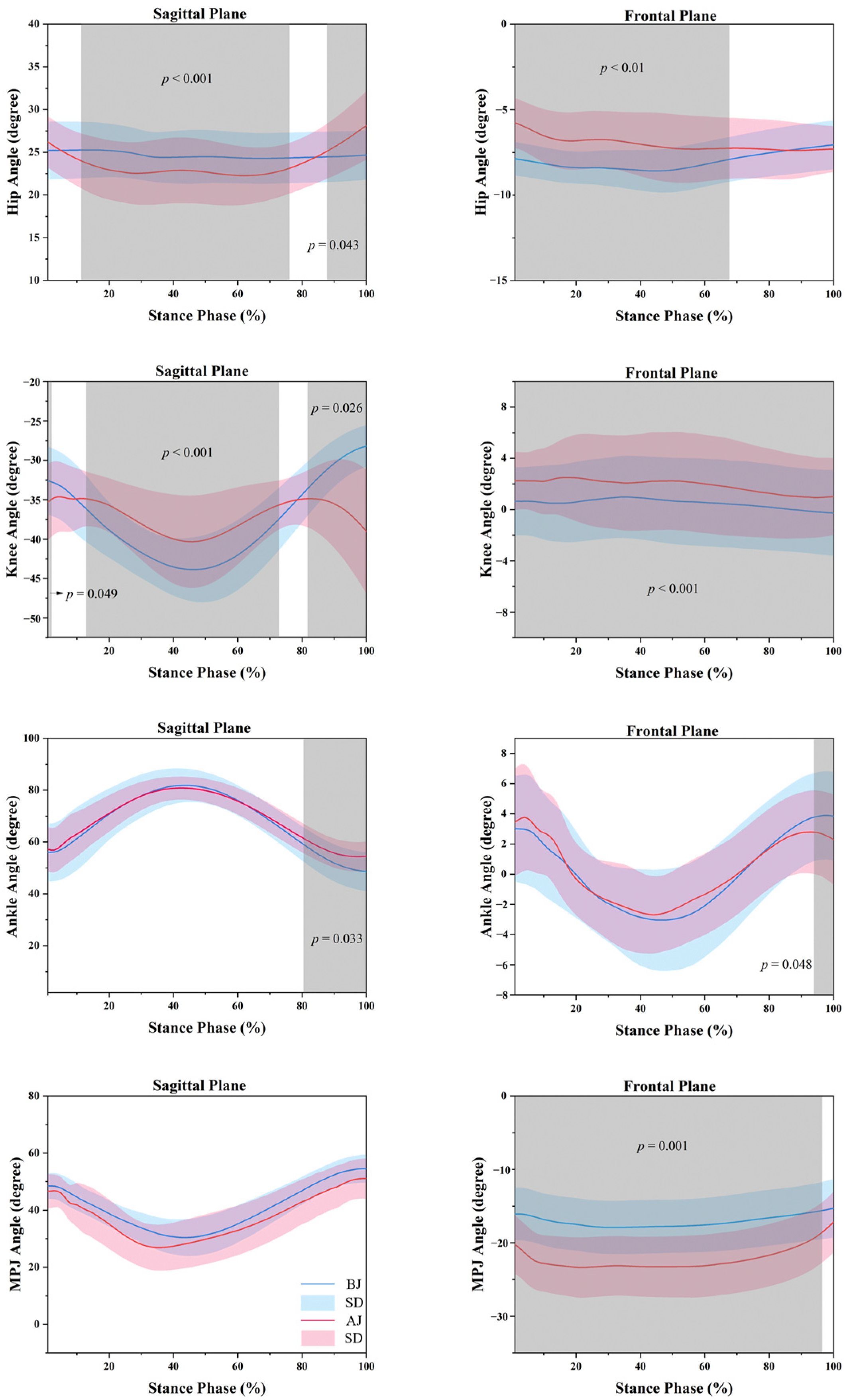
3.1. Kinematics
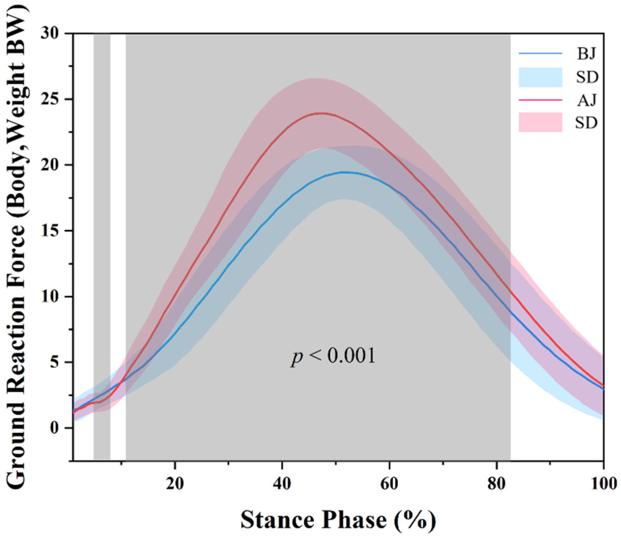
3.2. Kinetics
3.3. Surface EMG
4. Discussion
5. Conclusions
Author Contributions
Funding
Institutional Review Board Statement
Informed Consent Statement
Data Availability Statement
Conflicts of Interest
Abbreviations
| Acronyms | Meaning |
| ACL | Anterior cruciate ligament |
| AJ | Alternate jump |
| BJ | Bounced jump |
| BW | Bodyweight |
| EMG | Electromyography |
| LASI | Left anterior superior iliac spine |
| LPP | Left iliac crest |
| LTROC | Left greater trochanter |
| MPJ | Metatarsophalangeal joint |
| MVC | Maximum voluntary isometric contraction |
| RASI | Right anterior superior iliac spine |
| RLK | Right outer knee |
| RM1 | The first metatarsophalangeal joint |
| RM5 | The fifth metatarsophalangeal joint |
| RMK | Right inner knee |
| ROM | Range of motion |
| RPP | Right iliac crest |
| RTOE | Right big toe |
| RTROC | Right greater trochanter |
| SACR | Sacroiliac joint center |
| SD | Standard deviation |
| SPM | Statistical parametric mapping |
| VALR | Vertical impact loading rate |
| VGRF | Vertical ground reaction force |
References
- Cen, X.; Sun, D.; Rong, M.; Fekete, G.; Baker, J.S.; Song, Y.; Gu, Y. The Online Education Mode and Reopening Plans for Chinese Schools During the COVID-19 Pandemic: A Mini Review. Front. Public Health 2020, 8, 566316. [Google Scholar] [CrossRef] [PubMed]
- Bruce, O.L.; Ramsay, M.; Kennedy, G.; Edwards, W.B. Lower-limb joint kinetics in jump rope skills performed by competitive athletes. Sports Biomech. 2020, 1–14. [Google Scholar] [CrossRef] [PubMed]
- Hamid, A.; Jalali-Fard, A.; Abdinejad, H. A comparison of two aerobic training methods (running vs rope jumping) on health-related physical fitness in 10 to 12 years old boys. Phys. Act. Rev. 2016, 4, 9–17. [Google Scholar] [CrossRef]
- Buyze, M.T.; Foster, C.; Pollock, M.L.; Sennett, S.M.; Hare, J.; Sol, N. Comparative Training Responses to Rope Skipping and Jogging. Physician Sportsmed. 1986, 14, 65–69. [Google Scholar] [CrossRef] [PubMed]
- Eler, N.; Acar, H. The Effects of the Rope Jump Training Program in Physical Education Lessons on Strength, Speed and VO2 max in Children. Univers. J. Educ. Res. 2018, 6, 340–345. [Google Scholar] [CrossRef] [Green Version]
- Ha, A.S.; Ng, J.Y.Y. Rope skipping increases bone mineral density at calcanei of pubertal girls in Hong Kong: A quasi-experimental investigation. PLoS ONE 2017, 12, e0189085. [Google Scholar] [CrossRef] [Green Version]
- Bellver, M.; Drobnic, F.; Jovell, E.; Ferrer-Roca, V.; Abalos, X.; Del Rio, L.; Trilla, A. Jumping rope and whole-body vibration program effects on bone values in Olympic artistic swimmers. J. Bone Miner. Metab. 2021, 39, 858–867. [Google Scholar] [CrossRef]
- Trecroci, A.; Cavaggioni, L.; Caccia, R.; Alberti, G. Jump Rope Training: Balance and Motor Coordination in Preadolescent Soccer Players. J. Sports Sci. Med. 2015, 14, 792–798. [Google Scholar]
- Jump Rope for Heart. Available online: https://www.heartandstroke.ca/how-you-can-help/events/jump-rope-for-heart (accessed on 10 January 2022).
- Ha, A.S.; Burnett, A.; Sum, R.; Medic, N.; Ng, J.Y. Outcomes of the Rope Skipping ‘STAR’ Programme for Schoolchildren. J. Hum. Kinet. 2015, 45, 233–240. [Google Scholar] [CrossRef] [Green Version]
- Kumar, B.; Robinson, R.; Till, S. Physical activity and health in adolescence. Clin. Med. 2015, 15, 267–272. [Google Scholar] [CrossRef] [Green Version]
- Yang, X.; Lee, J.; Gu, X.; Zhang, X.; Zhang, T. Physical Fitness Promotion among Adolescents: Effects of a Jump Rope-Based Physical Activity Afterschool Program. Children 2020, 7, 95. [Google Scholar] [CrossRef] [PubMed]
- Aizawa, J.; Ohji, S.; Koga, H.; Masuda, T.; Yagishita, K. Correlations between sagittal plane kinematics and landing impact force during single-leg lateral jump-landings. J. Phys. Sci. 2016, 28, 2316–2321. [Google Scholar] [CrossRef] [PubMed] [Green Version]
- Yeow, C.H.; Lee, P.V.; Goh, J.C. An investigation of lower extremity energy dissipation strategies during single-leg and double-leg landing based on sagittal and frontal plane biomechanics. Hum. Mov. Sci. 2011, 30, 624–635. [Google Scholar] [CrossRef]
- Hobara, H.; Kobayashi, Y.; Kato, E.; Ogata, T. Differences in spring-mass characteristics between one- and two-legged hopping. J. Appl. Biomech. 2013, 29, 785–789. [Google Scholar] [CrossRef] [PubMed]
- Edwards, W.B. Modeling Overuse Injuries in Sport as a Mechanical Fatigue Phenomenon. Exerc. Sport Sci. Rev. 2018, 46, 224–231. [Google Scholar] [CrossRef] [PubMed]
- Wren, T.A.; Lindsey, D.P.; Beaupré, G.S.; Carter, D.R. Effects of creep and cyclic loading on the mechanical properties and failure of human Achilles tendons. Ann. Biomed. Eng. 2003, 31, 710–717. [Google Scholar] [CrossRef] [PubMed]
- Tian, Y. Biomechanical Properties of Multi-Swing and Single-Swing Rope Skipping Actions. Mol. Cell. Biomech. 2021, 18, 41–49. [Google Scholar] [CrossRef]
- Pittenger, V.M.; McCaw, S.T.; Thomas, D.Q. Vertical ground reaction forces of children during one- and two-leg rope jumping. Res. Q. Exerc. Sport 2002, 73, 445–449. [Google Scholar] [CrossRef]
- Chow, R.; Salleh, A.F.; Salim, M.S.; Radzi Bin Rusli, W.M.; Razak, N.A.; Sakeran, H. Effect of Rope Skipping Techniques on Kinematics and Dynamics of Motion. Int. Rev. Mech. Eng. 2014, 8, 1037. [Google Scholar] [CrossRef]
- Sun, D.; Fekete, G.; Mei, Q.; Gu, Y. The effect of walking speed on the foot inter-segment kinematics, ground reaction forces and lower limb joint moments. PeerJ 2018, 6, e5517. [Google Scholar] [CrossRef]
- Xu, D.; Zhou, H.; Baker, J.S.; István, B.; Gu, Y. An Investigation of Differences in Lower Extremity Biomechanics During Single-Leg Landing From Height Using Bionic Shoes and Normal Shoes. Front. Bioeng. Biotechnol. 2021, 9, 679123. [Google Scholar] [CrossRef] [PubMed]
- Xu, D.; Jiang, X.; Cen, X.; Baker, J.S.; Gu, Y. Single-Leg Landings Following a Volleyball Spike May Increase the Risk of Anterior Cruciate Ligament Injury More Than Landing on Both-Legs. Appl. Sci. 2020, 11, 130. [Google Scholar] [CrossRef]
- Pataky, T.C.; Goulermas, J.Y. Pedobarographic statistical parametric mapping (pSPM): A pixel-level approach to foot pressure image analysis. J. Biomech. 2008, 41, 2136–2143. [Google Scholar] [CrossRef]
- Pataky, T.C.; Robinson, M.A.; Vanrenterghem, J. Vector field statistical analysis of kinematic and force trajectories. J. Biomech. 2013, 46, 2394–2401. [Google Scholar] [CrossRef] [PubMed] [Green Version]
- Cen, X.; Lu, Z.; Baker, J.S.; István, B.; Gu, Y. A Comparative Biomechanical Analysis during Planned and Unplanned Gait Termination in Individuals with Different Arch Stiffnesses. Appl. Sci. 2021, 11, 1871. [Google Scholar] [CrossRef]
- Zhu, Z.; Fu, W.; Shao, E.; Li, L.; Song, L.; Wang, W.; Liu, Y. Acute Effects of Midsole Bending Stiffness on Lower Extremity Biomechanics during Layup Jumps. Appl. Sci. 2020, 10, 397. [Google Scholar] [CrossRef] [Green Version]
- Chowdhury, R.H.; Reaz, M.B.; Ali, M.A.; Bakar, A.A.; Chellappan, K.; Chang, T.G. Surface electromyography signal processing and classification techniques. Sensors 2013, 13, 12431–12466. [Google Scholar] [CrossRef]
- Jørgensen, K.; Fallentin, N.; Krogh-Lund, C.; Jensen, B. Electromyography and fatigue during prolonged, low-level static contractions. Eur. J. Appl. Physiol. Occup. Physiol. 1988, 57, 316–321. [Google Scholar] [CrossRef]
- Schwartz, C.; Tubez, F.; Wang, F.C.; Croisier, J.L.; Bruls, O.; Denoel, V.; Forthomme, B. Normalizing shoulder EMG: An optimal set of maximum isometric voluntary contraction tests considering reproducibility. J. Electromyogr. Kinesiol. 2017, 37, 1–8. [Google Scholar] [CrossRef] [Green Version]
- Allison, G.T.; Fujiwara, T. The relationship between EMG median frequency and low frequency band amplitude changes at different levels of muscle capacity. Clin. Biomech. 2002, 17, 464–469. [Google Scholar] [CrossRef]
- Armijo, P.R.; Flores, L.; Pokala, B.; Huang, C.K.; Siu, K.C.; Oleynikov, D. Gender equity in ergonomics: Does muscle effort in laparoscopic surgery differ between men and women? Surg. Endosc. 2022, 36, 396–401. [Google Scholar] [CrossRef] [PubMed]
- Yi, Z.; Yu, W. Changes in kinematics and electromyogrphy of lower limb while running barefoot. Sci. Technol. Eng. 2018, 18, 173–179. (In Chinese) [Google Scholar]
- Pataky, T.C. Generalized n-dimensional biomechanical field analysis using statistical parametric mapping. J. Biomech. 2010, 43, 1976–1982. [Google Scholar] [CrossRef] [PubMed]
- Funase, K.; Higashi, T.; Sakakibara, A.; Imanaka, K.; Nishihira, Y.; Miles, T.S. Patterns of muscle activation in human hopping. Eur. J. Appl. Physiol. 2001, 84, 503–509. [Google Scholar] [CrossRef] [PubMed]
- Yin, L.; Sun, D.; Mei, Q.C.; Gu, Y.D.; Baker, J.S.; Feng, N. The Kinematics and Kinetics Analysis of the Lower Extremity in the Landing Phase of a Stop-jump Task. Open Biomed. Eng. J. 2015, 9, 103–107. [Google Scholar] [CrossRef] [Green Version]
- Decker, M.J.; Torry, M.R.; Wyland, D.J.; Sterett, W.I.; Richard Steadman, J. Gender differences in lower extremity kinematics, kinetics and energy absorption during landing. Clin. Biomech. 2003, 18, 662–669. [Google Scholar] [CrossRef]
- Li, S.; Gu, Y. A Finite Element Study of the Stress on Metatarsals during Forefoot Strike at Different Angles. China Sport Sci. 2018, 38, 67–72, 97. (In Chinese) [Google Scholar] [CrossRef]
- Rodeo, S.A.; O’Brien, S.; Warren, R.F.; Barnes, R.; Wickiewicz, T.L.; Dillingham, M.F. Turf-toe: An analysis of metatarsophalangeal joint sprains in professional football players. Am. J. Sports Med. 1990, 18, 280–285. [Google Scholar] [CrossRef]
- Frimenko, R.E.; Lievers, W.; Coughlin, M.J.; Anderson, R.B.; Crandall, J.R.; Kent, R.W. Etiology and biomechanics of first metatarsophalangeal joint sprains (turf toe) in athletes. Crit. Rev. Biomed. Eng. 2012, 40, 43–61. [Google Scholar] [CrossRef]
- Mizuno, Y.; Kumagai, M.; Mattessich, S.M.; Elias, J.J.; Ramrattan, N.; Cosgarea, A.J.; Chao, E.Y. Q-angle influences tibiofemoral and patellofemoral kinematics. J. Orthop. Res. 2001, 19, 834–840. [Google Scholar] [CrossRef]
- Samaei, A.; Bakhtiary, A.H.; Elham, F.; Rezasoltani, A. Effects of genu varum deformity on postural stability. Int. J. Sports Med. 2012, 33, 469–473. [Google Scholar] [CrossRef] [PubMed]
- Zhang, Q.; Zhang, Y.; Huang, J.; Teo, E.C.; Gu, Y. Effect of Displacement Degree of Distal Chevron Osteotomy on Metatarsal Stress: A Finite Element Method. Biology 2022, 11, 127. [Google Scholar] [CrossRef] [PubMed]
- Austin, G.P.; Garrett, G.E.; Tiberio, D. Effect of added mass on human unipedal hopping. Percept. Mot. Ski. 2002, 94, 834–840. [Google Scholar] [CrossRef] [PubMed]
- Raffalt, P.C.; Alkjaer, T.; Simonsen, E.B. Intra-subject variability in muscle activity and co-contraction during jumps and landings in children and adults. Scand. J. Med. Sci. Sports 2017, 27, 820–831. [Google Scholar] [CrossRef]
- Jones, E.J.; Bishop, P.A.; Woods, A.K.; Green, J.M. Cross-sectional area and muscular strength: A brief review. Sports Med. 2008, 38, 987–994. [Google Scholar] [CrossRef] [PubMed]
- Golden, N.H.; Abrams, S.A.; Committeeon, N. Optimizing bone health in children and adolescents. Pediatrics 2014, 134, e1229–e1243. [Google Scholar] [CrossRef] [PubMed] [Green Version]
- Whiting, C.S.; Hoogkamer, W.; Kram, R. Metabolic cost of level, uphill, and downhill running in highly cushioned shoes with carbon-fiber plates. J. Sport Health Sci. 2021; in press. [Google Scholar] [CrossRef]
- Cigoja, S.; Fletcher, J.R.; Esposito, M.; Stefanyshyn, D.J.; Nigg, B.M. Increasing the midsole bending stiffness of shoes alters gastrocnemius medialis muscle function during running. Sci. Rep. 2021, 11, 749. [Google Scholar] [CrossRef]
- Roy, J.P.; Stefanyshyn, D.J. Shoe midsole longitudinal bending stiffness and running economy, joint energy, and EMG. Med. Sci. Sports Exerc. 2006, 38, 562–569. [Google Scholar] [CrossRef] [Green Version]
- Yu, H.B.; Tai, W.H.; Li, J.; Zhang, R.; Hao, W.Y.; Lin, J.Z. Effects of Shoe Midsole Hardness on Lower Extremity Biomechanics during Jump Rope in Healthy Males. Healthcare 2021, 9, 1394. [Google Scholar] [CrossRef]
- Nigg, B.M.; Gérin-Lajoie, M. Gender, age and midsole hardness effects on lower extremity muscle activity during running. Footwear Sci. 2011, 3, 3–12. [Google Scholar] [CrossRef]
- Sinclair, J.; Taylor, P.J. Effects of a Prophylactic Knee Sleeve on Anterior Cruciate Ligament Loading During Sport-Specific Movements. J. Sport Rehabil. 2019, 28, 1–7. [Google Scholar] [CrossRef] [PubMed]
- Moon, J.; Kim, H.; Lee, J.; Panday, S.B. Effect of wearing a knee brace or sleeve on the knee joint and anterior cruciate ligament force during drop jumps: A clinical intervention study. Knee 2018, 25, 1009–1015. [Google Scholar] [CrossRef] [PubMed]
- Emery, C.A.; Pasanen, K. Current trends in sport injury prevention. Best Pr. Res. Clin. Rheumatol. 2019, 33, 3–15. [Google Scholar] [CrossRef] [PubMed]
- Lephart, S.M.; Ferris, C.M.; Riemann, B.L.; Myers, J.B.; Fu, F.H. Gender differences in strength and lower extremity kinematics during landing. Clin. Orthop. Relat. Res. 2002, 162–169. [Google Scholar] [CrossRef] [PubMed]
- DiCesaro, S.F. Biomechanical Differences of the Lower Extremity during a Landing and Jumping Task in Prepubescent Girls and Boys; University of Pittsburgh: Pittsburgh, PA, USA, 2008. [Google Scholar]
- Shimokochi, Y.; Ambegaonkar, J.P.; Meyer, E.G.; Lee, S.Y.; Shultz, S.J. Changing sagittal plane body position during single-leg landings influences the risk of non-contact anterior cruciate ligament injury. Knee Surg. Sports Traumatol. Arthrosc. 2013, 21, 888–897. [Google Scholar] [CrossRef] [Green Version]
- Brown, T.N.; Palmieri-Smith, R.M.; McLean, S.G. Sex and limb differences in hip and knee kinematics and kinetics during anticipated and unanticipated jump landings: Implications for anterior cruciate ligament injury. Br. J. Sports Med. 2009, 43, 1049–1056. [Google Scholar] [CrossRef]


| Number | Age (Year) | Weight (kg) | Height (cm) |
|---|---|---|---|
| 16 | 13.2 ± 0.65 | 54.98 ± 5.60 | 169.18 ± 3.67 |
| Root Mean Square Amplitude Normalized Value % | ||
|---|---|---|
| BJ | AJ | |
| Tibialis anterior | 14.46 ± 4.45 * | 5.57 ± 2.15 |
| Gastrocnemius medial head | 41.59 ± 8.51 | 40.03 ± 13.67 |
| Gastrocnemius lateral head | 34.05 ± 7.79 * | 23.49 ± 5.14 |
| Iliopsoas | 5 ± 0.76 * | 51.38 ± 5.32 |
| Rectus femoris | 5.67 ± 0.58 | 10.67 ± 2.52 |
| Semitendinosus | 38.33 ± 2.52 * | 51.67 ± 4.04 |
Publisher’s Note: MDPI stays neutral with regard to jurisdictional claims in published maps and institutional affiliations. |
© 2022 by the authors. Licensee MDPI, Basel, Switzerland. This article is an open access article distributed under the terms and conditions of the Creative Commons Attribution (CC BY) license (https://creativecommons.org/licenses/by/4.0/).
Share and Cite
Lin, Y.; Lu, Z.; Cen, X.; Thirupathi, A.; Sun, D.; Gu, Y. The Influence of Different Rope Jumping Methods on Adolescents’ Lower Limb Biomechanics during the Ground-Contact Phase. Children 2022, 9, 721. https://doi.org/10.3390/children9050721
Lin Y, Lu Z, Cen X, Thirupathi A, Sun D, Gu Y. The Influence of Different Rope Jumping Methods on Adolescents’ Lower Limb Biomechanics during the Ground-Contact Phase. Children. 2022; 9(5):721. https://doi.org/10.3390/children9050721
Chicago/Turabian StyleLin, Yi, Zhenghui Lu, Xuanzhen Cen, Anand Thirupathi, Dong Sun, and Yaodong Gu. 2022. "The Influence of Different Rope Jumping Methods on Adolescents’ Lower Limb Biomechanics during the Ground-Contact Phase" Children 9, no. 5: 721. https://doi.org/10.3390/children9050721
APA StyleLin, Y., Lu, Z., Cen, X., Thirupathi, A., Sun, D., & Gu, Y. (2022). The Influence of Different Rope Jumping Methods on Adolescents’ Lower Limb Biomechanics during the Ground-Contact Phase. Children, 9(5), 721. https://doi.org/10.3390/children9050721