Analysis of Physeal Fractures from the United States National Trauma Data Bank
Abstract
:1. Introduction
2. Materials and Methods
3. Results
4. Discussion
5. Conclusions
Author Contributions
Funding
Institutional Review Board Statement
Informed Consent Statement
Data Availability Statement
Acknowledgments
Conflicts of Interest
References
- Mann, D.C.; Rajmaira, S. Distribution of physeal and nonphyseal fractures in 2650 long-bone fractures in children aged 0–16 years. J. Pediatr. Orthop. 1990, 10, 713–716. [Google Scholar] [CrossRef] [PubMed]
- Mizuta, T.; Benson, W.; Foster, B.K.; Pateron, D.C.; Morris, L.L. Statistical analysis of the incidence of physeal injuries. J. Pediatr. Orthop. 1987, 7, 518–523. [Google Scholar] [CrossRef] [PubMed]
- Peterson, H.A.; Madhok, R.; Benson, J.T.; Ilstrup, D.M.; Melton, L.J. Physeal fractures: Part 1. Epidemiology in Olmsted County, Minnesota, 1979–1988. J. Pediatr. Orthop. 1994, 14, 423–430. [Google Scholar] [CrossRef] [PubMed]
- Shaw, N.; Erickson, C.; Bryant, S.J.; Ferguson, V.L.; Krebs, M.D.; Hadley-Miller, N.; Payne, K. Regenerative Medicine Approaches for the Treatment of Pediatric Physeal Injuries. Tissue Eng. Part B. 2018, 24, 85–97. [Google Scholar] [CrossRef]
- Naranje, S.M.; Erali, R.A.; Warner, W.C.; Sawyer, J.R.; Kelly, D.M. Epidemiology of Pediatric Fractures Presenting to Emergency Departments in the United States. J. Pediatr. Orthop. 2016, 36, e45–e48. [Google Scholar] [CrossRef]
- Vescio, A.; Testa, G.; Sapienza, M.; Caldaci, A.; Montemagno, M.; Andreacchio, A.; Canavese, F.; Pavone, V. Is Obesity a Risk Factor for Loss of Reduction in Children with Distal Radius Fractures Treated Conservatively? Children 2022, 9, 425. [Google Scholar] [CrossRef]
- Arkader, A.; Warner, W.C.; Horn, B.D.; Shaw, R.N.; Wells, L. Predicting the outcome of physeal fractures of the distal femur. J. Pediatr. Orthop. 2007, 27, 703–708. [Google Scholar] [CrossRef]
- Barmada, A.; Gaynor, T.; Mubarak, S.J. Premature physeal closure following distal tibia physeal fractures: A new radiographic predictor. J. Pediatr. Orthop. 2003, 23, 733–739. [Google Scholar] [CrossRef]
- Barr, L. Paediatric supracondylar humeral fractures: Epidemiology, mechanisms and incidence during school holidays. J. Child. Orthop. 2014, 8, 167–170. [Google Scholar] [CrossRef] [Green Version]
- Larsen, M.C.; Bohm, K.C.; Rizkala, A.R.; Ward, C.M. Outcomes of nonoperative treatment of Salter-Harris II distal radius fractures: A systematic review. Hand 2016, 11, 29–35. [Google Scholar] [CrossRef] [Green Version]
- Leary, J.T.; Handling, M.; Talerico, M.; Yong, L.; Bowe, A. Physeal fractures of the distal tibia: Predictive factors of premature physeal closure and growth arrest. J. Pediatr. Orthop. 2009, 29, 356–361. [Google Scholar] [CrossRef] [PubMed]
- Rohmiller, M.T.; Gaynor, T.P.; Pawelek, J. Salter-Harris I and II fractures of the distal tibia: Does mechanism of injury relate to premature physeal closure? J. Pediatr. Orthop. 2006, 26, 322–328. [Google Scholar] [CrossRef] [PubMed] [Green Version]
- 2015 Sports, Fitness and Leisure Activity Topline Participation Report. Available online: https://sfia.org/research/ (accessed on 29 May 2022).
- Monget, F.; Sapienza, M.; McCracken, K.L.; Nectoux, E.; Fron, D.; Andreacchio, A.; Pavone, V.; Canavese, F. Clinical Characteristics and Distribution of Pediatric Fractures at a Tertiary Hospital in Northern France: A 20-Year-Distance Comparative Analysis (1999–2019). Medicina 2022, 58, 610. [Google Scholar] [CrossRef] [PubMed]
- National Trauma Data Bank [Database]; American College of Surgeons: Chicago, IL, USA, 2016; Updated 2016.
- Hebal, F.; Hu, Y.Y.; Raval, M.V. Resarch in using clinical registries in children’s surgical care. Semin. Pediat. Surg. 2018, 26, 345–352. [Google Scholar] [CrossRef] [PubMed]
- Belmont, P.J.; E’Stephan, J.G.; Romano, D.; Bader, J.O.; Nelson, K.J.; Schoenfeld, A.J. Risk factors for complications and in-hospital mortality following hip fractures: A study using the National Trauma Data Bank. Arch. Orthop. Trauma Surg. 2014, 134, 597–604. [Google Scholar] [CrossRef]
- Piatt, J.H. Pediatric spinal injury in the US: Epidemiology and disparities. J. Neurosurg. Pediatrics 2015, 16, 463–471. [Google Scholar] [CrossRef] [Green Version]
- Merkow, R.P.; Rademaker, A.W.; Bilimoria, K.Y. Practical guide to surgical data sets: National Cancer Database (NCDB). JAMA Surg. 2018, 153, 850–851. [Google Scholar] [CrossRef]
- Laor, T.; Cornwall, R. Describing pediatric fractures in the era of ICD-10. Pediatr. Radiol. 2020, 50, 761–775. [Google Scholar] [CrossRef]
- Hedström, E.M.; Svensson, O.; Bergström, U.; Michno, P. Epidemiology of fractures in children and adolescents: Increased incidence over the past decade: A population-based study from northern Sweden. Acta Orthop. 2010, 81, 148–153. [Google Scholar] [CrossRef]
- Rennie, L.; Court-Brown, C.M.; Mok, J.Y.; Beattie, T.F. The epidemiology of fractures in children. Injury 2007, 38, 913–922. [Google Scholar] [CrossRef]
- Kawamoto, K.; Kim, W.C.; Tsuchida, Y.; Tsuji, Y.; Fujioka, M.; Horii, M.; Mikami, Y.; Tokunaga, D.; Kubo, T. Incidence of physeal injuries in Japanese children. J. Pediatr. Orthop. B 2006, 15, 126–130. [Google Scholar] [CrossRef] [PubMed]
- Worlock, P.; Stower, M. Fracture patterns in Nottingham children. J. Pediatr. Orthop. 1986, 6, 656–660. [Google Scholar] [CrossRef] [PubMed]
- Thomas, R.A.; Hennrikus, W.L. Treatment and outcomes of distal tibia salter harris II fractures. Injury 2020, 51, 636–641. [Google Scholar] [CrossRef] [PubMed]


| Age | Femur n (%) | Tibia n (%) | Fibula n (%) | Humerus n (%) | Radius n (%) | Ulna n (%) |
|---|---|---|---|---|---|---|
| 0–4 | 2077 (5.8) | 417 (1.2) | 166 (0.5) | 2190 (6.1) | 461 (1.3) | 450 (1.3) |
| 5–8 | 987 (2.8) | 485 (1.4) | 296 (0.8) | 3820 (10.7) | 1610 (4.5) | 1524 (4.3) |
| 9–12 | 1012 (2.8) | 873 (2.4) | 616 (1.7) | 1089 (3.0) | 1811 (5.1) | 1618 (4.5) |
| 13–18 | 2657 (7.4) | 3624 (10.1) | 2299 (6.4) | 1284 (3.6) | 2290 (6.4) | 2059 (5.8) |
| Total | 6733 (18.9) | 5399 (15.1) | 3377 (9.5) | 8383 (23.5) | 6172 (17.3) | 5651 (15.8) |
| Age | Femur n (%) | Tibia n (%) | Fibula n (%) | Humerus n (%) | Radius n (%) | Ulna n (%) |
|---|---|---|---|---|---|---|
| 0–4 | 858 (4.5) | 341 (1.8) | 127 (0.7) | 2076 (10.9) | 315 (1.7) | 352 (1.9) |
| 5–8 | 497 (2.6) | 353 (1.9) | 206 (1.1) | 3308 (17.4) | 1059 (5.6) | 990 (5.2) |
| 9–12 | 422 (2.2) | 501 (2.6) | 313 (1.6) | 888 (4.7) | 903 (4.8) | 784 (4.1) |
| 13–18 | 999 (5.3) | 1155 (6.1) | 761 (4.0) | 553 (2.9) | 652 (3.4) | 596 (3.1) |
| Total | 2776 (14.6) | 2350 (12.4) | 1407 (7.4) | 6825 (35.9) | 2929 (15.4) | 2722 (14.3) |
| Bone n (%) | ||||||||||||
|---|---|---|---|---|---|---|---|---|---|---|---|---|
| Femur | Tibia | Fibula | Humerus | Radius | Ulna | |||||||
| Age | P | D | P | D | P | D | P | D | P | D | P | D |
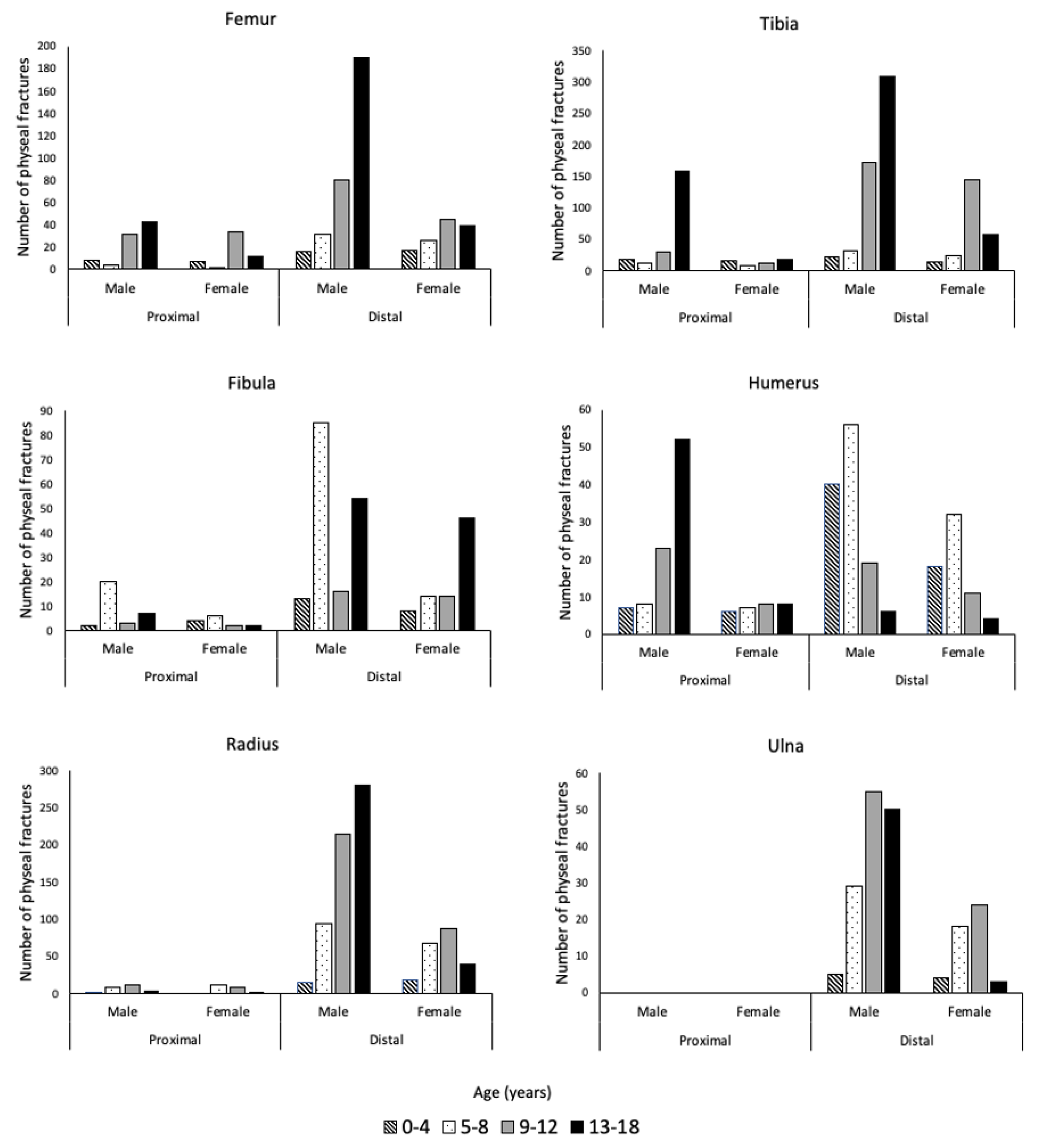
| 0–4 | 8 (0.3) | 16 (0.7) | 17 (0.7) | 22 (0.9) | 2 (0.1) | 13 (0.6) | 7 (0.3) | 40 (1.7) | 2 (0.1) | 15 (0.6) | 0 (0.0) | 5 (0.2) |
| 5–8 | 4 (0.2) | 31 (1.3) | 13 (0.6) | 31 (1.3) | 20 (0.9) | 85 (3.6) | 8 (0.3) | 56 (2.4) | 8 (0.3) | 94 (4.0) | 0 (0.0) | 29 (1.2) |
| 9–12 | 32 (1.4) | 80 (3.4) | 29 (1.2) | 173 (7.4) | 3 (0.1) | 16 (0.7) | 23 (1.0) | 19 (0.8) | 12 (0.5) | 214 (9.2) | 0 (0.0) | 55 (2.4) |
| 13–18 | 43 (1.8) | 190 (8.1) | 159 (6.8) | 308 (13.2) | 7 (0.3) | 54 (2.3) | 52 (2.2) | 6 (0.3) | 5 (0.2) | 282 (12.1) | 0 (0.0) | 50 (2.1) |
| Bone n (%) | ||||||||||||
|---|---|---|---|---|---|---|---|---|---|---|---|---|
| Femur | Tibia | Fibula | Humerus | Radius | Ulna | |||||||
| Age | P | D | P | D | P | D | P | D | P | D | P | D |
| 0–4 | 7 (0.7) | 17 (1.8) | 16 (1.7) | 14 (1.5) | 4 (0.4) | 8 (0.8) | 6 (0.6) | 18 (1.9) | 0 (0.0) | 18 (1.9) | 0 (0.0) | 4 (0.4) |
| 5–8 | 2 (0.2) | 26 (2.7) | 8 (0.8) | 24 (2.5) | 6 (0.6) | 14 (1.5) | 7 (0.7) | 32 (3.4) | 12 (1.3) | 68 (7.1) | 0 (0.0) | 18 (1.9) |
| 9–12 | 34 (3.6) | 45 (4.7) | 13 (1.4) | 144 (15.1) | 2 (0.2) | 14 (1.5) | 8 (0.8) | 11 (1.2) | 8 (0.8) | 88 (9.2) | 0 (0.0) | 24 (2.5) |
| 13–18 | 12 (1.3) | 39 (4.1) | 18 (1.9) | 58 (6.1) | 2 (0.2) | 46 (4.8) | 8 (0.8) | 4 (0.4) | 3 (0.3) | 40 (4.2) | 0 (0.0) | 3 (0.3) |
| SH I (%) | SH II (%) | SH III (%) | SH IV (%) | |
|---|---|---|---|---|
| Femur | 96 (4.3) | 231 (10.3) | 31 (1.4) | 16 (0.7) |
| Tibia | 36 (1.6) | 448 (20) | 119 (5.3) | 204 (9.1) |
| Fibula | 39 (1.7) | 120 (5.4) | 0 (0) | 0 (0) |
| Humerus | 32 (1.4) | 119 (5.3) | 0 (0) | 20 (0.9) |
| Radius | 50 (2.2) | 546 (24.4) | 13 (0.6) | 16 (0.7) |
| Ulna | 9 (0.4) | 79 (3.5) | 8 (0.4) | 4 (0.2) |
| Total | 262 (11.7) | 1543 (69.0) | 171 (7.6) | 260 (11.6) |
Publisher’s Note: MDPI stays neutral with regard to jurisdictional claims in published maps and institutional affiliations. |
© 2022 by the authors. Licensee MDPI, Basel, Switzerland. This article is an open access article distributed under the terms and conditions of the Creative Commons Attribution (CC BY) license (https://creativecommons.org/licenses/by/4.0/).
Share and Cite
Fuchs, J.R.; Gibly, R.F.; Erickson, C.B.; Thomas, S.M.; Hadley Miller, N.; Payne, K.A. Analysis of Physeal Fractures from the United States National Trauma Data Bank. Children 2022, 9, 914. https://doi.org/10.3390/children9060914
Fuchs JR, Gibly RF, Erickson CB, Thomas SM, Hadley Miller N, Payne KA. Analysis of Physeal Fractures from the United States National Trauma Data Bank. Children. 2022; 9(6):914. https://doi.org/10.3390/children9060914
Chicago/Turabian StyleFuchs, Joseph R., Romie F. Gibly, Christopher B. Erickson, Stacey M. Thomas, Nancy Hadley Miller, and Karin A. Payne. 2022. "Analysis of Physeal Fractures from the United States National Trauma Data Bank" Children 9, no. 6: 914. https://doi.org/10.3390/children9060914
APA StyleFuchs, J. R., Gibly, R. F., Erickson, C. B., Thomas, S. M., Hadley Miller, N., & Payne, K. A. (2022). Analysis of Physeal Fractures from the United States National Trauma Data Bank. Children, 9(6), 914. https://doi.org/10.3390/children9060914


