Article

Comparison of Digestion Methods Using Atomic Absorption Spectrometry for the Determination of Metal Levels in Plants

1
RME-Laboratory of Resources, Materials, and Ecosystems, Faculty of Sciences of Bizerte, University of Carthage, Bizerte 7021, Tunisia
2
Department of Technologies and Applied Sciences, School of Agriculture, Polytechnic Institute of Beja, 7800-295 Beja, Portugal
3
CERENA, Instituto Superior Técnico, University of Lisbon, 1049-001 Lisbon, Portugal
4
LR12SP13, Faculty of Medicine of Monastir, University of Monastir, Monastir 5000, Tunisia
5
MARE—Marine and Environmental Sciences Centre & ARNET—Aquatic Research Network Associated Laboratory, Faculdade de Ciências da Universidade de Lisboa, Campo Grande, 1749-016 Lisbon, Portugal
6
Departamento de Biologia Vegetal, Faculdade de Ciências da Universidade de Lisboa, Campo Grande, 1749-016 Lisboa, Portugal
*
Author to whom correspondence should be addressed.
Separations 2023, 10(1), 40; https://doi.org/10.3390/separations10010040
Submission received: 12 December 2022 / Revised: 5 January 2023 / Accepted: 6 January 2023 / Published: 9 January 2023
(This article belongs to the Section Environmental Separations)

Abstract

:
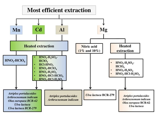
Trace metal elements (TMEs) are among the most important types of pollutants in the environment. Therefore, a precise determination of these contaminants in several environmental components is required for the safety assurance of living organisms. Spectroscopic analysis is an efficient technique employed to detect and determine TME contents in numerous samples. Hence, to achieve reliable and accurate results when using spectroscopic analysis, samples should be carefully prepared. In the present study, the comparison of eight digestion methods of five vegetal samples was carried out to quantify Cd, Mn, Al and Mg contents using the atomic absorption spectroscopy technique. According to the extraction techniques used in this study, results showed an outstanding difference in TME levels determined in the same vegetal sample. The results obtained indicated that the highest Mn concentrations were recorded when using the mixture of HNO3-HClO4 in the studied species: atriplex portulacoides, arthrocnemum indicum, olea europaea BCR-62, ulva lactuca and ulva lactuca BCR-279 compared to all other methods. Regarding the extraction of Cd, our results showed that heated extraction using different acids (HNO3-H2SO4-HClO4, HCl-HNO3, HNO3-HClO4, HNO3-H2SO4, HNO3-HCl-HClO4 and HNO3-HCl-H2SO4) was the most efficient in atriplex portulacoides, arthrocnemum indicum, olea europaea BCR-62, ulva lactuca and ulva lactuca BCR-279. Similarly, these heated acid digestion techniques (efficient for Cd) showed the highest levels of Al in atriplex portulacoides and arthrocnemum indicum. However, for the Mg extraction, our results revealed that the effectiveness of the method used depended on the plant species studied. Regarding these findings, the efficiency of metal quantification by AAS depends on the digestion procedure, the metallic ion to determine and the plant species.

Graphical Abstract

1. Introduction

Trace metal elements (TMEs), contained in various types of samples (water, soil and plant tissues, etc.) can be precisely measured by highly sensitive spectroscopic techniques, such as graphite furnace (GFAAS), hydride generation atomic absorption spectrometry (HGAAS) and flame (FAAS) [1]. Several techniques require aqueous samples to determine metal concentrations [2]; therefore, solid samples generally require conversion to soluble forms using digestion methods. For metal analysis, several methods have been employed for the digestion of plant tissue, such as drying and conventional digestion, ignition and microwave digestion, drying and microwave digestion, wet acid digestion, and a mixture of concentrated acid digestion, etc. During the dry ashing procedure, some of the TMEs in the sample might vanish via the adsorption of elements on the walls of the furnace, or volatilization, such as arsenic (As), chromium (Cr) and lead (Pb), which may be lost at ashing temperatures of 500–550 °C [3]. Usually, for the dissolution of herbal product samples, acid digestion methods are used before the elemental analysis [4]. Indeed, in spectroscopic elemental analysis, acid digestion of the sample is a crucial step of the completely analytical procedure. In highly complex matrices such as herb and plant materials, acid digestion has a considerable effect on the retrieval of several analyte contents [1]. Numerous studies have shown that acid digestion methods provide relevant results which, except for the metals associated with silicates, could reach up to 100% in the determination of the total elemental composition, depending on the studied element [5,6]. For metal extraction, a variety of acids or acid mixtures have been used, among which the most frequently used are nitric (HNO3), sulfuric (H2SO4), perchloric acids (HClO4), and hydrochloric acid (HCl) [7]. The choice of a combination of acids or an individual acid is based on the nature of the matrix to be decomposed [8]. In addition, there are multifarious methods that require the use of a combination of an acid with an oxidant, such as hydrogen peroxide.
Specific plant species can absorb and hyperaccumulate metal contaminants and/or excess nutrients from the growth substrate [9]. These latter will be absorbed and accumulated mainly in the roots, the primary biological metal pool in plants. Nevertheless, a small amount of the metals will be translocated into the shoots [10,11,12], except highly mobile metals such as zinc, manganese, cadmium, lanthanum and cerium [13,14,15]. Although these metal internalization processes take place within plant tissues, these elements are still prone to being exported during senescence to the adjacent ecosystems [16], entering the food chain [17], or even being remobilized to the sediment during decomposition processes [18]. Thus, considering the key biogeochemical role of plants in ecosystems, it becomes of extreme importance to disentangle the best procedures to analyze metals in plant tissues with the highest efficiency possible.
The research work carried out in this paper is a continuity of works published by Sleimi et al. [19] in the Sustainability journal. Indeed, it was demonstrated that cation contents varied with digestion methods used. The present study is intended to appraise the effectiveness of eight different acid digestion methods in order to recommend the most effective one which leads to the maximum recovery of other TMEs which were not studied by Sleimi et al. [19]. The analysis of cadmium (Cd), manganese (Mn), magnesium (Mg) and aluminum (Al) was conducted using flame atomic absorption spectrometry.

2. Materials and Methods

2.1. Sample Preparation

This study focused on different plants: atriplex portulacoides, arthrocnemum indicum, ulva lactuca (field samples) and two certified reference materials from the Community Bureau of Reference-BCR: olea europaea BCR-62 and ulva lactuca BCR-279. Regarding field samples, shoots were collected from twelve individuals per species and per station. The sampling area was limited to 20 m2 per species. Plant samples (aerial parts) were dried for two weeks (until constant weight) at 60 °C. Then, dry samples were pulverized using an agate ball mill.

2.2. Methods of Digestion

The samples were submitted to eight different acid digestion methods: cold extraction with nitric acid diluted at 1%, cold extraction with nitric acid diluted at 10% and heated extraction using different acid digestion, as detailed below, to recognize the most suitable digestion method to measure the contents of Cd, Mn, Al and Mg in the plant samples by atomic absorption spectrophotometer (AAS).

2.2.1. Nitric Acid Digestion (1% and 10%)

Cold extraction was carried out with two different concentrations of nitric acid of 1% (method A) and 10% (method B), according to the method of Larson et al. [20]. An aliquot of 25 mg of fine powder of the plant sample was placed in steel cups. After drying for 24 h at 60 °C, 10 mL of nitric acid was poured into the mixture and left at the temperature of the laboratory for 15 days. All the samples were agitated occasionally to ensure better contact between the powder plant and the acid solution. Finally, the mineral deposit was filtered through ashless filter paper (Whatman N°1, 90 mm diameter circles). The extracts were stored at 4 °C up to the time of the assay.

2.2.2. Acid Mixtures Digestion

The extraction was achieved through a heated attack using six different acid digestion methods, such as the following: nitric-sulfuric-perchloric acid digestion (method C) in a proportion of (10:1:0.5, v/v/v) [21,22,23]; hydrochloric-nitric acid digestion known as aqua regia mixture (method D) in a proportion of (3:1, v/v) [24]; nitric-perchloric acid digestion (method E) in a proportion of (3:1, v/v) [25]; nitric-sulfuric acid digestion (method F) in a proportion of (2:1, v/v) [26]; nitric-hydrochloric-perchloric acid digestion (method G) in a proportion of (5:1:0.5, v/v/v) [2] and nitric-hydrochloric-sulfuric acid digestion (method H) in a proportion of (5:1:1, v/v/v) [8] (Table 1). About 40 to 50 mg of plant powder, formerly desiccated in an incubator at 60 °C for 24 h, was poured into a Khjeldahl flask with 3 mL of the mixture of acids, as explained above, and was then placed on a heated ramp. To ensure good mineralization, the temperature was increased gradually, up to 150 °C by increments of 50 °C every 15 min, then to 350 °C by increments of 100 °C every 15 min. Incineration was subsequently maintained for one hour at 350 °C. After two hours of mineralization, a white ash was obtained that would be dissolved in 50 mL of nitric acid (0.05%). Hence, the solutions obtained were kept at 4 °C until the analysis.

2.3. Trace Metal Element Analysis

After the digestion procedure, the concentrations of Cd, Mn, Mg and Al contained in the final solutions were determined by means of the flame atomic absorption spectrometer (Perkin Elmer 900T, Waltham, MA, USA). For each element assessed, the spectrometer requires specific hollow cathode lamps (HCl) and a deuterium lamp for continuous background correction. Instrumental operating parameters for each element (e.g., wavelength, slit width etc.) are shown in Table 2. In order to prepare a series of composite standards, a calibration with standard solutions of 1000 mg·L−1 of Cd, Mn, Mg and Al was used.

2.4. Statistical Analysis

The analysis of variance (ANOVA) coupled with pairwise Tukey honest significant difference test (HSD) was carried out in order to assess the existence of significant differences between the concentrations attained by different extraction procedures. Values were presented as mean ± standard deviation, n = 10. Bars marked with different letters are significantly different at p < 0.05.

3. Results

3.1. Cadmium Assay

Generally, assay results of the Cd showed that heated extraction, using different acid combinations, produced the highest Cd recoveries in all species studied (Figure 1). In fact, the highest Cd contents recorded were 249.4, 246.6, 251.2, 246.6 and 242.5 µg·g−1 dry weight (DW) in atriplex portulacoides, arthrocnemum indicum, olea europaea BCR-62, ulva lactuca and ulva lactuca BCR-279, respectively.

3.2. Manganese Assay

Figure 2 shows the levels of Mn according to the extraction process in plants studied beforehand. Overall maximum Mn levels were obtained upon the application of extraction method E, presenting Mn concentration values of 681.1, 801.1, 1106.1, 878.7 and 1809.4 µg·g−1 DW in atriplex portulacoides, arthrocnemum indicum, olea europaea BCR-62, ulva lactuca and ulva lactuca BCR-279, respectively.

3.3. Magnesium Assay

Our results showed that the highest Mg levels were attained using method C in atriplex portulacoides and arthrocnemum indicum, and the quantified Mg contents were 2185.7 and 2296.6 µg·g−1 DW in atriplex portulacoides and arthrocnemum indicum, respectively (Figure 3).
Otherwise, it was revealed that the contents of Mg were higher using the extraction methods F and H in olea europaea BCR-62 and method H in ulva lactuca (Figure 3), while methods A and B were the most reliable for Mg extraction in ulva lactuca BCR-279.

3.4. Aluminum Assay

The determination of Al using different acid digestion methods showed that the heated extraction methods achieved the highest Al levels in atriplex portulacoides and arthrocnemum indicum (Figure 4). On the other hand, methods D and H proved to be more efficient for Al extraction in olea europaea BCR-62, ulva lactuca and ulva lactuca BCR-279 samples. For example, the highest Al contents recorded were 3227, 3951, 7329, 8377 and 8752 µg·g−1 DW in atriplex portulacoides, arthrocnemum indicum, olea europaea BCR-62, ulva lactuca and ulva lactuca BCR-279, respectively.

4. Discussion

The diversity of the techniques used for preparing plant materials for elemental analysis has engendered a great deal of interest as well as controversy. The mineralization of plant samples is often considered to be a necessary technical step that precedes the spectral determination of particular metals; it is a crucial procedure for the final quality of the entire analytical process. It should be accurately enhanced in accordance with either the matrix or the specific analyte [27]. It is important and also preferable to select a simple procedure of digestion that reduces the sample time, diminishes the danger of contamination during handling, and includes relatively safe procedures [28].
For the digestion of various samples for metal analysis, several methods have been suggested in previous studies. Aqua regia has been recommended as the most efficient method of digestion for samples with low organic matter or carbonate contents such as agricultural soils and sediments [29]. It was also suggested that nitric acid digestion presented the highest efficiency in recovering Cd, Mn, and Ni in the majority of compost samples [7]. For the recovery of TME in six types of composts, the comparison between nitric acid and aqua regia digestion revealed an equivalent analytical sensitivity. However, nitric acid digestion extracted more Pb, Cd, Cu and Zn, while aqua regia digestion recovered more Cr, Ni and Hg. Moreover, it is preferable to substitute aqua regia digestion by nitric acid digestion since the latter is less expensive and more environmentally benign [30].
According to Uddin et al. [1], the best acid mixture for the decomposition of herbal samples was hydrochloric-nitric acids HNO3–HCl in a ratio of 1:3, owing to the ability of the combination to release the metallic ions from such complex matrices of herbal materials and thereafter to reduce the level of the noise during the detection procedure.
Furthermore, the study of the quantification of potassium (K), phosphorus (P), calcium (Ca), iron (Fe), copper (Cu), Mg, Zn and Mn contents in rice (oryza sativa) and barley (hordeum vulgare) seedlings revealed that the digesting capacity of HNO3 acid and HNO3–HClO4 acid mixture was not significantly different [31].
In the present study, our data indicated that the highest Mn concentrations were recorded using method E (p < 0.05), which represented the mixture of HNO3-HClO4 in studied species: atriplex portulacoides, arthrocnemum indicum, olea europaea BCR-62, ulva lactuca and ulva lactuca BCR-279, compared to all other methods.
According to Shaibur et al. [31], in the scientific world of plant nutrition, nitric acid-perchloric acid mixture is considered to be the prominent digesting reagent. It was also revealed that using the nitric/perchloric acid procedure is better than using only nitric acid for digestion, due to the better element recovery. However, perchloric acid digestion can cause the loss of K and B by volatilization; in addition, the use of specially designed hoods is required, since perchloric acid introduces the risk of explosion [32]. Otherwise, Warman and Muizelaar proved that nitric/perchloric acid and nitric acid procedures presented the same analytical sensitivity, and there is no advantage in using the more expensive and hazardous nitric/perchloric acid digestion [33].
On the other hand, results showed that the heated extraction method led to the highest Cd level in atriplex portulacoides, arthrocnemum indicum, olea europaea BCR-62, ulva lactuca and ulva lactuca BCR-279 and the highest Al content in atriplex portulacoides, arthrocnemum indicum.
In addition, it was reported that the digestion method of organic matter is dictated by not only the elements to determine but also by the elemental content of the plant tissue [34]. According to Uddin et al. [1], the herbal samples have complex matrices since they consist of either one herb or a mixture of herbs obtained from different parts of the plant, such as seeds, roots, leaves, and flowers, which might have different chemical properties.
The results obtained also showed that all digestion methods tested achieved the highest Cd, Mn and Al levels compared to the certified reference values of 0.1 µg·g−1 DW, 57 µg·g−1 DW and 450 µg·g−1 DW, respectively, in olea europaea BCR-62 (Table 3). Likewise, we noticed that the digestion methods used in this study led to the greatest Cd level in ulva lactuca BCR-279 (Table 4).
The correlations between eight digestion methods used to determine Cd, Mn, Al and Mg contents in plant tissues are shown in Table 5. For Cd assay, the methods B and F were negatively correlated (p < 0.01). However, the methods C, D, E presented a significant positive correlation at p < 0.01 and p < 0.05. Also, method H was negatively correlated to method D and G (p < 0.05). All the methods used in this study to quantify Mn contents were positively correlated and the correlation was significant at p < 0.01 and p < 0.05. Regarding Al assay, a positive correlation was observed between the methods D and H (p < 0.01). In addition, method F was highly correlated to methods C and G (a positive significant correlation was recorded at p < 0.01), and method A was positively correlated to methods B, D, E and H (p < 0.05). For Mg, method A was positively correlated to methods B, E and G (p < 0.01) and methods D and F (p < 0.05). On the other hand, the methods D, E, F, G and H were positively correlated at p < 0.01 and p < 0.05 and all these methods were found to be positively correlated to method B (p < 0.01 and p < 0.05).
To sum up, the effectiveness of AAS in quantifying the different TME is influenced by the technique used for extraction, the element to be determined, and the plant tissue.

5. Conclusions

The shoots of atriplex portulacoides, arthrocnemum indicum, olea europaea BCR-62, ulva lactuca and ulva lactuca BCR-279 were collected, rinsed and oven-dried, and subsequently crushed and homogenized. Eight procedures of extraction were tested to show the best method of turning dry samples into solutions of Cd, Mn, Mg and Al.
Our results showed that the application of nitric–perchloric acid mixture (method E) was the most efficient digestion method for the extraction of Mn in atriplex portulacoides, arthrocnemum indicum, olea europaea BCR-62, ulva lactuca and ulva lactuca BCR-279 when compared to all other methods. In addition, all heated extraction methods, including methods C, D, E, F, G and H, were the most appropriate techniques for solubilizing Cd ions into solution from all studied plant species, and also for dissolving Al in atriplex portulacoides, arthrocnemum indicum. In addition, in atriplex portulacoides, arthrocnemum indicum, olea europaea BCR-62 and ulva lactuca, the maximum recovery of Mg was obtained by the heated extraction procedures. However, the methods A and B were the best extraction methods for ulva lactuca BCR-279. According to these findings, and despite the fact that the atomic absorption spectrophotometer is known to be efficient in elemental composition analysis and the quantification of TME in solution, the application of the appropriate digestion method is the most crucial key to achieving the extraction of the maximum amount of a specific metal containing in various samples.

Author Contributions

Conceptualization, N.S. and I.C.; Methodology, I.B., R.K. and N.D.; Formal analysis, I.B., N.D., R.F. and S.H.; Writing-original draft, I.B. and R.K.; Writing-review and editing, B.D., R.K., S.H. and R.F.; Visualization, B.D., N.S. and I.C.; Supervision, N.S. and I.C. All authors have read and agreed to the published version of the manuscript.

Funding

This research received no external funding.

Acknowledgments

The authors would like to thank Fundação para a Ciência e a Tecnologia (FCT) for funding MARE (Marine and Environmental Sciences Centre, UIDB/04292/2020 and UIDB/04292/2020), ARNET (Aquatic Research Network Associated Laboratory, LA/P/0069/2020) and CERENA (Centro de Recursos Naturais e Ambiente, UIDB/04028/2020). This work was supported by “Redesigning the rhizosphere microbiome of Salicornia for exploitation of bioproducts and improvement of bioprocesses” Rhizomis (FCT, PTDC/BIA-MIC/29736/201). B. Duarte was supported by research contracts (CEECIND/00511/2017 and 2022.01746.CEECIND).

Conflicts of Interest

The authors declare no conflict of interest.

References

  1. Uddin, A.H.; Khalid, R.S.; Alaama, M.; Abdualkader, A.M.; Kasmuri, A.; Abbas, S.A. Comparative study of three digestion methods for elemental analysis in traditional medicine products using atomic absorption spectrometry. J. Anal. Sci. Technol. 2017, 7, 6. [Google Scholar] [CrossRef] [Green Version]
  2. Micó, C.; Recatalá, L.; Peris, M.; Sanchez, J. A comparison of two digestion methods for the analysis of heavy metals by flame atomic absorption spectroscopy. Spectrosc. Eur. 2007, 19, 23–26. [Google Scholar]
  3. Azcue, J.; Mudroch, A. Comparison of different washing, ashing and digestion methods for the analysis of trace elements in vegetation. Int. J. Environ. Anal. Chem. 1994, 57, 151–262. [Google Scholar] [CrossRef]
  4. Duyusen, E.G.; Görkem, A. Comparison of acid digestion techniques to determine heavy metals in sediment and soil samples. Gazi Univ. J. Sci. 2011, 24, 29–34. [Google Scholar]
  5. Andersen, K.J.; Kisser, M.I. Digestion of Solid Matrices Desk Study. Horizontal 2004, 18, 1–39. [Google Scholar]
  6. Santoro, A.; Held, A.; Linsinger, T.P.; Perez, A.; Ricci, M. Comparison of total and aqua regia extractability of heavy metals in sewage sludge: The case study of a certified reference material. Trends Analyt. Chem. 2017, 89, 34–40. [Google Scholar] [CrossRef]
  7. Hseu, Z.Y. Evaluating heavy metal contents in nine composts using four digestion methods. Bioresour. Technol. 2004, 95, 53–59. [Google Scholar] [CrossRef]
  8. Asher, A.S.; Samuel, K.E.; Mary, D.S. Analytical method for comparison of suitable wet digestion methods for heavy metal analysis in soil around a cement industry. Int. J. Res. Innov. Sci. 2020, 7, 41–47. [Google Scholar]
  9. Tangahu, B.V.; Abdullah, S.R.S.; Basri, H.; Idris, M.; Anuar, N.; Mukhlisin, M. A Review on Heavy Metals (As, Pb, and Hg) Uptake by Plants through Phytoremediation. Int. J. Chem. Eng. 2011, 2011, 939161. [Google Scholar] [CrossRef]
  10. Weis, P.; Windham, L.; Burke, D.J.; Weis, J.S. Release into the environment of metals by two vascular salt plants. Mar. Environ. Res. 2002, 54, 325–329. [Google Scholar] [CrossRef]
  11. Windham, L.; Weis, J.S.; Weis, P. Uptake and distribution of metals in two dominant salt marsh macrophytes, spartina alterniflora (cordgrass) and phragmites australis (common reed). Estuar. Coast. Shelf Sci. 2003, 56, 63–72. [Google Scholar] [CrossRef]
  12. Caçador, I.; Valo, C.; Catarino, F. Seasonal variation of Zn, Pb, Cu and Cd concentrations in the roots-sediment system of spartima maritima and halimione portulacoides from Tagus estuary salt marches. Mar. Environ. Res. 2000, 49, 279–290. [Google Scholar] [CrossRef] [PubMed]
  13. Kouki, R.; Ayachi, R.; Ferreira, R.; Sleimi, N. Behavior of cucumis sativus L. in presence of Aluminum stress: Germination, plant growth and antioxidant enzymes. Food Sci. Nutr. 2021, 9, 3280–3288. [Google Scholar] [CrossRef] [PubMed]
  14. Dridi, N.; Ferreira, R.; Bouslimi, H.; Brito, P.; Martins-Dias, S.; Caçador, I.; Sleimi, N. Assessment of tolerance to lanthanum and cerium in helianthus annuus plant: Effect on growth, mineral nutrition and secondary metabolism. Plants 2022, 11, 988. [Google Scholar] [CrossRef]
  15. Dridi, N.; Brito, P.; Bouslimi, H.; Ferreira, R.; Martins-Dias, S.; Caçador, I.; Sleimi, N. Physiological and biochemical behaviours and antioxidant response of helianthus annuus under lanthanum and cerium stress. Sustainability 2022, 14, 4153. [Google Scholar] [CrossRef]
  16. Duarte, B.; Vaz, N.; Valentim, J.M.; Dias, J.M.; Silva, H.; Marques, J.C.; Sleimi, N.; Caçador, I. Revisiting the outwelling hypothesis: Modelling salt marsh detrital metal exports under extreme climatic events. Mar. Chem. 2017, 191, 24–33. [Google Scholar] [CrossRef]
  17. Fonseca, V.F.; França, S.; Duarte, B.; Caçador, I.; Cabral, H.N.; Mieiro, C.L.; Coelho, J.P.; Pereira, E.; Reis-Santos, P. Spatial variation in mercury bioaccumulation and magnification in a temperate estuarine food web. Front. Mar. Sci 2019, 6, 117. [Google Scholar] [CrossRef]
  18. Duarte, B.; Reboreda, R.; Caçador, I. Seasonal variation of Extracellular Enzymatic Activity (EEA) and its influence on metal speciation in a polluted salt marsh. Chemosphere 2008, 73, 1056–1063. [Google Scholar] [CrossRef]
  19. Sleimi, N.; Bankaji, I.; Kouki, R.; Dridi, N.; Duarte, B.; Caçador, I. Assessment of extraction methods of trace metallic elements in plants: Approval of a common method. Sustainability 2022, 14, 1428. [Google Scholar] [CrossRef]
  20. Larson, K.D.; Dejong, T.K.; Johnson, R.S. Physiological and growth responses of mature peach trees to postharvest water stress. J. Am. Soc. Hortic. Sci. 1988, 133, 296–300. [Google Scholar] [CrossRef]
  21. Sghaier, B.D.; Bankaji, I.; Pedro, S.; Caçador, I.; Sleimi, N. Photosynthetic behaviour and mineral nutrition of tamarix gallica cultivated under aluminum and NaCl combined stress. Phyton-Int. J. Exp. Bot. 2019, 88, 239–252. [Google Scholar] [CrossRef]
  22. Labidi, O.; Vives-Peris, V.; Gómez-Cadenas, A.; Pérez-Clemente, R.M.; Sleimi, N. Assessing of growth, antioxidant enzymes and phytohormone regulation in cucurbita pepo under cadmium stress. Food Sci. Nutr. 2021, 9, 2021–2031. [Google Scholar] [CrossRef] [PubMed]
  23. Sleimi, N.; Kouki, R.; Hadj Ammar, M.; Ferreira, R.; Perez-Clemente, R. Barium effect on germination, plant growth, and antioxidant enzymes in cucumis sativus L. plants. Food Sci. Nutr. 2021, 9, 2086–2094. [Google Scholar] [CrossRef] [PubMed]
  24. Abbruzzini, T.F.; Silva, C.A.; de Andrade, D.A.; de Oliveira Carneiro, W.J. Influence of digestion methods on the recovery of iron, zinc, nickel, chromium, cadmium and lead contents in 11 organic residues. Rev. Bras. Ciênc. Solo 2014, 38, 166–176. [Google Scholar] [CrossRef]
  25. Caçador, I.; Caetano, M.; Duarte, B.; Vale, C. Stock and losses of trace metals from salt marsh plants. Mar. Environ. Res. 2009, 67, 75–82. [Google Scholar] [CrossRef] [PubMed] [Green Version]
  26. Momen, A.A.; Zachariadis, G.A.; Anthemidis, A.N.; Stratis, J.A. Investigation of four digestion procedures for multi-element determination of toxic and nutrient elements in legumes by inductively coupled plasma-optical emission spectrometry. Anal. Chim. Acta 2006, 565, 81–88. [Google Scholar] [CrossRef]
  27. Adamczyk-Szabela, D.; Anielak, P.; Wolf, W.M. Influence of digestion procedure and residual carbon on manganese, copper, and zinc determination in herbal matrices by atomic absorption spectrometry. J. Anal. Meth. Chem. 2017, 2017, 6947376. [Google Scholar] [CrossRef] [Green Version]
  28. Zheljazkov, V.D.; Warman, P.R. Comparison of Three Digestion Methods For the Recovery of 17 Plant Essential Nutrients And Trace Elements from Six Composts. Compost. Sci. Util. 2002, 10, 197–203. [Google Scholar] [CrossRef]
  29. Sastre, J.; Sahuquillo, A.; Vidal, M.; Rauret, G. Determination of Cd, Cu, Pb and Zn in environmental samples microwave-assisted total digestion versus aqua regia and nitric acid extraction. Anal. Chim. Acta 2002, 462, 59–72. [Google Scholar] [CrossRef]
  30. Schmid, R. The effects of sample processing on amounts of nutrients and pollutants in composts. Assoc. Ger. Agric. Investig. Res. Inst. Ser. Congr. Rep. 1990, 32, 357–362. [Google Scholar]
  31. Shaibur, M.R.; Shamim, A.H.M.; ImamulHuq, S.M.; Kawai, S. Comparison of digesting capacity of nitric acid and nitric acid-perchloric acid mixture and the effect of lanthanum chloride on potassium measurement. Nat. Sci. 2010, 8, 157–162. [Google Scholar]
  32. Zarcinas, B.A.; Cartwright, B. Analysis of Soil and Plant Material by Inductively Coupled Plasma-Optical Emission Spectroscopy; Commonwealth Scientific and Industrial Research Organization: Canberra, Australia, 1983; p. 36.
  33. Warman, P.R.; Muizelaar, T. A comparison of the elemental analysis of compost using two standard methods. In Proceedings of the 5th Annual Meeting of the Composting Council of Canada, Aylmer, PQ, Canada, 1–3 November 1995.
  34. Sahrawat, K.L.; Kumar, G.R.; Rao, J.K. Evaluation of triacid and dry ashing procedures for determining potassium, calcium, magnesium, iron, zinc, manganese, and copper in plant materials. Commun. Soil Sci. Plant Anal. 2002, 33, 95–102. [Google Scholar] [CrossRef]
Figure 1. Contents of Cd (µg·g−1 DW) in atriplex portulacoides, arthrocnemum indicum, olea europea BCR-62, ulva lactuca and ulva lactuca BCR-279 using methods A, B, C, D, E, F, G and H. Values were presented as mean ± standard deviation, n = 10. Letters (a, b) show a significant difference (p < 0.05) between the concentrations attained by different extraction procedures.
Separations 10 00040 g001
Figure 2. Contents of Mn (µg·g−1 DW) in atriplex portulacoides, arthrocnemum indicum, olea europea BCR-62, ulva lactuca and ulva lactuca BCR-279 using methods A, B, C, D, E, F, G and H. Values were presented as mean ± standard deviation, n = 10. Letters (a, b, c, d) show a significant difference (p < 0.05) between the concentrations attained by different extraction procedures.
Separations 10 00040 g002
Figure 3. Contents of Mg (µg·g−1 DW) in atriplex portulacoides, arthrocnemum indicum, olea europea BCR-62, ulva lactuca and ulva lactuca BCR-279 using methods A, B, C, D, E, F, G and H. Values were presented as mean ± standard deviation, n = 10. Letters (a, b, c) show a significant difference (p < 0.05) between the concentrations attained by different extraction procedures.
Separations 10 00040 g003
Figure 4. Contents of Al (µg·g−1 DW) in atriplex portulacoides, arthrocnemum indicum, olea europea BCR-62, ulva lactuca and ulva lactuca BCR-279 using methods A, B, C, D, E, F, G and H. Values were presented as mean ± standard deviation, n = 10. Letters (a, b, c) show a significant difference (p < 0.05) between the concentrations attained by different extraction procedures.
Separations 10 00040 g004
Table 1. Acid digestion procedure.
MethodsReagentsProportionsTime/min
CHNO3/H2SO4/HClO410:1:0.5, v/v/v120
DHCl/HNO33:1, v/v120
EHNO3/HClO43:1, v/v120
FHNO3/H2SO42:1, v/v120
GHNO3/HCl/HClO45:1:0.5, v/v/v120
HHNO3/HCl/H2SO45:1:1, v/v/v120
Table 2. Instrumental parameters for flame atomic absorption spectrometer FAAS of TME analysis.
MetalWavelength (nm)Slit Width
(nm)
Background CorrectionOxidant: Acetylene
L·min−1
Lampe Type
Cd228.80.7YesAir: Acetylene
10:2.5
HCl
Mn279.480.2NoAir: Acetylene
10:2.5
HCl
Mg285.210.7NoAir: Acetylene
10:2.5
HCl
Al309.270.7NoN2O:Acetylene
06:7.5
HCl
Table 3. Comparison of results obtained using tested digestion methods with certified reference values in Olea europaea BCR-62.
Olea europaea BCR-62Cd (µg·g−1 DW)Mn (µg·g−1 DW)Al (µg·g−1 DW)
Method A181.5187.61345.9
Method B175.1100.81725.7
Method C233.3364.11144.6
Method D236.4442.37329.9
Method E251.21106.11691.9
Method F229.1443.72792.9
Method G231.7366.41498.2
Method H219.1286.17051.0
Certified reference value000.1057.00450.0
Table 4. Comparison of results obtained using tested digestion methods with certified reference values in ulva lactuca BCR-279.
Ulva lactuca BCR-279Cd (µg·g−1 DW)
Method A167.875
Method B170.833
Method C221.062
Method D210.322
Method E242.534
Method F234.414
Method G232,081
Method H235,996
Certified reference value000.274
Table 5. The correlation between the digestion methods for Cd, Mn, Al and Mg determined in plant tissues (Pearson correlation).
Cd
ABCDEFGH
A1
B0.1451
C0.206−0.6691
D0.412−0.2750.809 *1
E0.005−0.5690.944 **0.857 *1
F−0.343−0.950 **0.5800.2810.5801
G0.584−0.3530.1510.3360.0400.3231
H−0.4440.330−0.522−0.823 *−0.565−0.393−0.761 *1
Mn
ABCDEFGH
A1
B0.994 **1
C0.974 **0.973 **1
D0.858 *0.838 *0.912 **1
E0.952 **0.929 **0.962 **0.844 *1
F0.974 **0.965 **0.967 **0.803 *0.987 **1
G0.998 **0.993 **0.976 **0.882 *0.937 **0.958 **1
H0.992 **0.997 **0.961 **0.803 *0.933 **0.973 **0.987 **1
Al
ABCDEFGH
A1
B0.820 *1
C−0.4080.0091
D0.764 *0.670−0.6231
E0.741 *0.847 *0.2880.2741
F−0.2200.3120.918 **−0.3700.4361
G0.0500.5690.706−0.1300.6000.920 **1
H0.716 *0.494−0.812 *0.962 **0.107−0.599−0.3511
Mg
ABCDEFGH
A1
B0.968 **1
C−0.198−0.1851
D0.711 *0.840 *−0.0251
E0.973 **0.991 **−0.3010.814 *1
F0.732 *0.789 *−0.718 *0.6880.848 *1
G0.946 **0.971 **−0.4140.791 *0.992 **0.906 **1
H0.6620.810 *−0.4360.830 *0.813 *0.889 *0.849 *1
(*) Correlation is significant at 0.05; (**) Correlation is significant at 0.01.
Disclaimer/Publisher’s Note: The statements, opinions and data contained in all publications are solely those of the individual author(s) and contributor(s) and not of MDPI and/or the editor(s). MDPI and/or the editor(s) disclaim responsibility for any injury to people or property resulting from any ideas, methods, instructions or products referred to in the content.