Review

Health-Promoting Potential of Millet: A Review

1
Postharvest Technology Division, Bangladesh Agricultural Research Institute, Gazipur 1701, Bangladesh
2
College of Food Science and Nutritional Engineering, China Agricultural University, Beijing 100083, China
3
Food Engineering & Tea Technology, Shahjalal University of Science & Technology, Sylhet 3114, Bangladesh
4
Buddhist Tzu Chi Medical Foundation, Hualien 970, Taiwan
*
Authors to whom correspondence should be addressed.
Separations 2023, 10(2), 80; https://doi.org/10.3390/separations10020080
Submission received: 29 December 2022 / Revised: 19 January 2023 / Accepted: 23 January 2023 / Published: 24 January 2023
(This article belongs to the Special Issue Feature Papers in Separations from Editorial Board Members)

Abstract

:
Being a key source of animal food, millet production has been sharply increasing over the last few years in order to cope with the dietary requirements of the ever-increasing world population. It is a splendid source of essential nutrients such as protein, carbohydrates, fat, minerals, vitamins, and also some other bioactive compounds that eventually help through multiple biological activities, including antioxidant, anti-hyperglycemic, anti-cholesterol, anti-hypertensive, anthropometric effects and regulation of gut microbiota composition. These bioactive compounds, nutrients, and functions of cereal grains can be affected by processing techniques such as decortication, soaking, malting, milling, fermentation, etc. This study discusses the nutritional and functional properties of millet-incorporated foods and their impact on health, based on around 150 articles between 2015 and 2022 from the Web of Science, Google Scholar, Food and Agriculture Organization of the United Nations (FAO), Breeding Bid Survey (BBS), and FoodData Central (USDA) databases. Analyzing literature reviews, it is evident that the incorporation of millet and its constituents into foodstuffs could be useful against undernourishment and several other health diseases. Additionally, this review provides crucial information about the beneficial features of millet, which can serve as a benchmark of guidelines for industry, consumers, researchers, and nutritionists.

Graphical Abstract

1. Introduction

Millet belongs to the family Poaceae, which can grow well under dry, high-temperature conditions as grasses with small seeds, and has been used as fodder and human food for around 10,000 years [1]. According to the Food and Agriculture Organization of the United Nations (FAO), Asia and Africa are the predominant growers, while India, Niger, and China are the three most high-yield countries (Figure 1) [2]. Cultivated varieties of millet include pearl millet, finger millet, proso millet, foxtail millet (FM), etc. (Table 1). In millet, several macronutrients, minerals (iron, zinc, phosphorus, calcium, potassium), and vitamins are higher than those in rice and wheat [3]. Antinutrients such as phytates, polyphenols, and tannins reduce mineral bioavailability by chelating cations [4]. Nevertheless, phytochemicals of millets such phenolic compounds exhibit antioxidant action via scavenging reactive oxygen species (ROS), reducing power, and/or metal-chelating activity towards ferric and ferrous ions. The abundance of protease and amylase inhibitors affects the grains’ digestibility [5]. Some important characteristic features are inherent in millet, such as hypolipidemic, low-glycemic index, and antioxidative characteristics [6]. Because of its nutritional value, millet is used to make noodles, nutritious soups, hard drinks, pancakes, and cereal porridges worldwide [7]. Considering these several health benefits, this review work has been undertaken to summarize those benefits and provide a real picture of their impact on health.

2. Methodology

In order to give a whole perspective of millet’s health benefits, the present study carried out this comprehensive review based on the Web of Science, Google Scholar, Food and Agriculture Organization of the United Nations (FAO), and FoodData Central (USDA) databases from 2015 to 2022. More than 100 articles are reported in this review. The generalized data collection procedure is followed as presented in Figure 2.

3. Nutritional Quality of Millets

Millets are nutritionally equivalent to or even superior to several of the most important cereal grains. Millets are full of physiologically active substances and provide numerous health benefits, including a high antioxidant content, high fiber content, low glycemic index, and gluten-free protein. Considering the nutritional profile of these small-seeded crops, they are a great source of energy, protein, and minerals. Among its vitamins, millet grains are rich in niacin (B3), thiamine, riboflavin, and folic acid. It is estimated that nearly 70% of grains are composed of carbohydrates, with most being soluble carbohydrates and dietary fiber. A majority of millets’ polysaccharides are composed of amylopectin and amylose (70–80%). In addition to being rich in dietary fiber, they are also richer in polyphenols (0.2–0.5%). Millets’ polyphenols, tannins, and phytates provide the bulk of their antioxidant activity, and these substances have a role in regulating the aging process. In comparison to other grains, finger millet has the highest calcium level, at 344 mg/100 g [19].
A good amount of moisture (12%) is contained in sorghum millet, along with nearly 10.4% protein and 1.3% fat. The amount of fiber and minerals in grain sorghum is approximately 1.6%. Sorghum grain has a high carbohydrate content (73%), making it an excellent source of energy (349 kcal). Other carbohydrates found in sorghum include cellulose and hemicellulose. Sorghum also contains good amounts of calcium (25 mg), dietary fiber (14.3 g), iron (4.1 mg), and phosphorus (222 mg), respectively, per 100 g of edible portion [20].
Both little millet and kodo millet, which are classified as nutraceutical crops, contain between 37 and 38% dietary fiber. Millets are widely utilized as a snack, infant food, processed food, etc. due to their high nutrient content and so are considered a complete food. In developing countries, sorghum and millets contribute greatly to nutritional security. The millets are also known as miracle grains and nutria-cereals [21].
Furthermore, in comparison with wheat (1.5%) and rice (0.6%), millets contain several times more minerals (1–5 g/100 g). There is an abundance of iron in Pearl Millet and Barnyard Millet, which can fulfill the iron requirement of anemic individuals. There is a high content of zinc (4.1 mg/100 g) in foxtail millet, as well as a high content of iron [22]. The details of nutritional composition of millets have been presented in Table 2Table 3 and Table 4.

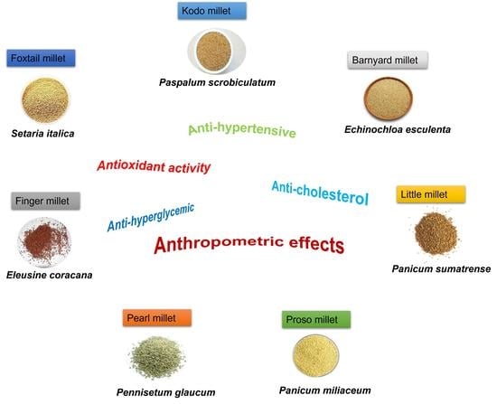
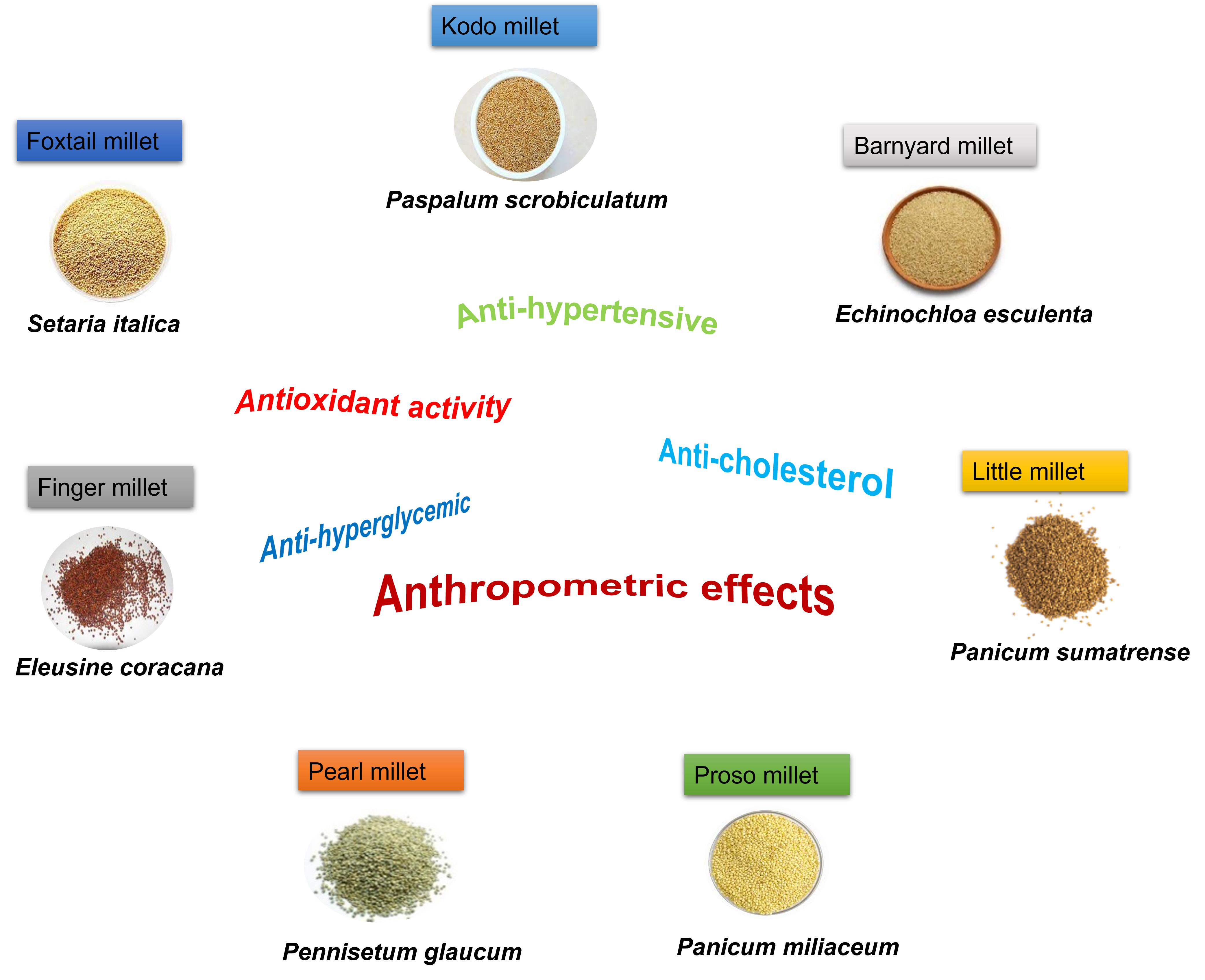
4. Beneficial Features of Millet

Millet is rich in nutrients and compounds, which offer multiple health benefits including.

4.1. Antioxidant Activity

It is evident from the literature that millet (little, pearl, proso, foxtail, finger, and kodo millets) whole grains contain antioxidants, reductants, and metal chelators in their soluble and insoluble phenolic extracts (Table 2). Nevertheless, whole millets may be beneficial as natural antioxidant sources depending on the variety.
There has been great interest in studying the nutraceutical and antioxidant properties of FM, pear millet, and finger millet varieties. According to recent studies, FM contains 3.34 mg tocopherol/100 g (wet basis) and 47 mg polyphenols/100. On the other hand, proso millet contains 2.22 mg tocopherol/100 g (wet basis) and 29 mg polyphenolics/100 g [30].
Currently, over 50 phenolic compounds belonging to several classes, namely, phenolic acids and their derivatives, dehydrotriferulates and dehydrodiferulates, dimers and flavan-3-ol monomers, flavanonols, flavones, and flavonols in four phenolics fractions of several whole millet grains (pearl, little, proso, foxtail, finger, and kodo millets) were positively or tentatively identified using high-performance liquid chromatography (HPLC) and HPLC-tandem mass spectrometry. However, in vitro tests revealed that kodo millet’s insoluble bound fraction showed the highest content of phenols as well as antioxidant activity. In light of this, and based upon published literature data, millet grains can be used as functional food ingredients as well as natural antioxidants [21].
Several studies have investigated the antioxidant potential of phenolics, and other bioactive components isolated from millet grains and their fractions. It was reported that an ethanol extract of barnyard millet grains contained one serotonin derivative, two flavonoids, and three antioxidative phenolic compounds. Furthermore, Abedin et al. [31] reported that the methanolic extracts of FM also showed a higher total antioxidant capacity (170 ± 4 mg ascorbic acid equivalents (AAE)/100 g) and total phenolic content (52 ± 2 mg gallic acid equivalents (GAE)/100 g). Additionally, kodo millet flour methanol extracts quenched 1,1,diphenyl-2-picrylhydrazyl (DPPH) by 70% compared to 15% to 53% in other millet extracts. In addition, the white varieties of sorghum, finger millet, and foxtail millet showed lower quenching than their colored counterparts, indicating that phenolics in the seed coat could be responsible for the antioxidant activities. Moreover, compared to wheat, rice, and other millet species, extracts from finger millet were found to have significantly stronger radical-scavenging activity [32].

4.2. Anti-Hyperglycemic Effects

Fiber and non-starchy polysaccharides, indigestible carbs that are present in millet, help to lower sugar levels in the blood [33]. It also has a low glycemic index (GI), which helps to reduce sugar levels in the blood [34].
A 12-week study with pre-diabetes (64 people) provided similar results. After 6 weeks of FM intervention, fasting blood glucose and glucose after 120 min decreased to 0.3 ± 0.7 mmol/L and 1.0 ± 2.7 mmol/L, respectively. Then both types of glucose remained steady for up to 12 weeks. There was an insignificant difference in fast insulin, fructosamine, and insulin content (after 120 min) throughout the study period. Homeostasis model assessment of insulin resistance (HOMA-IR) significantly decreased from an initial 3.6 ± 2.3 to 2.9 ± 1.7 in the 12th week, on the other hand, HOMA-IR increased from 0.4 ± 0.2 to 0.5 ± 0.6, respectively [35]. A slight decrease in fasting and post-meal blood sugar insulin resistance was observed by Ren et al. [35] after taking 50 g of FM/per day. In another study, Shobana et al. [36] also found lower fasting blood sugar levels and a fall in triglyceride and cholesterol levels in rats with diabetes while feeding them a diet containing 20% finger millet. Moreover, Fu et al. [37] found that prolamin from cooked foxtail millet (PCFM) ameliorated islet-cell impairment i.e., stimulated insulin secretion as evident from significantly high homeostasis model assessment of β cell function (HOMA-β). HOMA-IR of the model control group (MC) was significantly higher than the normal control group (NC), which indicates insulin resistance (Table 5). Moreover, the study of Seo et al. [38] demonstrated that N-p-coumaroyl serotonin, feruloyl serotonin of barnyard millet (Echinochloa utilis) were significantly reduced glucose content (72 and 51% at 0.2 mg/wells) in Caco-2 (human intestinal epithelial) cells and inhibited intestinal sucrase (IC50 of 4.0 and 9 μM) in mammalian rat. Furthermore, it was revealed in a recent study by Krishnan et al. [39] that pearl millet phenolics inhibit carbolytic enzymes and regulate GLUT and thus have anti-diabetic properties. Similarly, a significant reduction in fasting glucose and insulin levels, as well as HOMA-IR levels, was observed after pearl millet whole grain powder or ethanolic extract administration to obese rats fed a high-fat diet, which supports the hypoglycemic effects of pearl millet [40]. Moreover, proso millet has been found to improve blood sugar levels and insulin levels in genetically obese type 2 diabetic mice under conditions of high fat intake [41].

4.3. Anti-Cholesterol Effects

Consumption of millets reduces hyperlipidemia and raises the levels of high-density lipoprotein cholesterol (HDL-C), according to a recent systematic review and meta-analysis of the impacts of millets consumption on lipid profile, including triglyceride (TG), total cholesterol (TC), low-density lipoprotein cholesterol (LDL-C), HDL-C, and very-low-density lipoprotein cholesterol (VLDL-C). Consuming millets for periods ranging from 21 days to 4 months was shown to reduce levels of TC, TG, LDL-C, and VLDL-C by 8.0, 9.5, 10, and 9.0%, respectively, according to the findings of 19 research. Researchers in four separate investigations found that consuming millets resulted in normal levels of TG and TC (>150 and >200 mg/dl, respectively). Additionally, the HDL-C 4.0 increased by 6.0% after eating millet-based meals [42].
Lee et al. [43] found reduced triglyceride, compared with the control group that those fed foxtail and proso millet in rats. Additionally, millet protein may help lower cholesterol, which was claimed by Nishizawa et al. [44].
Choi et al. [45] found foxtail millet protein (FMP) diet group had a significantly higher total plasma cholesterol concentration than the casein diet group. HDL-C concentration in the FMP diet group was more than twice that of the casein diet group. The plasma insulin concentration of the FMP diet group was reduced by 57% in contrast to the casein diet, without any significant difference in plasma glucose concentration. The plasma adiponectin concentration of the FMP diet group was 64% greater than that of the casein diet group. In the liver, the concentration of cholesterol declined significantly in the case of the FMP diet when compared with that of the casein diet, but triglyceride content was greater in the FMP diet group. The plasma triglyceride content of the casein diet group was found to be significantly lower than the FMP diet group. HDL-cholesterol in the plasma of the casein diet group was significantly lower than the FMP diet group. Plasma glucose concentration was similar; however, insulin concentration in the FMP diet group was lower than in the casein diet group. The FMP diet group contained almost twice the plasma adiponectin than the casein diet group. Liver cholesterol concentrations were significantly lower for the FMP diet than the casein diet, but triglyceride content was similar.
Molla M. M., [23] found millet protein diets such as uncooked extracted (UCE), cooked extracted (CE), uncooked extracted enriched (UCEE), and cooked extracted enriched (CEE) groups significantly reduced the plasma and liver TC, TG, and LDL-C than uncooked (UC) and cooked FM (C FM) flour diets, in casein, control, and normal diet group (Table 6 and Table 7). It also significantly increased the plasma HDL-C compared to UC and C FM flour diets, casein, control, and normal diets. These results are strongly supported by Choi et al. [45]; Park et al. [41]; Nishizawa et al. [44]. UCE and UCEE FM protein diets increased the plasma HDL-C and reduced the plasma and liver TC, TG, and plasma LDL-C in comparison with other diets. Cholesterols lowering effects might be the result of antinutritional richness in the UCE and UCEE FM protein diet than other diets [46]. Higher antinutrients might help to reduce cholesterol levels, and the risk of stroke, coronary heart disease, certain cancer, and liver disorder through their antioxidant activity [47].
Millet protein (FM) increased the plasma HDL-C level whereas, decreased the plasma TC, TG, and LDL-C and liver TC and TG, which may be a good predictor to reduce the risk of liver injury and hyperlipidemia [23]. These outcomes are parallel to the findings of Choi et al. [45] who reported that Korean foxtail millet protein has a protective action for reducing atherosclerosis by increasing the HDL-C level. Nishizawa et al. [48] reported that proso millet protein supplemented with lysine and threonine have a favorable effect on cholesterol metabolism by elevating plasma HDL-C level and lowering LDL-C, TC, and TG level for both plasma and liver which is inversely related to the risk of coronary heart disease.

4.4. Anti-Hypertensive Effects

Ren et al. [35] found that after 12 weeks millet (FM) interventions, diastolic blood pressure (DBP) declined significantly from 84.9 ± 8.5 mmHg to 81.6 ± 7.8 mmHg. Hou et al. [49] found both systolic blood pressure (SBP) and DBP decreased significantly due to 12 weeks millet (FM) diet intervention from the initial (133.61 ± 1.94 mmHg) to 12th week (129.48 ± 2.14 mmHg). On average, SBP and DBP were reduced by 4.13 mmHg and 3.49 mmHg, respectively. Dietary fiber, protein, and minerals in whole grains are inversely associated with blood pressure (BP) [50]. FMP hydrolysates may significantly decrease BP in hypertensive rats [51]. Studies have revealed that phytochemicals in millet possess serum lipid-reducing properties [52]. Chen et al. [51] observed SBP declined significantly after 4 weeks of millet intervention, compared to the control (194.9 ± 3.5 mmHg). Whereas it was found 167.4 ± 2.5, 166.3 ± 3.5, 168.4 ± 6.3, and 179.0 ± 5.0 mmHg in captopril, raw foxtail millet protein hydrolysates (RPH), extruded foxtail millet protein hydrolysates (EPH), and fermented foxtail millet protein hydrolysates with R. oryzae (FRPH), respectively. SBP persistently declined in RPH, EPH, and captopril groups, except the FRPH group. The reduction was observed to be higher in the RPH (28.3 mmHg), EPH (24.8 mmHg), and captopril (23.6 mmHg) treatment groups, but in the FRPH group, it was only 13.6 mmHg.
Chen et al. [51] found reduced serum angiotensin-converting enzyme (ACE) activity significantly after 4 weeks of intervention in the control group (141.8 ± 14.6 U/L). For other treatments the values were; RPH (104.1 ± 7.4 U/L), FRPH (85.5 ± 16 U/L), EPH (88.9 ± 15.5 U/L), and captopril (95.2 ± 13.6 U/L). Additionally, a non-significant difference was detected between the three FMP hydrolysate groups and the captopril group. Plasma angiotensin II (Ang II) levels in control were 2.85 ± 0.29 ng/mL, which declined significantly when treated with FRPH (1.75 ± 0.42 ng/mL), RPH (1.68 ± 0.48 ng/mL), captopril group (1.50 ± 0.56 ng/mL) and EPH (1.44 ± 0.31 ng/mL).
Hou et al. [53] found the levels of Ang II, aldosterone, and ACE exhibited a downward drift during the study period. The antihypertensive effects of whole grains seem to be developed from better endothelial activity, which combined actions of several intrinsic factors such as obstructing the vasoconstrictors effect, induction of vasodilation, affecting vasorelaxation pathways. Chen et al. [51] indicated millet diets have an anti-hypertensive effect, resulting from the inhibition of serum ACE activity. He also found that ACE, Ang II, and aldosterone in serum declined, demonstrating a probable antihypertensive mechanism of millet inhibition of ACE activity with slight hypertension.
UCE and UCEE FM protein diet may have antioxidant activities, that are believed to break up the free radical chain of oxidation and donate hydrogen thereby lowering the cholesterol level. Therefore, it is said that FM protein has a protective action against degenerative diseases such as heart disease, stroke, liver disorders, and cancer [54].
Reactive oxygen species (ROS) are facilitators of cellular injury and play a vital role in hepatic damage in the case of D-galactosamine-induced hepatitis. This elimination of ROS in the liver is accelerated by the non-essential amino acid proline which is higher in the UCE and UCEE FM protein diet [55]. Millet (FM) protein diet is capable to suppress the hepatotoxicity of D-galactosamine-induced acute liver injury [56].

4.5. Anthropometric Effects

Intake of millet decreased body weight, body mass index (BMI), and degree of obesity. Body fat mass of 22.1 ± 7.1 kg declined to 21.1 ± 6.2 kg, which appears to be parallel to the body weight variation of initial 69.1 ± 11.6kg to final 68.2 ± 11.2 kg [35].
Choi et al. [45] could not find any significant variation in weight gain, epididymal adipose tissue weight, or plasma triglyceride concentration between the two dietary groups, but the liver weight was higher in the FMP diet group than that of the group fed with the casein diet.
Although there was no significant difference in body weight (BW) gain or liver weight between the two dietary groups, the epididymal adipose tissue weight was significantly lower in the FMP group than in the casein group.
Hou et al. [49] found that millet significantly reduced BMI, body fat percentage (BF%), body fat mass, and waist circumference (WC) during the study period, whereas the circumferences of the hip increased. Waist-hip ratio closely stayed constant. Higher BP might decrease bone density, eventually causing osteoporosis. The primary key to measuring and monitoring any intervention treatment is weight loss. BMI reduced significantly at 12 weeks of the study, which advised that millet might lessen BP secondarily by decreasing obesity degree. Body weight is intensely linked with hypertension, any upsurge in BW rises body fluid and peripheral resistance [57].
Chen et al. [51] found body weight of spontaneously hypertensive rats usually increased with age, but among the treatment groups, the difference was insignificant. Considering heart weight (%) in the control group (0.50 ± 0.03 g/100 g BW), it was significantly lower in captopril (0.41 ± 0.02 g/100 g BW), RPH (0.43 ± 0.01 g/100 g BW), EPH (0.40 ± 0.01 g/100 g BW), and FRPH (0.41 ± 0.01 g/100 g BW).
Fu et al. [37] found a steady BW increase in the NC group, while it decreased significantly in the MC group, which is considered a classic diabetes indicator. BW of mice increased slightly after 5 weeks of treatments with millet (PCFM), which advocated that millet intervention might inhibit diabetic BW loss. The PCFM group exhibited significantly lower fasting blood glucose (FBG) than the MC group, though it was still significantly higher than the NC group.
The millet diet (UCE and UCEE FM protein) significantly reduced body weight as compared to other diets [23]. The reason may be a lower gain of final body weight by the UCE and UCEE FM protein diet over the whole study period than other diets. High protein intake can contribute to losing weight more than other diets [58]. Lower energy intake associated with IIP intake possibly contributes to weight loss more than other low-protein diets [59]. It is also well reported that millet (FM) protein contains more fiber, antioxidants, and phenolic compounds which may contribute to lower body weight [60]. Liver and relative liver weight was significantly increased by the UCE, UCEE, and CEE FM protein diet than other diets [59].

4.6. Effects on Gut Microbiota Composition

Fu et al. [37] found Firmicutes and Bacteroides two major phyla in the gut microbiota, and their ratio (Bacteroides (B)/Firmicutes (F)) is positively related to plasma glucose level. MC group demonstrated significantly lower B/F than the NC group, which caused a higher level of FBG. Millet intervention slightly amplified the B/F ratio, though the dissimilarity witnessed between MC and PCFM groups was non-significant. Overall, at the phylum level in gut microbiota, millet administration has a non-significant effect. However, the PCFM group contained higher Odoribacter than the MC group. The relative abundance of the above-mentioned bacteria was changed by PCFM, which might be associated with the anti-diabetic effect. PCFM group also contained a higher amount of Blautia, Akkermansia and Odoribacter compared to NC and MC groups.
Molla M.M., [23] found a higher abundance of Bacteroidetes in the casein diet followed by the normal and control diet and that may be due to a high number of Prevotella and Bacteroides, increasing the inflammation (Table 8 and Table 9. Inflammation involves chronic liver diseases and develops progressive hepatic damage and fibrosis. It has been reported that Lactobacellus rhamnosus, Lactobacillus salivarius or Pediococcus pantosaceus prevent D-galactosamine-induced liver injury [61]. Firmicutes are more dominant bacteria in the UC FM diet than in the C FM diet. In contrast, Bacteroidetes predominate in the C FM diet followed by Firmicutes (Table 8 and Table 9). The higher abundance of the UC FM diet was largely due to a high number of Allobaculum genus and Lactobacillus genus at the phylum level of Firmicutes than the C FM diet (Table 8 and Table 9). Butyric acids have been reported to play a crucial role in upholding gut health, the energy source to the colonic mucosa, a regulator of gene expression, regulation, differentiation, and apoptosis in host cells [62]. Providing a UC FM diet could upsurge the bacterial production of butyric acids in the large intestine. These results are supported by the findings of Louis et al. [63]. The resistant starch or high protein diet provides an energy source for gut microbiota, which has been proposed as a potential prodrug for treating inflammatory bowel disease (IBD) and decrease DNA damage [64]. On the other hand, the higher abundance of Bacteroidetes in the C FM diet may be due to the higher presence of Prevotella genus which has been reported to cause various diseases (Table 8 and Table 9) [65]. Bacteroidetes was the most abundant phylum in the CE FM protein diet followed by Firmicutes (Table 8 and Table 9). The increase in Firmicutes involves producing several butyrate microbes, which have several health-beneficial effects [63].
However, the microbiome of the UCE FM protein diet contained a high proportion of Firmicutes and a low proportion of Actinobacteria. Murphy et al. [66] reported that a relative increase in Firmicutes and a decrease in Actinobacteria levels could contribute to lower blood glucose, TNF-ɑ, and triglyceride levels. Costa et al. [67] stated Firmicutes dominated followed by Bacteroidetes. Similar findings also have been made by Middelbos et al. [68] in dogs upon supplementation with dietary fiber. On the other hand, the UCE FM protein diet had higher Proteobacteria, while the CE FM protein diet had lower. The high abundance of Proteobacteria in the UCE FM protein diet may be due to a high number of Sutterella geniuses in the phylum of Proteobacteria (Table 8 and Table 9). In the CE FM protein diet, the abundance is high due to the higher number of Bacteroides at the phylum level of Bacteroidetes. Bacteroides with high abundance has been reported to influence inflammation, liver injury, and regeneration [69].
In the CEE and UCEE FM protein diet, Bacteroidetes was the most abundant phylum with high abundance in CEE FM protein diet (Table 8 and Table 9) followed by Firmicutes, Verrucomicrobia, Proteobacteria, and Actinobacteria. In contrast, the presence of Verrucomicrobia and Firmicutes was greater followed by Bacteroidetes, Actinobacteria, and Proteobacteria in the UCEE FM protein diet (Table 8 and Table 9). The high abundance of Bacteroidetes in the CEE FM protein diet may be due to a high number of Prevotella genus. An increase in the Prevotella genus at the phylum of Bacteroidetes might result in periodontal disease, human alcoholic liver disease, and IBD in patients [70]. Prevotellaceae might produce sulfatases that disrupt the mucosal barrier function; these enzymes are elevated in intestinal biopsies from IBD patients [71]. Higher Verrucomicrobia might be due to a high number of Akkermansia genus in the UCEE FM protein diet. It has been reported that Akkermansia is a much degrading bacterium in the mucus layer [56]. The presence of this bacterium is contrariwise related to body weight and type-2 diabetes. Data shows that the UCE FM protein diet led to the relative increase in Firmicutes and Proteobacteria compared with another phylum of Bacteria, in control and other diets. In the UCEE FM protein diet, there was a good combination of Verrucomicrobia and Firmicutes, which have several favorable effects on gut microbiota. Therefore, it is evident that increased Akkermansia and Setterella genus under the phylum of Verrucomicrobia and Proteobacteria and another Lactobacillus bacterium in UCE and UCEE FM protein diets were found beneficial for preventing acute liver injury after administration of D-galactosamine [72].

5. Conclusions

Millet enables better glycemic control both fasting and postprandial. Glucose-reducing virtue could be a combined outcome of greater leptin concentrations, lesser insulin resistance, and fewer inflammations. Only the Millet diet is discussed here, nonetheless, other whole grains might have a similar glucose-reducing effect.
It seems millet protein advances the sensitivity of insulin and metabolism of cholesterol by increasing adiponectin concentration; therefore, it might serve as beneficial for preventing obesity, type-2 diabetes, and cardiac diseases.
Daily consumption of the millet diet significantly reduced BP, BMI, BF(%), and fat mass without affecting usual dietary habits. FM might enable the collective effects of dietary fiber, protein, minerals, and micronutrients, reducing fat and cholesterol intake, which eventually reduces BP. This finding might be a methodical benchmark for directing daily consumption of it to reduce BP.
The incorporation of myofibrillar proteins hydrolysates was highly effective in reducing BP. It can also obstruct the serum ACE activity and drop the levels of Ang II. Antioxidant activity exploitation might be an alternate way to prevent hypertension and it might be enhanced by extrusion processing. Moreover, RPH, EPH, and FRPH reduced the heart weight (%) decreased in spontaneously hypertensive rats. It also reduces left ventricular pressure and cardiac damage. Based on the results, it might be proposed that FMP hydrolysates intake (RPH and EPH) ameliorates hypertension. More conclusive studies incorporating animals as a subject are essential to elucidate their precise mode of action regarding the quantitative structure–activity relationship of ACE inhibitory peptides.
Glucose homeostasis disorders triggered by diabetes can be ameliorated by millet. This study provides pragmatic evidence for introducing millet and derivatives as a prospective food constituent directing diabetes attenuation.
Intake of millet protein especially UCE and UCEE FM protein helps to reduce body weight and increase the liver’s relative weight. It also directed that higher serum AST, ALT, and LDH activities by D-galactosamine were attenuated by the incorporation of millet especially UCE and UCEE millet protein. Plasma TC, TG, and LDL-C, liver, TC, and TG were reduced, whereas HDL-C was increased by the ingestion of UCE and UCEE millet protein. The lipid depressing capability of MDA was also attributed to the ingestion of UCE and UCEE millet protein. Liver histopathological studies exhibited that UCE and UCEE millet protein have a significant effect to lighten D-galactosamine-induced hepatocellular injury. CEE and CEE millet protein contributed more to decreasing serum AST, ALT, and LDH activities, plasma, and liver cholesterol than casein and normal diet. Therefore, this study suggests that millet protein might be considered to avert hepatic disease, acute liver injury, and hyperlipidemia.

Author Contributions

Conceptualization: Q.Z. and Q.S.; methodology: A.A.S. and M.R.R.; investigation: M.M.M. and H.H.K.; Data Curation: A.A.S.; writing—original draft preparation: A.A.S. and T.A.; writing—Review and Editing: M.R.R. and N.I.; visualization: G.F.C.; funding acquisition: Q.Z. and Q.S. All authors have read and agreed to the published version of the manuscript.

Funding

This study was financially supported by China Agriculture Research System of MOF and MARA (CARS-06-14.5).

Data Availability Statement

Not applicable.

Acknowledgments

Not applicable.

Conflicts of Interest

The authors declare no conflict of interest.

Abbreviations

FAO: Food and Agriculture Organization of the United Nations; BBS: Breeding Bid Survey; USDA: FoodData Central; FM: Foxtail millet; ROS: Reactive oxygen species, GI: Glycemic index; HOMA-IR: Homeostasis model assessment of insulin resistance; PCFM: Prolamin from cooked foxtail millet; HOMA-β: Homeostasis model assessment of β cell function; MC: Model control group; NC: Normal control group; TG: Triglyceride; FMP: Foxtail millet protein; HDL-C: High-density lipoprotein-cholesterol; UCE: Uncooked extracted; CE: Cooked extracted; CEE: Cooked extracted enriched; TC: Total cholesterol; LDL-C: Low-density lipoprotein cholesterol; VLDL-C: Very-low-density lipoprotein cholesterol; UC: Uncooked; C FM: Cooked FM; AST: Aspartate transaminase; ALT: Alanine transaminase; LDH: Lactate dehydrogenase; MDA: Malondialdehyde; DBP: Diastolic blood pressure; SBP: Systolic blood pressure; BP: Blood Pressure; RPH: Raw foxtail millet protein hydrolysates; EPH: Extruded foxtail millet protein hydrolysates; FRPH: Fermented foxtail millet protein hydrolysates; ACE: Angiotensin-converting enzyme; Ang II: Angiotensin II; ROS: Reactive oxygen species; BMI: Body Mass Index; BW: Body weight; BF: Body fat percentage; WC: Waist circumference; FBG: Fasting blood glucose; HPLC: high-performance liquid chromatography.

References

  1. Tripathi, M.K.; Jadam, R.S.; Kumar, A. Quality Management System in Millet and Sorghum. In Millets and Millet Technology; Springer: Berlin/Heidelberg, Germany, 2021; pp. 363–379. [Google Scholar] [CrossRef]
  2. FAO. World Food and Agriculture—Statistical Yearbook; FAO: Rome, Italy, 2020. [Google Scholar]
  3. Saini, S.; Saxena, S.; Samtiya, M.; Puniya, M.; Dhewa, T. Potential of underutilized millets as Nutri-cereal: An overview. J. Food Sci. Technol. 2021, 58, 4465–4477. [Google Scholar] [CrossRef] [PubMed]
  4. Ertop, M.H.; Bektaş, M. Enhancement of bioavailable micronutrients and reduction of antinutrients in foods with some processes. Food Health 2018, 4, 159–165. [Google Scholar] [CrossRef]
  5. Yin, D.; Yin, X.; Wang, X.; Lei, Z.; Wang, M.; Guo, Y.; Aggrey, S.E.; Nie, W.; Yuan, J. Supplementation of amylase combined with glucoamylase or protease changes intestinal microbiota diversity and benefits for broilers fed a diet of newly harvested corn. J. Anim. Sci. Biotechnol. 2018, 9, 24. [Google Scholar] [CrossRef] [PubMed]
  6. Sharma, N.; Niranjan, K. Foxtail millet: Properties, processing, health benefits, and uses. Food Rev. Int. 2017, 34, 329–363. [Google Scholar] [CrossRef]
  7. Asrani, P.; Ali, A.; Tiwari, K. Millets as an alternative diet for gluten-sensitive individuals: A critical review on nutritional components, sensitivities and popularity of wheat and millets among consumers. Food Rev. Int. 2022, 38, 1–30. [Google Scholar] [CrossRef]
  8. Yang, X.; Zhang, S.; He, C.; Xue, P.; Zhang, L.; He, Z.; Zang, L.; Feng, B.; Sun, J.; Zheng, M. METTL14 suppresses proliferation and metastasis of colorectal cancer by down-regulating oncogenic long non-coding RNA XIST. Mol. Cancer 2020, 19, 46. [Google Scholar] [CrossRef] [Green Version]
  9. Ren, X.; Chen, J.; Wang, C.; Molla, M.M.; Diao, X.; Shen, Q. In vitro starch digestibility, degree of gelatinization and estimated glycemic index of foxtail millet-derived products: Effect of freezing and frozen storage. J. Cereal Sci. 2016, 69, 166–173. [Google Scholar] [CrossRef]
  10. Akinola, S.A.; Badejo, A.A.; Osundahunsi, O.F.; Edema, M.O. Effect of preprocessing techniques on pearl millet flour and changes in technological properties. Int. J. Food Sci. Technol. 2017, 52, 992–999. [Google Scholar] [CrossRef]
  11. Ganguly, S.; Sabikhi, L.; Singh, A.K. Effect of whey-pearl millet-barley based probiotic beverage on Shigella-induced pathogenicity in murine model. J. Funct. Foods 2019, 54, 498–505. [Google Scholar] [CrossRef]
  12. Sarita; Singh, E. Potential of Millets: Nutrients Composition and Health Benefits. J. Sci. Innov. Res. 2016, 5, 46–50. [Google Scholar] [CrossRef]
  13. Srilekha, K.; Kamalaja, T.; Maheswari, K.U.; Rani, R.N. Nutritional Composition of Little Millet Flour. Int. Res. J. Pure Appl. Chem. 2019, 31, 1–4. [Google Scholar] [CrossRef]
  14. Tyl, C.; Marti, A.; Hayek, J.; Anderson, J.; Ismail, B.P. Effect of growing location and variety on nutritional and functional properties of proso millet (Panicum miliaceum) grown as a double crop. Cereal Chem. 2018, 95, 288–301. [Google Scholar] [CrossRef]
  15. Das, S.; Khound, R.; Santra, M.; Santra, D.K. Beyond Bird Feed: Proso Millet for Human Health and Environment. Agriculture 2019, 9, 64. [Google Scholar] [CrossRef] [Green Version]
  16. Almaski, A.; Thondre, S.; Lightowler, H.; Coe, S. Determination of the polyphenol and antioxidant activity of different types and forms of millet. Proc. Nutr. Soc. 2017, 76, E5. [Google Scholar] [CrossRef] [Green Version]
  17. Ramadoss, D.P.; Sivalingam, N. Vanillin extracted from Proso and Barnyard millets induce apoptotic cell death in HT-29 human colon cancer cell line. Nutr. Cancer 2019, 72, 1422–1437. [Google Scholar] [CrossRef]
  18. Anis, M.A.; Sreerama, Y.N. Inhibition of protein glycoxidation and advanced glycation end-product formation by barnyard millet (Echinochloa frumentacea) phenolics. Food Chem. 2020, 315, 126265. [Google Scholar] [CrossRef]
  19. Tripathi, M.K.; Mohapatra, D.; Jadam, R.S.; Pandey, S.; Singh, V.; Kumar, V.; Kumar, A. Nutritional Composition of Millets. In Millets and Millet Technology; Springer: Berlin/Heidelberg, Germany, 2021; pp. 101–119. [Google Scholar]
  20. Abah, C.R.; Ishiwu, C.N.; Obiegbuna, J.E.; Oladejo, A.A. Nutritional Composition, Functional Properties and Food Applications of Millet Grains. Asian Food Sci. J. 2020, 14, 9–19. [Google Scholar] [CrossRef]
  21. Saleh, A.S.; Zhang, Q.; Chen, J.; Shen, Q. Millet Grains: Nutritional Quality, Processing, and Potential Health Benefits. Compr. Rev. Food Sci. Food Saf. 2013, 12, 281–295. [Google Scholar] [CrossRef]
  22. Amadou, I.; Gounga, M.E.; Le, G.-W. Millets: Nutritional Composition, Some Health Benefits and Processing—A Review. Emir. J. Food Agric. 2013, 25, 501–508. [Google Scholar] [CrossRef]
  23. Molla, M.M. Effect of Foxtail Millet Diet on Liver Injury and Blood Lipid Profile Induced by D-Galactosamine in Mice. Ph.D. Thesis, China Agricultural University (CAU), Beijing, China, 2016. [Google Scholar]
  24. Ciulu, M.; Cádiz-Gurrea, M.d.l.L.; Segura-Carretero, A. Extraction and Analysis of Phenolic Compounds in Rice: A Review. Molecules 2018, 23, 2890. [Google Scholar] [CrossRef] [Green Version]
  25. Xiang, J.; Apea-Bah, F.B.; Ndolo, V.U.; Katundu, M.C.; Beta, T. Profile of phenolic compounds and antioxidant activity of finger millet varieties. Food Chem. 2018, 275, 361–368. [Google Scholar] [CrossRef] [PubMed]
  26. Slama, A.; Cherif, A.; Sakouhi, F.; Boukhchina, S.; Radhouane, L. Fatty acids, phytochemical composition and antioxidant potential of pearl millet oil. J. Consum. Prot. Food Saf. 2019, 15, 145–151. [Google Scholar] [CrossRef]
  27. Yuan, Y.; Xiang, J.; Zheng, B.; Sun, J.; Luo, D.; Li, P.; Fan, J. Diversity of phenolics including hydroxycinnamic acid amide derivatives, phenolic acids contribute to antioxidant properties of proso millet. Lwt 2021, 154, 112611. [Google Scholar] [CrossRef]
  28. Xiang, J.; Zhang, M.; Apea-Bah, F.B.; Beta, T. Hydroxycinnamic acid amide (HCAA) derivatives, flavonoid C-glycosides, phenolic acids and antioxidant properties of foxtail millet. Food Chem. 2019, 295, 214–223. [Google Scholar] [CrossRef]
  29. Saha, D.; Gowda, M.V.C.; Arya, L.; Verma, M.; Bansal, K.C. Genetic and Genomic Resources of Small Millets. Crit. Rev. Plant Sci. 2016, 35, 56–79. [Google Scholar] [CrossRef]
  30. Chandrasekara, A.; Naczk, M.; Shahidi, F. Effect of processing on the antioxidant activity of millet grains. Food Chem. 2011, 133, 1–9. [Google Scholar] [CrossRef]
  31. Abedin, J.; Abdullah, A.T.M.; Satter, M.A.; Farzana, T. Physical, functional, nutritional and antioxidant properties of foxtail millet in Bangladesh. Heliyon 2022, 8, e11186. [Google Scholar] [CrossRef]
  32. Liang, S.; Liang, K. Millet grain as a candidate antioxidant food resource: A review. Int. J. Food Prop. 2019, 22, 1652–1661. [Google Scholar] [CrossRef] [Green Version]
  33. Sharma, B.; Gujral, H.S. Influence of nutritional and antinutritional components on dough rheology and in vitro protein & starch digestibility of minor millets. Food Chem. 2019, 299, 125115. [Google Scholar] [CrossRef]
  34. Anitha, S.; Kane-Potaka, J.; Tsusaka, T.W.; Botha, R.; Rajendran, A.; Givens, D.I.; Parasannanavar, D.J.; Subramaniam, K.; Prasad, K.D.V.; Vetriventhan, M.; et al. A Systematic Review and Meta-Analysis of the Potential of Millets for Managing and Reducing the Risk of Developing Diabetes Mellitus. Front. Nutr. 2021, 8, 386. [Google Scholar] [CrossRef]
  35. Ren, X.; Yin, R.; Hou, D.; Xue, Y.; Zhang, M.; Diao, X.; Zhang, Y.; Wu, J.; Hu, J.; Hu, X.; et al. The Glucose-Lowering Effect of Foxtail Millet in Subjects with Impaired Glucose Tolerance: A Self-Controlled Clinical Trial. Nutrients 2018, 10, 1509. [Google Scholar] [CrossRef] [PubMed] [Green Version]
  36. Shobana, S.; Harsha, M.R.; Platel, K.; Srinivasan, K.; Malleshi, N.G. Amelioration of Hyperglycaemia and Its Associated Complications by Finger Millet (Eleusine Coracana L.) Seed Coat Matter in Streptozotocin-Induced Diabetic Rats. Br. J. Nutr. 2010, 104, 1787–1795. [Google Scholar] [CrossRef] [PubMed] [Green Version]
  37. Fu, Y.; Yin, R.; Liu, Z.; Niu, Y.; Guo, E.; Cheng, R.; Diao, X.; Xue, Y.; Shen, Q. Hypoglycemic Effect of Prolamin from Cooked Foxtail Millet (Setaria italic) on Streptozotocin-Induced Diabetic Mice. Nutrients 2020, 12, 3452. [Google Scholar] [CrossRef] [PubMed]
  38. Seo, K.-H.; Ra, J.-E.; Lee, S.-J.; Lee, J.H.; Kim, S.R.; Lee, J.H.; Seo, W.D. Anti-hyperglycemic activity of polyphenols isolated from barnyard millet (Echinochloa utilis L.) and their role inhibiting α-glucosidase. J. Korean Soc. Appl. Biol. Chem. 2015, 58, 571–579. [Google Scholar] [CrossRef]
  39. Krishnan, V.; Verma, P.; Saha, S.; Singh, B.; Vinutha, T.; Kumar, R.R.; Kulshreshta, A.; Singh, S.P.; Sathyavathi, T.; Sachdev, A.; et al. Polyphenol-Enriched Extract from Pearl Millet (Pennisetum Glaucum) Inhibits Key Enzymes Involved in Post Prandial Hyper Glycemia (α-Amylase, α-Glucosidase) and Regulates Hepatic Glucose Uptake. Biocatal. Agric. Biotechnol. 2022, 43, 102411. [Google Scholar] [CrossRef]
  40. Alzahrani, N.S.; Alshammari, G.M.; El-Ansary, A.; Yagoub, A.E.A.; Amina, M.; Saleh, A.; Yahya, M.A. Anti-Hyperlipidemia, Hypoglycemic, and Hepatoprotective Impacts of Pearl Millet (Pennisetum Glaucum L.) Grains and Their Ethanol Extract on Rats Fed a High-Fat Diet. Nutrients 2022, 14, 1791. [Google Scholar] [CrossRef]
  41. Park, K.-O.; Ito, Y.; Nagasawa, T.; Choi, M.-R.; Nishizawa, N. Effects of Dietary Korean Proso-Millet Protein on Plasma Adiponectin, HDL Cholesterol, Insulin Levels, and Gene Expression in Obese Type 2 Diabetic Mice. Biosci. Biotechnol. Biochem. 2008, 72, 2918–2925. [Google Scholar] [CrossRef]
  42. Anitha, S.; Botha, R.; Kane-Potaka, J.; Givens, D.I.; Rajendran, A.; Tsusaka, T.W.; Bhandari, R.K. Can Millet Consumption Help Manage Hyperlipidemia and Obesity? A Systematic Review and Meta-Analysis. Front. Nutr. 2021, 8, 478. [Google Scholar] [CrossRef]
  43. Lee, S.H.; Chung, I.-M.; Cha, Y.-S.; Park, Y. Millet consumption decreased serum concentration of triglyceride and C-reactive protein but not oxidative status in hyperlipidemic rats. Nutr. Res. 2010, 30, 290–296. [Google Scholar] [CrossRef]
  44. Nishizawa, N.; Togawa, T.; Park, K.-O.; Sato, D.; Miyakoshi, Y.; Inagaki, K.; Ohmori, N.; Ito, Y.; Nagasawa, T. Dietary Japanese Millet Protein Ameliorates Plasma Levels of Adiponectin, Glucose, and Lipids in Type 2 Diabetic Mice. Biosci. Biotechnol. Biochem. 2009, 73, 351–360. [Google Scholar] [CrossRef] [Green Version]
  45. Choi, Y.-Y.; Osada, K.; Ito, Y.; Nagasawa, T.; Choi, M.-R.; Nishizawa, N. Effects of Dietary Protein of Korean Foxtail Millet on Plasma Adiponectin, HDL-Cholesterol, and Insulin Levels in Genetically Type 2 Diabetic Mice. Biosci. Biotechnol. Biochem. 2005, 69, 31–37. [Google Scholar] [CrossRef] [PubMed]
  46. Lidon, F.; Silva, M.M. An Overview on Applications and Side Effects of Antioxidant Food Additives. Emir. J. Food Agric. 2016, 28, 823. [Google Scholar] [CrossRef]
  47. Gul, K.; Yousuf, B.; Singh, A.K.; Singh, P.; Wani, A.A. Rice bran: Nutritional values and its emerging potential for development of functional food—A review. Bioact. Carbohydr. Diet. Fibre 2015, 6, 24–30. [Google Scholar] [CrossRef]
  48. Nishizawa, N.; Oikawa, M.; Hareyama, S. Effect of Dietary Protein from Proso Millet on the Plasma Cholesterol Metabolism in Rats. Agric. Biol. Chem. 1990, 54, 229–230. [Google Scholar]
  49. Hou, D.; Chen, J.; Ren, X.; Wang, C.; Diao, X.; Hu, X.; Zhang, Y.; Shen, Q. A whole foxtail millet diet reduces blood pressure in subjects with mild hypertension. J. Cereal Sci. 2018, 84, 13–19. [Google Scholar] [CrossRef]
  50. Damsgaard, C.T.; Biltoft-Jensen, A.; Tetens, I.; Michaelsen, K.F.; Lind, M.V.; Astrup, A.; Landberg, R. Whole-Grain Intake, Reflected by Dietary Records and Biomarkers, Is Inversely Associated with Circulating Insulin and Other Cardiometabolic Markers in 8-to 11-Year-Old Children. J. Nutr. 2017, 147, 816–824. [Google Scholar] [CrossRef] [Green Version]
  51. Chen, J.; Duan, W.; Ren, X.; Wang, C.; Pan, Z.; Diao, X.; Shen, Q. Effect of foxtail millet protein hydrolysates on lowering blood pressure in spontaneously hypertensive rats. Eur. J. Nutr. 2016, 56, 2129–2138. [Google Scholar] [CrossRef] [PubMed]
  52. Samtiya, M.; Aluko, R.E.; Dhewa, T.; Moreno-Rojas, J. Potential Health Benefits of Plant Food-Derived Bioactive Components: An Overview. Foods 2021, 10, 839. [Google Scholar] [CrossRef]
  53. Hou, D.; Zhao, Q.; Yousaf, L.; Khan, J.; Xue, Y.; Shen, Q. Consumption of mung bean (Vigna radiata L.) attenuates obesity, ameliorates lipid metabolic disorders and modifies the gut microbiota composition in mice fed a high-fat diet. J. Funct. Foods 2019, 64, 103687. [Google Scholar] [CrossRef]
  54. Román, G.C.; Jackson, R.E.; Gadhia, R.; Román, A.N.; Reis, J. Mediterranean Diet: The Role of Long-Chain ω-3 Fatty Acids in Fish; Polyphenols in Fruits, Vegetables, Cereals, Coffee, Tea, Cacao and Wine; Probiotics and Vitamins in Prevention of Stroke, Age-Related Cognitive Decline, and Alzheimer Disease. Rev. Neurol. 2019, 175, 724–741. [Google Scholar] [CrossRef]
  55. Liu, B.; Ding, C.; Tang, W.; Zhang, C.; Gu, Y.; Wang, Z.; Yu, T.; Li, Z. Hepatic ROS Mediated Macrophage Activation Is Responsible for Irinotecan Induced Liver Injury. Cells 2022, 11, 3791. [Google Scholar] [CrossRef]
  56. Molla, M.M.; Ren, X.; Rahman, E.; Kamal, M.; Sabuz, A.A.; Khatun, A.; Chao, W.; Shen, Q. Use of Chou’s 5-steps Rule to Study the Effect of Cereal Dietary Protein on Liver and Coronary Heart Disease Prevention. Curr. Nutr. Food Sci. 2020, 17, 11–27. [Google Scholar] [CrossRef]
  57. Tuan, N.T.; Adair, L.S.; Stevens, J.; Popkin, B.M. Prediction of hypertension by different anthropometric indices in adults: The change in estimate approach. Public Health Nutr. 2009, 13, 639–646. [Google Scholar] [CrossRef] [PubMed] [Green Version]
  58. Moon, J.; Koh, G. Clinical Evidence and Mechanisms of High-Protein Diet-Induced Weight Loss. J. Obes. Metab. Syndr. 2020, 29, 166–173. [Google Scholar] [CrossRef] [PubMed]
  59. Wu, Y.; Li, B.; Li, L.; Mitchell, S.E.; Green, C.L.; D’Agostino, G.; Wang, G.; Wang, L.; Li, M.; Li, J.; et al. Very-Low-Protein Diets Lead to Reduced Food Intake and Weight Loss, Linked to Inhibition of Hypothalamic MTOR Signaling, in Mice. Cell Metab. 2021, 33, 888–904. [Google Scholar] [CrossRef]
  60. Chauhan, M.; Sonawane, S.K.; Arya, S.S. Nutritional and Nutraceutical Properties of Millets: A Review. Clin. J. Nutr. Diet. 2018, 1, 1–10. [Google Scholar]
  61. Shi, D.; Lv, L.; Fang, D.; Wu, W.; Hu, C.; Xu, L.; Chen, Y.; Guo, J.; Hu, X.; Li, A.; et al. Administration of Lactobacillus salivarius LI01 or Pediococcus pentosaceus LI05 prevents CCl4-induced liver cirrhosis by protecting the intestinal barrier in rats. Sci. Rep. 2017, 7, 6927. [Google Scholar] [CrossRef] [Green Version]
  62. Massier, L.; Blüher, M.; Kovacs, P.; Chakaroun, R.M. Impaired Intestinal Barrier and Tissue Bacteria: Pathomechanisms for Metabolic Diseases. Front. Endocrinol. 2021, 12, 616506. [Google Scholar] [CrossRef]
  63. Louis, D.N.; Ohgaki, H.; Wiestler, O.D.; Cavenee, W.K.; Burger, P.C.; Jouvet, A.; Scheithauer, B.W.; Kleihues, P. The 2007 WHO Classification of Tumours of the Central Nervous System. Acta Neuropathol. 2007, 114, 97–109. [Google Scholar] [CrossRef] [Green Version]
  64. Wong, S.H.; Yu, J. Gut microbiota in colorectal cancer: Mechanisms of action and clinical applications. Nat. Rev. Gastroenterol. Hepatol. 2019, 16, 690–704. [Google Scholar] [CrossRef]
  65. Rinninella, E.; Cintoni, M.; Raoul, P.; Lopetuso, L.R.; Scaldaferri, F.; Pulcini, G.; Miggiano, G.A.D.; Gasbarrini, A.; Mele, M.C. Food Components and Dietary Habits: Keys for a Healthy Gut Microbiota Composition. Nutrients 2019, 11, 2393. [Google Scholar] [CrossRef] [PubMed]
  66. Murphy, E.F.; Cotter, P.D.; Hogan, A.; O’Sullivan, O.; Joyce, A.; Fouhy, F.; Clarke, S.F.; Marques, T.M.; O’Toole, P.W.; Stanton, C.; et al. Divergent metabolic outcomes arising from targeted manipulation of the gut microbiota in diet-induced obesity. Gut 2013, 62, 220–226. [Google Scholar] [CrossRef]
  67. Costa, M.C.; Arroyo, L.G.; Allen-Vercoe, E.; Stämpfli, H.R.; Kim, P.T.; Sturgeon, A.; Weese, J.S. Comparison of the Fecal Microbiota of Healthy Horses and Horses with Colitis by High Throughput Sequencing of the V3-V5 Region of the 16S rRNA Gene. PLoS ONE 2012, 7, e41484. [Google Scholar] [CrossRef] [PubMed] [Green Version]
  68. Middelbos, I.S.; Vester Boler, B.M.; Qu, A.; White, B.A.; Swanson, K.S.; Fahey Jr, G.C. Phylogenetic Characterization of Fecal Microbial Communities of Dogs Fed Diets with or without Supplemental Dietary Fiber Using 454 Pyrosequencing. PLoS ONE 2010, 5, e9768. [Google Scholar] [CrossRef] [Green Version]
  69. Liu, Y.-M.; Shen, J.-D.; Xu, L.-P.; Li, H.-B.; Li, Y.-C.; Yi, L.-T. Ferulic acid inhibits neuro-inflammation in mice exposed to chronic unpredictable mild stress. Int. Immunopharmacol. 2017, 45, 128–134. [Google Scholar] [CrossRef] [PubMed]
  70. Chung, W.S.F.; Walker, A.W.; Bosscher, D.; Garcia-Campayo, V.; Wagner, J.; Parkhill, J.; Duncan, S.H.; Flint, H.J. Relative Abundance of the Prevotella Genus within the Human Gut Microbiota of Elderly Volunteers Determines the Inter-Individual Responses to Dietary Supplementation with Wheat Bran Arabinoxylan-Oligosaccharides. BMC Microbiol. 2020, 20, 283. [Google Scholar] [CrossRef]
  71. Zhang, Y.; Tan, L.; Li, C.; Wu, H.; Ran, D.; Zhang, Z. Sulforaphane alter the microbiota and mitigate colitis severity on mice ulcerative colitis induced by DSS. AMB Express 2020, 10, 119. [Google Scholar] [CrossRef]
  72. Stojanov, S.; Berlec, A.; Štrukelj, B. The Influence of Probiotics on the Firmicutes/Bacteroidetes Ratio in the Treatment of Obesity and Inflammatory Bowel disease. Microorganisms 2020, 8, 1715. [Google Scholar] [CrossRef]
Figure 1. World production of millet (million tonnes) [2].
Separations 10 00080 g001
Figure 2. Data collection procedure.
Separations 10 00080 g002
Table 1. Different types of millet and their potential features.
Millet TypeFeaturesReferences
Foxtail milletReduces risk of colon cancer.
Lessen cholesterol and possesses anti-diabetic capability.
Attenuates ethanol-induced hepatic damage.
[8,9]
Pearl millet Gluten-free property averts celiac disease.
The immune system improves by inhibiting pathogenicity induced by Shigella.
[10,11]
Finger milletReduces damage to soft tissue and facilitates the healing process.
Reduces plasma triglycerides, thus reducing the risk of cardiovascular disease.
[12]
Kodo milletMinimize glycemic index and diabetes occurrence, and have antioxidant actions as well.[13]
Proso milletCeliac disease can be prevented due to gluten-free properties.
Being a low-glycemic index (GI) food reduces type 2 diabetes risks.
[14,15]
Little milletPolyphenol content helps to prevent various metabolic disorders.[13,16]
Barnyard milletDamaging apoptotic cells reduces colorectal cancer risk.
Inhibits protein glycation and glycoxidation, which improves the state of diabetes.
[17,18]
Table 2. Comparison of amino acid content between millets and other major cereals (mg/g of N) 1.
Food GrainArgHisLysTrpPheTyrMetCysThrLeuIleVal
Rice4801302308028029015090230500300280
Wheat2901301707028018090140180410220280
Bajra300140190110290200150110140750260330
Sorghum2401601507030018010090210880270340
Finger millet300130220100310220210140240690400480
Kodo millet27013015050430-180110197648360410
Proso millet29011019050310-160-150760410410
Foxtail millet22013014060420-1801001901040480430
Little millet25012011060330-18090190760370350
Barnyard millet27012015050430-180110200650360410
1 Data adapted from [1,23].
Table 3. Phenolic content (mg/100 g) and reducing capacity (%) of millets and rice.
Food GrainPhenolic Acid (mg/100 g)Reducing Capacity (%)Reference
VanillicProto
Catechuic
P-Hydroxy BenzoicSyringicGentisicGallicCoumaricCaffeicFerulicSinapinicCinnamic
Rice0.540.900.490.55NA1.381.620.6111.482.080.30-[24]
Finger millet1.522.310.890.776.15NA5.691.6667.97NA3.505.7 ± 1.05[25]
Pearl millet1.631.182.201.739.63NA26.822.1338.70NA34.53-[26]
Proso milletNANANA3.05NANA8.357.5523.56NANA2.6 ± 0.20[27]
Foxtail millet8.71NA1.469.362.15NA213.371.0675.58NA78.174.8 ± 1.15[28]
Table 4. Proximate composition comparison between millets and other major cereals(/100 g) 1.
Food GrainCHO (g)Protein (g)Fat (g)Crude Fiber (g)Dietary Fiber (g)Energy (Kcal)Mineral (g)Calcium (mg)Potassium (mg)Iron (mg)Phosphorus (mg)Magnesium
(mg)
Sodium
(mg)
Zinc
(mg)
Thiamin
(mg)
Niacin
(mg)
Riboflavin
(mg)
Carotene
(mg)
VB6
(mg)
Folic AcidVE
(mg)
Rice78.26.80.50.25.23450.6101600.716090-1.40.414.30.040-8.0-
Wheat71.211.81.51.212.93461.5413065.330613817.12.70.415.10.1640.5736.6-
Bajra67.511.65.01.2-3612.3422968.030713710.93.10.382.80.21132-45.519.0
Sorghum72.610.41.31.614.33491.6252224.12661717.31.60.384.30.15470.2120.012.0
Finger millet72.07.31.33.618.83282.73442833.928313711.02.30.421.10.1942-18.322.0
Kodo millet65.98.31.49.0153092.6271880.51881474.60.70.152.00.090-23.1-
Proso millet70.412.51.12.214.23411.9142060.82061538.21.40.414.50.280---
Foxtail millet60.912.34.38.0143313.3312902.8290814.62.40.593.20.1132-15.031.0
Little millet67.07.74.77.612.23411.5172209.32201338.13.70.303.20.090-9.0-
Barnyard millet65.56.22.29.813.73074.4202805.028082-3.00.334.20.10---
1 Data adapted from [1,23,29].
Table 5. Anti-hyperglycemic effects of prolamin from cooked foxtail millet (PCFM) on type 2 diabetic mice.
Diet GroupFasting Blood Glucose (mmol/L)Insulin (mu/L)Islet β Cell Function (HOMA-β)Blood Glucose
0, 30, 60 and 120 min (mmol/L)
Area under Curve (AUC)Serum Triglyceride (mmol/L)Liver Triglyceride (TG) (mmol/L)Aspartate Amino-Transferase (U/L)Alanine Amino-Transferase (U/L)
NC7.2016.20135.455, 10, 8, 718.000.701.2056.0030.00
MC27.3016.3515.2427, 32, 31, 3060.001.603.0080.0077.00
PCFM23.4018.2520.2321, 29, 25, 2356.001.002.0054.0056.00
Table 6. Effect of different dietary FM protein formulations on activities of serum enzymes, the concentration of cholesterols and triglyceride in plasma and liver injected with D-Galactosamine in mice 1.
ParameterDietary Group
G0T0GNT0GNT1G1T1G2T2G3T3G4T4G5T5G6T6
Activities of serum enzyme (U/L)AST340.83 ± 34.94 d113.33 ± 25.21 ab261.25 ± 21.13 c241.10 ± 7.30 b243.16 ± 211.32 b81.33 ± 5.24 a94.66 ± 3.14 a67.66 ± 14.27 a85.50 ± 11.39 a
ALT55.0 ± 18.56 d48.33 ± 23.44 cd50.33 ± 10.59 c50.33 ± 21.36 c51.66 ± 3.14 c18.16 ± 7.38 ab22.16 ± 2.40 b13.0 ± 4.0 a21.66 ± 1.50 b
LDH2832.16 ± 347.53 d1531.83 ± 307.60 bc2585.16 ± 1020.30 c2232.5 ± 461.59 cd2298.66 ± 259.77 cd1114.33 ± 314.91 ab1283.83 ± 87.44 b1047.0 ± 95.33 a 1107.66 ± 193.95 ab
Plasma components (mmol/L)TC4.34 ± 0.19 d3.45 ± 021 bc4.12 ± 1.39 c3.98 ± 0.42 bc3.47 ± 0.28 bc2.62 ± 0.16 ab3.06 ± 0.07 b2.05 ± 0.32 a2.85 ± 0.10 ab
TG1.25 ± 0.20 d1.13 ± 0.23 cd1.20 ± 0.15 c0.84 ± 0.14 b0.85 ± 0.08 b0.82 ± 0.08 ab0.83 ± 0.15 b0.81 ± 0.16 a0.83 ± 0.07 b
HDL-C0.72 ± 0.541.34 ± 1.190.75 ± 0.351.55 ± 1.631.67 ± 1.802.11 ± 1.582.07 ± 1.463.17 ± 1.482.96 ± 1.83
LDL-C2.81 ± 1.28 d1.81 ± 1.28 bc2.33 ± 1.13 c1.94 ± 1.03 b2.01 ± 1.03 bcd0.48 ± 0.39 a0.68 ± 0.60 a0.47 ± 0.22 a0.68 ± 0.23 a
Liver lipids (mmol/L)TC0.92 ± 0.560.44 ± 0.140.51 ± 0.360.42 ± 0.150.38 ± 0.230.36 ± 0.140.36 ± 0.220.16 ± 0.150.26 ± 0.16
TG1.50 ± 0.981.08 ± 0.601.67 ± 1.150.92 ± 0.561.27 ± 1.480.74 ± 0.340.85 ± 0.300.38 ± 0.260.76 ± 0.55
Liver MDA (nmol/mg protein)5.21 ± 0.65 d4.35 ± 1.46 cd4.56 ± 0.67 c4.68 ± 1.13 c4.58 ± 1.37 c 2.55 ± 1.65 b2.89 ± 0.32 b0.68 ± 0.21 a 2.79 ± 0.08 b
1 Data adapted from [23]. AST: aspartate transaminase, ALT: alanine transaminase, LDH: lactate dehydrogenase, MDA: Malondialdehyde, G0T0 = Casein, GNT0 = Normal feed (without inject), GNT1 = Normal feed, G1T1 = Uncooked FM flour, G2T2 = Cooked FM flour, G3T3 = Uncooked extracted FM protein, G4T4 = Cooked extracted FM protein, G5T5 = Uncooked extracted enriched FM protein, G6T6 = Cooked extracted enriched FM protein. Values are means ± SD. Different letters (a, b, c, d) indicates significant results (p < 0.05); no letter implies non-significant results (p < 0.05).
Table 7. Effect of different dietary FM protein formulations on activities of serum enzymes, the concentration of cholesterols and triglyceride in plasma and liver without injecting D-galactosamine in mice 1.
ParameterDietary Group
G0T0GNT0GNT1G1T1G2T2G3T3G4T4G5T5G6T6
Activities of serum enzyme (U/L)AST133.16 ± 56.33126.00 ± 23.09128.66 ± 34.56125.66 ± 38.68111.83 ± 36.21121.16 ± 22.56120.33 ± 24.32106.33 ± 27.50102.66 ± 21.78
ALT62.16 ± 12.4443.00 ± 52.5048.50 ± 17.6240.83 ± 11.9742.83 ± 40.5734.50 ± 12.5829.16 ± 15.3125.50 ± 12.3732.66 ± 10.30
LDH1338.66 ± 323.801210.16 ± 286.141334.50 ± 328.571104.66 ± 277.131117.66 ± 226.02904.33 ± 425.28960.83 ± 151.03903.16 ± 95.101019.50 ± 244.42
Plasma components (mmol/L)TC3.79 ± 0.62 d3.11 ± 0.33 bc3.44 ± 0.53 c3.03 ± 0.50 bc3.04 ± 1.04 bc2.46 ± 0.69 ab2.68 ± 0.36 b2.11 ± 0.17 a2.61 ± 0.92 b
TG1.50 ± 0.741.27 ± 0.801.28 ± 0.451.21 ± 0.421.20 ± 0.120.95 ± 0.221.00 ± 0.140.87 ± 0.220.93 ± 0.27
HDL-C2.75 ± 0.64 a3.10 ± 0.45 ab3.05 ± 1.01 ab3.17 ± 0.45 ab3.12 ± 2.48 ab4.11 ± 0.81 b4.08 ± 1.05 b8.06 ± 6.97 c7.65 ± 2.34 bc
LDL-C10.63 ± 7.61 c10.41 ± 4.05c10.51 ± 3.26 c5.59 ± 1.85 b4.90 ± 3.05 b0.20 ± 0.04 a0.26 ± 0.09 a0.10 ± 0.0 a0.14 ± 0.05 a
Liver lipids(mmol/L)TC0.91 ± 0.280.77 ± 0.150.83 ± 0.160.81 ± 0.200.76 ± 0.210.62 ± 0.060.72 ± 0.160.54 ± 0.280.72 ± 0.16
TG6.74 ± 0.50 d3.51 ± 1.03 b4.61 ± 2.66 bc3.74 ± 2.26 b5.26 ± 6.04 c1.30 ± 1.03 ab1.68 ± 0.61 ab0.52 ± 0.60 a0.96 ± 0.80 ab
Liver MDA (nmol/mg protein)5.31 ± 1.22 d3.03 ± 1.16 cd3.13 ± 1.58 cd3.43 ± 1.46 c3.47 ± 1.43 c1.38 ± 0.87 ab1.81 ± 0.77 b0.58 ± 0.14 a1.27 ± 0.84 ab
1 Data adapted from [23]. AST: aspartate transaminase, ALT: alanine transaminase, LDH: lactate dehydrogenase, MDA: Malondialdehyde, G0T0 = Casein, GNT0 = Normal feed, GNT1 = Normal feed, G1T1 = Uncooked FM flour, G2T2 = Cooked FM flour, G3T3 = Uncooked extracted FM protein, G4T4 = Cooked extracted FM protein, G5T5 = Uncooked extracted enriched FM protein, G6T6 = Cooked extracted enriched FM protein. Values are means ± SD. Different letters (a, b, c, d) indicates significant results (p < 0.05); no letter denotes non-significant results (p < 0.05).
Table 8. Relative abundance of fecal bacteria at the phylum level 1.
DietBacteroidetes
%
Firmicutes
%
Verrucomicrobia
%
Proteobacteria
%
Actinobacteria
%
Cyanobacteria
%
Tenericutes
%
Other
%
Normal54.8231.239.242.91.050.1100.65
Casein85.9412.610.010.390.090.010.010.94
Control57.8916.8024.440.300.330.010.030.20
UC FM flour46.1049.88-0.870.840.01-2.30
C FM flour80.815.63-3.658.30--1.61
UCE FM protein68.0712.870.0418.180.670.01-0.16
CE FM protein79.3514.26-5.620.140.01-0.62
CEE FM protein69.4622.484.531.790.58--1.16
UCEE FM protein0.9634.5563.400.500.57--0.02
1 Data adapted from [23].
Table 9. Relative abundance of fecal bacteria at genus level.
DietAkkermansia (%)Allobaculum (%)Prevotella (%)Bacteroides (%)Lactobacillus (%)Oscilliospria (%)Anaerotruncus (%)Sutterella (%)
Normal9.2424.140.852.991.84%0.340.29-
Casein0.010.0110.965.530.190.650.01-
Control24.45-1.681.2110.930.380.040.12
UC FM flour-38.390.840.134.960.170.040.26
C FM flour-0.9112.050.810.700.140.013.52
UCE FM protein0.04-6.334.820.014.842.1314.02
CE FM protein--0.5129.87-1.940.430.18
CEEFM protein4.53-54.923.64-0.541.121.56
UCEE FM protein63.40--0.250.011.500.380.01
1 Data adapted from [23].
Disclaimer/Publisher’s Note: The statements, opinions and data contained in all publications are solely those of the individual author(s) and contributor(s) and not of MDPI and/or the editor(s). MDPI and/or the editor(s) disclaim responsibility for any injury to people or property resulting from any ideas, methods, instructions or products referred to in the content.

