Review

Current Trends and Technological Advancements in the Study of Honey Bee-Derived Peptides with an Emphasis on State-of-the-Art Approaches: A Review

by
Wissam K. Al-Rubaie
1,
Dhia F. Al-Fekaiki
1,
Alaa Kareem Niamah
1,*,
Deepak Kumar Verma
2,*,
Smita Singh
3 and
Ami R. Patel
4
1
Department of Food Science, College of Agriculture, University of Basrah, Basra City 61004, Iraq
2
Agricultural and Food Engineering Department, Indian Institute of Technology Kharagpur, Kharagpur 721302, West Bengal, India
3
Department of Allied Health Sciences, Chitkara School of Health Sciences, Chitkara University, Rajpura 140401, Punjab, India
4
Division of Dairy Microbiology, Mansinhbhai Institute of Dairy and Food Technology—MIDFT, Dudhsagar Dairy Campus, Mehsana 384002, Gujarat, India
*
Authors to whom correspondence should be addressed.
Separations 2024, 11(6), 166; https://doi.org/10.3390/separations11060166
Submission received: 28 March 2024 / Revised: 18 May 2024 / Accepted: 23 May 2024 / Published: 27 May 2024
(This article belongs to the Section Analysis of Food and Beverages)

Abstract

:
Honey is a natural product that is used by a large number of people because of its distinctive compositional constituents, which have a considerable impact on its market value. The distinctive combination of amino acids and sugars found in honey’s composition, along with its peptide content, could potentially provide several benefits to human health. During the past few years, cutting-edge techniques have been developed and used for the purpose of investigating, identifying, and characterizing peptides that are produced from honey bees. Therefore, the purpose of this review is to examine current trends and technological advancements in the study of honey bee-derived peptides, focusing on innovative and cutting-edge methods. Furthermore, this review explores various attributes of honey and its components, including the honey bee-derived peptide defensin-1. In addition, this review investigates various methods for separating and purifying peptides, as well as the factors that affect these methods. Additionally, defensin-1, a peptide produced by honey bees, is discussed along with its antioxidant and antimicrobial capabilities. In addition, this review focuses on cutting-edge and innovative omic methods used to study honey bee peptides, as well as the significance of artificial intelligence tools in their investigation. Consequently, the review paper delves into various significant obstacles faced by researchers and scientists studying honey bee peptides, while also offering an extensive range of fascinating opportunities and possibilities for future research for those interested in groundbreaking discoveries in this area.

1. Introduction

There are an infinite number of natural resources available for human uses, which have been utilized for ages as food, medicine, agriculture, etc. Honey is one of the many commodities that have been used by people for generations, owing to its unique properties. Honey is a kind of naturally occurring sweet material generated by bees. Bees may produce honey in the following ways: (1) by sucking nectar from flowers; (2) by secretions from live plant parts; and/or (3) by the excrement of plant-sucking insects on living plant parts. In the natural process of honey production, bees often perch above blooms to gather nectar, which they subsequently change by mixing it with their unique chemicals. After storing this mixture, the bees transport it to their honeycomb to ripen, where they leave it until it reaches maturity [1].
Honey has been used by humans for a very long time. It was the very first sweetener that was discovered and is also considered a healthy food. Even as far back as 5500 BC, the ancient literature of Egypt, India, and China made mention of honey. Several of the great works of ancient Greek literature and philosophy, including Homer’s Iliad and Odyssey and the works of Plato, Aristotle, and others, discuss the value of honey to humankind. The medicinal benefits of honey have been documented in the Egyptian literature dating back 5000 years; the Papyrus Ebers is particularly effusive in its praise of honey’s healing abilities. The use of honey in Ayurvedic medicine dates back at least 4000 years in India. A Sumerian clay tablet (about 2500 BC) documents the use of honey for medicinal purposes in wound treatment; similar evidence dates back to the Xin dynasty in China (c. 2000 BC). Honey’s healing properties come from its anti-inflammatory and antioxidant phenolic components such as flavonoids and phenolic acids [2]. Honey has been shown to have antibacterial, antiviral, antifungal, anticancer, and anti-diabetic properties in a number of in vitro and in vivo investigations. It has also been shown to have a beneficial effect on the immune system and to reduce the risk of cardiovascular disease, nervous system disorders, respiratory disorders, and gastrointestinal issues [3,4]. The ingestion of honey is depicted visually in Figure 1, revealing its biological activity and its preventative role against serious illnesses and disorders.
Antibiotic peptides such as abaecin, defensin-1, apidaecin, and hymenoptaecin have been found in honey, according to certain reports [5]. Some researchers have established a novel technique for the determination of the honey bee-derived defensin-1 peptide using the ELISA test [6]. This is a new method for honey quality control. Honey bee hypopharyngeal glands are responsible for the production of a peptide known as bee defensin-1. Since it is found in royal jelly and is also known by the name royalysin, it most likely plays an important part in the maintenance of the bee larvae’s health [3,7,8]. Gram-positive bacteria, such as Paenibacillus larvae, Bacillus subtilis, and Staphylococcus aureus, are susceptible to its antibacterial effects [9].
Based on the in-depth evaluation in the preceding paragraphs, the purpose of this review is to explore the current breakthroughs and advancements in the field of honey bee-derived peptides, with a focus on innovative and state-of-the-art approaches. Furthermore, this review investigates various aspects of honey, including its characteristics and constituents along with the honey bee-derived peptide defensin-1. In addition, this review discusses various methods for separating and purifying peptides, as well as the factors that affect these methods. Additionally, there has been a discussion on the properties of honey bee-derived peptide defensin-1, specifically its antioxidant and antimicrobial properties. Moreover, this review focuses on recent and advanced omic methods used to study honey bee peptides, as well as the significance of artificial intelligence tools in their analysis capabilities. Finally, the review paper discusses various noteworthy obstacles encountered by scientists and researchers in the study of honey bee peptides, as well as numerous intriguing possibilities and research directions for those interested in pioneering studies and breakthroughs in this field.

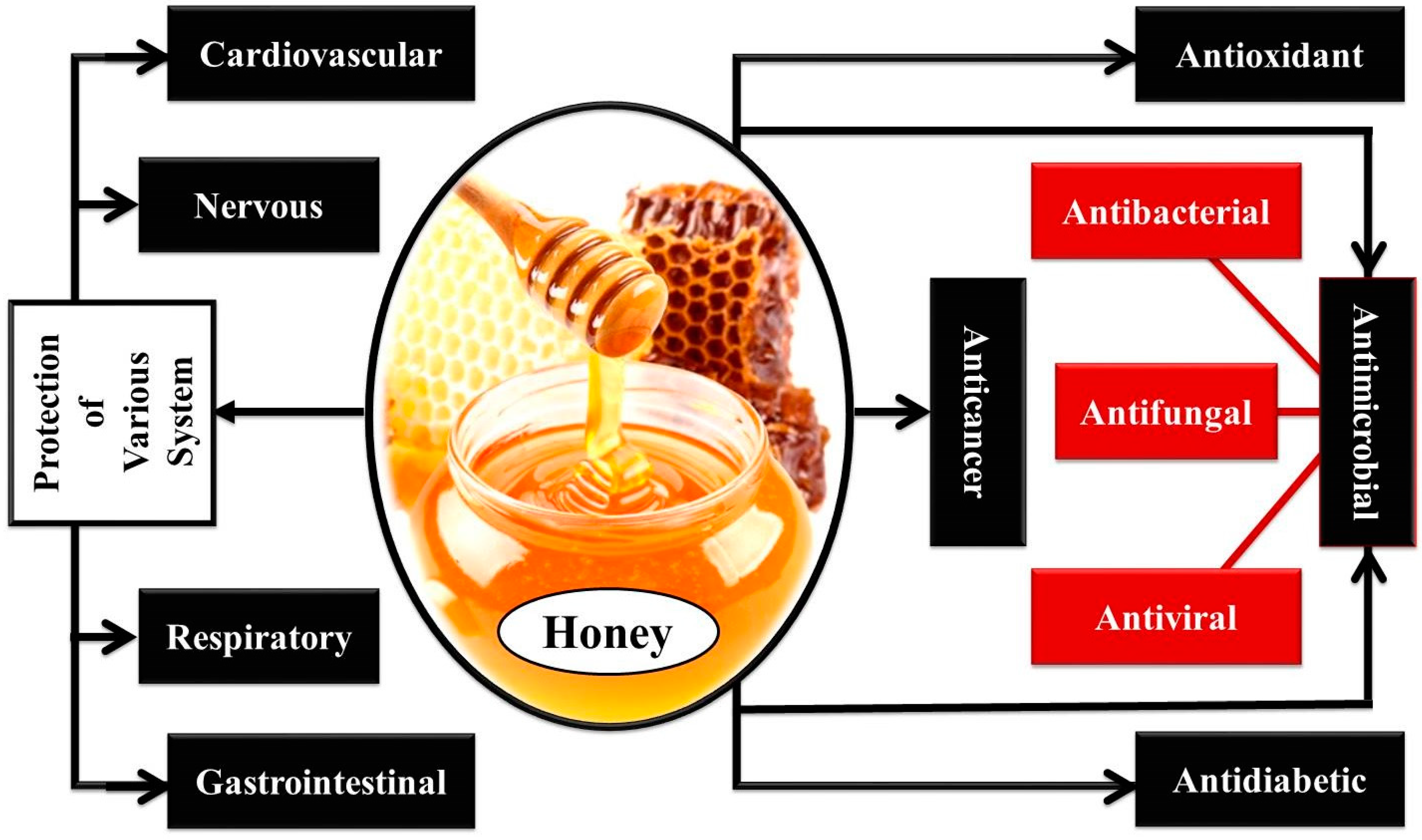
2. Honey, Its Characteristics and Constituents: A Brief Overview

Honey bee females are equipped with a tube-like tongue called a viproboscis, which they use to collect nectar from blossoms. The nectar is then mixed with saliva and enzymes, and the resultant mixture is stored in honey sacks. The contents of the honey sacks are regurgitated into cells, left to dry up to roughly 16% moisture content, and then stored for later use. Among insects, only the honey bee can produce food that can be consumed by humans. Apis mellifera, along with its various subspecies like A. mellifera anatolica, A. mellifera carnica, A. mellifera caucasica, A. mellifera ssp. sicula, etc., plays a crucial role in producing the majority of honey that is consumed by humans. On the other hand, various species including A. ligustica, A. indica, A. florea, A. dorsata, A. cerana, A. caucasica, and A. andreniformis contribute to the production of honey for human consumption [2].
Honey produced naturally by bees is composed of hundreds of distinct compounds belonging to dozens of different chemical classes [10]. The two most essential types of carbohydrates are called fructose and glucose, both of which are monosaccharides. In addition to the two monosaccharides, around 25 distinct oligosaccharides have been identified. Several of these oligosaccharides are related to nutrition, such as panose, 1-ketose, 6-kestose, and palatinose 110 and 297. The disaccharides sucrose, maltose, trehalose, and turanose make up the majority of the oligosaccharides that can be found in blossom honey. Honey made from honeydew may include a greater number of oligosaccharides and trisaccharides than honey made from blossoms, including melezitose and raffinose [11]. About 0.5% of honey comprises protein, most of which is in the form of enzymes and amino acids. As shown in Table 1A, honeydew and blossom honey both include vital nutritional components that are necessary for human health. Furthermore, honey also has trace amounts of vital minerals which are known to play an important part in maintaining human health. The minerals found in honey are shown in Table 1B, together with their typical concentration and RDI (recommended daily intake) in terms of their needs for human health.
Honey contains the enzyme diastase (amylase), which helps break down complex sugars like starch and glycogen into simpler sugars. Honey also contains the enzyme invertase (sucrase, glucosidase), which helps break down sucrose into fructose and glucose. Finally, honey contains the enzyme glucose oxidase, which helps produce hydrogen peroxide and gluconic acid from glucose. The enzyme glucose oxidase, which is contained in honey, is responsible for the production of hydrogen peroxide, which may have antibacterial effects in the mouth [11]. Honey has such a small number of vitamins and minerals that it only provides a very small amount to the recommended daily intake (RDI) for any of the many trace constituents. In addition to being a good source of energy, honey is also rich in a wide variety of minerals, including potassium, chlorine, sulfur, calcium, sodium, phosphorus, magnesium, silicon, iron, manganese, and copper [12]. The presence of acids and amino acids in honey has a significant impact on its aroma. Over the last several decades, a small amount of study on the fragrance components of honey has been carried out, and as a result, more than 500 distinct volatile compounds have been found in various varieties of honey. The fragrant compounds that are included in honey may significantly differ from the various varieties of honey based on the botanical source of the honey [13].
Another significant category of compounds, polyphenols, are essential both in terms of their structure and their functions. Honey has a total polyphenol level ranging from 56 mg/kg to 500 mg/kg. This total polyphenol content completely depends on the kind of honey [14]. Honey contains a variety of polyphenols, the most common of which are flavonoids (such as apigenin, chrysin, galangin, kaempferol, luteolin, and quercetin), phenolic acids, and other phenolic acid-related compounds (derivatives of phenolic acids). It is believed that the presence of polyphenols in honey is responsible for its antioxidant properties [15,16].
Honey has been shown to have a range of bioactivities, one of the most notable of which is its antimicrobial activity, which has been shown in a large number of study papers that have been published by scientists. Some studies suggest that the honey’s antimicrobial activity and capacity to heal wounds are also extremely specialized. This specialized strength of honey is determined by several different aspects, including the condition of the region chosen, the condition of the season chosen, and the type of flower from which the nectar is collected. Due to differences in pH, sugar content, the concentration of the bioactive components (antioxidants, defensin-1, hydrogen peroxide, methylglyoxal, phenolics, etc.), storage circumstances, and bacterial strain susceptibility, honey types may vary widely in their antimicrobial efficacy [17].
Table 1. (A) Honeydew and blossom honey with their own unique set of nutritional components [18,19,20]. (B) The minerals that are found in honey, along with their typical amount and the recommended daily intake (RDI) suggested for human consumption [21,22].
(A)
ComponentsComposition in g/100 g
HoneydewBlossom Honey
Moisture16.317.2
Fructose31.838.2
Glucose26.131.3
Sucrose0.50.7
Other disaccharides45
Melezitose40.1
Erlose10.8
Other oligosaccharides13.13.6
Acids1.10.5
(B)
Typical Amount and RDI of HoneyMineral Composition in Honey
CaClCuFeMgPKNaZn
Amount (mg/100 g)4–302–200.01–0.11–3.40.7–132–6010–4700.6–400.2–0.5
RDI (mg)1000-2184001000--15
Abbreviations: Ca: calcium; Cl: chlorine; Cu: copper; Fe: iron; K: potassium; Mg: magnesium; Na: sodium; P: phosphorous; Zn: zinc.

Honey-Derived Peptide Defensin-1 Produced by Bees

Peptides are subunits of proteins that are responsible for certain biological processes. The correct folding and stability of peptides occur through intermolecular interactions such as hydrogen bonding, electrostatic forces, and van der Waals forces. These interactions are important for peptides to exert their biological function. Moreover, peptides play a crucial role in signaling pathways, facilitating communication within and between cells during important biological processes. In addition, they play a crucial role in various biological processes, such as growth regulation, immune response, and neurotransmission, which are essential for numerous aspects of life. There is a symphony of life that begins with short peptides, which play a crucial role in human nutrition and the immunological defense system [23,24]. Enzyme catalysis can lead to the formation of a peptide bond through various mechanisms, such as the reverse hydrolysis reaction of amides and transpeptidation. The reverse hydrolysis reaction operates through the principle of microscopic reversibility. It is evident that the formation and hydrolysis of the peptide bond originate from a common intermediate. Therefore, the reaction conditions are adjusted to favor the formation of peptide bonds. These peptides are encoded within the structure of the parent proteins. There are few reported bioactive peptides that can be chemically synthesized. Bioactive peptides have a wide range of effects on human health, including the gastrointestinal, endocrine, cardiovascular, immunological, and neurological systems. Sánchez and Vázquez [25] argued that bioactive peptides represent the future of physiologically active regulators. These peptides help in the treatment of a broad variety of illnesses and ailments, which all contribute to an enhanced overall quality of life, as well as lowering the risk of food going rancid, owing to oxidation and microbial breakdown.
The homology model of the honey peptide (defensin-1), derived from Apis mellifera bees [26], is shown in Figure 2 as a three-dimensional visual representation. The function of defensin-1 is comparable to that of honey. It controls the production of keratinocytes and the release of matrix metalloproteinase-9, which are both essential for the healing process [27]. Furthermore, it has been demonstrated that small amounts of this peptide at the micromolar level are capable of inducing notable effects. Furthermore, according to the findings of previous studies, the mechanisms that are responsible for preventing the growth of fungus were identified [28,29]. These mechanisms include (1) modifications in fungal gene expression, (2) permeabilization of the membrane, (3) synthesis inhibition of (1,3)-βD-glucan, and (4) the induction of apoptosis by ROS-mediated mitochondria/caspase-dependent pathways [28,29].
The possible role of royal jelly as an antibacterial agent has been the subject of investigation in several scientific studies that have been published. In addition, both in vitro and in vivo research has been conducted on the antimicrobial peptide defensin-1 to investigate its potential wound-healing effects. According to published reports from previous studies in connection to the concerned investigations, the antimicrobial activity of royal jelly was explored by a group of researchers and scientists against the following bacteria: E. coli, Klebsiella pneumoniae, Proteus aeruginosa, P. mirabilis, Staphylococcus aureus, S. enteritidis, S. epidermidis, and S. pneumoniae [30,31]. The results of this earlier research revealed that royal jelly was efficient against every strain of bacteria that was examined. The identification of individual honey components was the topic of earlier and equally significant research that reported on a potential strategy for honey quality control. This method was used to isolate enzymes from bee salivary secretions, making honey a unique substance. Defensin-1 was another unique honey component [32]. Defensin-1, a peptide produced by bees, has been found in honey at concentrations ranging from 0.04 to 5.17 μg/g. If adulteration is conducted on honey, the reported levels of the bee peptide defensin-1 might decrease. This test was performed by Bucekova et al. [33] to screen for defensin-1 in honey, and the results were highly sensitive and specific. The identification of proteins that are produced by honey bees could be a method that might be employed to confirm the authenticity of honey. Additionally, the ELISA test for defensin-1 might be used to swiftly examine the effectiveness of medicinal honey. In addition, Szweda [9], found that the antibacterial action of honey is exceedingly complicated and is not yet identified in its entirety. It has been proven that certain components of royal jelly, such as bee defensin-1, a peptide that is produced by the hypopharyngeal glands of honey bees, play an important role in antibacterial capabilities. Furthermore, it is thought to be crucial to the well-being of bee larvae as a component of royal jelly (also known as royalysin). Gram-positive bacteria, such as B. subtilis, P. larvae, and S. aureus, are susceptible to its antibacterial effects. They are also thought to be the etiological agent for a significant illness affecting bee larvae known as American foulbrood. All honey indeed has antibacterial properties due to its high sugar content and low pH; however, it has been shown that the quantity of this peptide varies greatly between different samples of honey and royal jelly. This is the case even though all honeys have these antibacterial properties. Additionally, bees produce at least three additional antimicrobial peptides as part of their innate immune system, which helps them fight against pathogens. However, no evidence of their presence in honey has been found as of yet.
Honey’s antibacterial properties are dependent on several distinct components that may function alone or in conjunction with one another. The most important of these include phenolic chemicals, hydrogen peroxide, the honey’s pH, and the osmotic pressure that is produced by the honey itself [26]. Moreover, research has also shown that the presence of methylglyoxal in manuka honey is directly responsible for the significant antibacterial effect that it has. This is the direct cause of the honey’s antibacterial properties. The “unique manuka factor” refers to the non-peroxide antibacterial action that occurs as a result of the presence of methylglyoxal in honey.

3. Characterization of Peptides

3.1. Molecular Weight

The molecular weight of peptides is a critically important physicochemical characteristic that influences their biological activities. Honey, like other products derived from bees, contains a wide array of biologically active proteins and peptides. These proteins and peptides possess antimicrobial and antioxidant properties, as evidenced by previous research [26]. The usual chain length of biologically active peptides is between 2 and 20, which makes them short enough to be absorbed by the gastrointestinal system or to successfully reach the active sites of certain enzymes [34,35]. Electrophoresis was used as a method to effectively extract and identify the proteins that were present in Malaysian honey [36]. Acacia, Tualang, and multifloral honey were used as samples for this study. This honey was reported to include major royal jelly proteins (MRJPs), such as MRJP-1, MRJP-2, MRJP-5, and MRJP-7. These proteins had molecular weights ranging from 20.12 to 76.67 kDa, 23.91 to 79.52 kDa, and 30.65 to 56.31 kDa, respectively, and were characterized by their potency as antioxidants and free radical scavengers [36]. Valachova et al. [6], employed an ELISA test, which was a new method in honey quality control, to isolate and determine the amount of bee-derived peptide defensin-1. This study was conducted using bee honey from Slovakia. The molecular weight of the peptide was determined to be 6.5 kDa [6]. HPLC technology was utilized by Furusawa et al. [7] in their investigation of the quantitative analysis of the peptide apisin, the primary and distinctive protein in royal jelly generated from young worker bees. Satisfactory identification and quantification of apisin were achieved. Then, after electrophoresis in the presence of SDS-PAGE revealed an estimated molecular weight of 5 kDa and purification with the volumetric exclusion method was conducted, its antioxidant capabilities were assessed, and its efficacy in scavenging free radicals was analyzed [7]. A quality control standard based on apisin was suggested for use in the future. Electrophoresis was used by Borutinskaite et al. [24], to determine the molecular weight of a group of proteins that had been isolated from samples of buckwheat honey. The molecular weights of the proteins reached 10.07, 11.6, 19.39, 19.43, and 20.24 kDa. The results were different depending on the production conditions, so Borutinskaite and their co-workers determined the molecular weight of the group. Furthermore, Biopeptide was isolated from honey bees using chromatographic methods [37], and its molecular weight was 5.5 kDa. It was recognized as an essential molecule in the elimination of bacteria when combined with other biological chemicals like flavonoids and hydrogen peroxide [37]. In a recent study that looked into the effect of these peptides as antioxidants, the biopeptides that were isolated and purified from various sources of honey using the gel filtration chromatography method with the Sephacryl S-100 column were found to have molecular weights of 14, 17, and 24 kDa, respectively [38].

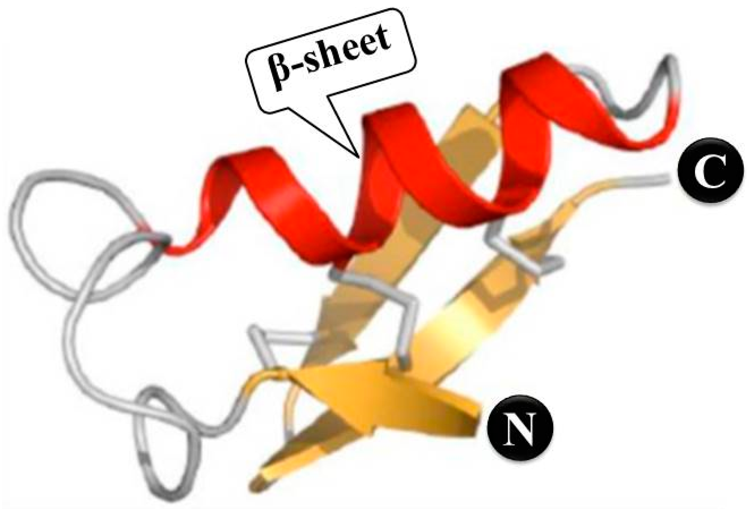
3.2. Amino Acid Analysis

Amino acids are essential for the synthesis of peptide chains [26]. In addition, amino acids play a crucial role in determining the characteristics of biological peptides, acting as compounds with antioxidant and antimicrobial properties. The properties of peptides and proteins are influenced by the specific amino acids used and their arrangement in the sequence [26]. MPRJ1 peptides that were extracted from the Apis cerana bee strain were found to exhibit an amino acid sequence that began at the N-terminal of the amino acid sequence (N(S)-Ile-Leu-Arg-Gly-Glu-Ser-Leu-Asp-Lys). This discovery was made by Srisuparbh et al. [39]. Royal jelly peptides (bee defensin-1), which were isolated and purified from Apis mellifera honey using ultrafiltration technology as well as chromatography techniques, have a vital activity in capturing and binding free radicals and preventing their spread, as stated by Bose et al. [37]. This is because of the presence of the amino terminus of the N-terminal hydrophilic peptide chain of the amino acid sequence (N(S)-Ile-Leu-Arg-Gly-Glu-Ser-Leu-Asn-Lys-Ser-Leu-Pro-Ile-Leu) [37]. The most prevalent amino acids in honey were determined by analyzing forty samples of monotype honey, which included honey from ilex, oak, heather, and nut trees [40]. These amino acids were as follows: Pro, Asp, Glu, Asn, Ser, Gln, Thr, Arg, Ala, Tyr, Met, Val, Phe, Ileu, Leu, and Lys. These amino acids were determined by employing the HPLC technique in the presence of OPA and FMOC reagents [40]. Koike et al. [40], found a class of biological peptides that can be distinguished from the hydrolysis of BoNTA-derived proteins. Because this molecule included an amino sequence in the peptide chain that translated as Leu-Tyr-Gly-Ile-Ala-Ile-Asn-Pro-Asn-Arg, the study suggested that it might be used as a distinguishing feature and a quick way for determining the purity level of honey.

4. Separation and Purification of Peptides

The separation, identification, and purification of biological peptides require a multi-step procedure that, ultimately, relies on the peptides’ physicochemical properties to distinguish one from the other (solubility, ionic charge, size of molecules, adsorption properties, and binding with vital molecules). The nature of the raw material is what ultimately decides the purification methods utilized, and extraction is the initial phase of purification procedures before the efficacy and functionality of any peptide or protein can be determined [41,42]. It might be difficult to isolate peptides from foods using the technologies that are now available. Synthesized studies need a high level of purity since the substances being studied are often found in complicated mixtures that include a variety of components, such as organic acids, free amino acids, sugars, and salts. To obtain peptides characterized by purity in addition to their effective biological properties, these separation and purification processes require efficient techniques that depend on the separation of peptides based on molecular weight and on whether or not they are hydrophilic or hydrophobic. Therefore, purification procedures often include chromatography and ultrafiltration techniques [26,43].

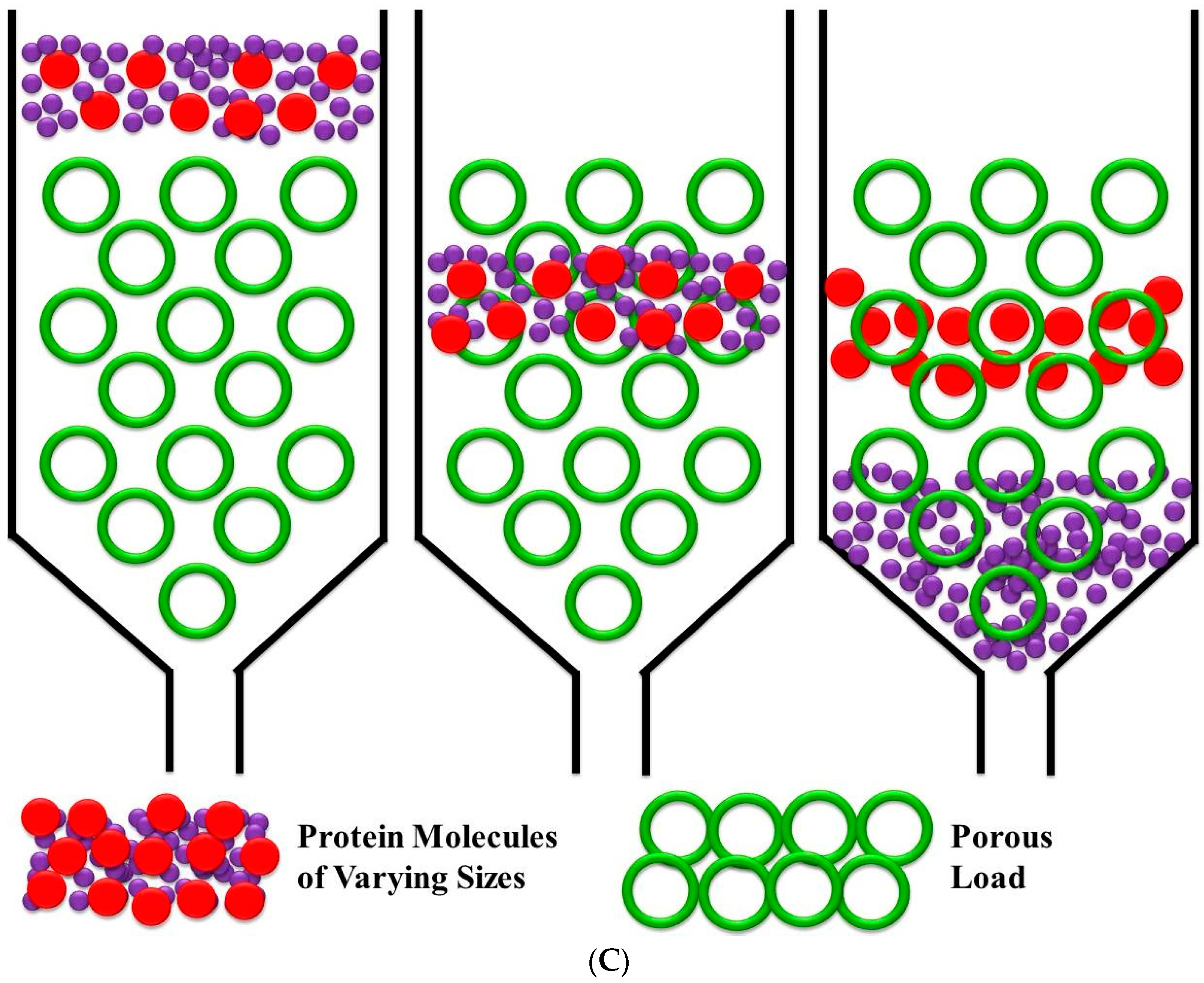
4.1. Ultrafiltration

Methods that rely on membranes may also be used to separate peptides of varying molecular weights. These procedures are often referred to by their more popular names, microfiltration and ultrafiltration. Within the context of this procedure, the concept of separation is determined by the molecular weight of the peptides as well as the porosity of the membranes. These membranes are used to prevent larger molecules from passing through while allowing smaller ones to do so [44]. As can be seen in Figure 3A, ultrafiltration procedures provide many benefits, some of which include their scalability, the avoidance of a need for additional chemical agents, and the capability of conjunction with other processes [43]. The most recent and important studies on ultrafiltration, which were aimed at separating and purifying honey-derived peptides produced from bees, are summarized in Table 2A. In research carried out by Chua and their co-workers, the extraction of honey proteins included both physical and chemical processes [36]. A semipermeable membrane with a molecular weight cutoff (MWCO) is used as a physical method in the process of separating proteins from other molecules. Researchers are attracted by the proteins found in honey because they may have applications in the pharmacological and therapeutic fields. Utilizing ultracentrifugation in conjunction with certain chemical processes (deposition) on a variety of honey samples, it has become possible to successfully separate honey proteins both in the laboratory and on a commercial scale. The primary protein found in royal jelly, which was named MRJP1 and was also described as bee defensin-1, has a molecular weight of 5.5 kDa and a hydrophilic amino terminus at the end of the peptide chain [37]. Bose et al. [37] were successful in isolating and purifying this protein by using methods that were suitable for homogenous ultrafiltration and chromatography. The N-terminus contained the hydrophilic sequence that was present in the greatest abundance.

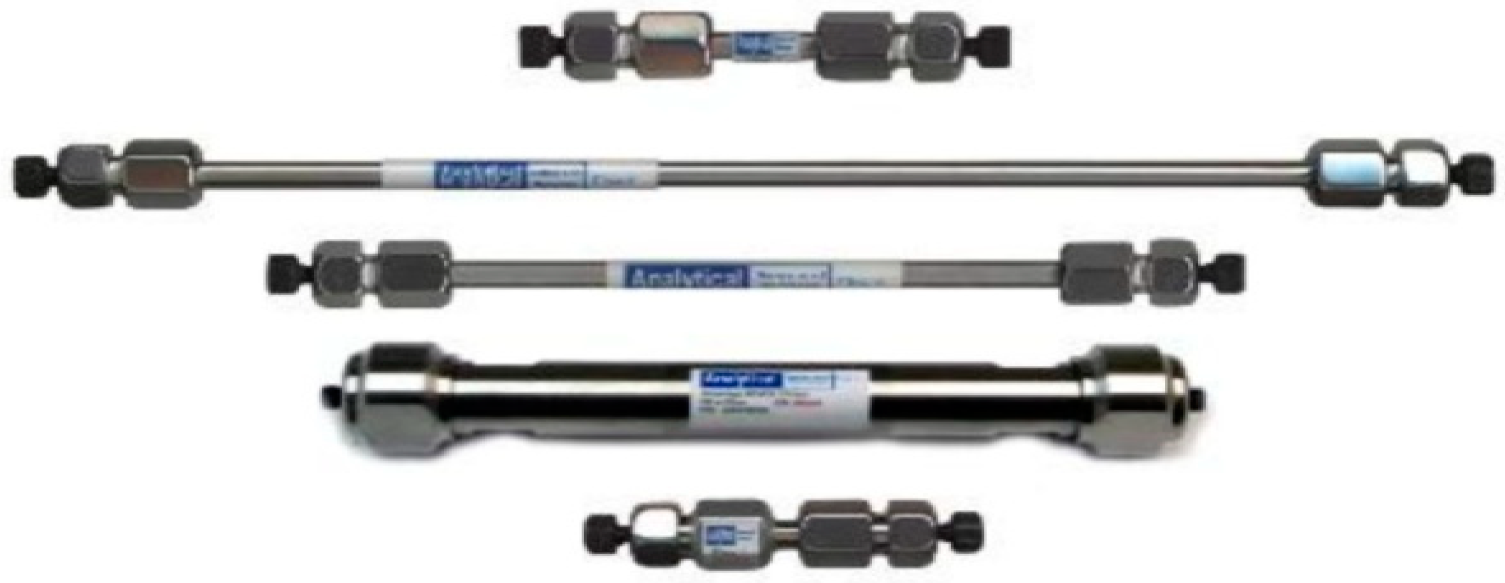
4.2. Reversed-Phase HPLC

In recent years, RP-HPLC has emerged as an indispensable device for the isolation and investigation of proteins and peptides. The biotechnology industry relies heavily on this methodology since it is used to examine proteins and peptides for their identification as well as any impurities that may be present. In the process of preparing samples for protein identification by mass spectrometry, one of the most important steps is to separate digested proteomes and peptides, and RP-HPLC plays an important part in this stage. Since the principle for isolation is the characteristics of hydrophobic compounds including water, this is used in the purification of proteins on a large scale. During the research process, it is also utilized to separate a wide variety of proteins and peptides [45]. Because the success of this method is contingent upon first dissolving the sample in an appropriate solvent (referred to as the stationary phase) and then transporting it through the separation column, which operates at a pressure ranging from 50 to 200 bars, the components of the sample are split up between the two phases according to the polarity of their respective charge states. In RP-HPLC technology, the stationary phase is non-polar, while the mobile phase is polar. This configuration is the most prevalent form of HPLC technology used in detection and separation. The separation and purification processes of RP-HPLC are shown in Figure 3B. If the ratios of the mobile phase consisting of water and acetonitrile are at a ratio of 1:1, this indicates that the separation is carried out under isocratic conditions (the separation is carried out without changing the components of the mobile phase). On the other hand, the separation is referred to as gradient elution if the components of the mobile phase are mixed in a mixture with varying proportions [46]. The most recent and important studies on reversed-phase HPLC, which were aimed at separating and purifying honey-derived peptides produced from bees, are summarized in Table 2B.
Table 2. Recent research on key approaches for the separation and purification of honey-derived peptides produced from bees. (A) Ultrafiltration. (B) Reversed-phase HPLC. (C) Gel filtration chromatography.
(A)
Sample TypeConditions Employed in the Separation and Purification MethodConcentration of CompoundReferences
Separation ConditionPurification Condition
Needful Chemical and SolventTime (min)Temperature (°C)Column TypeColumn/Plate Dimension
Litchi chinensis honeytris-HCl buffer at a concentration of 0.01 M (pH 7.4)2525–30a Q Sepharose (anion exchange) column16 × 20 mm5.12 mg/mL[37]
Honey bee pupae (Apis mellifera)Linear gradient of acetonitrile with concentrations ranging from 5 to 45% and containing 0.1% TFA2525–305-C18 semi-preparation column4.6 × 250 mm135 µg/mL[47]
Apis mellifera Carnica colonies in Slovakia were used to collect honey.TBST buffer comprising 50 mM tris-HCl, 7.5 pH, 200 mM sodium chloride (NaCl), and 0.05% Tween 205020–25Sephadex G-100 column (GE Healthcare, UK)16 × 20 mm125 µg/mL[48]
Netherlands honeyLoading buffer consisting 3 M urea dissolved in 5% acetic acid, and methyl green added for reference purposes4525–30Cylindrical gel3.7 × 6 cm5.0 mg/mL[49]
Royal jelly proteintris-HCl buffer at a concentration of 20 mM (pH 8.0)5020–25TSKgel DEAE-5PW column7.5 × 75 mm229 µg/mL[50]
(B)
Sample TypeConditions Employed in the Separation and Purification MethodConcentration of CompoundReferences
Separation ConditionPurification Condition
Needful Chemical and SolventTime (min)Temperature (°C)Column TypeColumn/Plate DimensionParticle Size (µm)Injected Volume (µL)
Honey produced by Apis mellifera in JapanMixture of 0.1% formic acid (A) and methanol that already contains 0.1% formic acid (B). A gradient program was established in the following manner: beginning, 5% B; from 0 to 1 min, 5% B; from 1 to 15 min, 50% B; from 15 to 25 min, 95% B; and from 25 to 30 min, 5% B4525–30YMC Triart C18 analytical column(YMC Co., Ltd., Kyoto, Japan)100 × 2.1 mm3250 µg mL[40]
Honey bee pupae (Apis mellifera)Linear gradient of acetonitrile with concentrations ranging from 5% to 45% that includes 0.1% TFA2525–305-C18 semi-preparation column4.6 × 250 mm55135 µg/mL[47]
Honey derived from Ziziphus species made by Apis mellifera at bee farms located in the Himalayan area.It took 5 min at 0%, 40 min at 70%, and 45 min at 0% for the acetonitrile to be used in the gradient elutions2525–30C-18 (Purospher STAR, RP-18 end-capped: Merck, Darmstadt, Germany150 × 4.6 mm55120 nM[51]
Manuka honeyElution using a linear gradient of deionized water and acetonitrile (0–100%) containing 0.05% trifluoroacetic acid2020–25C18 reverse phase (RP) column250 × 4.6 mm 55135 µg/mL[52]
Royal jelly (RJ) A. melliferaIsocratic elution carried out using 55% (v/v) acetonitrile that contains 0.04% (v/v) trifluoracetic acid4530C-18 TOSOH-ODS column150 × 4.6 mm55145 µg/mL[53]
(C)
Sample TypeSeparation MethodConditions Employed in the Separation and Purification MethodConcentration of CompoundReferences
Separation ConditionPurification Condition
Needful Chemical and SolventTime (min)Temperature (°C)Column TypeColumn/Plate Dimension
Honey major proteinGel Filtration Chromatography12.5 Mm Pyridine-acetate buffer2037Sephacryl S-100 column2.5 × 85 mm155 µg/mL[38]
Litchi chinensis honeyGel Filtration Chromatographytris-HCl buffer used at a concentration of 0.01 M (pH 7.4)2525–30Q Sepharose (anion exchange) column16 × 20 mm5.12 mg/mL[37]
Bee honey Gel Filtration Chromatography0.05 M phosphate buffer with a pH of 6.6 in buffer A, then the sample was eluted with 0.5 M sodium chloride in buffer A4525–30Sepharose FF column (GE Healthcare, UK)10 × 300 mm135 µg/mL[54]
Netherlands honeyGel Filtration ChromatographyLoading buffer consisting of 3 M urea dissolved in 5% acetic acid, and methyl green added for reference purposes4525–30Cylindrical gel3.7 × 6 cm5.0 mg/mL[49]
Honey of A. cerena colonyGel Filtration Chromatographytris-HCl bufferused with a linear gradient of NaCl concentrations ranging from 0.0 to 0.3 M4525–30Sephadex G-2002.5 × 85 mm165 µg/mL[39]

4.2.1. Chromatographic Column

The columns that are used in HPLC are always made of stainless steel (314 L, stainless steel), and the thickness of their walls is designed to be able to withstand pressures that are greater than 30 KPSI. This is a higher pressure than the one that is used not only in the analysis but also in the column-filling process. In the case of columns that are used in the process of chemical analysis, the typical diameter is 4.6 mm, and the length of the column may typically be from 5 to 25 cm. Although the column’s length may exceed 40 cm and its width could be more than 1 cm, such columns are not used for studies. The diameter of the packing granules has a direct correlation to the length of the column; the smaller the diameter of the packing granules, the shorter the length of the column. This is because there is an increase in the amount of counter pressure that occurs when the granules are reduced in size [55,56]. The several types of columns that are used in the chromatographic analysis are shown in Figure 4.

4.2.2. Mechanism of Protein/Peptide Retention

During the RP-HPLC process, hydrocarbon groups are chemically bonded to the surface of the particle, which results in the particle surface being very hydrophobic. The “hydrophobic foot” of a protein is what binds to the hydrophobic surface, which is how the protein adheres to it (Figure 5). Due to the thickness of the hydrophobic surface, proteins are too large to be fully adsorbed by it. This prevents proteins from adhering to hydrophobic surfaces. The vast majority of protein molecules are in direct fluid contact with the surface and thus are revealed. Because hydrophobic adsorption leads to a high net interaction, the protein remains adsorbed on the surface until a certain concentration of organic solvent has been reached. At this stage, the protein desorbs from the surface and elutes from the column [57].

4.2.3. Column Characteristics

Particles Support

The process of separating peptides and proteins involves interacting with the hydrophobic surface of particles packed in columns. This is performed to obtain the desired results. Silica is frequently used as the material for the particles that make up the column because it is physically durable, it is stable under the conditions of most solvents (except at pH levels higher than 6.5), and it can be formed into spherical particles of various sizes with holes of varying diameters. For these reasons, silica is frequently used as the material for the particles. It is also important to keep in mind that separation performance is also influenced by the quality of the silica that is used in HPLC columns [58,59].

Pore Diameter

In RP-HPLC, the usage of silica with pores smaller than 100 Å leads to subpar protein separations. The separation of proteins is greatly improved by using silica with pores wider than 300 Å in diameter. Since proteins are unable to pass through holes of this size, separation takes place only on the very superficial surface. Because proteins and even bigger peptides can penetrate the pore and completely interact with the surface of wide-pore silica, the peak shape and resolution are much improved. In modern protein separation processes, wide-pore silica is almost exclusively employed. Because tiny peptides, such as those that are produced as a consequence of protease digests, can penetrate the pores of small pore silica and interact with the surface, small pore silica has the potential to be used in the process of separating protein digests. Wide-pore silica, on the other hand, can effectively separate peptides and leads to varying degrees of selectivity and resolution [60]. Mesaik et al. [51] conducted research in which they used RP-HPLC technology for the separation and determination of honey glycoproteins and glycopeptides, as well as the assessment of immunomodulatory properties. Researchers employed a C-18 column (Purospher STAR, RP-18 end-capped at 5 μm, 150-4.6, Merck, Darmstadt, Germany). After equilibrating the column with 0.1% TFA, a flow of 1.0 mL/min was used to introduce 100 μL of a filtered protein sample into the column. The acetonitrile was used to produce gradient elutions at the following times and percentages: 5 min at 0%, 40 min at 70%, and 45 min at 0%. The peaks were then collected, concentrated, and kept at a temperature of −20 °C until their subsequent usage. While the non-toxic peptides originating in the honey of bees, known as BoNTA, were isolated and identified in nineteen samples from various locations using liquid chromatography procedures, they were regarded as a tool to test the product’s safety [40]. Using the RP-HPLC technique on a ShimadzuLC-10 HPLC system at room temperature and using a separation column type (C18 Jupiter column, USA), Da Silva et al. [61] characterized a broad spectrum of biological peptides derived from one of the beneficial bacteria. They obtained one peak for the biopeptide, with a holding time of 32 min. The analysis was performed at a wavelength of 225 nm using 0.05% TFA, 20–80% acetonitrile and ultrapure water as the mobile phase [62,63].

4.3. Gel Filtration Chromatography

There is a mechanism that can be used for the separation and purification of peptides according to their molecular sizes that is called size-exclusion chromatography. This is one of the mechanisms that are being explored for the separation and purification of peptides because it is both an easy-to-use approach for the separation of molecules with various molecular sizes and a mechanism that can separate and purify peptides according to their molecular sizes. In gel filtration, the degree of separation achieved is directly proportional to the particle size distribution in the sample solution, as depicted in Figure 3C. Components that have a high molecular weight are unable to pass through the gel pore and are instead ejected out with a dead volume. On the other hand, components that have a low molecular weight will enter through the gel pore, remain inside the column, and then be eliminated to the outside of the gel, depending on the type of gel that is being used [43].
The most recent and important studies on gel filtration chromatography, which were aimed at separating and purifying honey-derived peptides produced from bees, are summarized in Table 2C. In a study that was conducted by Srisuparbh et al. [39], the researchers separated and purified the defensing-1 antimicrobial peptide of honey bees (royal jelly protein, which plays an important role in eliminating bacteria in synergy with other biological compounds in honey) from the A. cerena colony of honey bees using a gel filtration technique with a column Sephadex G-200 chromatographic separation. The study that was conducted by Steinhorn et al. [64], found that the purification process of honey proteins from clover and manuka honey, conducted through the size-exclusion process and separated by ultrafiltration using 10 (KDa MWCO) porous separation membranes, which included five samples of New Zealand honey, showed a protein that is effective in modifying and improving immunity. Ibrahim et al. [38] used a gel filtration chromatography technique with Sephacryl S-100 column chromatography that performed the elution process of the sample using a pyridine–acetate buffer at a concentration of 12.5 mM to isolate and purify the biological active peptides that were derived from the main protein in honey. This allowed them to successfully isolate and purify the peptides.

5. Factors Influencing the Separation and Purification of Honey Bee Peptides

Various factors influence the separation and purification of honey bee peptides, including the characteristics of the peptides, the properties of the extraction and purification methods, and the intended application of the peptides [37,65,66,67]. Several important factors have been explored in Table 3 that impact the separation and purification process of honey bee peptides. These factors include the characteristics of the peptides, the complexity of the sample, the method of extraction, the strategy for purification, the conditions of chromatography, the methods of detection, the scale of the process, the throughput, and the specific requirements of the application. Through careful consideration of these factors and the fine-tuning of experimental conditions, researchers can attain a high level of efficiency and effectiveness in the separation and purification of honey bee peptides. This process is crucial for a wide range of scientific, industrial, and biomedical applications.

6. Honey-Derived Peptide Defensin-1 Produced by Bees as an Antioxidant and Antimicrobial

Antioxidants are molecules, ions, or radicals that are generally stable yet may delay or prevent the oxidation of other molecules. Antioxidants are known to be compounds that can stop or slow down the progression of chain reactions that lead to the production of free radicals. Additionally, antioxidants can halt the oxidation process before it even begins, which is another way that antioxidants can prevent the oxidation process from starting. They can do this by donating an electron to the free radical, which subsequently oxidizes outside of the body’s cells, producing radicals that are feeble, inactive, and relatively stable [79,80]. Honey is a rich source of bioactive compounds, including antioxidants and antimicrobials, due to the presence of both enzymatic and non-enzymatic antioxidants within its composition. Honey has been shown to have antioxidant and antibacterial action, which is due to the fact that it includes a vast range of bioactive compounds in addition to bee peptides. The naturally occurring antibacterial agents abaecin, defensin-1, apidaecin, and hymenoptaeci are all included in these bee peptides. Additionally, honey incorporates bee pollen into its composition [81,82].
The hydrolysis of food proteins results in the production of bioactive peptides, which not only have the ability to perform physiological activity but can also absorb nitrogen from the diet. According to the findings of several studies, the bioavailability of peptides is much higher than that of proteins or free amino acids [33,67]. The nature of the structure of peptides, in terms of the type and sequence of amino acids contained within the peptide chain, as well as their partial weight, is what gives peptides their properties as biological compounds with antimicrobial and antioxidant activity [83]. Consequently, because of their capacity to get rid of reactive oxygen species (ROS) and to prevent oxidized metal ions from forming, biological peptides play an important part in the process of getting rid of the free radicals that are responsible for inflammation, aging, and chronic illnesses [38]. Based on the research conducted by Bose et al. [37], protein is identified as the main biomolecule in honey with significant biological functions. In addition, peptides have strong antimicrobial and antioxidant effects. The antimicrobial mechanisms of honey-derived peptides from bees, as well as the possible effects of these peptides on human health, are shown in Figure 6. Peptides exhibit a more potent reducing activity in comparison to proteins due to their unique structure consisting of amino acids and their smaller size relative to proteins with a higher molecular weight. The findings of Bose et al. [37], demonstrate that in addition to being an effective reducing agent, the peptides are also excellent electron donors. The presence of hydrophobic amino acids and/or the presence of long side chains of amino acids might be responsible for this.
Honey and its pure protein, as well as peptides, have been shown to be active antioxidants by Bose et al. [37], who also found that these substances may scavenge free radicals (DPPH). The findings showed that honey, honey peptides, and proteins all had different levels of antioxidant activity in neutralizing free radicals: 73.16%, 68.55%, and 59.71%, respectively. Unprocessed honey was claimed to have the highest antioxidant potential due to the high concentration of other antioxidant-rich components that it contains. These components include enzymes, phenolic compounds, carotenoids, vitamin C, and organic acids. There is a peptide in honey which is a remarkable compound, due to its unique structure that is determined by the specific arrangement and sequence of amino acids in its peptide chain. Additionally, it has a relatively low molecular weight compared to proteins, making it an intriguing subject of study.

6.1. Antioxidant Properties of the Peptide Defensin-1: Final Remarks

The peptide defensin-1, derived from honey produced by bees, does indeed possess antioxidant properties. Defensin-1 is a specific antimicrobial peptide that can be found in honey, specifically in honey bee-derived products such as honey, royal jelly, and bee pollen [28,30,54,84]. Defensin-1 has a dual role in the honey bee’s biology. It serves as a crucial component of the immune system, safeguarding against harmful pathogens [85,86]. Additionally, defensin-1 exhibits antioxidant properties by effectively neutralizing free radicals and preventing oxidative stress-related damage [38,81]. Studies have demonstrated that defensin-1 plays a role in enhancing the antioxidant capacity of honey and other products derived from bees [81]. Research has shown its effectiveness in counteracting ROS and decreasing oxidative stress in different cellular and animal models [87,88]. Defensin-1 plays a crucial role in safeguarding cells and tissues against oxidative damage, which is commonly linked to aging, inflammation, and chronic diseases [87,88,89]. It achieves this by neutralizing free radicals and preventing lipid peroxidation. Honey-derived defensin-1 exhibits antioxidant properties that position it as a promising contender for therapeutic applications in addressing conditions associated with oxidative stress [87,90]. Additional investigation is required to gain a comprehensive understanding of how it works, its ability to be absorbed by the body, and its safety record. This will help unlock its full therapeutic potential and determine its suitability for use in functional foods, nutraceuticals, and pharmaceutical formulations that aim to support health and prevent diseases associated with oxidative stress.

6.2. Antimicrobial Properties of the Peptide Defensin-1: Final Remarks

Defensin-1, a peptide derived from honey, has been found to possess strong antimicrobial properties. Defensin-1 is a vital element in the immune system of bees, playing a crucial role in safeguarding bee colonies against harmful pathogens. Defensin-1 demonstrates a wide range of antimicrobial activity, effectively targeting bacteria, fungi, and certain viruses [61,62,63,81,82]. Defensin-1 exhibits antimicrobial properties by disrupting the cell membranes of microorganisms, interfering with the synthesis of their cell walls, and inhibiting crucial cellular processes [82,84,91]. Defensin-1 can effectively eliminate or hinder the development of harmful microorganisms by specifically targeting microbial membranes and cell structures [38,82,84]. This crucial action plays a significant role in preserving the overall health and stability of bee colonies. In addition, defensin-1 has shown minimal harm to mammalian cells, making it a promising candidate for treating microbial infections in humans [84,92,93]. Scientists are currently investigating the potential uses of defensin-1 as a natural antimicrobial agent in a range of areas, such as medicine, agriculture, and food preservation [54,84,90,94]. In broad terms, defensin-1 derived from honey plays a crucial role in protecting bees from pathogens and presents exciting possibilities to establish new antimicrobial approaches to combat microbial infections that affect human health [84].

7. Emerging, Promising, and Cutting-Edge Omic Methods for Studying Honey Bee Peptides

Certain approaches in the field of technology and science have shown great potential in studying honey bee peptides. These methods are considered to be at the forefront of research and have garnered interest from researchers, scientists, and individuals in the industrial sector who are seeking new and innovative opportunities. Within this context, the study of honey bee peptides can greatly benefit from a range of advanced techniques and scientific tools. These tools provide a holistic understanding of the composition, structure, function, and interactions of peptides [95,96]. Several important tools and techniques can be utilized in honey bee peptide research (Table 4). By incorporating various omics and cutting-edge tools, scientists can delve into the intricate world of honey bee peptides and their impact on bee well-being, behavior, and ecological relationships. Additionally, they can investigate the potential uses of these peptides in fields such as medicine, agriculture, and biotechnology [28,44]. Based on the information presented in Table 4, it is evident that these omic approaches are highly advanced and hold great potential for studying honey bee peptides. They can provide fresh and groundbreaking possibilities in this field of study.

8. Artificial Intelligence: A Promising Approach for Investigating Honey Bee Peptides

Artificial intelligence (AI) is rapidly becoming a valuable tool in the study of honey bee peptides. Its emergence in this field is opening up new possibilities for research and analysis [119,120,121]. Therefore, it is evident that AI has the potential to revolutionize the study of honey bee peptides. It provides novel methods for data analysis, modeling, and prediction, showcasing its immense promise as an emerging tool in this field [119,122,123]. Table 5 addresses different approaches through which AI can contribute to research on honey bee peptides. Through the utilization of AI, scientists can expedite the exploration, analysis, and utilization of honey bee peptides, propelling further understanding of bee biology, health, and ecological relationships [121,122,123]. Moreover, utilizing AI-driven approaches enhances traditional experimental methods by providing novel perspectives and predictive abilities for peptide-based research, not only in apiculture but also in agriculture and biomedicine [119,121,124,125,126].

9. Challenges and Research Opportunities in Honey Bee Peptides

Exploring the realm of honey bee peptides offers a fascinating avenue for scientific investigation, with its fair share of obstacles and promising avenues for research. Table 6 and Table 7 present several significant challenges and promising research opportunities. In a nutshell, the study of honey bee peptides offers challenges and research opportunities that span across various fields including biochemistry, biotechnology, pharmacology, and biomedical science [71,77,90,96,105,131,149]. Through understanding these challenges and the exploration of these opportunities, researchers can tap into the therapeutic, nutritional, and biotechnological possibilities of honey bee peptides, ultimately enhancing human health and well-being [11,26,71,77,87,90,158]. Consequently, after an in-depth review and discussion in Table 6 and Table 7, we can confidently assert that the suggestions and recommendations provided are trustworthy and highly beneficial for professionals working in the realms of research, science, and industry. Furthermore, this will assist people engaged in the exploration of cutting-edge solutions for sustainable practices on various aspects of honey bee peptides. In addition, people will uncover new possibilities for utilizing the diverse array of processes associated with honey bee peptides.

10. Concluding Remarks

Honey contains a diverse range of bioactive components, such as amino acids, proteins, enzymes, essential minerals, vitamins, and the bee-derived peptide defensin-1. These bioactive molecules have been found to exhibit interesting biological properties, both in vitro and in vivo, such as antioxidant and antimicrobial activities. At present, membrane separation, RP-HPLC, and gel filtration chromatography are the main approaches used to separate and purify honey-derived peptides produced by bees. However, there is an urgent need for faster and more efficient techniques to analyze the diverse properties of honey produced by bees. Moreover, there is an unprecedented need from consumers for fast, reliable, and cost-effective methods to detect adulteration in honey. This paper provides an overview of the latest advancements in honey quality assessment procedures, serving as a valuable resource for future research in this field. Based on our study, it is evident that while the current methods are precise and accurate, there is still an opportunity for enhancement to expedite the testing process and enhance the reliability of results for naturally occurring honey used in industrial and commercial applications. Exploring the fascinating realm of honey bee peptides and their profound effects on bee health, behavior, and ecological interactions can be achieved using advanced techniques like proteomics, genomics, transcriptomics, and metabolomics. Furthermore, exploring potential applications of these peptides in fields like medicine, agriculture, and biotechnology could be helpful. In addition, the application of artificial intelligence (AI) can greatly enhance the speed and efficiency of studying honey bee peptides. This can lead to a deeper comprehension of bee biology, health, and ecological interactions. As an example, AI tools such as machine learning and neural networks can simulate the interactions between peptides and honey bee proteins, receptors, or enzymes. Additional planned systematic research investigations can aid in the advancement of drug delivery systems and formulations based on peptides. Addressing the therapeutic and nutraceutical potential of honey bee peptides in human health and disease prevention opens up opportunities for drug discovery, as well as the development of functional foods and dietary supplements.

Author Contributions

Investigation, A.K.N., D.F.A.-F., D.K.V. and A.R.P. have conceptualized, interpreted, corrected, and compiled literature and technically sound final versions of the manuscript; A.K.N., D.F.A.-F. and W.K.A.-R. have compiled the tables for manuscripts; D.K.V., A.K.N., A.R.P., W.K.A.-R. and S.S. have read the manuscript and provided suggestions and corrections for the final submission. All authors have read and agreed to the published version of the manuscript.

Funding

This research received no external funding.

Acknowledgments

The authors would like to acknowledge the financial assistance of the Food Research Consumer Protection Unit Lab., Department of Food Science, College of Agriculture, University of Basrah. Furthermore, the authors would like to express their gratitude to the staff of the “Food Research Consumer Protection Unit Lab” for their constant support.

Conflicts of Interest

The authors state that there are no conflicts of interest.

References

  1. CXS 12-19811; Codex Standard 12. Revised Codex Standard for Honey, Standards and Standard Methods. World Health Organization: Geneva, Switzerland, 2001; p. 11.
  2. Israili, Z.H. Antimicrobial properties of honey. Am. J. Ther. 2014, 21, 304–323. [Google Scholar] [CrossRef] [PubMed]
  3. Coppock, R.W. Bee products as nutraceuticals to nutraceuticals for bees. In Nutraceuticals; Academic Press: Cambridge, MA, USA, 2021; pp. 813–833. [Google Scholar] [CrossRef]
  4. Cianciosi, D.; Forbes-Hernández, T.Y.; Afrin, S.; Gasparrini, M.; Reboredo-Rodriguez, P.; Manna, P.P.; Zhang, J.; Bravo Lamas, L.; Martínez Flórez, S.; Agudo Toyos, P.; et al. Phenolic compounds in honey and their associated health benefits: A review. Molecules 2018, 23, 2322. [Google Scholar] [CrossRef] [PubMed]
  5. Yang, Y.; Battesti, M.J.; Djabou, N.; Muselli, A.; Paolini, J.; Tomi, P.; Costa, J. Melissopalynological origin determination and volatile composition analysis of Corsican “chestnut grove” honeys. Food Chem. 2012, 132, 2144–2154. [Google Scholar] [CrossRef]
  6. Valachová, I.; Bučeková, M.; Majtán, J. Quantification of bee-derived peptide defensin-1 in honey by competitive enzyme-linked immunosorbent assay, a new approach in honey quality control. Czech J. Food Sci. 2016, 34, 233–243. [Google Scholar] [CrossRef]
  7. Furusawa, T.; Arai, Y.; Kato, K.; Ichihara, K. Quantitative analysis of Apisin, a major protein unique to royal jelly. Evidence-Based Complementary and Alternative. Medicine 2016, 2016, 5040528. [Google Scholar] [CrossRef] [PubMed]
  8. Buttstedt, A.; Moritz, R.F.; Erler, S. Origin and function of the major royal jelly proteins of the honeybee (Apis mellifera) as members of the yellow gene family. Biol. Rev. 2014, 89, 255–269. [Google Scholar] [CrossRef] [PubMed]
  9. Szweda, P. Antimicrobial Activity of Honey. In Honey Analysis; IntechOpen: London, UK, 2017. [Google Scholar] [CrossRef]
  10. Puścion-Jakubik, A.; Borawska, M.H.; Socha, K. Modern methods for assessing the quality of bee honey and botanical origin identification. Foods 2020, 9, 1028. [Google Scholar] [CrossRef] [PubMed]
  11. Gündoğdu, E.; Çakmakçı, S.; Şat, İ.G. An overview of honey: Its composition, nutritional and functional properties. J. Food Sci. Eng. 2019, 9, 10–14. [Google Scholar] [CrossRef]
  12. Aili, S.R.; Touchard, A.; Escoubas, P.; Padula, M.P.; Orivel, J.; Dejean, A.; Nicholson, G.M. Diversity of peptide toxins from stinging ant venoms. Toxicon 2014, 92, 166–178. [Google Scholar] [CrossRef] [PubMed]
  13. Bogdanov, S.; Ruoff, K.; Oddo, L.P. Physico-chemical methods for the characterisation of unifloral honeys: A review. Apidologie 2004, 35 (Suppl. S1), S4–S17. [Google Scholar] [CrossRef]
  14. Al-Mamary, M.; Al-Meeri, A.; Al-Habori, M. Antioxidant activities and total phenolics of different types of honey. Nutr. Res. 2002, 22, 1041–1047. [Google Scholar] [CrossRef]
  15. Kenjerić, D.; Mandić, M.L.; Primorac, L.; Bubalo, D.; Perl, A. Flavonoid profile of Robinia honeys produced in Croatia. Food Chem. 2007, 102, 683–690. [Google Scholar] [CrossRef]
  16. Bobiş, O.; Bonta, V.; Cornea-Cipcigan, M.; Nayik, G.A.; Dezmirean, D.S. Bioactive molecules for discriminating Robinia and helianthus honey: High-performance liquid chromatography–electron spray ionization–mass spectrometry polyphenolic profile and physicochemical determinations. Molecules 2021, 26, 4433. [Google Scholar] [CrossRef] [PubMed]
  17. Kumar, P.; Sindhu, R.K.; Narayan, S.; Singh, I. Honey collected from different floras of Chandigarh Tricity: A comparative study involving physicochemical parameters and biochemical activities. J. Diet. Suppl. 2010, 7, 303–313. [Google Scholar] [CrossRef] [PubMed]
  18. Choi, S.H.; Nam, M.S. Classification of honeydew and blossom honeys by principal component analysis of physicochemical parameters. Korean J. Agric. Sci. 2020, 47, 67–81. [Google Scholar]
  19. Bergamo, G.; Seraglio, S.K.; Gonzaga, L.V.; Fett, R.; Costa, A.C. Physicochemical characteristics of bracatinga honeydew honey and blossom honey produced in the state of Santa Catarina: An approach to honey differentiation. Food Res. Int. 2019, 116, 745–754. [Google Scholar] [CrossRef]
  20. Pita-Calvo, C.; Vázquez, M. Differences between honeydew and blossom honeys: A review. Trends Food Sci. Technol. 2017, 59, 79–87. [Google Scholar] [CrossRef]
  21. Solayman, M. Minerals and Trace Elements. In Honey: Composition and Health Benefits; Wiley: Hoboken, NJ, USA, 2023; Volume 24, pp. 80–101. [Google Scholar] [CrossRef]
  22. Lanjwani, M.F.; Channa, F.A. Minerals content in different types of local and branded honey in Sindh, Pakistan. Heliyon 2019, 5, e02042. [Google Scholar] [CrossRef] [PubMed]
  23. Apostolopoulos, V.; Bojarska, J.; Chai, T.T.; Elnagdy, S.; Kaczmarek, K.; Matsoukas, J.; New, R.; Parang, K.; Lopez, O.P.; Parhiz, H.; et al. A global review on short peptides: Frontiers and perspectives. Molecules 2021, 26, 430. [Google Scholar] [CrossRef]
  24. Borutinskaite, V.; Treigyte, G.; Čeksteryte, V.; Kurtinaitiene, B.; Navakauskiene, R. Proteomic identification and enzymatic activity of buckwheat (Fagopyrum esculentum) honey based on different assays. J. Food Nutr. Res. 2018, 57, 57–69. [Google Scholar]
  25. Sánchez, A.; Vázquez, A. Bioactive peptides: A review. Food Qual. Saf. 2017, 1, 29–46. [Google Scholar] [CrossRef]
  26. Alvarez-Suarez, J.M.; Tulipani, S.; Romandini, S.; Bertoli, E.; Battino, M. Contribution of honey in nutrition and human health: A review. Mediterr. J. Nutr. Metab. 2010, 3, 15–23. [Google Scholar] [CrossRef]
  27. Kurek-Górecka, A.; Górecki, M.; Rzepecka-Stojko, A.; Balwierz, R.; Stojko, J. Bee products in dermatology and skin care. Molecules 2020, 25, 556. [Google Scholar] [CrossRef] [PubMed]
  28. Dumitru, C.D.; Neacsu, I.A.; Grumezescu, A.M.; Andronescu, E. Bee-derived products: Chemical composition and applications in skin tissue engineering. Pharmaceutics 2022, 14, 750. [Google Scholar] [CrossRef] [PubMed]
  29. Memariani, H.; Memariani, M. Anti-fungal properties and mechanisms of melittin. Appl. Microbiol. Biotechnol. 2020, 104, 6513–6526. [Google Scholar] [CrossRef] [PubMed]
  30. Maželienė, Ž.; Aleksandravičienė, A.; Pašvenskaitė, M.; Viliušienė, I.; Šakienė, D.; Dailidaitė, E. Antimicrobial activity of royal jelly, honey, and their mixture. Biologija 2022, 68, 159–164. [Google Scholar] [CrossRef]
  31. Lee, S.; Lee, K.S.; Ok, M.; Kim, B.Y.; Jin, B.R. Antimicrobial activity of major royal jelly protein 8 and 9 of honeybee (Apis mellifera) venom. J. Asia-Pac. Entomol. 2022, 25, 101964. [Google Scholar] [CrossRef]
  32. Pauliuc, D.; Dranca, F.; Oroian, M. Antioxidant activity, total phenolic content, individual phenolics and physicochemical parameters suitability for Romanian honey authentication. Foods 2020, 9, 306. [Google Scholar] [CrossRef] [PubMed]
  33. Bucekova, M.; Godocikova, J.; Kohutova, L.; Danchenko, M.; Barath, P.; Majtan, J. Antibacterial activity and bee-derived protein content of honey as important and suitable complementary tools for the assessment of honey quality. J. Food Compos. Anal. 2023, 123, 105610. [Google Scholar] [CrossRef]
  34. Zhang, Y.; Liu, H.; Hong, H.; Luo, Y. Purification and identification of dipeptidyl peptidase IV and angiotensin-converting enzyme inhibitory peptides from silver carp (Hypophthalmichthys molitrix) muscle hydrolysate. Eur. Food Res. Technol. 2019, 245, 243–255. [Google Scholar] [CrossRef]
  35. Sato, K.; Miyasaka, S.; Tsuji, A.; Tachi, H. Isolation and characterization of peptides with dipeptidyl peptidase IV (DPPIV) inhibitory activity from natto using DPPIV from Aspergillus oryzae. Food Chem. 2018, 261, 51–56. [Google Scholar] [CrossRef] [PubMed]
  36. Chua, L.S.; Lee, J.Y.; Chan, G.F. Characterization of the proteins in honey. Anal. Lett. 2015, 48, 697–709. [Google Scholar] [CrossRef]
  37. Bose, D.; Padmavati, M.; Banerjee, R. Isolation, purification and characterization of protein from Litchi chinensis honey and generation of peptides. J. Addict. Recovery 2020, 3, 1–6. [Google Scholar] [CrossRef]
  38. Ibrahim, H.R.; Nanbu, F.; Miyata, T. Potent antioxidant peptides derived from honey major protein enhance tolerance of eukaryotic cells toward oxidative stress. In Food Production, Processing and Nutrition; Springer: Berlin/Heidelberg, Germany, 2021; pp. 1–10. [Google Scholar] [CrossRef]
  39. Srisuparbh, D.; Klinbunga, S.; Wongsiri, S.; Sittipraneed, S. Isolation and characterization of major royal jelly cDNAs and proteins of the honey bee (Apis cerana). BMB Rep. 2003, 36, 572–579. [Google Scholar] [CrossRef]
  40. Koike, H.; Kanda, M.; Hayashi, H.; Matsushima, Y.; Yoshikawa, S.; Ohba, Y.; Hashimoto, T. Development of an alternative approach for detecting botulinum neurotoxin type A in honey: Analysis of non-toxic peptides with a reference labelled protein via liquid chromatography-tandem mass spectrometry. Food Addit. Contam. Part A 2020, 37, 1359–1373. [Google Scholar] [CrossRef]
  41. Lee, C.H. A simple outline of methods for protein isolation and purification. Endocrinol. Metab. 2017, 32, 18–22. [Google Scholar] [CrossRef] [PubMed]
  42. Pal, G.K.; Suresh, P.V. Sustainable valorisation of seafood by-products: Recovery of collagen and development of collagen-based novel functional food ingredients. Innov. Food Sci. Emerg. Technol. 2016, 37, 201–215. [Google Scholar] [CrossRef]
  43. Cermeño, M.; Kleekayai, T.; Amigo-Benavent, M.; Harnedy-Rothwell, P.; FitzGerald, R.J. Current knowledge on the extraction, purification, identification, and validation of bioactive peptides from seaweed. Electrophoresis 2020, 41, 1694–1717. [Google Scholar] [CrossRef] [PubMed]
  44. Zaky, A.A.; Abd El-Aty, A.M.; Ma, A.; Jia, Y. An overview on antioxidant peptides from rice bran proteins: Extraction, identification, and applications. Crit. Rev. Food Sci. Nutr. 2022, 62, 1350–1362. [Google Scholar] [CrossRef]
  45. De Luca, C.; Lievore, G.; Bozza, D.; Buratti, A.; Cavazzini, A.; Ricci, A.; Catani, M. Downstream processing of therapeutic peptides by means of preparative liquid chromatography. Molecules 2021, 26, 4688. [Google Scholar] [CrossRef]
  46. Herraiz, T. Sample preparation and reversed phase-high performance liquid chromatography analysis of food-derived peptides. Anal. Chim. Acta 1997, 352, 119–139. [Google Scholar] [CrossRef]
  47. Yang, X.; Chen, K.; Liu, H.; Zhang, Y.; Luo, Y. Purification and identification of peptides with high angiotensin-I converting enzyme (ACE) inhibitory activity from honeybee pupae (Apis mellifera) hydrolysates with in silico gastrointestinal digestion. Eur. Food Res. Technol. 2019, 245, 535–544. [Google Scholar] [CrossRef]
  48. Majtan, J.; Klaudiny, J.; Bohova, J.; Kohutova, L.; Dzurova, M.; Sediva, M.; Majtan, V. Methylglyoxal-induced modifications of significant honeybee proteinous components in manuka honey: Possible therapeutic implications. Fitoterapia 2012, 83, 671–677. [Google Scholar] [CrossRef] [PubMed]
  49. Kwakman, P.H.; Velde, A.A.T.; de Boer, L.; Speijer, D.; Christina Vandenbroucke-Grauls, M.J.; Zaat, S.A. How honey kills bacteria. FASEB J. 2010, 24, 2576–2582. [Google Scholar] [CrossRef] [PubMed]
  50. Guo, H.; Kouzuma, Y.; Yonekura, M. Isolation and Properties of Antioxidative Peptides from Water-Soluble Royal Jelly Protein Hydrolysate. Food Sci. Technol. Res. 2005, 11, 222–230. [Google Scholar] [CrossRef]
  51. Mesaik, M.A.; Dastagir, N.; Uddin, N.; Rehman, K.; Azim, M.K. Characterization of immunomodulatory activities of honey glycoproteins and glycopeptides. J. Agric. Food Chem. 2015, 63, 177–184. [Google Scholar] [CrossRef] [PubMed]
  52. Lee, H.; Churey, J.J.; Worobo, R.W. Purification and structural characterization of bacillomycin F produced by a bacterial honey isolate active against Byssochlamys fulva H25. J. Appl. Microbiol. 2008, 105, 663–673. [Google Scholar] [CrossRef] [PubMed]
  53. Fontana, R.; Mendes, M.A.; De Souza, B.M.; Konno, K.; César, L.M.M.; Malaspina, O.; Palma, M.S. Jelleines: A family of antimicrobial peptides from the Royal Jelly of honeybees (Apis mellifera). Peptides 2004, 25, 919–928. [Google Scholar] [CrossRef]
  54. Bucekova, M.; Sojka, M.; Valachova, I.; Martinotti, S.; Ranzato, E.; Szep, Z.; Majtan, J. Bee-derived antibacterial peptide, defensin-1, promotes wound reepithelialisation in vitro and in vivo. Wound Health S. Afr. 2017, 10, 25–35. [Google Scholar]
  55. Ali, A.H. High-performance liquid chromatography (HPLC): A review. Ann. Adv. Chem. 2022, 6, 10–20. [Google Scholar] [CrossRef]
  56. Kanu, A.B. Recent developments in sample preparation techniques combined with high-performance liquid chromatography: A critical review. J. Chromatogr. A 2021, 1654, 462444. [Google Scholar] [CrossRef] [PubMed]
  57. Li, W.; Huang, J.; Zheng, L.; Liu, W.; Fan, L.; Sun, B.; Zhao, M. A fast stop-flow two-dimensional liquid chromatography tandem mass spectrometry and its application in food-derived protein hydrolysates. Food Chem. 2023, 406, 135000. [Google Scholar] [CrossRef] [PubMed]
  58. Al-Sulaimi, S.; Kushwah, R.; Abdullah Alsibani, M.; El Jery, A.; Aldrdery, M.; Ashraf, G.A. Emerging developments in separation techniques and analysis of chiral pharmaceuticals. Molecules 2023, 28, 6175. [Google Scholar] [CrossRef] [PubMed]
  59. Luo, C.; DeStefano, J.J.; Langlois, T.J.; Boyes, B.E.; Schuster, S.A.; Godinho, J.M. Fundamental to achieving fast separations with high efficiency: A review of chromatography with superficially porous particles. Biomed. Chromatogr. 2021, 35, e5087. [Google Scholar] [CrossRef] [PubMed]
  60. Reuhs, B.L. High-Performance Liquid Chromatography. In Food Analysis. Food Science Text Series; Nielsen, S.S., Ed.; Springer: Cham, Switzerland, 2017; pp. 213–226. [Google Scholar] [CrossRef]
  61. Da Silva, B.S.; Díaz-Roa, A.; Yamane, E.S.; Hayashi, M.A.; Junior, P.I.S. Doderlin: Isolation and characterization of a broad-spectrum antimicrobial peptide from Lactobacillus acidophilus. Res. Microbiol. 2023, 174, 103995. [Google Scholar] [CrossRef]
  62. Al-sahlany, S.T.G.; Altemimi, A.B.; Al-Manhel, A.J.A.; Niamah, A.K.; Lakhssassi, N.; Ibrahim, S.A. Purification of bioactive peptide with antimicrobial properties produced by Saccharomyces cerevisiae. Foods 2020, 9, 324. [Google Scholar] [CrossRef] [PubMed]
  63. Naimah, A.K.; Al-Manhel, A.J.A.; Al-Shawi, M.J. Isolation, purification and characterization of antimicrobial peptides produced from Saccharomyces boulardii. Int. J. Pept. Res. Ther. 2018, 24, 455–461. [Google Scholar] [CrossRef]
  64. Steinhorn, G.; Sims, I.M.; Carnachan, S.M.; Carr, A.J.; Schlothauer, R. Isolation and characterisation of arabinogalactan-proteins from New Zealand kanuka honey. Food Chem. 2011, 128, 949–956. [Google Scholar] [CrossRef]
  65. Chua, L.S.; Lee, J.Y.; Chan, G.F. Honey protein extraction and determination by mass spectrometry. Anal. Bioanal. Chem. 2013, 405, 3063–3074. [Google Scholar] [CrossRef]
  66. Luo, X.; Dong, Y.; Gu, C.; Zhang, X.; Ma, H. Processing technologies for bee products: An overview of recent developments and perspectives. Front. Nutr. 2021, 8, 727181. [Google Scholar] [CrossRef]
  67. Jose, A.; Binu, A.M.; Syrus, E.C.; Baiju, J.E.; Jose, S.; Abraham, A.M.; Jacob, J. Purification and characterization of proteins from Manuka honey. Mater. Today Proc. 2023; in press. [Google Scholar] [CrossRef]
  68. Issaq, H.J.; Conrads, T.P.; Janini, G.M.; Veenstra, T.D. Methods for fractionation, separation and profiling of proteins and peptides. Electrophoresis 2002, 23, 3048–3061. [Google Scholar] [CrossRef] [PubMed]
  69. Bíliková, K.; Wu, G.; Šimúth, J. Isolation of a peptide fraction from honeybee royal jelly as a potential antifoulbrood factor. Apidologie 2001, 32, 275–283. [Google Scholar] [CrossRef]
  70. Fekete, S.; Veuthey, J.L.; Guillarme, D. New trends in reversed-phase liquid chromatographic separations of therapeutic peptides and proteins: Theory and applications. J. Pharm. Biomed. Anal. 2012, 69, 9–27. [Google Scholar] [CrossRef] [PubMed]
  71. Agyei, D.; Ongkudon, C.M.; Wei, C.Y.; Chan, A.S.; Danquah, M.K. Bioprocess challenges to the isolation and purification of bioactive peptides. Food Bioprod. Process. 2016, 98, 244–256. [Google Scholar] [CrossRef]
  72. Ares, A.M.; Valverde, S.; Bernal, J.L.; Nozal, M.J.; Bernal, J. Extraction and determination of bioactive compounds from bee pollen. J. Pharm. Biomed. Anal. 2018, 147, 110–124. [Google Scholar] [CrossRef]
  73. Wang, X.; Yang, S.; Li, Y.; Zhang, J.; Jin, Y.; Zhao, W.; Zhou, J. Optimization and application of parallel solid-phase extraction coupled with ultra-high performance liquid chromatography–tandem mass spectrometry for the determination of 11 aminoglycoside residues in honey and royal jelly. J. Chromatogr. A 2018, 1542, 28–36. [Google Scholar] [CrossRef]
  74. Sahlan, M.; Mahira, K.F.; Wiratama, I.; Mahadewi, A.G.; Yohda, M.; Hermansyah, H.; Noguchi, K. Purification and characterization of proteins in multifloral honey from kelulut bee (stingless bee). Heliyon 2019, 5, e02835. [Google Scholar] [CrossRef]
  75. Zhu, S.; Wang, S.; Wang, L.; Huang, D.; Chen, S. Identification and characterization of an angiotensin-I converting enzyme inhibitory peptide from enzymatic hydrolysate of rape (Brassica napus L.) bee pollen. LWT 2021, 147, 111502. [Google Scholar] [CrossRef]
  76. Al-Rubaie, W.K.; Al-Fekaiki, D.F. Isolation and Characterization of Bee-Derived Peptide in Iraqi Honey by Size-exclusion Chromatograph. J. Glob. Sci. Res. 2022, 7, 2789–2798. [Google Scholar] [CrossRef]
  77. Ferrazzano, L.; Catani, M.; Cavazzini, A.; Martelli, G.; Corbisiero, D.; Cantelmi, P.; Tolomelli, A. Sustainability in peptide chemistry: Current synthesis and purification technologies and future challenges. Green Chem. 2022, 24, 975–1020. [Google Scholar] [CrossRef]
  78. Akhtari, N.; Banan, K.; Fatahian, F.; Vatanpour, H.; Rezadoost, H.; Ghorbani-Bidkorpeh, F. A validated extraction technique followed by high-performance liquid chromatography-ultraviolet analysis for the assay of melittin as an indicator component of honey bee venom in cosmeceutical products. Sep. Sci. Plus 2024, 2300231. [Google Scholar] [CrossRef]
  79. Nagmoti, D.M.; Khatri, D.K.; Juvekar, P.R.; Juvekar, A.R. Antioxidant activity free radical-scavenging potential of Pithecellobium dulce Benth seed extracts. Free Radic. Antioxid. 2012, 2, 37–43. [Google Scholar] [CrossRef]
  80. Balkrishna, A.; Rohela, A.; Kumar, A.; Kumar, A.; Arya, V.; Thakur, P.; Kuca, K. Mechanistic insight into antimicrobial and antioxidant potential of Jasminum species: A herbal approach for disease management. Plants 2021, 10, 1089. [Google Scholar] [CrossRef]
  81. Stefanis, C.; Stavropoulou, E.; Giorgi, E.; Voidarou, C.; Constantinidis, T.C.; Vrioni, G.; Tsakris, A. Honey’s antioxidant and antimicrobial properties: A bibliometric study. Antioxidants 2023, 12, 414. [Google Scholar] [CrossRef]
  82. Bouali, N.; Ahmad, I.; Patel, H.; Alhejaili, E.B.; Hamadou, W.S.; Badraoui, R.; Noumi, E. GC–MS screening of the phytochemical composition of Ziziphus honey: ADME properties and in vitro/in silico study of its antimicrobial activity. J. Biomol. Struct. Dyn. 2024, 42, 1368–1380. [Google Scholar] [CrossRef]
  83. Tsavea, E.; Vardaka, F.P.; Savvidaki, E.; Kellil, A.; Kanelis, D.; Bucekova, M.; Mossialos, D. Physicochemical characterization and biological properties of pine honey produced across Greece. Foods 2022, 11, 943. [Google Scholar] [CrossRef]
  84. Amerikova, M.; Pencheva El-Tibi, I.; Maslarska, V.; Bozhanov, S.; Tachkov, K. Antimicrobial activity, mechanism of action, and methods for stabilisation of defensins as new therapeutic agents. Biotechnol. Biotechnol. Equip. 2019, 33, 671–682. [Google Scholar] [CrossRef]
  85. Jarczak, J.; Kościuczuk, E.M.; Lisowski, P.; Strzałkowska, N.; Jóźwik, A.; Horbańczuk, J.; Bagnicka, E. Defensins: Natural component of human innate immunity. Hum. Immunol. 2013, 74, 1069–1079. [Google Scholar] [CrossRef]
  86. Hora, Z.A.; Altaye, S.Z.; Wubie, A.J.; Li, J. Proteomics improves the new understanding of honeybee biology. J. Agric. Food Chem. 2018, 66, 3605–3615. [Google Scholar] [CrossRef]
  87. Cornara, L.; Biagi, M.; Xiao, J.; Burlando, B. Therapeutic properties of bioactive compounds from different honeybee products. Front. Pharmacol. 2017, 8, 261216. [Google Scholar] [CrossRef]
  88. Guo, L.; Tang, J.; Tang, M.; Luo, S.; Zhou, X. Reactive oxygen species are regulated by immune deficiency and Toll pathways in determining the host specificity of honeybee gut bacteria. Proc. Natl. Acad. Sci. USA 2023, 120, e2219634120. [Google Scholar] [CrossRef]
  89. Bagameri, L.; Botezan, S.; Bobis, O.; Bonta, V.; Dezmirean, D.S. Molecular Insights into Royal Jelly Anti-Inflammatory Properties and Related Diseases. Life 2023, 13, 1573. [Google Scholar] [CrossRef]
  90. Roy, D.; Arati, C.; Manikandan, B.; Abinash, G.; Nisa, N.; Bhanushree, B.; Gurusubramanian, G. Pharmacological and therapeutic potential of honey bee antimicrobial peptides. Indian J. Biochem. Biophys. 2023, 60, 365–384. [Google Scholar] [CrossRef]
  91. Lehrer, R.I.; Bevins, C.L.; Ganz, T. Defensins and other antimicrobial peptides and proteins. In Mucosal Immunology; Elsevier: Amsterdam, The Netherlands, 2005; p. 95. [Google Scholar] [CrossRef]
  92. Sahl, H.G.; Pag, U.; Bonness, S.; Wagner, S.; Antcheva, N.; Tossi, A. Mammalian defensins: Structures and mechanism of antibiotic activity. J. Leukoc. Biol. 2005, 77, 466–475. [Google Scholar] [CrossRef]
  93. Khurshid, Z.; Zafar, M.S.; Naseem, M.; Khan, R.S.; Najeeb, S. Human oral defensins antimicrobial peptides: A future promising antimicrobial drug. Curr. Pharm. Des. 2018, 24, 1130–1137. [Google Scholar] [CrossRef]
  94. Gao, X.; Ding, J.; Liao, C.; Xu, J.; Liu, X.; Lu, W. Defensins: The natural peptide antibiotic. Adv. Drug Deliv. Rev. 2021, 179, 114008. [Google Scholar] [CrossRef]
  95. Erban, T.; Shcherbachenko, E.; Talacko, P.; Harant, K. The unique protein composition of honey revealed by comprehensive proteomic analysis: Allergens, venom-like proteins, antibacterial properties, royal jelly proteins, serine proteases, and their inhibitors. J. Nat. Prod. 2019, 82, 1217–1226. [Google Scholar] [CrossRef]
  96. Bo, W.; Chen, L.; Qin, D.; Geng, S.; Li, J.; Mei, H.; Liang, G. Application of quantitative structure-activity relationship to food-derived peptides: Methods, situations, challenges and prospects. Trends Food Sci. Technol. 2021, 114, 176–188. [Google Scholar] [CrossRef]
  97. Ji, T.; Liu, Z.; Shen, J.; Shen, F.; Liang, Q.; Wu, L.; Corona, M. Proteomics analysis reveals protein expression differences for hypopharyngeal gland activity in the honeybee, Apis mellifera carnica Pollmann. BMC Genom. 2014, 15, 665. [Google Scholar] [CrossRef]
  98. Buchberger, A.; Yu, Q.; Li, L. Advances in mass spectrometric tools for probing neuropeptides. Annu. Rev. Anal. Chem. 2015, 8, 485–509. [Google Scholar] [CrossRef]
  99. Boonen, K.; De Haes, W.; Van Houtven, J.; Verdonck, R.; Baggerman, G.; Valkenborg, D.; Schoofs, L. Quantitative peptidomics with isotopic and isobaric tags. In Peptidomics: Methods and Strategies; Schrader, M., Fricker, L., Eds.; Humana Press: New York, NY, USA, 2018; pp. 141–159. [Google Scholar] [CrossRef]
  100. Zhang, W.; Wang, X.; Yang, S.; Niu, Q.; Wu, L.; Li, Y.; Zhou, J. Simultaneous quantification of five biogenic amines based on LC–MS/MS and its application in honeybee venom from different subspecies. Biomed. Chromatogr. 2020, 34, e4740. [Google Scholar] [CrossRef]
  101. Bong, J.; Middleditch, M.; Loomes, K.M.; Stephens, J.M. Proteomic analysis of honey. Identification of unique peptide markers for authentication of NZ mānuka (Leptospermum scoparium) honey. Food Chem. 2021, 350, 128442. [Google Scholar] [CrossRef]
  102. Chen, Y.P.; Pettis, J.S.; Zhao, Y.; Liu, X.; Tallon, L.J.; Sadzewicz, L.D.; Evans, J.D. Genome sequencing and comparative genomics of honey bee microsporidia, Nosema apis reveal novel insights into host-parasite interactions. BMC Genom. 2013, 14, 451. [Google Scholar] [CrossRef]
  103. Moran, N.A. Genomics of the honey bee microbiome. Curr. Opin. Insect Sci. 2015, 10, 22–28. [Google Scholar] [CrossRef]
  104. McAfee, A.; Harpur, B.A.; Michaud, S.; Beavis, R.C.; Kent, C.F.; Zayed, A.; Foster, L.J. Toward an upgraded honey bee (Apis mellifera L.) genome annotation using proteogenomics. J. Proteome Res. 2016, 15, 411–421. [Google Scholar] [CrossRef]
  105. Trapp, J.; McAfee, A.; Foster, L.J. Genomics, transcriptomics and proteomics: Enabling insights into social evolution and disease challenges for managed and wild bees. Mol. Ecol. 2017, 26, 718–739. [Google Scholar] [CrossRef]
  106. Diao, Q.; Sun, L.; Zheng, H.; Zeng, Z.; Wang, S.; Xu, S.; Wu, J. Genomic and transcriptomic analysis of the Asian honeybee Apis cerana provides novel insights into honeybee biology. Sci. Rep. 2018, 8, 822. [Google Scholar] [CrossRef]
  107. Lariviere, P.J.; Leonard, S.P.; Horak, R.D.; Powell, J.E.; Barrick, J.E. Honey bee functional genomics using symbiont-mediated RNAi. Nat. Protoc. 2023, 18, 902–928. [Google Scholar] [CrossRef]
  108. Mahato, D.K.; Verma, D.K.; Billoria, S.; Kopari, M.; Prabhakar, P.K.; Kumar, A.; Srivastav, P.P. Applications of nuclear magnetic resonance in food processing and packaging management. In Developing Technologies in Food Science; Apple Academic Press: Palm Bay, FL, USA, 2017; pp. 109–142. [Google Scholar]
  109. Klupczynska, A.; Plewa, S.; Dereziński, P.; Garrett, T.J.; Rubio, V.Y.; Kokot, Z.J.; Matysiak, J. Identification and quantification of honeybee venom constituents by multiplatform metabolomics. Sci. Rep. 2020, 10, 21645. [Google Scholar] [CrossRef]
  110. Díaz-Galiano, F.J.; Murcia-Morales, M.; Fernández-Alba, A.R. From sound check to encore: A journey through high-resolution mass spectrometry-based food analyses and metabolomics. Compr. Rev. Food Sci. Food Saf. 2024, 23, e13325. [Google Scholar] [CrossRef]
  111. Wiltgen, M. Algorithms for structure comparison and analysis: Homology modelling of proteins. Encycl. Bioinform. Comput. Biol. ABC Bioinform. 2018, 1, 3. [Google Scholar]
  112. Pattabhiramaiah, M.; Ramesh, K.; Kv, V.; Muniswamy Reddy, S. Computational analysis of PhospholipaseA2 in the honey bee venom. J. Apic. Res. 2020, 59, 706–721. [Google Scholar] [CrossRef]
  113. López-Pedrouso, M.; Lorenzo, J.M.; Alché, J.D.D.; Moreira, R.; Franco, D. Advanced proteomic and bioinformatic tools for predictive analysis of allergens in novel foods. Biology 2023, 12, 714. [Google Scholar] [CrossRef]
  114. Hasan, M.E.; Samir, A.; Khalil, M.M.; Shafaa, M.W. Bioinformatics approach for prediction and analysis of the Non-Structural Protein 4B (NSP4B) of the Zika virus. J. Genet. Eng. Biotechnol. 2024, 22, 100336. [Google Scholar] [CrossRef]
  115. Jónsdóttir, S.Ó.; Jørgensen, F.S.; Brunak, S. Prediction methods and databases within chemoinformatics: Emphasis on drugs and drug candidates. Bioinformatics 2005, 21, 2145–2160. [Google Scholar] [CrossRef]
  116. Saikia, S.; Bordoloi, M. Molecular docking: Challenges, advances and its use in drug discovery perspective. Curr. Drug Targets 2019, 20, 501–521. [Google Scholar] [CrossRef]
  117. Nisa, N.; Rasmita, B.; Arati, C.; Uditraj, C.; Siddhartha, R.; Dinata, R.; Gurusubramanian, G. Repurposing of phyto-ligand molecules from the honey bee products for Alzheimer’s disease as novel inhibitors of BACE-1: Small molecule bioinformatics strategies as amyloid-based therapy. Environ. Sci. Pollut. Res. 2023, 30, 51143–51169. [Google Scholar] [CrossRef]
  118. Park, D.; Jung, J.W.; Lee, M.O.; Lee, S.Y.; Kim, B.; Jin, H.J.; Kwon, H.W. Functional characterization of naturally occurring melittin peptide isoforms in two honey bee species, Apis mellifera and Apis cerana. Peptides 2014, 53, 185–193. [Google Scholar] [CrossRef]
  119. Aronica, P.G.; Reid, L.M.; Desai, N.; Li, J.; Fox, S.J.; Yadahalli, S.; Verma, C.S. Computational methods and tools in antimicrobial peptide research. J. Chem. Inf. Model. 2021, 61, 3172–3196. [Google Scholar] [CrossRef]
  120. Lefin, N.; Herrera-Belén, L.; Farias, J.G.; Beltrán, J.F. Review and perspective on bioinformatics tools using machine learning and deep learning for predicting antiviral peptides. Mol. Divers. 2023; ahead of print. [Google Scholar] [CrossRef]
  121. Sanyal, A.; Ghosh, A.; Roy, C.; Mazumder, I.; Marrazzo, P. Revolutionizing the use of honeybee products in healthcare: A focused review on using bee pollen as a potential adjunct material for biomaterial functionalization. J. Funct. Biomater. 2023, 14, 352. [Google Scholar] [CrossRef]
  122. Robles-Loaiza, A.A.; Pinos-Tamayo, E.A.; Mendes, B.; Ortega-Pila, J.A.; Proaño-Bolaños, C.; Plisson, F.; Almeida, J.R. Traditional and computational screening of non-toxic peptides and approaches to improving selectivity. Pharmaceuticals 2022, 15, 323. [Google Scholar] [CrossRef]
  123. Yan, J.; Cai, J.; Zhang, B.; Wang, Y.; Wong, D.F.; Siu, S.W. Recent progress in the discovery and design of antimicrobial peptides using traditional machine learning and deep learning. Antibiotics 2022, 11, 1451. [Google Scholar] [CrossRef]
  124. Simone-Finstrom, M. Proceedings of the 2018 American Bee Research Conference. Bee World 2018, 95, 47–72. [Google Scholar] [CrossRef]
  125. Hu, F.L.; Bíliková, K.; Casabianca, H.; Daniele, G.; Salmen Espindola, F.; Feng, M.; Zhou, J.H. Standard methods for Apis mellifera royal jelly research. J. Apic. Res. 2019, 58, 1–68. [Google Scholar] [CrossRef]
  126. Zeng, J.; Jia, X. Systems Theory-Driven Framework for AI Integration into the Holistic Material Basis Research of Traditional Chinese Medicine. Engineering, 2024; in press. [Google Scholar] [CrossRef]
  127. Azari, S. Evolutionary Algorithms for Improving De Novo Peptide Sequencing. Doctoral Dissertation, Open Access Te Herenga Waka-Victoria University of Wellington, Wellington, New Zealand, 2020. [Google Scholar]
  128. Yoshida, M.; Hinkley, T.; Tsuda, S.; Abul-Haija, Y.M.; McBurney, R.T.; Kulikov, V.; Cronin, L. Using evolutionary algorithms and machine learning to explore sequence space for the discovery of antimicrobial peptides. Chem 2018, 4, 533–543. [Google Scholar] [CrossRef]
  129. Perpetuo, L.; Klein, J.; Ferreira, R.; Guedes, S.; Amado, F.; Leite-Moreira, A.; Vitorino, R. How can artificial intelligence be used for peptidomics? Expert Rev. Proteom. 2021, 18, 527–556. [Google Scholar] [CrossRef]
  130. McDonnell, K.; Howley, E.; Abram, F. Critical evaluation of the use of artificial data for machine learning based de novo peptide identification. Comput. Struct. Biotechnol. J. 2023, 21, 2732–2743. [Google Scholar] [CrossRef]
  131. Fontaine, F.; Overman, J.; François, M. Pharmacological manipulation of transcription factor protein-protein interactions: Opportunities and obstacles. Cell Regen. 2015, 4, 2. [Google Scholar] [CrossRef]
  132. Lee, E.Y.; Lee, M.W.; Fulan, B.M.; Ferguson, A.L.; Wong, G.C. What can machine learning do for antimicrobial peptides, and what can antimicrobial peptides do for machine learning? Interface Focus 2017, 7, 20160153. [Google Scholar] [CrossRef]
  133. Pasrija, P.; Jha, P.; Upadhyaya, P.; Khan, M.; Chopra, M. Machine learning and artificial intelligence: A paradigm shift in big data-driven drug design and discovery. Curr. Top. Med. Chem. 2022, 22, 1692–1727. [Google Scholar] [CrossRef]
  134. Will, I.; Beckerson, W.C.; de Bekker, C. Using machine learning to predict protein–protein interactions between a zombie ant fungus and its carpenter ant host. Sci. Rep. 2023, 13, 13821. [Google Scholar] [CrossRef]
  135. Swan, A.L.; Mobasheri, A.; Allaway, D.; Liddell, S.; Bacardit, J. Application of machine learning to proteomics data: Classification and biomarker identification in postgenomics biology. Omics A J. Integr. Biol. 2013, 17, 595–610. [Google Scholar] [CrossRef]
  136. Ramazi, S.; Mohammadi, N.; Allahverdi, A.; Khalili, E.; Abdolmaleki, P. A review on antimicrobial peptides databases and the computational tools. Database 2022, 2022, baac011. [Google Scholar] [CrossRef]
  137. Le, T.D.; Suttikhana, I.; Ashaolu, T.J. Unconventional production strategies, action mechanisms, and structure-functional attributes of food-derived peptides. Food Hydrocoll. 2023, 144, 109054. [Google Scholar] [CrossRef]
  138. Satpathy, R. Bioinspired Algorithms in Solving Three-Dimensional Protein Structure Prediction Problems. In Bio-Inspired Computing for Information Retrieval Applications; IGI Global: Hershey, PA, USA, 2017; pp. 316–337. [Google Scholar]
  139. Correa, L.D.L.; Dorn, M. A knowledge-based artificial bee colony algorithm for the 3-D protein structure prediction problem. In Proceedings of the 2018 IEEE Congress on Evolutionary Computation (CEC), Rio de Janeiro, Brazil, 8–13 July 2018; IEEE: Piscataway, NJ, USA, 2018; pp. 1–8. [Google Scholar]
  140. Lee, A.C.L.; Harris, J.L.; Khanna, K.K.; Hong, J.H. A comprehensive review on current advances in peptide drug development and design. Int. J. Mol. Sci. 2019, 20, 2383. [Google Scholar] [CrossRef]
  141. Wu, Q.; Ke, H.; Li, D.; Wang, Q.; Fang, J.; Zhou, J. Recent progress in machine learning-based prediction of peptide activity for drug discovery. Curr. Top. Med. Chem. 2019, 19, 4–16. [Google Scholar] [CrossRef]
  142. Basith, S.; Manavalan, B.; Hwan Shin, T.; Lee, G. Machine intelligence in peptide therapeutics: A next-generation tool for rapid disease screening. Med. Res. Rev. 2020, 40, 1276–1314. [Google Scholar] [CrossRef]
  143. Sepčić, S. Application of Machine Learning in Peptide Design. Doctoral Dissertation, Department of Biotechnology, University of Rijeka, Rijeka, Croatia, 2020. [Google Scholar]
  144. Aguilera-Puga, M.D.C.; Cancelarich, N.L.; Marani, M.M.; de la Fuente-Nunez, C.; Plisson, F. Accelerating the discovery and design of antimicrobial peptides with artificial intelligence. In Computational Drug Discovery and Design; Springer: New York, NY, USA, 2023; pp. 329–352. [Google Scholar]
  145. Hummon, A.B.; Richmond, T.A.; Verleyen, P.; Baggerman, G.; Huybrechts, J.; Ewing, M.A.; Sweedler, J.V. From the genome to the proteome: Uncovering peptides in the Apis brain. Science 2006, 314, 647–649. [Google Scholar] [CrossRef]
  146. Starr, A.E.; Deeke, S.A.; Li, L.; Zhang, X.; Daoud, R.; Ryan, J.; Figeys, D. Proteomic and metaproteomic approaches to understand host–microbe interactions. Anal. Chem. 2018, 90, 86–109. [Google Scholar] [CrossRef]
  147. Mann, M.; Kumar, C.; Zeng, W.F.; Strauss, M.T. Artificial intelligence for proteomics and biomarker discovery. Cell Syst. 2021, 12, 759–770. [Google Scholar] [CrossRef]
  148. Miller, D.L. Functional Ecology and Genomics of a Honey Bee Defensive Symbiont, Bombella apis. Doctoral Dissertation, Indiana University, Terre Haute, IN, USA, 2023. [Google Scholar]
  149. Coelho, L.P.; Santos-Júnior, C.D.; de la Fuente-Nunez, C. Challenges in computational discovery of bioactive peptides in’omics data. Proteomics 2024, 2024, 2300105. [Google Scholar] [CrossRef]
  150. Schrader, M. Origins, Technological Advancement, and Applications of Peptidomics. In Peptidomics: Methods and Strategies; Schrader, M., Fricker, L.D., Eds.; Humana: New York, NY, USA, 2024; Volume 2758. [Google Scholar] [CrossRef]
  151. Segata, N.; Boernigen, D.; Tickle, T.L.; Morgan, X.C.; Garrett, W.S.; Huttenhower, C. Computational meta’omics for microbial community studies. Mol. Syst. Biol. 2013, 9, 666. [Google Scholar] [CrossRef]
  152. Sudhakar, P.; Machiels, K.; Verstockt, B.; Korcsmaros, T.; Vermeire, S. Computational biology and machine learning approaches to understand mechanistic microbiome-host interactions. Front. Microbiol. 2021, 12, 618856. [Google Scholar] [CrossRef]
  153. Jiang, Y.; Luo, J.; Huang, D.; Liu, Y.; Li, D.D. Machine learning advances in microbiology: A review of methods and applications. Front. Microbiol. 2022, 13, 925454. [Google Scholar] [CrossRef]
  154. Bjerge, K.; Frigaard, C.E.; Mikkelsen, P.H.; Nielsen, T.H.; Misbih, M.; Kryger, P. A computer vision system to monitor the infestation level of Varroa destructor in a honeybee colony. Comput. Electron. Agric. 2019, 164, 104898. [Google Scholar] [CrossRef]
  155. Marchal, P.; Buatois, A.; Kraus, S.; Klein, S.; Gomez-Moracho, T.; Lihoreau, M. Automated monitoring of bee behaviour using connected hives: Towards a computational apidology. Apidologie 2020, 51, 356–368. [Google Scholar] [CrossRef]
  156. Besson, M.; Alison, J.; Bjerge, K.; Gorochowski, T.E.; Høye, T.T.; Jucker, T.; Clements, C.F. Towards the fully automated monitoring of ecological communities. Ecol. Lett. 2022, 25, 2753–2775. [Google Scholar] [CrossRef]
  157. Bernardes, R.C.; Botina, L.L.; da Silva, F.P.; Fernandes, K.M.; Lima MA, P.; Martins, G.F. Toxicological assessment of agrochemicals on bees using machine learning tools. J. Hazard. Mater. 2022, 424, 127344. [Google Scholar] [CrossRef]
  158. Alvarez-Suarez, M.J.; Giampieri, F.; Battino, M. Honey as a source of dietary antioxidants: Structures, bioavailability and evidence of protective effects against human chronic diseases. Curr. Med. Chem. 2013, 20, 621–638. [Google Scholar] [CrossRef]
  159. Silva, B.; Costa, A.C.O.; Tchewonpi, S.S.; Bönick, J.; Huschek, G.; Gonzaga, L.V.; Rawel, H.M. Comparative quantification and differentiation of bracatinga (Mimosa scabrella Bentham) honeydew honey proteins using targeted peptide markers identified by high-resolution mass spectrometry. Food Res. Int. 2021, 141, 109991. [Google Scholar] [CrossRef]
  160. Eisenberg, D. Three-dimensional structure of membrane and surface proteins. Annu. Rev. Biochem. 1984, 53, 595–623. [Google Scholar] [CrossRef]
  161. Leipart, V.; Montserrat-Canals, M.; Cunha, E.S.; Luecke, H.; Herrero-Galán, E.; Halskau, Ø.; Amdam, G.V. Structure prediction of honey bee vitellogenin: A multi-domain protein important for insect immunity. FEBS Open Bio 2022, 12, 51–70. [Google Scholar] [CrossRef]
  162. Lewis, A.L.; Richard, J. Challenges in the delivery of peptide drugs: An industry perspective. Ther. Deliv. 2015, 6, 149–163. [Google Scholar] [CrossRef]
  163. Dubey, S.K.; Parab, S.; Dabholkar, N.; Agrawal, M.; Singhvi, G.; Alexander, A.; Kesharwani, P. Oral peptide delivery: Challenges and the way ahead. Drug Discov. Today 2021, 26, 931–950. [Google Scholar] [CrossRef] [PubMed]
  164. Giampieri, F.; Quiles, J.L.; Cianciosi, D.; Forbes-Hernández, T.Y.; Orantes-Bermejo, F.J.; Alvarez-Suarez, J.M.; Battino, M. Bee products: An emblematic example of underutilized sources of bioactive compounds. J. Agric. Food Chem. 2022, 70, 6833–6848. [Google Scholar] [CrossRef]
  165. Peng, H.; Wang, J.; Chen, J.; Peng, Y.; Wang, X.; Chen, Y.; Wang, Q. Challenges and opportunities in delivering oral peptides and proteins. Expert Opin. Drug Deliv. 2023, 20, 1349–1369. [Google Scholar] [CrossRef]
  166. Boyacioglu, D.; Samanci, A.E.T.; Samanci, T. Future prospects of propolis, bee pollen, royal jelly, and bee venom. In Bee Products and Their Applications in the Food and Pharmaceutical Industries; Academic Press: Cambridge, MA, USA, 2022; pp. 411–440. [Google Scholar]
Figure 1. A schematic diagram of the positive outcomes associated with regular ingestion of honey.
Separations 11 00166 g001
Figure 2. A three-dimensional pictorial presentation on the homology model of the honey peptide (defensin-1), which has been obtained from Apis mellifera bees.
Separations 11 00166 g002
Figure 3. The principle and instrumentation of various methods for the separation and purification of honey-derived peptides produced by bees. (A) Membrane separation using ultrafiltration approach; (B) RP-HPLC method; and (C) gel filtration chromatography.
Separations 11 00166 g003aSeparations 11 00166 g003b
Figure 4. Pictorial representations on different types of columns that have been used in chromatographic apparatus.
Separations 11 00166 g004
Figure 5. The mobile phase, where the polypeptide is injected into the column. The hydrophobic “foot” of the polypeptide adsorbs onto the hydrophobic surface of the reversed-phase material, where it remains for the amount of time necessary for the organic modifier concentration to reach a critical concentration and desorb from the polypeptide. The polypeptide is desorbed when the concentration of the organic modifier reaches a critical concentration.
Separations 11 00166 g005
Figure 6. The mechanism of action of the antimicrobial activities of honey-derived peptides from bees and their potential impacts on human health.
Separations 11 00166 g006
Table 3. Different factors that impact the separation and purification of honey bee peptides [35,37,47,62,65,66,67,68,69,70,71,72,73,74,75,76,77,78].
Key FactorsObservation and Remarks
Peptide Characteristics
  • The physicochemical properties of peptides derived from honey bees are crucial factors in their separation and purification processes.
  • These properties include molecular weight, charge, hydrophobicity, and solubility.
  • Peptides with varying properties may necessitate distinct separation methods, including size exclusion chromatography, ion exchange chromatography, reversed-phase chromatography, or affinity chromatography, to attain the most effective separation and purification.
Sample Complexity
  • The intricate nature of the honey bee peptide sample, which includes various proteins, peptides, sugars, and lipids, can impact the effectiveness and specificity of separation and purification techniques.
  • Various methods can be used to eliminate unwanted substances and enhance the concentration of desired peptides before separation.
  • These methods include protein precipitation, ultrafiltration, and solid-phase extraction.
Extraction Method
  • The selection of the extraction technique for honey bee peptides can have an impact on their subsequent separation and purification processes.
  • Different extraction methods can be used based on the stability and solubility of the peptides and the desired level of purity.
  • These methods include acid extraction, enzymatic hydrolysis, and solid-phase extraction.
Purification Strategy
  • The choice of purification strategy relies on the unique properties of the honey bee peptides and the desired level of purity.
  • Various purification methods, such as chromatography, electrophoresis, precipitation, or membrane filtration, can be employed to isolate and purify peptides from complex matrices.
Chromatographic Conditions
  • The separation and purification of honey bee peptides can be influenced by various chromatographic parameters, including the type of column, chemistry of the stationary phase, composition of the mobile phase, flow rate, and temperature.
  • Ensuring optimal chromatographic conditions is crucial for achieving superior resolution, peak purity, and reproducibility in peptide separation.
Detection and Analysis Methods
  • The accuracy and efficiency of peptide separation and purification can be influenced by the sensitivity and selectivity of detection and analysis methods.
  • These methods include UV-visible spectroscopy, mass spectrometry, and amino acid analysis.
  • The incorporation of additional detection methods improves the ability to identify and measure honey bee peptides in intricate samples.
Scale and Throughput
  • The size of the separation and purification process, which can vary from a small-scale analysis to larger-scale preparation, affects the selection of equipment, materials, and operational conditions.
  • Advanced methods like automated chromatography systems or parallel processing techniques greatly enhance productivity and throughput in peptide purification.
Application Requirements
  • The desired use of the honey bee peptides, such as conducting bioactivity assays, studying their structure, or developing therapeutics, can influence the standards for separating and purifying them in terms of purity, yield, and quality.
  • The fine-tuning of separation and purification parameters is customized to fulfill the specific needs of the downstream application.
Table 4. Some omic approaches as emerging, promising, and cutting-edge tools for studying honey bee peptides that can attract attention from researchers, scientists, and industry professionals who are looking forward to new and innovative opportunities.
Omic ApproachesScientific Observation, Remarks, and SuggestionsReferences
Proteomics
  • Proteomics allow for the analysis of proteins and peptides in honey bee samples, providing insights into their identification, quantification, and characterization.
  • Liquid chromatography–mass spectrometry (LC-MS/MS) is a widely utilized method for efficiently identifying peptides.
  • In the field of quantitative proteomics, techniques like label-free quantification and isobaric labeling (e.g., TMT, iTRAQ) offer valuable information on peptide abundance and differential expression.
[86,97,98,99,100,101]
Genomics and Transcriptomics
  • An in-depth analysis of honey bees, such as Apis mellifera, allows for the discovery of genes that encode peptides and the understanding of how the expression of these peptides is regulated at the transcriptional level.
  • RNA sequencing (RNA-seq) and genome-wide association studies (GWAS) provide valuable insights into the genetic factors that contribute to the diversity and regulation of peptides in honey bee populations.
[97,102,103,104,105,106,107]
Metabolomics
  • Metabolomics allow for a thorough analysis of small molecules, such as peptides, in honey bee samples.
  • Through the use of mass spectrometry-based metabolomics and nuclear magnetic resonance (NMR) spectroscopy, valuable information can be obtained regarding the metabolic pathways and bioactive compounds linked to honey bee peptides.
  • This enables a deeper understanding of their biosynthesis, regulation, and physiological roles.
[98,108,109,110]
Microbial and Functional Genomics
  • Functional genomic methods, such as metagenomics and functional expression screening, investigate the involvement of microorganisms associated with honey bees in the production and metabolism of peptides.
  • Through the analysis of the microbiome and metatranscriptomics of honey bee gut and hive environments, scientists have discovered the significant role of microbes in peptide synthesis, degradation, and bioactivity.
[97,103,104,105,106,107]
Bioinformatics and Computational Tools
  • Peptide sequence analyses, structure predictions, and functional annotations are made easier with the help of bioinformatics tools and databases.
  • Sequence alignment algorithms, like BLAST and Clustal Omega, are used to identify peptides that are similar in structure, while structure prediction software, such as Phyre2 and I-TASSER, can be utilized to predict the structures and binding sites of peptides.
  • In addition, there are databases, such as UniProt and PeptideAtlas, that offer carefully curated collections of peptide sequences and annotations.
[102,104,111,112,113,114]
Chemoinformatics and Molecular Docking
  • Chemoinformatic methods allow for the examination and prediction of interactions between peptides and receptors, as well as the relationships between their structures and activities.
  • Software for molecular docking, like AutoDock and GOLD, can predict how peptides bind to target proteins and determine their affinity.
  • This enables the design and optimization of peptide-based therapeutics and inhibitors.
[96,115,116,117]
High-Throughput Screening Assays
  • Through the use of high-throughput screening assays, researchers can quickly assess the bioactivity and functionality of peptides.
  • Cell-based assays, enzyme assays, and receptor binding assays evaluate the biological effects of honey bee peptides on cellular processes, enzymatic activities, and molecular targets, offering valuable insights into their therapeutic potential and mode of action.
[87,118]
Emerging Analytical Techniques
  • New and advanced analytical methods, like single-cell proteomics, mass spectrometry imaging (MSI), and cryo-electron microscopy (cryo-EM), provide fresh perspectives on the spatial distribution, subcellular localization, and structural organization of honey bee peptides in tissues and organs.
  • These sophisticated tools enhance traditional omic approaches, offering a more comprehensive insight into peptide biology and function.
[86,98,101]
Table 5. The role of artificial intelligence as a new and powerful tool for revolutionizing the study of honey bee peptides.
Approaches for Implementing AI ToolsObservation, Remarks, and SuggestionsReferences
Peptide Sequencing and Identification
  • AI algorithms, like deep learning models, can help with the precise sequencing and identification of honey bee peptides from mass spectrometry data.
  • These algorithms can analyze patterns from extensive peptide databases and make highly accurate predictions of peptide sequences, even for peptides that are new or have been modified.
[127,128,129,130]
Predictive Modeling of Peptide–Bee Interactions
  • AI techniques, such as machine learning and neural networks, can simulate the interactions between peptides and honey bee proteins, receptors, or enzymes.
  • Through the analysis of sequence–structure–function relationships, these models can make predictions about the bioactivity, binding affinity, and specificity of honey bee peptides towards their molecular targets.
[123,131,132,133,134]
Functional Annotation and Classification
  • AI algorithms can categorize honey bee peptides by analyzing their functional annotations, biological activities, or structural motifs.
  • Using advanced techniques in natural language processing (NLP), scientists can extract valuable information from scientific literature and databases.
  • This information is then used to categorize peptides into different functional groups, including antimicrobial, antioxidant, and immunomodulatory peptides.
[18,38,51,81,121,123,132,135,136,137]
Prediction of Peptide Structures and Properties
  • Advanced techniques like generative adversarial networks (GANs) and reinforcement learning have revolutionized the field of structure prediction.
  • These AI-driven methods can accurately predict the intricate three-dimensional structures of honey bee peptides solely based on their amino acid sequences.
  • These advanced models allow for the investigation of peptide conformational dynamics, stability, and physicochemical properties.
[16,18,137,138,139]
Design and Optimization of Peptide Therapeutics
  • AI-based optimization algorithms, like genetic algorithms and reinforcement learning, can design and optimize peptide-based therapeutics to achieve specific properties and activities.
  • These algorithms are designed to navigate through a wide range of chemical combinations to identify peptides that have enhanced effectiveness, selectivity, and pharmacokinetic properties for a variety of biomedical uses.
[140,141,142,143,144]
Mining of Omics Data for Peptide Discovery
  • Advanced data mining techniques powered by AI can be utilized to analyze omics datasets, which encompass genomic, proteomic, and metabolomic data.
  • The objective is to uncover previously unknown honey bee peptides and the biosynthetic pathways they are associated with.
  • Through the integration of various data sources, AI algorithms can identify peptides that show promise in terms of their bioactivities and therapeutic relevance.
[120,129,135,145,146,147,148,149,150]
Prediction of Peptide-Microbiome Interactions
  • AI-powered models can provide insights into the connections between peptides produced by honey bees and the microbiome that they share with gut microbes and hive pathogens.
  • These predictive models can evaluate the effects of peptides on the dynamics of microbial communities, virulence factors, and interactions between hosts and the microbiome.
  • They can provide valuable insights for strategies to modulate the microbiome and manage diseases.
[146,148,151,152,153]
Automated Image Analysis and Phenotyping
  • AI algorithms can be used to analyze images and videos of honey bee behavior, physiology, and morphology to phenotype bees and evaluate the impact of peptides on bee health and performance.
  • Computer vision and pattern recognition methods can be used to measure various characteristics of bees, including their foraging behavior, immune response, and developmental stages. These techniques allow for efficient and non-invasive analyses.
[124,154,155,156,157]
Table 6. Several significant challenges faced by scientists and researchers when studying honey bee peptides.
Key ChallengesImportant RemarksReferences
Complexity of Peptide Mixtures
  • Peptides derived from honey bees are commonly found in intricate mixtures alongside various proteins, enzymes, and compounds. Extracting and analyzing particular peptides from this intricate mixture can pose difficulties because of their limited presence and the possibility of interference from other components.
[38,125,159]
Identification and Structural Characterization
  • Understanding the biological functions and potential applications of honey bee peptides relies heavily on determining their primary sequence and three-dimensional structure. Nevertheless, conventional sequencing techniques may not always be adequate for de novo sequencing or structural elucidation, particularly when dealing with novel or modified peptides.
[36,37,39,160,161]
Functional Diversity and Biological Activity
  • Honey bee peptides demonstrate a wide range of biological activities, encompassing antimicrobial, antioxidant, immunomodulatory, and neuroprotective properties. Understanding the mechanisms of action and physiological roles of these peptides necessitates thorough functional studies and bioassays in appropriate biological systems.
[2,9,40,51,83,90,158]
Stability and Bioavailability
  • It is crucial to maintain the stability and bioavailability of honey bee peptides to effectively utilize them for therapeutic or nutritional purposes. Ensuring the stability of peptides in the gastrointestinal tract, serum, and target tissues, and finding ways to improve their bioavailability and delivery, present significant hurdles for peptide-based interventions.
[66,158,162,163,164,165]
Regulatory and Safety Considerations
  • It is crucial to address the regulatory requirements and safety assessments for honey bee peptides intended for human consumption or therapeutic use. Establishing the safety, effectiveness, and quality of peptide-based products through preclinical and clinical studies is crucial for obtaining regulatory approval and bringing them to market.
[12,25,26,166]
Table 7. Numerous fascinating prospects and research perspectives for scientists and researchers who are looking forward to the innovative study and discovery of honey bee peptides.
Area for Innovative Study and DiscoveryRemarks, Suggestions, and Recommendations
Discovery of Novel Peptides
  • Investigating the honey bee proteome and peptidome through cutting-edge omics technologies, like mass spectrometry, genomics, and transcriptomics, presents exciting possibilities for uncovering new peptides with distinct structures and bioactivities.
  • Examining bee-derived products, like honey, royal jelly, propolis, and venom, for bioactive peptides offers a valuable pool of potential therapeutic agents.
Structure–Activity Relationship Studies
  • Studying the connections between the structure and activity of honey bee peptides allows for the development and improvement of peptide-based therapeutics that are more effective and targeted.
  • Utilizing peptide synthesis, structural modeling, and in silico design approaches allows for the rational engineering and modification of peptides to achieve specific biological activities.
Functional Characterization and Mechanistic Studies
  • Understanding the biological functions and mechanisms of honey bee peptides in relevant biological models offers valuable insights into their potential therapeutic applications and how they work.
  • Functional characterization and mechanistic studies of honey bee peptides in diverse physiological contexts are made possible through the use of cell-based assays, animal models, and molecular biology techniques.
Bioinformatics and Computational Approaches
  • Utilizing bioinformatics tools and computational modeling techniques enables the prediction, assessment, and optimization of honey bee peptides for a wide range of applications.
  • Peptide design, screening, and virtual screening of peptide libraries for bioactivity and specificity are supported by sequence analysis, structure prediction, and molecular docking studies.
Biotechnological and Pharmaceutical Innovations
  • By utilizing advanced biotechnological methods, such as recombinant peptide production, peptide synthesis, and formulation optimization, it becomes possible to develop peptide-based products that possess enhanced stability, bioavailability, and therapeutic capabilities.
  • Peptide-based drug delivery systems, formulations, and biomaterials provide cutting-edge solutions for precise drug delivery, tissue engineering, and regenerative medicine.
Biomedical and Nutraceutical Applications
  • Investigating the therapeutic and nutraceutical uses of honey bee peptides in human health and disease presents possibilities for drug discovery, the development of functional foods, and dietary supplementation.
  • By focusing on particular health conditions like infectious diseases, inflammation, neurodegeneration, and metabolic disorders, utilizing peptide-based interventions can effectively address medical needs that have not been met and contribute to overall wellness.
Disclaimer/Publisher’s Note: The statements, opinions and data contained in all publications are solely those of the individual author(s) and contributor(s) and not of MDPI and/or the editor(s). MDPI and/or the editor(s) disclaim responsibility for any injury to people or property resulting from any ideas, methods, instructions or products referred to in the content.

