
Isolation of Arborescin from Artemisia absinthium L. and Study of Its Antioxidant and Antimicrobial Potential by Use of In Vitro and In Silico Approaches

 ,
,  , , , ,
  ,
,  , and
, and
Abstract
1. Introduction
2. Materials and Methods
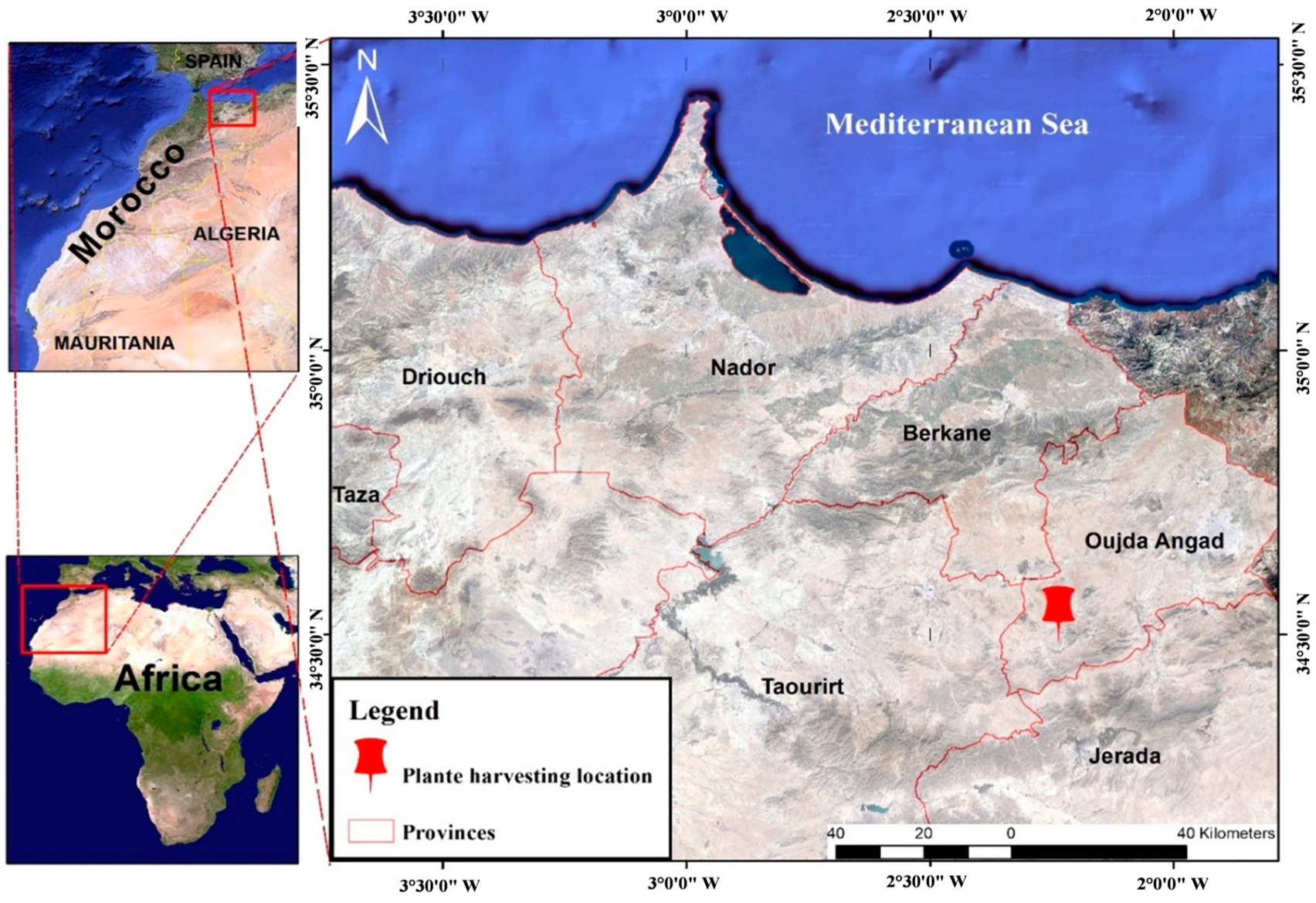
2.1. Plant Material
2.2. Isolation Processus
2.3. High-Performance Liquid Chromatography
2.4. Elemental Analysis
2.5. Fourier Transform Infrared Spectroscopy FT-IR
2.6. Mass Spectroscopy
2.7. MEB/EDX
2.8. Density Function Theory Study
2.9. Antioxidant Activity
2.9.1. Scavenging 2,2-Diphenyl-1-picrylhydrazyl Radical Test
2.9.2. β-Carotene Bleaching Test
2.10. Antimicrobial Activity
- 1.
- Bacterial strains
- 2.
- Fungal strains
- 3.
- Determination of MIC, MBC, and MFC
2.11. Molecular Docking Protocol
- Subsequent to this step, we employed the PyMoL 2.3 tool to preprocess all macromolecules, eliminating water molecules and extraneous protein residues. In order to guarantee structural integrity, nonpolar hydrogen atoms were integrated into the refined proteins. Sequentially, the Swiss PDB viewer, renowned for its energy minimization capabilities [45], was employed to minimize their energy states to the lowest possible level. After these purification and optimization processes, the macromolecules were archived in PDB format and primed for subsequent analysis.
- Throughout the computational interaction process, we implemented a semi-flexible modeling approach utilizing the extensively employed PyRx AutoDock Vina molecular docking software. Within PyRx, the target proteins were prepared and designated as macromolecules [46]. The 3D conformers of all ligands, initially in SDF format, were introduced into PyRx and subjected to energetic optimization. Subsequently, they were converted to pdbqt format using the Open Babel tool within the PyRx AutoDock Vina software, with a selection of the most optimal hit [47]. The results of the docking analysis were projected, and the outcomes, in conjunction with the docked macromolecules and ligands, were exported as output files in pdbqt format. These ligand and macromolecule files were merged and preserved in PDB format for further scrutiny using PyMol software. Ultimately, 2D visualizations were generated utilizing Discovery Studio Visualizer (version 4.6).
3. Results and Discussion
3.1. High-Performance Liquid Chromatography Analysis
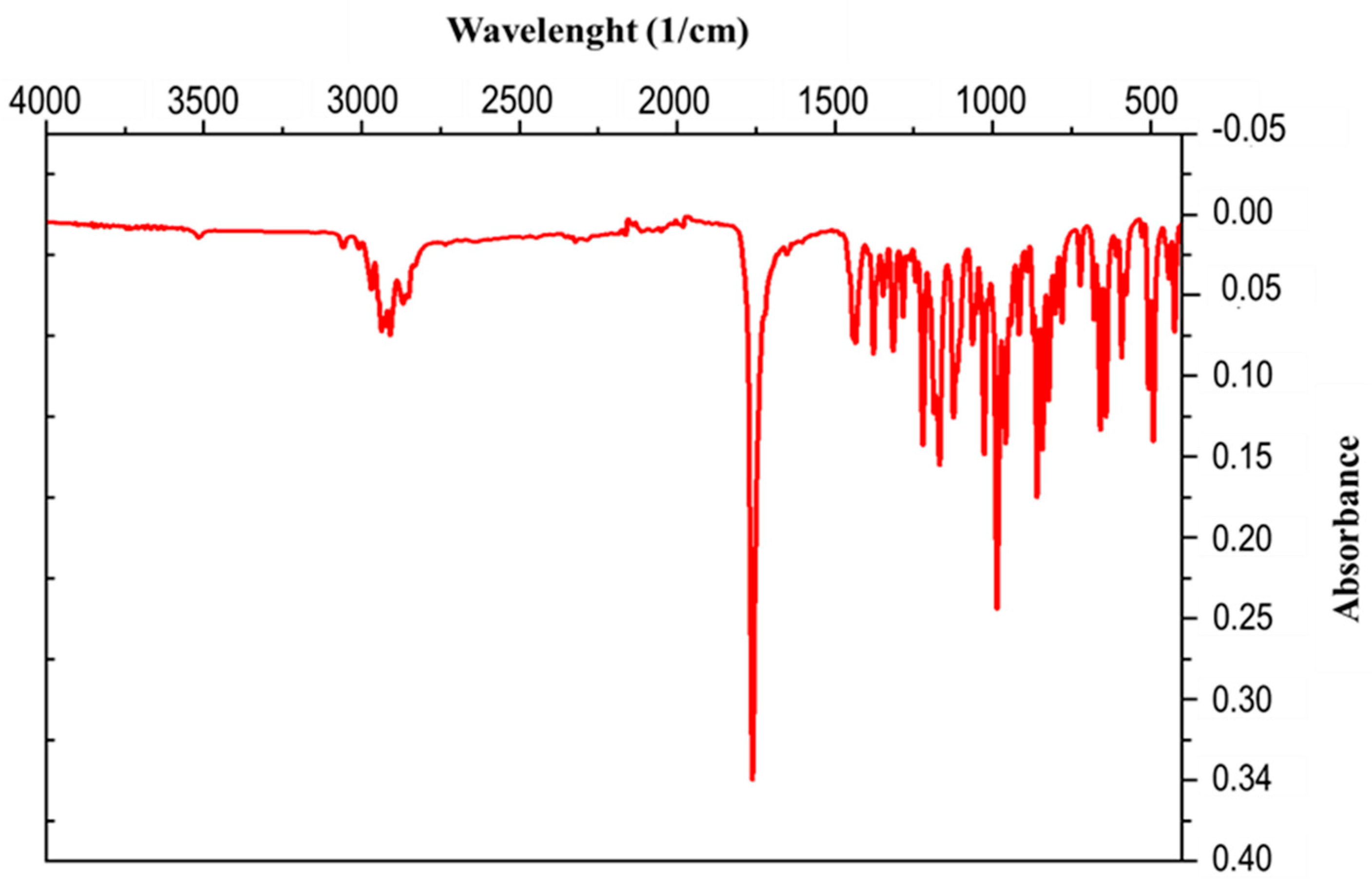
3.2. Infrared Spectroscopy
3.3. Scanning Electron Microscopy and Energy-Dispersive Spectroscopy
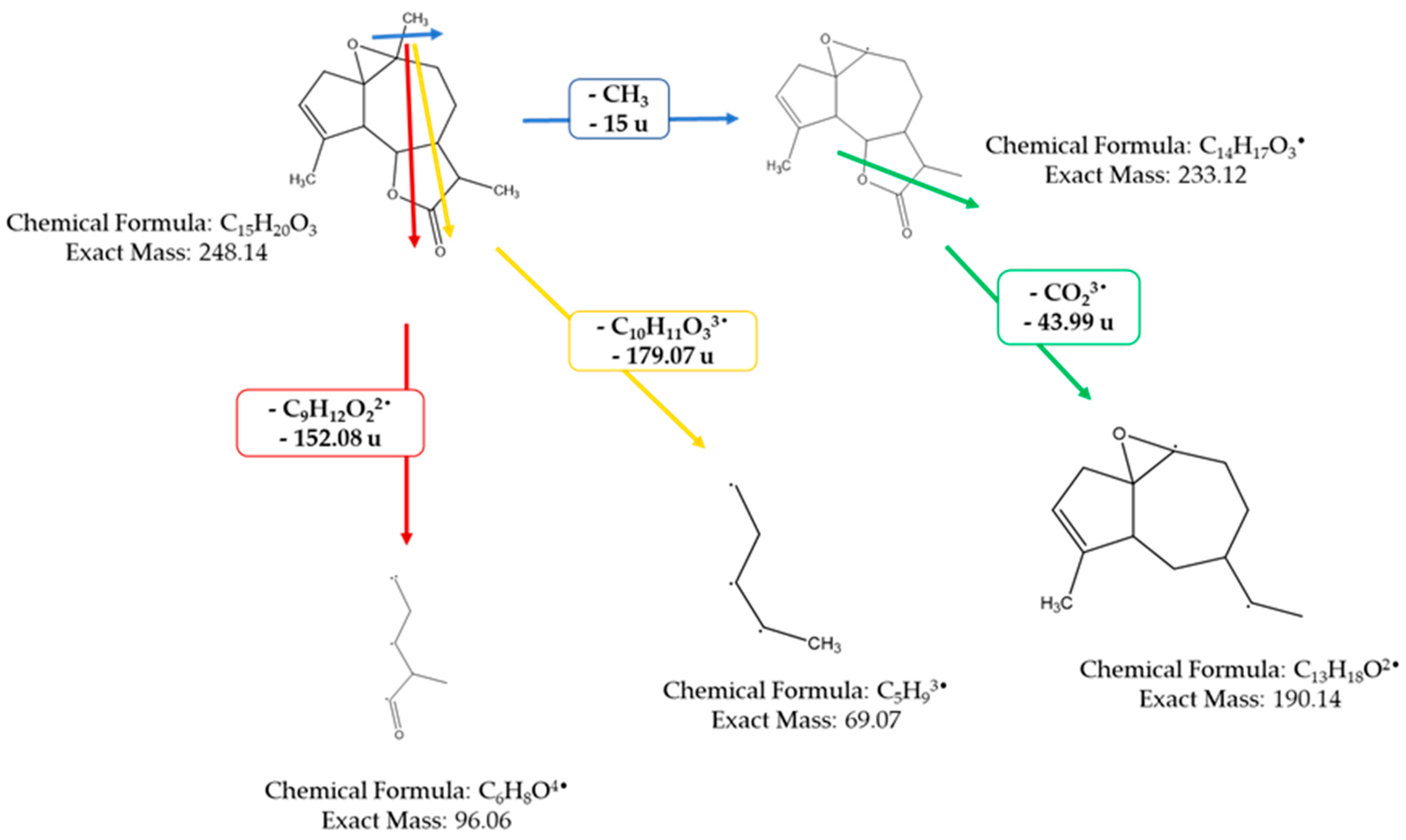
3.4. Mass Spectrometer
3.5. Elemental Analysis CNHSO
3.6. Quantum Chemistry Calculations
3.7. Antioxidant Activity
3.7.1. DPPH Radical Trapping Test
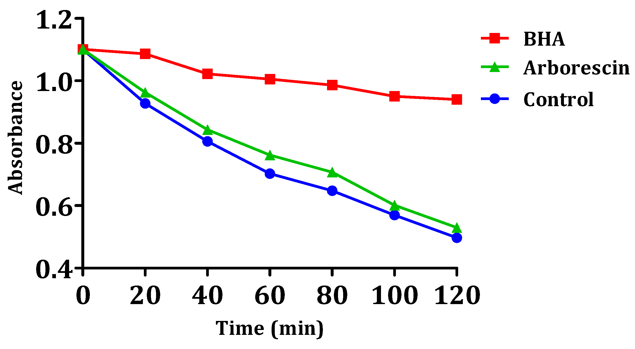
3.7.2. Bleaching Kinetics of the β-Carotene Assay
3.8. Antibacterial Activity
3.9. Antifungal Activity
3.10. Molecular Docking Predictions Revealing Potential Mechanisms of Action for Arborescin
3.10.1. Interactions with Xanthine Oxidoreductase (PDB: 3AX7): Antioxidant Activity
3.10.2. Interactions with Dihydrofolate Reductase (PDB: 4M6J): Antibacterial Activity
4. Conclusions
Author Contributions
Funding
Data Availability Statement
Acknowledgments
Conflicts of Interest
References
- Hao, D.-C.; Xiao, P.-G. Pharmaceutical resource discovery from traditional medicinal plants: Pharmacophylogeny and pharmacophylogenomics. Chin. Herb. Med. 2020, 12, 104–117. [Google Scholar] [CrossRef] [PubMed]
- Fabricant, D.S.; Farnsworth, N.R. The value of plants used in traditional medicine for drug discovery. Environ. Health Perspect. 2001, 109 (Suppl. S1), 69–75. [Google Scholar] [PubMed]
- Mohan, S.; Elhassan Taha, M.M.; Makeen, H.A.; Alhazmi, H.A.; Al Bratty, M.; Sultana, S.; Ahsan, W.; Najmi, A.; Khalid, A. Bioactive natural antivirals: An updated review of the available plants and isolated molecules. Molecules 2020, 25, 4878. [Google Scholar] [CrossRef] [PubMed]
- Gorlenko, C.L.; Kiselev, H.Y.; Budanova, E.V.; Zamyatnin, A.A., Jr.; Ikryannikova, L.N. Plant secondary metabolites in the battle of drugs and drug-resistant bacteria: New heroes or worse clones of antibiotics? Antibiotics 2020, 9, 170. [Google Scholar] [CrossRef] [PubMed]
- Kaoudoune, C.; Benchikh, F.; Benabdallah, H.; Loucif, K.; Mehlous, S.; Amira, S. Gastroprotective effect and in vitro Antioxidant Activities of the Aqueous Extract from Artemisia absinthium L. Aerial Parts. J. Drug Deliv. Ther. 2020, 10, 153–156. [Google Scholar] [CrossRef]
- Ahmad, W.; Hasan, A.; Abdullah, A.; Tarannum, T. Medicinal importance of Artemisia absinthium Linn (Afsanteen) in Unani medicine: A review. Hipp. J. Unani Med. 2010, 5, 117–125. [Google Scholar]
- Batiha, G.E.; Olatunde, A.; El-Mleeh, A.; Hetta, H.F.; Al-Rejaie, S.; Alghamdi, S.; Zahoor, M.; Magdy Beshbishy, A.; Murata, T.; Zaragoza-Bastida, A.; et al. Bioactive Compounds, Pharmacological Actions, and Pharmacokinetics of Wormwood (Artemisia absinthium). Antibiotics 2020, 9, 353. [Google Scholar] [CrossRef] [PubMed]
- Slepetys, J. Biology and biochemistry of wormwood. VIII. Accumulation dynamics of tannins, ascorbic acid and carotene (Russian). Труды Академии Наук Литoвскoй Сср Серия С Биoлoгические Науки 1975, 1, 43–48. [Google Scholar]
- Canadanovic-Brunet, J.M.; Djilas, S.M.; Cetkovic, G.S.; Tumbas, V.T. Free-radical scavenging activity of wormwood (Artemisia absinthium L.) extracts. J. Sci. Food Agric. 2005, 85, 265–272. [Google Scholar] [CrossRef]
- Akzhigitova, Z.; Baiseitova, A.; Dyusebaeva, M.; Ye, Y.; Jenis, J. Investigation of chemical constituents of Artemisia absinthium. Int. J. Biol. Chem. 2018, 11, 169–177. [Google Scholar] [CrossRef]
- Bhat, R.R.; Rehman, M.U.; Shabir, A.; Mir, M.U.R.; Ahmad, A.; Khan, R.; Masoodi, M.H.; Madkhali, H.; Ganaie, M.A. Chemical composition and biological uses of Artemisia absinthium (wormwood). In Plant and Human Health; Springer: Berlin/Heidelberg, Germany, 2019; Volume 3, pp. 37–63. [Google Scholar]
- Bora, K.S.; Sharma, A. Phytochemical and pharmacological potential of Artemisia absinthium Linn. and Artemisia asiatica Nakai: A review. J. Pharm. Res. 2010, 3, 325–328. [Google Scholar]
- da Silva, J.A.T. Mining the essential oils of the Anthemideae. Afr. J. Biotechnol. 2004, 3, 706–720. [Google Scholar]
- Picman, A.K. Biological activities of sesquiterpene lactones. Biochem. Syst. Ecol. 1986, 14, 255–281. [Google Scholar] [CrossRef]
- Wright, C.W. Artemisia، medicinal and aromatic plants-Industrial Profiles. Chapter 2002, 1, 10–22. [Google Scholar]
- Ghantous, A.; Gali-Muhtasib, H.; Vuorela, H.; Saliba, N.A.; Darwiche, N. What made sesquiterpene lactones reach cancer clinical trials? Drug Discov. Today 2010, 15, 668–678. [Google Scholar] [CrossRef] [PubMed]
- Schmidt, T.J. Structure-activity relationships of sesquiterpene lactones. Stud. Nat. Prod. Chem. 2006, 33, 309–392. [Google Scholar]
- Rodriguez, E.; Towers, G.; Mitchell, J. Biological activities of sesquiterpene lactones. Phytochemistry 1976, 15, 1573–1580. [Google Scholar] [CrossRef]
- Aberham, A.; Cicek, S.S.; Schneider, P.; Stuppner, H. Analysis of sesquiterpene lactones, lignans, and flavonoids in wormwood (Artemisia absinthium L.) using high-performance liquid chromatography (HPLC)−mass spectrometry, reversed phase HPLC, and HPLC−solid phase extraction−nuclear magnetic resonance. J. Agric. Food Chem. 2010, 58, 10817–10823. [Google Scholar] [CrossRef] [PubMed]
- Ru, G.; Manns, D.; Wilbert, S. Homoditerpene peroxides from Artemisia absinthium. Phytochemistry 1992, 31, 340–342. [Google Scholar]
- Ahamad, J.; Naquvi, K.; Ali, M.; Mir, S. Isoflavone glycosides from aerial parts of Artemisia absinthium. Chem. Nat. Compd. 2014, 49, 996–1000. [Google Scholar] [CrossRef]
- Hbika, A.; Bouyanzer, A.; Saadi, M.; El Ammari, L.; Benali, M.; Majidi, L.; Zarrouk, A. Structural study and thermal stability of Artemetin extracted from Artemisia absinthium L. Chem. Data Collect. 2022, 40, 100880. [Google Scholar] [CrossRef]
- Sridevi, V.K.; Chouhan, H.S.; Singh, N.K.; Singh, S.K. Antioxidant and hepatoprotective effects of ethanol extract of Vitex glabrata on carbon tetrachloride-induced liver damage in rats. Nat. Prod. Res. 2012, 26, 1135–1140. [Google Scholar] [CrossRef] [PubMed]
- Liu, K.C.-S.C.; Yang, S.-L.; Roberts, M.; Elford, B.; Phillipson, J. Antimalarial activity of Artemisia annua flavonoids from whole plants and cell cultures. Plant Cell Rep. 1992, 11, 637–640. [Google Scholar] [CrossRef] [PubMed]
- Hu, J.; Ma, W.; Li, N.; Wang, K.-J. Antioxidant and anti-inflammatory flavonoids from the flowers of Chuju, a medical cultivar of Chrysanthemum morifolim Ramat. J. Mex. Chem. Soc. 2017, 61, 282–289. [Google Scholar] [CrossRef]
- Li, W.-X.; Cui, C.-B.; Cai, B.; Wang, H.-Y.; Yao, X.-S. Flavonoids from Vitex trifolia L. inhibit cell cycle progression at G2/M phase and induce apoptosis in mammalian cancer cells. J. Asian Nat. Prod. Res. 2005, 7, 615–626. [Google Scholar] [CrossRef] [PubMed]
- Talmon, M.; Bosso, L.; Quaregna, M.; Lopatriello, A.; Rossi, S.; Gavioli, D.; Marotta, P.; Caprioglio, D.; Boldorini, R.; Miggiano, R. Anti-inflammatory activity of absinthin and derivatives in human bronchoepithelial cells. J. Nat. Prod. 2020, 83, 1740–1750. [Google Scholar] [CrossRef] [PubMed]
- Kasymov, S.Z.; Abdullaev, N.; Sidyakin, G.; Yagudaev, M. Anabsin—A new diguaianolide from Artemisia absinthium. Chem. Nat. Compd. 1979, 15, 430–435. [Google Scholar] [CrossRef]
- Ortet, R.; Prado, S.; Mouray, E.; Thomas, O.P. Sesquiterpene lactones from the endemic Cape Verdean Artemisia gorgonum. Phytochemistry 2008, 69, 2961–2965. [Google Scholar] [CrossRef] [PubMed]
- Adekenov, S. Chemical study of Artemisia austriaca Jacq. Int. J. Biol. Chem. 2021, 14, 156–163. [Google Scholar] [CrossRef]
- Bohlmann, F.; Hartono, L.; Jakupovic, J.; Huneck, S. Guaianolides related to arborescin from Artemisia adamsii. Phytochemistry 1985, 24, 1003–1007. [Google Scholar] [CrossRef]
- Hbika, A.; Bouyanzer, A.; Jalal, M.; Setti, N.; Loukili, E.; Aouniti, A.; Kerroum, Y.; Warad, I.; Hammouti, B.; Zarrouk, A.M. The Inhibiting Effect of Aqueous Extracts of Artemisia absinthium L. (Wormwood) on the Corrosion of Mild Steel in HCl 1 M. Anal. Bioanal. Electrochem. 2023, 15, 17–35. [Google Scholar]
- Janssens, M.L. Fundamental measurement techniques. In Flammability Testing of Materials Used in Construction, Transport and Mining; Elsevier: Amsterdam, The Netherlands, 2022; pp. 23–61. [Google Scholar]
- Ma, S.; Chowdhury, S.K.; Alton, K.B. Application of mass spectrometry for metabolite identification. Curr. Drug Metab. 2006, 7, 503–523. [Google Scholar] [CrossRef] [PubMed]
- Miceli, N.; Filocamo, A.; Ragusa, S.; Cacciola, F.; Dugo, P.; Mondello, L.; Celano, M.; Maggisano, V.; Taviano, M.F. Chemical characterization and biological activities of phenolic-rich fraction from cauline leaves of Isatis tinctoria L. (Brassicaceae) growing in Sicily, Italy. Chem. Biodivers. 2017, 14, e1700073. [Google Scholar] [CrossRef] [PubMed]
- Ouahabi, S.; Loukili, E.H.; Daoudi, N.E.; Chebaibi, M.; Ramdani, M.; Rahhou, I.; Bnouham, M.; Fauconnier, M.-L.; Hammouti, B.; Rhazi, L. Study of the Phytochemical Composition, Antioxidant Properties, and In Vitro Anti-Diabetic Efficacy of Gracilaria bursa-pastoris Extracts. Mar. Drugs 2023, 21, 372. [Google Scholar] [CrossRef] [PubMed]
- Kartal, N.; Sokmen, M.; Tepe, B.; Daferera, D.; Polissiou, M.; Sokmen, A. Investigation of the antioxidant properties of Ferula orientalis L. using a suitable extraction procedure. Food Chem. 2007, 100, 584–589. [Google Scholar] [CrossRef]
- Ouahabi, S.; Loukili, E.H.; Elbouzidi, A.; Taibi, M.; Bouslamti, M.; Nafidi, H.-A.; Salamatullah, A.M.; Saidi, N.; Bellaouchi, R.; Addi, M. Pharmacological properties of chemically characterized extracts from mastic tree: In vitro and in silico assays. Life 2023, 13, 1393. [Google Scholar] [CrossRef]
- Taibi, M.; Elbouzidi, A.; Ou-Yahia, D.; Dalli, M.; Bellaouchi, R.; Tikent, A.; Roubi, M.; Gseyra, N.; Asehraou, A.; Hano, C. Assessment of the antioxidant and antimicrobial potential of ptychotis verticillata duby essential oil from eastern Morocco: An in vitro and in silico analysis. Antibiotics 2023, 12, 655. [Google Scholar] [CrossRef]
- Taibi, M.; Elbouzidi, A.; Haddou, M.; Loukili, E.H.; Bellaouchi, R.; Asehraou, A.; Douzi, Y.; Addi, M.; Salamatullah, A.M.; Nafidi, H.-A. Chemical Profiling, Antibacterial Efficacy, and Synergistic Actions of Ptychotis verticillata Duby Essential Oil in Combination with Conventional Antibiotics. Nat. Prod. Commun. 2024, 19, 1934578X231222785. [Google Scholar] [CrossRef]
- Elbouzidi, A.; Ouassou, H.; Aherkou, M.; Kharchoufa, L.; Meskali, N.; Baraich, A.; Mechchate, H.; Bouhrim, M.; Idir, A.; Hano, C. LC–MS/MS phytochemical profiling, antioxidant activity, and cytotoxicity of the ethanolic extract of Atriplex halimus L. against breast cancer cell lines: Computational studies and experimental validation. Pharmaceuticals 2022, 15, 1156. [Google Scholar] [CrossRef] [PubMed]
- El Hachlafi, N.; Fikri-Benbrahim, K.; Al-Mijalli, S.H.; Elbouzidi, A.; Jeddi, M.; Abdallah, E.M.; Assaggaf, H.; Bouyahya, A.; Alnasser, S.M.A.; Attar, A. Tetraclinis articulata (Vahl) Mast. essential oil as a promising source of bioactive compounds with antimicrobial, antioxidant, anti-inflammatory and dermatoprotective properties: In vitro and in silico evidence. Heliyon 2023, 10, e23084. [Google Scholar] [CrossRef] [PubMed]
- Khokra, S.L.; Khan, S.A.; Thakur, P.; Chowdhary, D.; Ahmad, A.; Husain, A. Synthesis, molecular docking and potential antioxidant activity of di/trisubstituted pyridazinone derivatives. J. Chin. Chem. Soc. 2016, 63, 739–750. [Google Scholar] [CrossRef]
- Khatun, M.C.S.; Muhit, M.A.; Hossain, M.J.; Al-Mansur, M.A.; Rahman, S.A. Isolation of phytochemical constituents from Stevia rebaudiana (Bert.) and evaluation of their anticancer, antimicrobial and antioxidant properties via in vitro and in silico approaches. Heliyon 2021, 7, e08475. [Google Scholar] [CrossRef] [PubMed]
- Guex, N.; Peitsch, M.C. SWISS-MODEL and the Swiss-Pdb Viewer: An environment for comparative protein modeling. Electrophoresis 1997, 18, 2714–2723. [Google Scholar] [CrossRef] [PubMed]
- Pawar, R.P.; Rohane, S.H. Role of autodock vina in PyRx molecular docking. Asian J. Res. Chem. 2021, 5, 132–134. [Google Scholar]
- O’Boyle, N.M.; Banck, M.; James, C.A.; Morley, C.; Vandermeersch, T.; Hutchison, G.R. Open Babel: An open chemical toolbox. J. Cheminf. 2011, 3, 33. [Google Scholar] [CrossRef] [PubMed]
- Ferreira, J.F.; Gonzalez, J.M. Analysis of underivatized artemisinin and related sesquiterpene lactones by high-performance liquid chromatography with ultraviolet detection. Phytochem. Anal. 2009, 20, 91–97. [Google Scholar] [CrossRef]
- Green, M.D.; Mount, D.L.; Wirtz, R.A.; White, N.J. A colorimetric field method to assess the authenticity of drugs sold as the antimalarial artesunate. J. Pharm. Biomed. Anal. 2000, 24, 65–70. [Google Scholar] [CrossRef] [PubMed]
- Christen, P.; Veuthey, J. New trends in extraction, identification and quantification of artemisinin and its derivatives. Curr. Med. Chem. 2001, 8, 1827–1839. [Google Scholar] [CrossRef] [PubMed]
- Ivanescu, B.; Miron, A.; Corciova, A. Sesquiterpene lactones from Artemisia genus: Biological activities and methods of analysis. J. Anal. Methods Chem. 2015, 2015, 247685. [Google Scholar] [CrossRef] [PubMed]
- Arjunan, V.; Devi, L.; Subbalakshmi, R.; Rani, T.; Mohan, S. Synthesis, vibrational, NMR, quantum chemical and structure-activity relation studies of 2-hydroxy-4-methoxyacetophenone. Spectrochim. Acta Part A Mol. Biomol. Spectrosc. 2014, 130, 164–177. [Google Scholar] [CrossRef] [PubMed]
- El-Gammal, O.; Rakha, T.; Metwally, H.; El-Reash, G.A. Synthesis, characterization, DFT and biological studies of isatinpicolinohydrazone and its Zn (II), Cd (II) and Hg (II) complexes. Spectrochim. Acta Part A Mol. Biomol. Spectrosc. 2014, 127, 144–156. [Google Scholar] [CrossRef] [PubMed]
- Regti, A.; El Ayouchia, H.B.; Laamari, M.R.; Stiriba, S.E.; Anane, H.; El Haddad, M. Experimental and theoretical study using DFT method for the competitive adsorption of two cationic dyes from wastewaters. Appl. Surf. Sci. 2016, 390, 311–319. [Google Scholar] [CrossRef]
- Choudhary, V.; Bhatt, A.; Dash, D.; Sharma, N. DFT calculations on molecular structures, HOMO–LUMO study, reactivity descriptors and spectral analyses of newly synthesized diorganotin (IV) 2-chloridophenylacetohydroxamate complexes. J. Comput. Chem. 2019, 40, 2354–2363. [Google Scholar] [CrossRef] [PubMed]
- Tzanova, M.; Grozeva, N.; Gerdzhikova, M.; Argirova, M.; Pavlov, D.; Terzieva, S. Flavonoid content and antioxidant activity of Betonica bulgarica Degen et Neič. Bulg. Chem. Commun. 2018, 50, 90–97. [Google Scholar]
- Benchikh, F.; Amira, S.; Benabdallah, H. The evaluation of antioxidant capacity of different fractions of Myrtus communis L. leaves. Annu. Res. Rev. Biol. 2018, 22, 1–14. [Google Scholar] [CrossRef]
- Martins, A.; Mignon, R.; Bastos, M.; Batista, D.; Neng, N.R.; Nogueira, J.M.; Vizetto-Duarte, C.; Custódio, L.; Varela, J.; Rauter, A.P. In vitro antitumoral activity of compounds isolated from Artemisia gorgonum Webb. Phytother. Res. 2014, 28, 1329–1334. [Google Scholar] [CrossRef] [PubMed]
- Elbouzidi, A.; Taibi, M.; Laaraj, S.; Loukili, E.H.; Haddou, M.; El Hachlafi, N.; Naceiri Mrabti, H.; Baraich, A.; Bellaouchi, R.; Asehraou, A. Chemical profiling of volatile compounds of the essential oil of grey-leaved rockrose (Cistus albidus L.) and its antioxidant, anti-inflammatory, antibacterial, antifungal, and anticancer activity in vitro and in silico. Front. Chem. 2024, 12, 1334028. [Google Scholar] [CrossRef] [PubMed]
- Harrison, R. Structure and function of xanthine oxidoreductase: Where are we now? Free Radic. Biol. Med. 2002, 33, 774–797. [Google Scholar] [CrossRef] [PubMed]
- Harrison, R. Physiological roles of xanthine oxidoreductase. Drug Metab. Rev. 2004, 36, 363–375. [Google Scholar] [CrossRef] [PubMed]
- Scherz-Shouval, R.; Elazar, Z. Regulation of autophagy by ROS: Physiology and pathology. Trends Biochem. Sci. 2011, 36, 30–38. [Google Scholar] [CrossRef] [PubMed]
- Juan, C.A.; Pérez de la Lastra, J.M.; Plou, F.J.; Pérez-Lebeña, E. The chemistry of reactive oxygen species (ROS) revisited: Outlining their role in biological macromolecules (DNA, lipids and proteins) and induced pathologies. Int. J. Mol. Sci. 2021, 22, 4642. [Google Scholar] [CrossRef]
- Vickneson, K.; George, J. Xanthine oxidoreductase inhibitors. In Reactive Oxygen Species: Network Pharmacology and Therapeutic Applications; Springer: Berlin/Heidelberg, Germany, 2021; pp. 205–228. [Google Scholar]
- Andrews, B.A.; Dyer, R.B. Comparison of the Role of Protein Dynamics in Catalysis by Dihydrofolate Reductase from E. coli and H. sapiens. J. Phys. Chem. B 2022, 126, 7126–7134. [Google Scholar] [CrossRef] [PubMed]
- Chawla, P.; Teli, G.; Gill, R.K.; Narang, R.K. An insight into synthetic strategies and recent developments of dihydrofolate reductase inhibitors. ChemistrySelect 2021, 6, 12101–12145. [Google Scholar] [CrossRef]
- Bhagat, K.; Kumar, N.; Kaur Gulati, H.; Sharma, A.; Kaur, A.; Singh, J.V.; Singh, H.; Bedi, P.M.S. Dihydrofolate reductase inhibitors: Patent landscape and phases of clinical development (2001–2021). Expert Opin. Ther. Pat. 2022, 32, 1079–1095. [Google Scholar] [CrossRef] [PubMed]
- Bhabha, G.; Ekiert, D.C.; Jennewein, M.; Zmasek, C.M.; Tuttle, L.M.; Kroon, G.; Dyson, H.J.; Godzik, A.; Wilson, I.A.; Wright, P.E. Divergent evolution of protein conformational dynamics in dihydrofolate reductase. Nat. Struct. Mol. Biol. 2013, 20, 1243–1249. [Google Scholar] [CrossRef] [PubMed]
- Wróbel, A.; Arciszewska, K.; Maliszewski, D.; Drozdowska, D. Trimethoprim and other nonclassical antifolates an excellent template for searching modifications of dihydrofolate reductase enzyme inhibitors. J. Antibiot. 2020, 73, 5–27. [Google Scholar] [CrossRef] [PubMed]
- Bayazeed, A.; Alenazi, N.A.; Alsaedi, A.M.; Ibrahim, M.H.; Al-Qurashi, N.T.; Farghaly, T.A. Formazan analogous: Synthesis, antimicrobial activity, dihydrofolate reductase inhibitors and docking study. J. Mol. Struct. 2022, 1258, 132653. [Google Scholar] [CrossRef]
- Moujir, L.; Callies, O.; Sousa, P.M.; Sharopov, F.; Seca, A.M. Applications of sesquiterpene lactones: A review of some potential success cases. Appl. Sci. 2020, 10, 3001. [Google Scholar] [CrossRef]
- Lai, H.C.; Singh, N.P.; Sasaki, T. Development of artemisinin compounds for cancer treatment. Investig. New Drugs 2013, 31, 230–246. [Google Scholar] [CrossRef] [PubMed]
- White, N.J. Qinghaosu (artemisinin): The price of success. Science 2008, 320, 330–334. [Google Scholar] [CrossRef] [PubMed]
- Stavrianidi, A. A classification of liquid chromatography mass spectrometry techniques for evaluation of chemical composition and quality control of traditional medicines. J. Chromatogr. A 2020, 1609, 460501. [Google Scholar] [CrossRef]
- Beer, M.F.; Bivona, A.E.; Sánchez Alberti, A.; Cerny, N.; Reta, G.F.; Martín, V.S.; Padrón, J.M.; Malchiodi, E.L.; Sülsen, V.P.; Donadel, O.J. Preparation of sesquiterpene lactone derivatives: Cytotoxic activity and selectivity of action. Molecules 2019, 24, 1113. [Google Scholar] [CrossRef] [PubMed]
- Sokovic, M.; Ciric, A.; Glamoclija, J.; Skaltsa, H. Biological activities of sesquiterpene lactones isolated from the genus Centaurea L. (Asteraceae). Curr. Pharm. Des. 2017, 23, 2767–2786. [Google Scholar] [CrossRef] [PubMed]
- Ma, C.; Meng, C.-W.; Zhou, Q.-M.; Peng, C.; Liu, F.; Zhang, J.-W.; Zhou, F.; Xiong, L. New sesquiterpenoids from the stems of Dendrobium nobile and their neuroprotective activities. Fitoterapia 2019, 138, 104351. [Google Scholar] [CrossRef] [PubMed]
- Cheriti, A.; Belboukhari, N. Terpenoids of the Saharan Medicinal Plants Launaea Cass. Genus (Asteraceae) and Their Biological Activities; Nova Science Publishers Inc.: New York, NY, USA, 2015; pp. 51–70. [Google Scholar]
- Li, Q.; Wang, Z.; Xie, Y.; Hu, H. Antitumor activity and mechanism of costunolide and dehydrocostus lactone: Two natural sesquiterpene lactones from the Asteraceae family. Biomed. Pharmacother. 2020, 125, 109955. [Google Scholar] [CrossRef] [PubMed]
- Pandey, M.M.; Rastogi, S.; Rawat, A.K.S. Saussurea costus: Botanical, chemical and pharmacological review of an ayurvedic medicinal plant. J. Ethnopharmacol. 2007, 110, 379–390. [Google Scholar] [CrossRef] [PubMed]
- Kim, D.Y.; Choi, B.Y. Costunolide—A bioactive sesquiterpene lactone with diverse therapeutic potential. Int. J. Mol. Sci. 2019, 20, 2926. [Google Scholar] [CrossRef]
- Lin, X.; Peng, Z.; Su, C. Potential anti-cancer activities and mechanisms of costunolide and dehydrocostuslactone. Int. J. Mol. Sci. 2015, 16, 10888–10906. [Google Scholar] [CrossRef] [PubMed]
- Wang, J.; Su, S.; Zhang, S.; Zhai, S.; Sheng, R.; Wu, W.; Guo, R. Structure-activity relationship and synthetic methodologies of α-santonin derivatives with diverse bioactivities: A mini-review. Eur. J. Med. Chem. 2019, 175, 215–233. [Google Scholar] [CrossRef] [PubMed]


| Element | Mass % | Atom % |
|---|---|---|
| C | 78.7 ± 0.46 | 83.2 ± 0.49 |
| O | 21.2 ± 0.71 | 16.8 ± 0.56 |
| Total | 100.00 | 100.00 |
| Arborescin C15H20O3 | ||
|---|---|---|
| Mass | Experimental | By Formula |
| C% | 73.1 | 72.6 |
| O% | 7.86 | 8.06 |
| H% | 19.0 | 19.3 |
| N% | 0.00 | 0.00 |
| S% | 0.00 | 0.00 |
| C/O | 9.29 | 9.00 |
| Descriptors of Global Reactivity | Arborescin |
|---|---|
| Total global energy (ET) | −809.5 (u a) |
| Lowest unoccupied molecular orbital (ELUMO) | −0.04 (eV) |
| Highest occupied molecular orbital (EHOMO) | −6.77 (eV) |
| Energy gap (ΔEgap) | 6.73 (eV) |
| Absolute electronegativity (χ) | 3.41 (eV) |
| Overall hardness (Ƞ) | 3.36 (eV) |
| Electron affinity (Af) | 0.04 (eV) |
| Ionization (I) | 6.77 (eV) |
| Electrophilicity (ω) | 1.72 (eV) |
| Overall softness (σ) | 0.30 (eV) |
| Dipole moment (ᶙ) | 5.47 (D) |
| Chemical potential (µ) | −3.41 (eV) |
| DPPH (mg/mL) | β-Carotene (RAA %) | |
|---|---|---|
| BHA | - | 73.4 |
| Ascorbic acid | 0.16 ± 0.003 | - |
| Arborescin | 5.04 ± 0.12 | 3.64 |
| Bacterial Strains | E. coli | S. aureus | L. innocua | P. aeruginosa | |
|---|---|---|---|---|---|
| Arborescin | MIC (µg/mL) | 83 | 166 | 166 | 83 |
| MBC (µg/mL) | 333 | ≥666 | ≥666 | 333 |
| Molecule | Fungal Strains | Aspergillus niger | Candida glabrata | Penicillium digitatum |
|---|---|---|---|---|
| Arborescin | MIC (µg/mL) | 41 | 83 | 41 |
| MBC (µg/mL) | 166 | 41 | 166 |
| N° | Compounds | 3AX7 (Antioxidant) | 4M6J |
|---|---|---|---|
| (Antibacterial) | |||
| Free Binding Energy (kcal/mol) | |||
| - | Native Ligand | −8.0 (Oxypurinol) | −8.0 (Ciproflaxacin) |
| 1 | Arborescin | −8.0 | −8.1 |
Disclaimer/Publisher’s Note: The statements, opinions and data contained in all publications are solely those of the individual author(s) and contributor(s) and not of MDPI and/or the editor(s). MDPI and/or the editor(s) disclaim responsibility for any injury to people or property resulting from any ideas, methods, instructions or products referred to in the content. |
© 2024 by the authors. Licensee MDPI, Basel, Switzerland. This article is an open access article distributed under the terms and conditions of the Creative Commons Attribution (CC BY) license (https://creativecommons.org/licenses/by/4.0/).
Share and Cite
Hbika, A.; Elbouzidi, A.; Taibi, M.; Ouahabi, S.; Loukili, E.H.; Bouyanzer, A.; Yahyaoui, M.I.; Asehraou, A.; El Hachlafi, N.; Salamatullah, A.M.; et al. Isolation of Arborescin from Artemisia absinthium L. and Study of Its Antioxidant and Antimicrobial Potential by Use of In Vitro and In Silico Approaches. Separations 2024, 11, 209. https://doi.org/10.3390/separations11070209
Hbika A, Elbouzidi A, Taibi M, Ouahabi S, Loukili EH, Bouyanzer A, Yahyaoui MI, Asehraou A, El Hachlafi N, Salamatullah AM, et al. Isolation of Arborescin from Artemisia absinthium L. and Study of Its Antioxidant and Antimicrobial Potential by Use of In Vitro and In Silico Approaches. Separations. 2024; 11(7):209. https://doi.org/10.3390/separations11070209
Chicago/Turabian StyleHbika, Asmae, Amine Elbouzidi, Mohamed Taibi, Safae Ouahabi, El Hassania Loukili, Abdelhamid Bouyanzer, Meryem Idrissi Yahyaoui, Abdeslam Asehraou, Naoufal El Hachlafi, Ahmad Mohammad Salamatullah, and et al. 2024. "Isolation of Arborescin from Artemisia absinthium L. and Study of Its Antioxidant and Antimicrobial Potential by Use of In Vitro and In Silico Approaches" Separations 11, no. 7: 209. https://doi.org/10.3390/separations11070209
APA StyleHbika, A., Elbouzidi, A., Taibi, M., Ouahabi, S., Loukili, E. H., Bouyanzer, A., Yahyaoui, M. I., Asehraou, A., El Hachlafi, N., Salamatullah, A. M., Bourhia, M., Ibenmoussa, S., Addi, M., & Gharibi, E. (2024). Isolation of Arborescin from Artemisia absinthium L. and Study of Its Antioxidant and Antimicrobial Potential by Use of In Vitro and In Silico Approaches. Separations, 11(7), 209. https://doi.org/10.3390/separations11070209