Article

Study of Bacterial Elution from High-Efficiency Glass Fiber Filters

1
School of Light Industry and Engineering, South China University of Technology, Guangzhou 510641, China
2
State Key Laboratory of NBC Protection for Civilian, Beijing 100191, China
*
Authors to whom correspondence should be addressed.
Separations 2025, 12(5), 110; https://doi.org/10.3390/separations12050110 (registering DOI)
Submission received: 10 March 2025 / Revised: 17 April 2025 / Accepted: 22 April 2025 / Published: 25 April 2025

Abstract

:
Antibacterial filter materials have been effectively utilized for controlling biological contaminants and purifying indoor air, with the market for such materials experiencing continuous expansion. Currently, textile antibacterial testing standards are widely adopted to evaluate the antimicrobial efficacy of filter materials, yet no dedicated assessment protocols specifically tailored for filtration media have been established. This study aims to investigate the applicability of textile antibacterial testing methods to high-efficiency glass fiber filter materials (filtration efficiency > 99.9%), as well as to explore the factors that affect the rate of bacterial elution from high-efficiency glass fiber filter materials. By referencing the textile antibacterial testing standard (absorption method), significant discrepancies in bacterial recovery counts were observed between the high-efficiency glass fiber materials and the various textile control samples, with the former exhibiting a markedly lower recovery rate (approximately 10%). Pore structure and wettability analyses revealed the underlying causes of these differences. To ensure the accuracy of the antibacterial evaluation results, the effects of oscillation elution parameters (time and intensity) and material incubation conditions (duration, sealing and humidity) on bacterial recovery rates in glass fiber filter materials were systematically investigated to optimize the elution methodology. The results indicate that specimen type, size, elution method, incubation duration (4 h or 24 h), sealing conditions, and environmental humidity (10% or 30%, 60% and 95% RH) collectively influence bacterial recovery efficiency. The highest recovery efficiency (55%) was achieved when the filter materials were incubated in a sealed environment with humidity maintained at ≥60% RH. These findings emphasize the critical need to establish clear and specialized antibacterial performance testing standards for filter materials. The study provides essential guidance for developing material-specific evaluation protocols to ensure a reliable and standardized assessment of antimicrobial efficacy in high-efficiency filtration systems.

1. Introduction

Airborne microorganisms, serving as the principal biological contaminants in ambient air, present a latent menace to human well-being. They are capable of instigating or aggravating infectious maladies such as influenza, pneumonia, and the coronavirus (COVID-19) [1]. In response, antibacterial air filtration materials, which can not only mitigate the dissemination and harm of infectious diseases but also purify indoor air, have captured the attention of researchers. Scrutinizing the literature within related domains, a plethora of endeavors have been dedicated to devising active control technologies for airborne bacteria. These encompass ultraviolet irradiation [2], electrostatic mechanisms, microwave irradiation [3], plasma-based approaches [4], lysozyme-mediated processes, photocatalysis, and the application of airborne bactericides [5]. Among these strategies, the synthesis of antibacterial air filtration materials through the incorporation of antibacterial agents has emerged as a preeminent research focus. This is attributable to its remarkable efficiency and convenience in eradicating airborne biological pollutants [6]. Empirical findings have demonstrated that treating fibrous air filters with antibacterial agents can effectively impede the proliferation of microorganisms. Moreover, the utilization of such agents exerts negligible influence on the filtration efficiency of bioaerosols or inert test aerosols [7]. As one of the primary air filtration materials, high-efficiency glass fiber filter materials are predominantly utilized for the removal of airborne particulate matter in applications such as air conditioning filtration systems within commercial buildings and public transportation [8].
Notwithstanding the burgeoning research on antibacterial air filtration materials, a standardized and well-defined methodology for evaluating their antibacterial performance remains elusive. The conventional antibacterial performance testing protocols designed for textiles are frequently repurposed to assess the antibacterial efficacy of antibacterial filter media. Prominent standard-setting entities, including the American Association of Textile Chemists and Colorists (AATCC) [9], the Standardization Administration of China (SAC) [10], and the International Organization for Standardization (ISO) [11], have formulated pertinent standards for gauging the antibacterial properties of textiles. These standards are firmly rooted in the concept of colony-forming units (CFUs).
The absorption method, a cornerstone technique in textile antibacterial performance assessment, adheres to the following operational principle: A precisely calibrated concentration of the test bacterial solution is concurrently introduced to both the antibacterial-treated textiles and the standard blank specimens. Following a designated cultivation interval, each sample is meticulously rinsed with an eluent. Subsequently, the eluent undergoes serial dilution and is subjected to plate pouring culture and bacterial enumeration. This absorption method has found extensive application in evaluating the antibacterial performance of air filters treated with antibacterial agents. For example, R.B. Simmons and S.A. Crow [12] investigated the fungal colonization of novel and aged cellulose air filters (including those treated with antibacterial agents) deployed in heating, ventilation, and air conditioning systems. They achieved this by suspending the filters in containers with a relative humidity spanning from 55% to 99%. Miaśkiewicz Pęska and Ewa B. [13] conducted examinations on four distinct filter media and estimated the bacterial count via culturing techniques. The results showed a conspicuous reduction in the bacterial population within the treated filters. Katarzyna and Majchrzycka et al. [14] analyzed the influence of environmental sweat, along with filter paper and nonwoven fabrics impregnated with fungicides, on microbial activity. Their research revealed that the addition of sweat to the surface of nonwoven fabrics resulted in a substantial decline in microbial biological activity, albeit the activity still persisted at a relatively elevated level. Worrawit and Nakpan et al. [15] investigated the synergistic inactivation effect of ultraviolet irradiation and gaseous iodine on bacteria and on fungal spores collected on flat panel filters. All these studies utilized the absorption method to recover and quantify the bacteria on the samples, thereby deriving antibacterial outcomes. Nevertheless, despite the widespread adoption of the absorption method in evaluating the antibacterial efficacy of air filter media, there is limited research regarding the compatibility of this method for assessing the antibacterial performance of glass fiber filter media. Additionally, the potential ramifications of nebulous culture conditions on the bacterial recovery efficiency of samples remain largely unexplored.
In summary, procedures involving the absorption method across various national standards exhibit ambiguous and inconsistent requirements for experimental parameters. These parameters encompass the sample dimensions [16], the selection of standard blank samples, the method of oscillatory elution for detaching bacteria from the samples, and the cultivation humidity [17]. Such a scenario is likely to result in unreliable antibacterial test outcomes. Within the context of international trade, buyers typically require sellers to provide antibacterial test reports issued by third-party testing institutions. Owing to the current imperfections in the antibacterial testing regime and the dearth of a unified standard, manufacturers frequently face significant challenges. Consequently, the international trade of antibacterial filter media has been affected to a certain extent [18]. Therefore, by drawing on the antibacterial performance testing method for textiles (the absorption method), we compared the bacterial elution effects of high-efficiency glass fiber filter paper and textiles under the same test conditions. The results showed that the glass fiber filter paper exhibited bacterial elution issues. The underlying causes of these problems were revealed through pore size and wettability analyses. Finally, we investigated the influence of different experimental conditions on the bacterial recovery efficiency of high-efficiency glass fiber filter materials and identified the optimal bacterial elution conditions for such materials. These findings can provide guidance for establishing standardized antibacterial testing methods for high-efficiency glass fiber.

2. Materials and Methods

2.1. Procurement of Testing Materials

Material Sources

Chopped glass fibers were acquired from Chongqing International Composite Materials Co., Ltd. (Chongqing, China), and ultrafine glass fibers were obtained from Yulin Tianshengyuan Glass Fiber Technology Co., Ltd. (Yulin, China). Polypropylene nonwoven fabric (PP nonwoven fabric) was procured from Suzhou Chengyi Purification Technology Co., Ltd. (Suzhou, China), and 100% cotton fabric was purchased from Zhaoqing High tech Zone Yikang Fabric Co., Ltd. (Zhaoqing, China). Thermosetting acrylic resin and thermoplastic acrylic resin (SWP-9070, SWK-6269) were sourced from Kunshan Sanwang Resin Co., Ltd. (Kunshan, China). The non-ionic water repellent agent (MF-668) was obtained from Hangzhou Longmier Chemical Co., Ltd. (Hangzhou, China). Additionally, phosphate-buffered saline (PBS, 10×, pH 7.4) was supplied by Beijing Solarbio Science & Technology Co., Ltd. (Beijing, China).

2.2. Fabrication of Filter Media

The synthesis of the glass fiber base paper filter media (Material B) was achieved through a wet-laying process. Initially, the chopped glass fibers and ultrafine glass fibers were introduced into an acidic aqueous solution. Subsequently, a pulp disintegrator (IMT-SJ02, Dongguan Inter-Nessen Precision Instrument Co., Ltd. Dongguan, China) was utilized to homogeneously agitate the mixture at a precisely controlled rotational speed of 30,000 revolutions per minute (rpm) for a duration of 10 min. Following this, the well-dispersed mixture was transformed into glass fiber material (GFM) sheets using a sheet former (IMT-CP01A, Dongguan Inter Nessen Precision Instrument Co., Ltd. Dongguan, China). The formed GFM sheets were then subjected to a drying process at a temperature of 105 °C for 30 min to obtain the glass fiber base paper, Material B.
Material B was immersed in an aqueous solution of acrylic resin, with a thermosetting resin to thermoplastic resin ratio of 1:1. After a 3 min immersion period, the paper was dried at 105 °C for 30 min, resulting in the production of Material A. Moreover, when Material B was immersed in a mixed aqueous solution of acrylic resin and a water repellent agent, with a carefully calibrated ratio of thermosetting resin–thermoplastic resin–water repellent agent of 2:2:1, Material C was successfully prepared.

2.3. Bacterial Cultivation and Experimental Protocols

E. coli (ATCC 8739) bacterial cells were cultured in a nutrient broth medium at a temperature of 37 °C for 24 h to reach a cell count of approximately 109 colony-forming units per milliliter (CFU/mL). Subsequently, the bacterial suspension was diluted to a concentration of 107 CFU/mL using phosphate-buffered saline (PBS).
The test samples were precisely cut into a size of 2 × 2 cm as the original dimensions and aseptically placed in a standard six-well culture dish. A volume of 0.1 mL of the bacterial solution was evenly dispensed onto either the sample or the empty plate (for the non-sample control group, denoted as Group F, which only received 0.1 mL of pure bacterial solution). To control the cultivation environment during the experiment, the standard six-well culture dishes were either sealed by wrapping them multiple times with cling film or left unsealed.
The samples were then incubated in a 37 °C intelligent artificial climate incubator (PRX-150C, Ningbo Saifu Experimental Instrument Co., Ltd. Ningbo, China) for a specific cultivation time. Different humidity conditions were created by adjusting the humidity parameters of this incubator.
After the cultivation period, in accordance with the bacterial elution method specified in GB/T 20944.2 [9], the cultured sample or 0.1 mL of the bacterial solution was transferred into a test tube with 20 mL of PBS. The bacteria adhered to the sample were eluted into the eluent either by manual shaking with an amplitude of 30 cm for 30 s or by vortex oscillator shaking at a speed of 2500 revolutions per minute (rpm) for 30 s. The resulting bacterial eluent was then proportionally diluted onto nutrient agar (PCA) plates and cultured at 37 °C for 24 h. Post cultivation, the number of bacterial colonies (CFUs) was accurately counted using a colony counting software, ensuring the reliability and accuracy of the data. The pore size analysis and surface morphology imaging of the filter paper were performed using a capillary flow porometer (CFP-1100AE, Porous Materials Inc, Ithaca, NY, USA) and a scanning electron microscope (Gemini SEM 300, Carl Zeiss Microscopy GmbH, Jena, Germany), respectively. Bacterial morphology imaging of the bacteria-loaded filter paper was conducted with a field-emission scanning electron microscope (SU8010, Hitachi High-Tech Corporation, Tokyo, Japan). The particle filtration efficiency of the filter paper was obtained using a fully automated filter material testing system (Model 8130, TSI Incorporated, Shoreview, MN, USA). The material thickness was measured using a digital thickness gauge (Model IMT-HK210D, Inteernisen Precision Instrument Co., Ltd., Dongguan, China).
For data analysis and interpretation, we assumed that the theoretical growth quantity of bacteria on the filter material was equivalent to the bacterial recovery quantity of 0.1 mL of the bacterial solution, with Group F representing the theoretical bacterial growth quantity. Group S was designated to represent the initial number of bacteria dropped onto the material. The bacterial elution rate of the material was defined as the percentage of bacterial recovery quantity from the material to the bacterial recovery quantity from 0.1 mL of the bacterial solution (Group F).

2.4. Statistical Analysis

A independent samples t-test was utilized to analyze the two elution methods. To examine differences in the number of bacteria recovered among the various material types, as well as differences in the number of bacteria recovered from Material A and grown in Group F under varying humidity and sealing conditions, Tukey’s post hoc test was employed. All data were processed and analyzed using the SPSS statistical software (SPSS 27.0, IBM Corporation, Armonk, NY, USA). Significant differences between the groups are indicated by lowercase letters (a, b, c, etc.). Groups sharing the same letter show no significant difference (p > 0.05), while groups with different letters are significantly different (p < 0.05). Additionally, “ns” denotes no significant difference between two groups (p > 0.05).

3. Results and Discussion

3.1. Applicability of Textile Testing Standards to High-Efficiency Glass Fiber Filter Materials

3.1.1. Basic Material Properties and Comparison of International Textile Standards

In standardized testing protocols for antimicrobial textiles, the absence of appropriate blank controls (textiles without antimicrobial additives) has often necessitated the specification of alternative reference materials across various international standards. Specifically, ISO 20743 [11] recommends utilizing 100% additive-free cotton fabric as control specimens, while the Chinese national standard GB 21551.2 [19] specifies polypropylene nonwoven fabric as an appropriate reference material. These standardized substitutes enable comparative evaluation when original untreated counterparts are unavailable. Table 1 systematically compares the methodological variations inherent in different antimicrobial assessment approaches for textile products. Based on Table 1, which presents the blank control selection and bacterial elution methods (hand oscillation or vortex oscillation) recommended by international textile standards, we then compared, in Section 3.1.2, the bacterial elution efficiency of cotton fabric, polypropylene nonwoven fabric, and in-house fabricated high-efficiency glass fiber filter materials (Materials A, B, and C) using both the hand oscillation and vortex oscillation techniques.
Table 2 provides a systematic characterization of the fundamental properties of the selected materials in this experimental study. The results demonstrate that the filtration efficiencies of all the in-house fabricated materials (A, B, and C) exceeded 99.9%, confirming the successful preparation of high-efficiency glass fiber filter papers (where Material B represents low-strength glass fiber base paper, Material A denotes glass fiber filter paper with moderate strength, and Material C exhibits both moderate strength and water resistance).
The filtration efficiency and resistance of the materials were determined using a fully automated filter material testing system (Model 8130, TSI Incorporated, Shoreview, MN, USA), in accordance with the European standard EN 143 [21] “Respiratory protective devices-Particle filters-Requirements, testing, marking”.

3.1.2. Effects of Elution Method and Material Type on Bacterial Elution

Figure 1 comparatively illustrates the bacterial recovery efficiencies of five test materials at baseline (“0 h”) and post a 4-h incubation. The immediate recovery analysis (“0 h” condition, defined as bacterial elution conducted immediately after inoculum deposition) demonstrated no statistically significant differences (p > 0.05) in the recovered bacterial counts across all materials, with recovery rates achieving 100% equivalence to the initial inoculum (Group S).
Following the 4-h microbial incubation, significant inter-material variations emerged (p < 0.05), with all the experimental groups exhibiting bacterial counts surpassing the initial inoculum. This amplification phenomenon confirms active bacterial proliferation on material surfaces, corroborating Fisher and Shaffer’s findings regarding bacterial viability and growth on filtration media and surgical masks [22,23]. Material E (cotton fabric) displayed the most pronounced bacterial recovery (10-fold higher than Group F), a characteristic attributed to accelerated microbial colonization within fibrous textile matrices, as evidenced in previous studies [24].
Materials B (glass fiber base paper), C (water-repellent-treated paper), and D (polypropylene nonwoven fabric) showed statistically indistinguishable recovery efficiencies (p > 0.05) compared to the pure bacterial suspension control (Group F). This equivalence arises from distinct mechanisms, such as complete fiber dispersion of Material B during vortex elution due to insufficient internal bonding forces. In contrast, Material A (glass fiber paper treated with reinforcing resin) maintained its structural integrity without fiber dispersion under identical mechanical agitation. Surface-level bacterial retention within aqueous droplets was observed on Materials C and D, as these materials prevented substrate penetration. Consequently, bacterial recovery quantities in these groups approximated those obtained from direct suspension measurements.
Material A exhibited a markedly reduced recovery efficiency (0.1 × Group F). Potential explanatory factors may include suboptimal vortex intensity/duration for bacterial dislodgement; inadequate elution time; and the excessively small pores of the filter material, which prevent bacteria inside the filter material from being eluted [25,26].
In summary, among the three high-efficiency glass fiber filter materials (Materials A, B, and C) and two textile materials (polypropylene nonwoven fabric (D) and cotton fabric (E)), only Material A exhibited bacterial elution difficulties. Therefore, in Section 3.1.3, we analyze the causes of the bacterial elution problems in Material A.

3.1.3. Causes of Bacterial Recovery Differences Among Materials

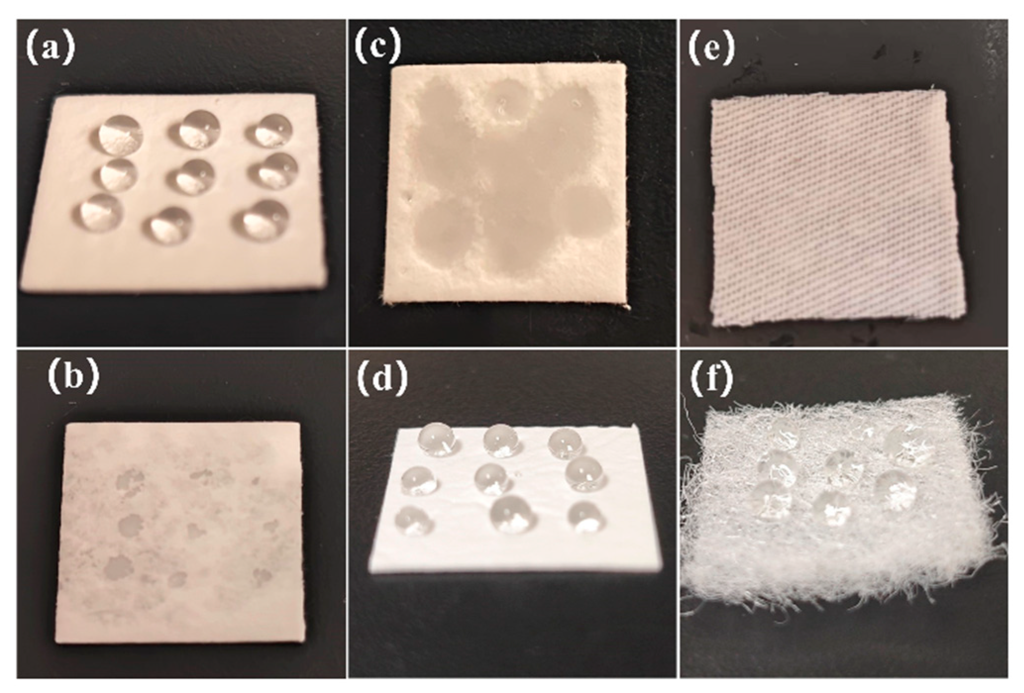
Figure 2 illustrates the wetting behavior of 0.1 mL bacterial suspension droplets on distinct substrate surfaces. As demonstrated in Figure 2, Materials A (glass fiber paper treated with reinforced resin), B (untreated glass fiber base paper), and E (cotton fabric) exhibited complete liquid absorption, forming continuous wet zones. In contrast, Materials C (glass fiber paper modified with water-repellent agent and reinforced resin) and D (polypropylene nonwoven fabric) showed no measurable liquid absorption, maintaining discrete droplet morphology throughout the observation period. Notably, Material A initially exhibited hydrophobicity upon immediate droplet deposition, with full liquid penetration occurring progressively after a 2-min incubation phase, suggesting time-dependent changes in wettability in reinforced resin-treated substrates. In summary, Figure 2 explains why Material A exhibited bacterial elution problems, while Materials C, D and E showed no such problems, due to the absence of bacterial penetration into the material’s interior thereby preventing elution issues.
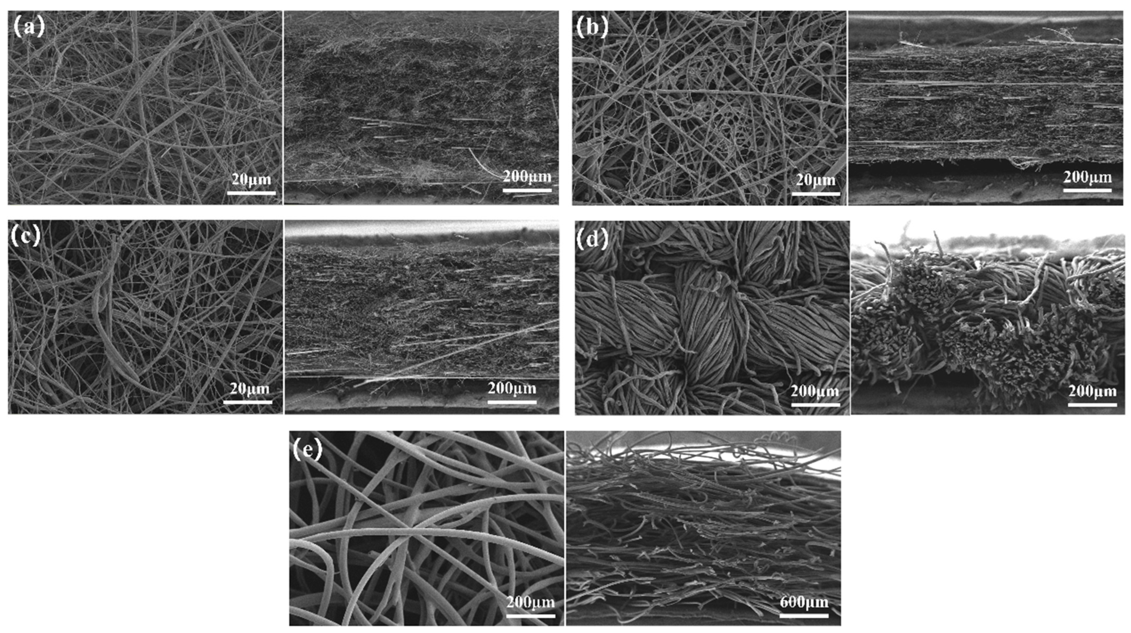
Table 3 summarizes the pore sizes of the various materials, with Figure 3 and Figure 4 presenting their SEM micrographs and pore size distributions, respectively. Figure 3 reveals that Materials A, B, and C exhibit significantly denser fiber packing than Materials D and E, featuring fiber diameters below 2 μm, compared to approximately 40 μm (Material D, polypropylene nonwoven fabric) and 30 μm (Material E, cotton fabric). Data from Table 3 and Figure 4 indicate that the high-efficiency glass fiber filters (Materials A, B, and C) possess average pore sizes < 3.5 μm, substantially smaller than 37.92 μm for Material D and 27.56 μm for Material E. Notably, since pore analyzers measure open and interconnected pores, any closed pores in the glass fiber filters may exhibit even smaller dimensions. Given Escherichia coli’s size (0.5 × 2–3 μm), these structural characteristics suggest bacterial retention in the glass fiber filters. Consequently, when applying conventional textile antibacterial testing methods to evaluate high-efficiency glass fiber filter materials, the presence of bacterial elution issues may lead to inaccurate test results. Conventional textile antimicrobial testing methods prove unsuitable for evaluating antibacterial performance in such microporous filtration systems.
An independent samples t-test was conducted on the two bacterial elution methods of hand oscillation and vortex oscillation. The results show that there is no significant difference (p > 0.05) in the number of bacteria recovered by the two methods. Therefore, in Section 3.2 and Section 3.3, we aim to investigate the factors influencing the bacterial recovery efficiency of high-efficiency glass fiber filter Material A by optimizing vortex oscillator parameters, elution time, and sample cultivation conditions, thereby improving the bacterial recovery rate of the filter material.

3.2. Impact of Vortex Oscillation Parameters and Material Dimensions on Bacterial Recovery Efficiency

Figure 5a,b illustrate variations in the bacterial recovery counts for Material A under different vortex oscillation times and intensities following the 4-hour incubation. At a fixed intensity of 2500 rpm (Figure 5a), bacterial recovery initially increased with oscillation time, peaking at 2 min (7.04 × 10⁵ CFU), followed by a gradual decline. This biphasic trend suggests potential bacterial inactivation under prolonged high-intensity mechanical agitation, consistent with findings by Wang [23], who demonstrated superior bacterial recovery efficiency using vortex oscillation compared to low-frequency oscillation or ultrasonic methods for polypropylene filters.
Figure 5b reveals a positive correlation between oscillation intensity (0–2500 rpm) and bacterial recovery at the optimized 2 min duration, achieving maximum recovery (9.30 × 10⁵ CFU) at 2500 rpm. This enhancement likely stems from intensified centrifugal forces generated at higher vortex intensities, which promote efficient bacterial dislodgement through enhanced fluid shear dynamics [27].
The influence of material dimensions was further investigated (Figure 5c). Bacterial recovery for Material A (initial size: 2 × 2 cm) exhibited a non-linear relationship with progressive subdivision; the recovery counts initially increased upon bisection and quartering (peak: 4.25 × 10⁵ CFU at quartered state), then decreased with nano-sectioning. These results underscore the critical role of standardized specimen sizing in optimizing recovery protocols [28]. Both oversized specimens and excessive fragmentation (e.g., nano-sectioning) were observed to reduce recovery efficacy.

3.3. Influence of Cultivation Humidity and Sealing Conditions on Bacterial Recovery Performance

3.3.1. Bacterial Recovery Rate in 4 h Unsealed Conditions

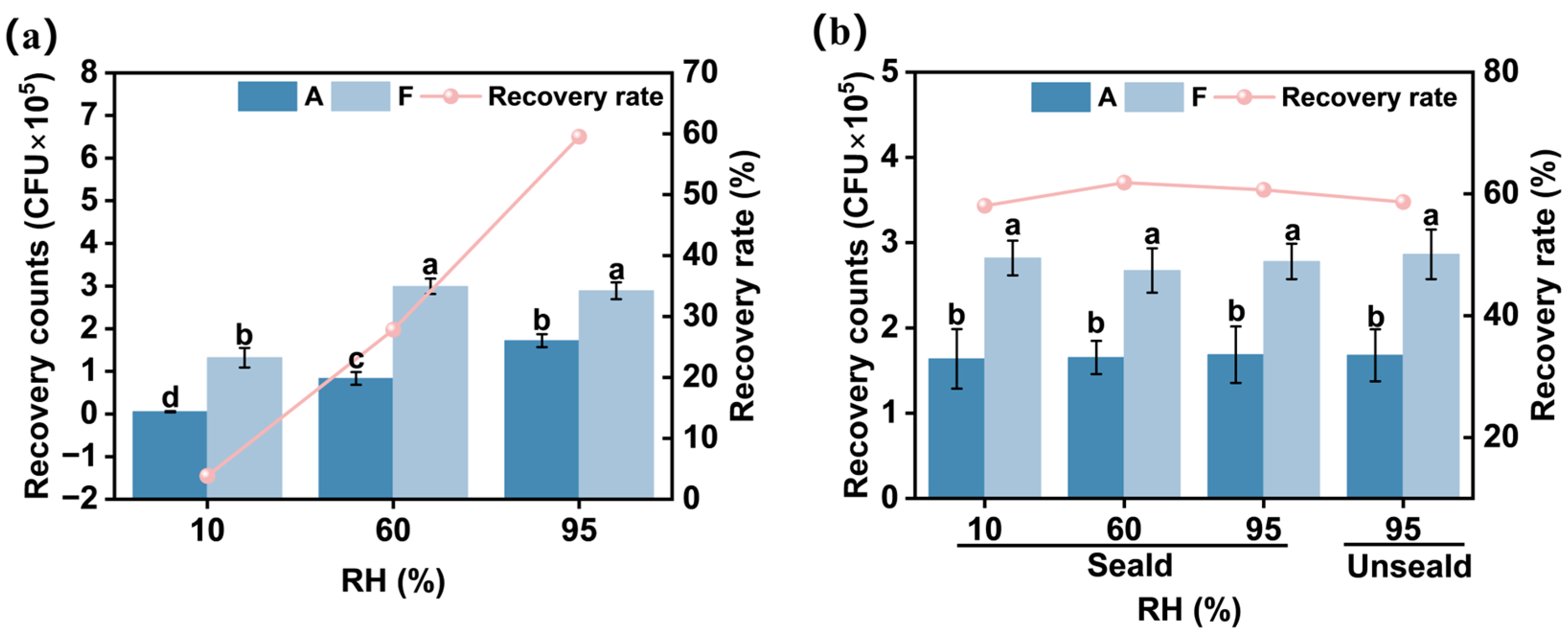
Figure 6a presents the bacterial recovery dynamics of Material A under varying humidity levels in unsealed conditions. Significant inter-humidity variations (p < 0.05) were observed in both the bacterial growth counts from Group F and the recovery counts from Material A. Minimal bacterial growth (1.32 × 10⁵ CFU) occurred at 10% humidity, while equivalent microbial loads (2.90 × 10⁵ CFU; p > 0.05) were recorded at 60% and 95% humidity. These findings collectively demonstrate that controlled humidity optimization is critical for maintaining microbial viability on filtration substrates, with arid conditions (10% humidity) substantially suppressing bacterial metabolic activity [29,30].
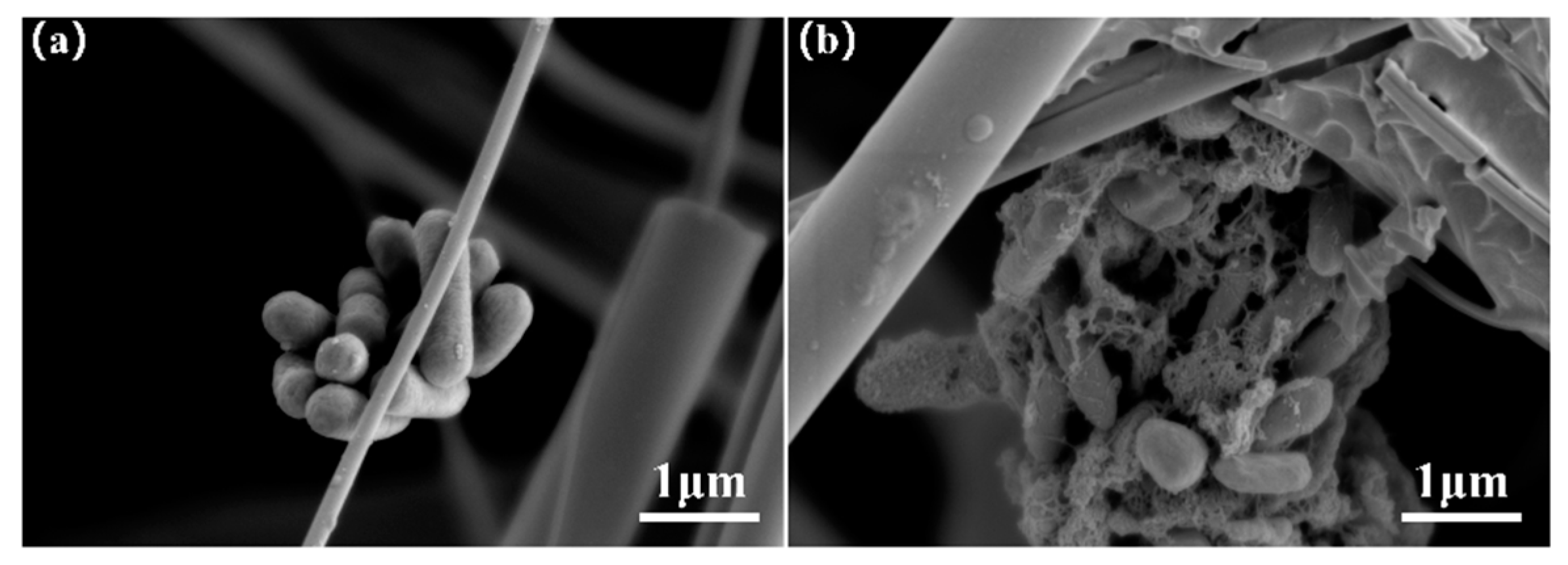
Material A exhibited humidity-dependent recovery behavior; the lowest recovery count (0.05 × 10⁵ CFU) at 10% humidity contrasted sharply with the peak recovery (1.72 × 10⁵ CFU) under 95% humidity. This reduction correlated with humidity-impaired bacterial growth at 10%. Notably, while comparable bacterial growth occurred at 60% and 95% humidity (p > 0.05), Material A displayed significantly higher recovery efficiency at 95% humidity (p < 0.05). This discrepancy suggests enhanced bacterial detachment from fibrous matrices under elevated humidity conditions, potentially mediated by humidity-modulated reductions in microbial adhesion forces to fiber surfaces [31]. The influencing effect of high humidity on bacterial elution can be explained by Figure 7. The morphological evolution of E. coli on Material A during the 0–4 h incubation demonstrates biofilm formation after 4 h cultivation. This biofilm development enhances bacterial adhesion to the material surface, consequently impeding effective bacterial elution.
Consequently, humidity elevation from 60% to 95% increased Material A’s bacterial recovery efficiency from 27.84% to 59.52% in unsealed conditions. The observed humidity–recovery correlation underscores the dual regulatory role of environmental moisture in governing both microbial growth and detachment mechanics in filtration media.

3.3.2. Comparative Bacterial Recovery in 4 h Sealed vs. Unsealed Conditions

As indicated by the previous experimental results (Figure 6a), Material A exhibited the highest bacterial recovery rate under 95% humidity in unsealed conditions. Consequently, Figure 6b compares differences in the bacterial recovery counts and rates between the sealed (varying humidity levels) and unsealed conditions (fixed 95% humidity). The results demonstrate no statistically significant differences (p > 0.05) in bacterial growth between the pure bacterial suspensions (Group F) under sealed and unsealed conditions. This suggests that the sealed environment mitigates moisture evaporation within the material matrix, maintaining optimal bacterial growth conditions.
Notably, Material A showed comparable bacterial recovery counts and recovery rates (p > 0.05) regardless of sealing status. This implies that humidity exerts no measurable influence on bacterial recovery efficiency during the 4-h sealed cultivation. Furthermore, unsealed cultivation at 95% humidity achieved equivalent bacterial recovery rates (59.81%) to sealed cultivation across all tested humidity levels. These findings align with prior conclusions confirming that controlled humidity facilitates the elution of bacteria from filter materials.

3.3.3. Bacterial Recovery Rates in 24 h Cultures with Varying Humidity and Sealing Conditions

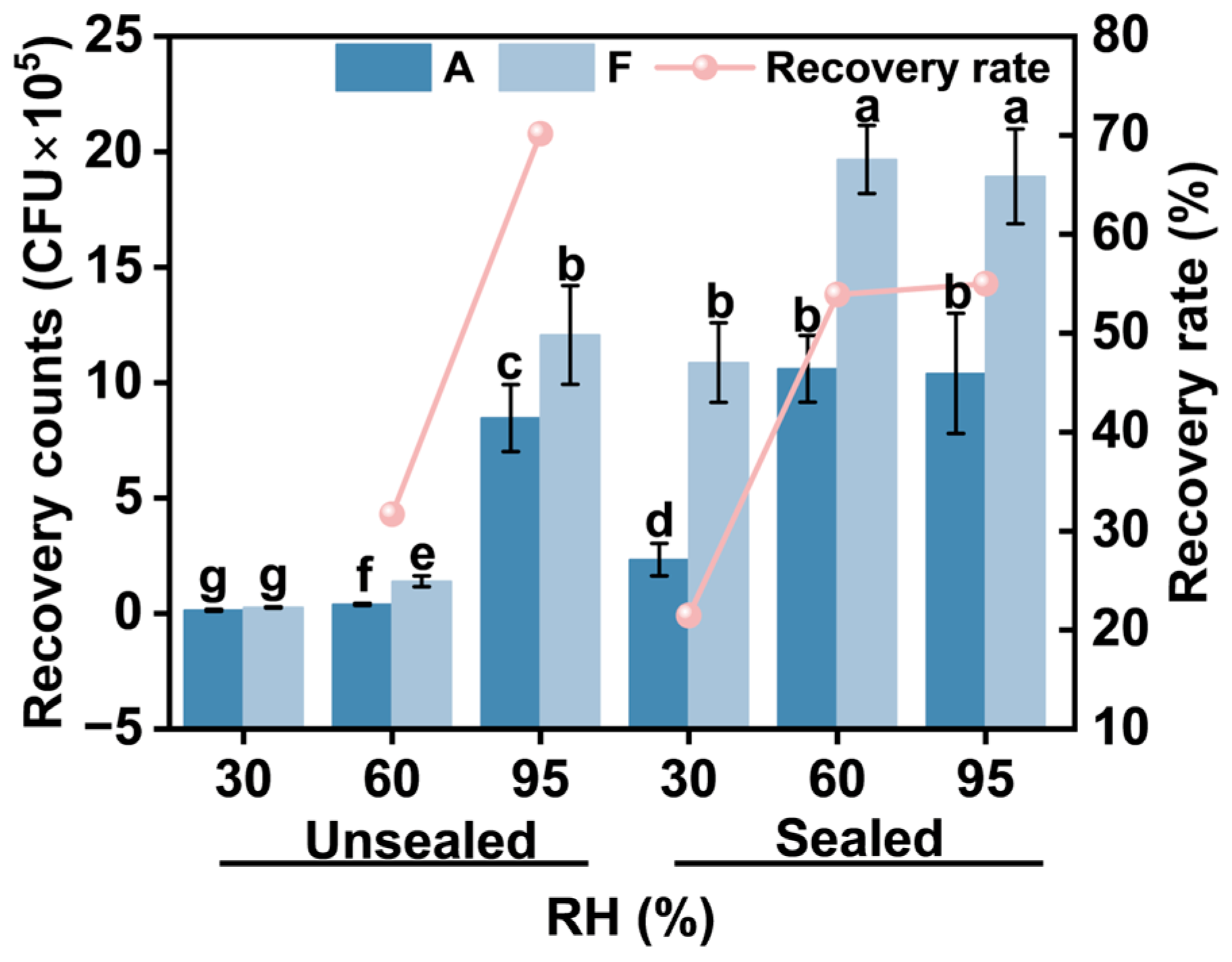
Figure 8 presents the bacterial growth for Group F and the recovery data for Material A under sealed and unsealed conditions at varying humidity levels over a 24-h cultivation period. In contrast to the 4-h results, both bacterial growth counts (Group F) and recovery counts (Material A) exhibited statistically significant humidity-dependent variations under sealed/unsealed conditions (p < 0.05).
Under unsealed conditions, the bacterial growth counts increased progressively with rising humidity, yielding 0.26 × 10⁵ CFU at 30% humidity, 1.40 × 10⁵ CFU at 60% humidity, and 12.07 × 10⁵ CFU at 95% humidity. Similarly, sealed conditions showed humidity-dependent growth enhancement. Notably, bacterial growth at 30% humidity (10.87 × 10⁵ CFU) was significantly lower than at 60% and 95% humidity (p < 0.05), while no significant differences were observed between 60% (19.67 × 10⁵ CFU) and 95% humidity (18.93 × 10⁵ CFU) (p > 0.05). These findings align with the previous conclusions regarding humidity-mediated regulation of bacterial growth.
Due to insufficient bacterial recovery counts under unsealed conditions at 30% humidity, this group was excluded from subsequent analysis (no recovery rate calculation was performed). In unsealed environments, bacterial recovery counts and recovery rates increased proportionally with humidity, reaching a maximum recovery rate of 70.17% at 95% humidity. Under sealed conditions, no statistically significant differences (p > 0.05) were detected in the bacterial recovery counts, recovery rates, or growth counts between 60% and 95% humidity. At identical humidity levels, both the bacterial growth counts and recovery counts under the sealed conditions significantly surpassed those under the unsealed conditions (p < 0.05). The optimal cultivation parameters for the 24 h test were identified as sealed conditions at ≥60% humidity, achieving a bacterial recovery rate of approximately 50%.

4. Conclusions

This study evaluated the bacterial recovery efficiency of high-efficiency glass fiber filter materials versus textile materials (e.g., cotton fabric) using the bacterial elution methods specified in GB/T 20944.2-2007. Significant differences in bacterial recovery were observed between these materials under two elution approaches (hand shaking vs. vortex shaking). SEM imaging and pore size analysis revealed that the smaller pore dimensions of high-efficiency glass fiber filters hindered bacterial elution. Upon investigating the factors influencing bacterial elution efficiency in glass fiber filter materials, we established the optimal parameters for bacterial recovery from a glass fiber filter; a 4 cm2 sample was divided into four equal sections, incubated in a sealed environment with ≥60% humidity, and then vortexed at 2500 rpm for 2 min to maximize bacterial growth and recovery counts. The principal findings are summarized as follows.
Notable disparities in bacterial growth were observed among the different filter materials (water-resistant vs. non-water-resistant) and textiles (cotton fabric). After the 4 h cultivation, bacterial recovery from the non-water-resistant filter Material A (0.72 × 10⁵ CFU) exhibited markedly lower efficiency (~10%) compared to the other materials (~7.28 × 10⁵ CFU, comparable to pure bacterial suspension), potentially compromising antimicrobial test accuracy.
Comparative analysis demonstrated similar performance between the two elution methods (hand shaking vs. 500 rpm vortex shaking) defined in GB/T 20944.2-2007. Parameter optimization revealed that bacterial recovery initially increased then decreased with prolonged shaking duration, peaking at 2 min. Increasing shaking intensity from 500 rpm to 2500 rpm enhanced recovery from 2.03 × 10⁵ CFU to 9.30 × 10⁵ CFU. Quartered specimens exhibited optimal recovery efficiency (applicable to the high-efficiency glass fiber filter materials prepared in this study).
Furthermore, we investigated the cultivation conditions that influence recovery rates. For extended cultivation (24 h), environmental sealing critically affected bacterial recovery and growth. Elevated humidity and sealed conditions promoted bacterial proliferation and elution, with recovery rates increasing from 0% to 55% as humidity rose from 30% to 95%. Under short-term cultivation (4 h), humidity variations (10–95%) minimally affected bacterial growth (~60% recovery) in sealed systems but significantly impacted unsealed specimens (5–60% recovery). We therefore recommend culturing filter materials in sealed environments with ≥ 60% humidity for optimal recovery.
This study demonstrates that high-efficiency glass fiber filters exhibit problematic bacterial retention (with bacteria penetrating into the material matrix and becoming difficult to elute) during textile-based antibacterial testing—an issue not observed in textile materials. This finding clearly indicates that the currently prevalent textile absorption method is inadequate for the accurate antimicrobial assessment of commercial filter materials, highlighting the need for the establishment of dedicated testing protocols specifically designed for filtration media. Furthermore, our investigation of factors influencing bacterial elution from glass fiber filters reveals that the development of standardized methods must include detailed specifications for the following parameters: sample type, dimensions, incubation duration, humidity control, and environmental sealing conditions.

Author Contributions

Methodology, L.R. and M.T.; validation, L.R.; investigation, Z.L. and H.W.; data curation, L.R.; writing—original draft preparation, L.R.; writing—review and editing, L.W. and M.T.; supervision, Y.L.; project administration, L.W. and D.W. All authors have read and agreed to the published version of the manuscript.

Funding

This research received no external funding.

Data Availability Statement

The original contributions presented in this study are included in the article. Further inquiries can be directed to the corresponding author(s).

Conflicts of Interest

The authors declare no conflicts of interest.

References

  1. Giordano, S.; Spagnuolo, V.; Capozzi, F. Biomonitoring of Air Pollution. Atmosphere 2021, 12, 433. [Google Scholar] [CrossRef]
  2. Yang, Y.; Zhang, H.; Nunayon, S.S.; Chan, V.; Lai, A.C. Disinfection Efficacy of Ultraviolet Germicidal Irradiation on Airborne Bacteria in Ventilation Ducts. Indoor Air 2018, 28, 806–817. [Google Scholar] [CrossRef]
  3. Wu, Y.; Yao, M. Inactivation of Bacteria and Fungus Aerosols Using Microwave Irradiation. J. Aerosol Sci. 2010, 41, 682–693. [Google Scholar] [CrossRef]
  4. Bahri, M.; Haghighat, F. Plasma-Based Indoor Air Cleaning Technologies: The State of the Art-Review. CLEAN—Soil. Air Water 2014, 42, 1667–1680. [Google Scholar] [CrossRef]
  5. Lee, S.H. Development of Photocatalyst Plasma Air Cleaning Filter Used in Air Conditioner. J. Adv. Oxid. Technol. 2003, 6, 66–69. [Google Scholar] [CrossRef]
  6. Li, Y.; Miao, Q.; Wang, X. Antibacterial Capability of Air Filter Fiber Materials Treated with Triclosan against Indoor Environmental Microbes. Atmosphere 2022, 13, 1104. [Google Scholar] [CrossRef]
  7. Foarde, K.K. Methodology to Perform Clean Air Delivery Rate Type Determinations with Microbiological Aerosols. Aerosol Sci. Technol. 1999, 30, 235–245. [Google Scholar] [CrossRef]
  8. Luo, Y.; Zhai, F.; Zhang, Y.; Chen, Z.; Ding, M.; Qin, D.; Yang, J.; Feng, G.; Li, L. A Superfine Glass Fibre Air Filter with Rapid Response to Photocatalytic Antibacterial Properties under Visible Light by Loading rGO/ZnO. R. Soc. Open Sci. 2021, 8, 202285. [Google Scholar] [CrossRef]
  9. AATCC 100-2019; Antibacterial Finishes on Textile Materials: Assessment of. American Association of Textile Chemists and Colorists (AATCC) AATCC: Research Triangle Park, NC, USA, 2019.
  10. GB/T 20944.2-2007; Textiles—Evaluation for Antibacterial Activity—Part 2: Absorption Method. Standardization Administration of China Standards Press of China: Beijing, China, 2007.
  11. ISO 20743:2021; Textiles—Determination of Antibacterial Activity of Textile Products. International Organization for Standardization: Geneva, Switzerland, 2021.
  12. Simmons, R.B.; Crow, S.A. Fungal Colonization of Air Filters for Use in Heating, Ventilating, and Air Conditioning (HVAC) Systems. J. Ind. Microbiol. 1995, 14, 41–45. [Google Scholar] [CrossRef]
  13. Miaśkiewicz-Pęska, E.B.; Słomczyński, T. Survival of Bacteria in Respiratory Protective Filters. Pol. J. Microbiol. 2017, 65, 475–477. [Google Scholar] [CrossRef]
  14. Majchrzycka, K.; Gutarowska, B.; Brochocka, A. Aspects of Tests and Assessment of Filtering Materials Used for Respiratory Protection Against Bioaerosols. Part II: Sweat in the Environment, Microorganisms in the Form of a Bioaerosol. Int. J. Occup. Saf. Ergon. 2010, 16, 275–280. [Google Scholar] [CrossRef]
  15. Nakpan, W.; Yermakov, M.; Indugula, R.; Reponen, T.; Grinshpun, S.A. Inactivation of Bacterial and Fungal Spores by UV Irradiation and Gaseous Iodine Treatment Applied to Air Handling Filters. Sci. Total Environ. 2019, 671, 59–65. [Google Scholar] [CrossRef]
  16. Pinho, E.; Magalhães, L.; Henriques, M.; Oliveira, R. Antimicrobial Activity Assessment of Textiles: Standard Methods Comparison. Ann. Microbiol. 2011, 61, 493–498. [Google Scholar] [CrossRef]
  17. Cunliffe, A.J.; Askew, P.D.; Stephan, I.; Iredale, G.; Cosemans, P.; Simmons, L.M.; Verran, J.; Redfern, J. How Do We Determine the Efficacy of an Antibacterial Surface? A Review of Standardised Antibacterial Material Testing Methods. Antibiotics 2021, 10, 1069. [Google Scholar] [CrossRef]
  18. Haase, H.; Jordan, L.; Keitel, L.; Keil, C.; Mahltig, B. Comparison of Methods for Determining the Effectiveness of Antibacterial Functionalized Textiles. PLoS ONE 2017, 12, e0188304. [Google Scholar] [CrossRef]
  19. GB 21551.2-2010; Antibacterial and Cleaning Function for Household and Similar Electrical Appliances—Special Requirements for Antibacterial Materials. Standardization Administration of China (SAC): Beijing, China, 2010.
  20. JIS L 1902:2015; Testing for Antibacterial Activity and Efficacy on Textile Products. Japanese Industrial Standards Committee: Tokyo, Japan, 2015.
  21. EN 143:2000; Respiratory Protective Devices—Particle Filters—Requirements, Testing, Marking. European Committee for Standardization (CEN): Brussels, Belgium, 2000.
  22. Fisher, E.M.; Shaffer, R.E. Considerations for Recommending Extended Use and Limited Reuse of Filtering Facepiece Respirators in Health Care Settings. J. Occup. Environ. Hyg. 2014, 11, D115–D128. [Google Scholar] [CrossRef]
  23. Wang, Z. Survival of Bacteria on Respirator Filters. Aerosol Sci. Technol. 1999, 30, 300–308. [Google Scholar] [CrossRef]
  24. Møretrø, T.; Almli, V.L.; Åsli, A.W.; Kummen, C.; Galler, M.; Langsrud, S. Kitchen Cloths: Consumer Practices, Drying Properties and Bacterial Growth and Survival. Food Control 2022, 142, 109195. [Google Scholar] [CrossRef]
  25. Teufel, L.; Pipal, A.; Schuster, K.C.; Staudinger, T.; Redl, B. Material-dependent Growth of Human Skin Bacteria on Textiles Investigated Using Challenge Tests and DNA Genotyping. J. Appl. Microbiol. 2010, 108, 450–461. [Google Scholar] [CrossRef]
  26. Takashima, M.; Shirai, F.; Sageshima, M.; Ikeda, N.; Okamoto, Y.; Dohi, Y. Distinctive Bacteria-Binding Property of Cloth Materials. Am. J. Infect. Control 2004, 32, 27–30. [Google Scholar] [CrossRef]
  27. Burton, N.C.; Adhikari, A.; Grinshpun, S.A.; Hornung, R.; Reponen, T. The Effect of Filter Material on Bioaerosol Collection of Bacillus Subtilis Spores Used as a Bacillus Anthracis Simulant. J. Environ. Monit. 2005, 7, 475–480. [Google Scholar] [CrossRef] [PubMed]
  28. Behnke, R.; Stahl, F.; Duske, K.; Warkentin, M.; Schwartz, M.; Hinz, B.; Walther, U. Influence of Test Specimen Geometry and Water Soaking on the In Vitro Cytotoxicity of Orthocryl®, Orthocryl® LC, Loctite® EA 9483 and Polypropylene. Molecules 2022, 27, 3949. [Google Scholar] [CrossRef]
  29. Maus, R.; Goppelsröder, A.; Umhauer, H. Survival of Bacterial and Mold Spores in Air Filter Media. Atmos. Environ. 2001, 35, 105–113. [Google Scholar] [CrossRef]
  30. Majchrzycka, K.; Okrasa, M.; Skóra, J.; Gutarowska, B. Evaluation of the Survivability of Microorganisms Deposited on Filtering Respiratory Protective Devices under Varying Conditions of Humidity. Int. J. Environ. Res. Public Health 2016, 13, 98. [Google Scholar] [CrossRef]
  31. Dannemiller, K.C.; Weschler, C.J.; Peccia, J. Fungal and Bacterial Growth in Floor Dust at Elevated Relative Humidity Levels. Indoor Air 2017, 27, 354–363. [Google Scholar] [CrossRef]
Figure 1. Bacterial recovery of different materials after culturing for “0 h” and 4 h: (a) number of bacteria recovered/grown at “0 h”; (b) number of bacteria recovered/grown at 4 h (Groups labeled with the same letter are not significantly different p > 0.05; those with different letters show significant differences p < 0.05).
Separations 12 00110 g001
Figure 2. State of 0.1 mL of bacterial solution added dropwise onto different materials: (a) Material A—“0” h, (b) Material A—4 h; (c) Material B—4 h; (d) Material C—4 h; (e) Material E—4 h; (f) Material D—4 h.
Separations 12 00110 g002
Figure 3. Planar and cross-sectional SEM images of different materials: (a) Material B; (b) Material A; (c) Material C; (d) Material E; and (e) Material D.
Separations 12 00110 g003
Figure 4. Pore size distributions of different materials: (a) Materials A, B, and C; (b) Materials D and E.
Separations 12 00110 g004
Figure 5. Influence of oscillation time, oscillation intensity, and sample size on bacterial recovery: (a) Oscillation time; (b) Oscillation intensity; (c) Sample cutting size.
Separations 12 00110 g005
Figure 6. Influence of different humidity conditions on the number of bacteria recovered from Material A during a 4 h cultivation period: (a) Without sealing; (b) With sealing and without sealing at 95% humidity. (Groups labeled with the same letter are not significantly different p > 0.05; those with different letters show significant differences p < 0.05).
Separations 12 00110 g006
Figure 7. Morphology of E. coli on Material A at different incubation times: (a) 0 h; (b) 4 h.
Separations 12 00110 g007
Figure 8. Influence of varying humidity and sealed/unsealed conditions on the number of bacteria recovered from Material A during a 24 h cultivation period. (Groups labeled with the same letter are not significantly different p > 0.05; those with different letters show significant differences p < 0.05).
Separations 12 00110 g008
Table 1. Differences in testing methods for antimicrobial properties of textiles.
Name of StandardISO 20743:2021 [11]AATCC 100-2019 [9]JISL 1902-2015 [20]GB/T 20944.2-2007 [10]GB
21551.2-2010 [19]
Material TypeTextilesTextilesTextilesTextilesTextiles; Microporous material
Additional Control Sample100% Cotton fabricNo mention100% Cotton fabric100% Cotton fabric100% Polypropylene nonwoven
Test SizeSuitable size(4.8 ± 0.1) cm cycle;
(3.8 × 3.8) cm square
Suitable sizeSuitable size5 cm Cycle
Test Time
(h)
18–242418–2418–2418–24
Incubation Humidity
(%)
No mentionNo mentionNo mentionNo mentionNo mention
Sealing ConditionsSealSealSealSealSeal
Incubation
Temperature
(°C)
37 ± 237 ± 237 ± 237 ± 237 ± 1
Elution MethodHand: 30 cm for 30 s;
Vortex: 5 sin 5 cycles
VortexHand: 30 cm arc for 30 turns;
Vortex: 5 sin 5 cycles
Hand: 30 cm for 30 s;
Vortex: 5 times (5 s each)
200 r/min for 1 min
Table 2. Basic information on different materials.
Serial NumberType
(Name)
Grammage
(g/m2)
Thickness (mm)Filtration Efficiency
(%)
Filtration
Resistance (Pa)
Aglass fiber paper treated with reinforcing resin73.30.39899.93223.5
Bglass fiber base paper69.70.28199.96298.2
Cglass fiber paper treated with water-resistant agent and reinforcing resin79.80.39899.95351.2
Dpolypropylene nonwoven fabric96.20.5697.401.1
Ecotton fabric221.01.0915.8556.9
Table 3. Pore size characterization of various materials.
Material NameMean Pore Diameter
(μm)
Maximum Pore Diameter
(μm)
A2.467.01
B3.417.88
C2.435.40
D37.92139.03
E27.5678.20
Disclaimer/Publisher’s Note: The statements, opinions and data contained in all publications are solely those of the individual author(s) and contributor(s) and not of MDPI and/or the editor(s). MDPI and/or the editor(s) disclaim responsibility for any injury to people or property resulting from any ideas, methods, instructions or products referred to in the content.