Abstract
(1) Background: Well-known monosodium glutamate (E-621, MSG), originally used as a food flavor enhancer, was approved approximately in all countries, but the toxicity versus the safety of (MSG) are still unclear due to variable scientific toxicological reports. Moreover, it was reported to trigger elevated frequencies of nausea and headaches in humans and provide deleterious effects on laboratory animals. The objectives of the present study were to (i) estimate the possible toxic effects of the food additive MSG (ii) and the ameliorating protective effects of the dietary supplement spirulina (Spirulina platensis) on the biochemical parameters of blood and the damage produced in organs of Swiss mice after applying a supplementary daily dose of MSG for 4 weeks. (2) Methods: The present study was conducted on 20 mature Swiss mice, which were randomly organized into four groups of five Swiss mice. The treatments were (I) the control group, in which Swiss mice were fed only animal feed and drinking water; group II MSG1, which received 1 mL of MSG; group III MSG0.5, which was treated with 0.5 mL of MSG; and (IV) the group MSGS, which was treated with 1 mL of monosodium glutamate and 1 mL of spirulina (aiming to reduce the MSG toxicity). (3) Results: At the end of the experiment, Swiss mice treated with MSG demonstrated a passiveness regarding behavioral aspects. As we hypothesized, the parameters of the spirulina group reached similar values to the control group, and no histopathological observations have been found. Altogether, our findings evidenced that monosodium glutamate leads to histopathological changes in Swiss mice kidneys and caused important modifications for all biochemical parameters of the blood serum. Noticeably, the potential protective effect of Spirulina platensis was proved and was described by using the FTIR spectroscopy technique. (4) Conclusions: A diet rich in antioxidants and other plant-derived bioactive compounds may provide healthy nutrition, alleviating the potential side effects of some food additives.
1. Introduction
The current trend among consumers is focused on high-quality food with an insignificant addition of synthetic additives, and the food industry aims to bring products to a different level by focusing on natural preservation and stabilization approached [1,2,3]. Flavor enhancers are playing a crucial key role in the gastronomic/culinary field of different countries. Different food additives categories (e.g., flavor enhancers) ought to be submitted to numerous investigations such as biochemical, mutagenic, and acute toxicity experimental studies in order to guarantee maximal consumer safety [4].
Various kinds of food additives (e.g., tartrazine, nitrites, monosodium glutamate) have been employed to improve the taste, tint, constancy, quality, and price of foods. For example, nitrites are a very well-described problem in food, because as a preservative, it causes many negative effects on human health [5]. Well-known monosodium glutamate (E-621, MSG) is a food additive; initially, it was used as a food flavor enhancer attributing a new kind of taste to some foods and was named “umami” [6]. It was discovered by a Japanese researcher more than a century ago and was extracted from dried kelp, which is a brown algae seaweed belonging to the Laminariales order [7]. Glutamate is one of the most abundant amino acids that exist in nature. Moreover, it is a fundamental element for the majority of tissues, peptides, proteins, and plays a crucial role in human metabolism, acting as a neurotransmitter [8]. Generally, glutamate arrives from different exogenous sources and mainly from the dietary flavoring agent MSG. Then, MSG is metabolized, providing free glutamate that does not differ biochemically from the endogenous ligand [9]. Hence, MSG has been approved approximately in all countries, but the toxicity versus the safety of MSG seems to be controversial. Some studies provided safety reports about that food enhancer [10,11]. However, other studies demonstrate the deleterious impact on laboratory animals [6,8,9,11,12,13,14,15,16,17,18,19]. Moreover, in humans, the ingestion of repeated high doses of MSG can trigger an elevated frequency of nausea and headaches [20].
Another interesting seaweed product is spirulina. The common term spirulina is derived from the final products achieved from the pure cultures of prokaryotic cyanobacteria, namely Arthrospira spp. [21]. In South America and Africa, it was grown as a part of the food diet constituent since ancient times [22]. This blue-green cyanobacterium prefers natural habitats such as alkaline lakes with an increased pH. The name spirulina was formerly given due to the spiral shape of its filaments and was originally considered eukaryotic algae. Spirulina is a rich source of protein with approximatively 60–70% protein content and is considered a functional product without any proven toxic effects compared to other cyanobacteria. The bioactive compounds from spirulina (Spirulina platensis) are well studied, and a diet supplemented with spirulina can provide benefits such as stimulating the immune system; reducing hyperlipidemia, hypercholesterolemia, and obesity; exhibiting anticancer, neuroprotective, and hepatoprotective properties; counteracting the effects of radiation, heavy metals, and drug toxicity levels; and preventing allergies, inflammatory diseases, viral infections, and other metabolic diseases [23,24,25,26,27,28]. Spirulina is formed by several active bio-compounds, especially phycocyanin and β-carotene, which have a powerful antioxidant and anti-inflammatory activity [29]. The antioxidant and anti-inflammatory properties of phycocyanin were first reported in 1998, and then, it was confirmed in numerous research studies [30]. Phycocyanin can neutralize free radicals such as alkoxyl, peroxyl, and hydroxyl. It also lowers nitrite production and inhibits microsomal peroxidation of liver lipids. Based on these research studies and the fact that spirulina is a rich source of antioxidants and might produce an important protective effect in Swiss mice, in this study, a fine powder was introduced in order to investigate the ameliorative effects of spirulina versus the impact produced by MSG [28,31,32,33,34,35,36,37]. Furthermore, the protective effects of spirulina administrated to the rodents have been reported by various authors [28,36,37,38,39,40,41,42,43].
The objectives of the present study were to estimate the possible toxic effects of the food additive MSG and the ameliorating protective effects of dietary supplement spirulina (Spirulina platensis) powder on biochemical parameters of blood and the damage produced in organs of Swiss mice after applying a supplementary daily dose of MSG during four weeks. Additionally, in the treated mice, the spectra of MSG and spirulina powders were determined, while the marker bands of these groups were identified using FTIR spectroscopy.
2. Materials and Methods
2.1. Animals
In the present study, we used Swiss mice. The experiment lasted 4 weeks. During this period, Swiss mice received monosodium glutamate and spirulina at different concentrations. Two weeks before the experiment started, Swiss mice were kept under the following conditions: 12/12 light–dark cycle, humidity 50 ± 10%, temperature 22 ± 1 °C, in the Establishment for Laboratory Animals of the University of Agricultural Science and Veterinary Medicine, Cluj, Romania. In cages, Swiss mice have had the possibility to freely access standard food and water. The experiment complied with the procedures established by the Directive 2010/63/EU and national legislation (Law No. 43/2014) [44,45].
2.2. Experimental Design
Rodents were organized into four groups. Each experimental group consisted of 5 Swiss mice, and the groups were kept in individual cages. The experimental groups were: (I)—the control group (C) received only animal feed and drinking water for the entire term of the experiment; (II) group MSG1, which received 1 mL of MSG; (III) group MSG0.5 that was treated with 0.5 mL of MSG; and (IV) group MSGS—that was treated with 1 mL of monosodium glutamate and 1 mL of spirulina. Treatment of Swiss mice with MSG and spirulina were administered by oral gavage.
2.3. Chemicals
In the present study, food-grade monosodium glutamate (Figure 1), with molecular formula C5H8NaNO4 H2O was purchased from Ajinomoto® and spirulina (Spirulina plantensis) was purchased from Herbavit SA (Oradea, Romania). Each mouse was tagged on the tail in order to differentiate the individuals.
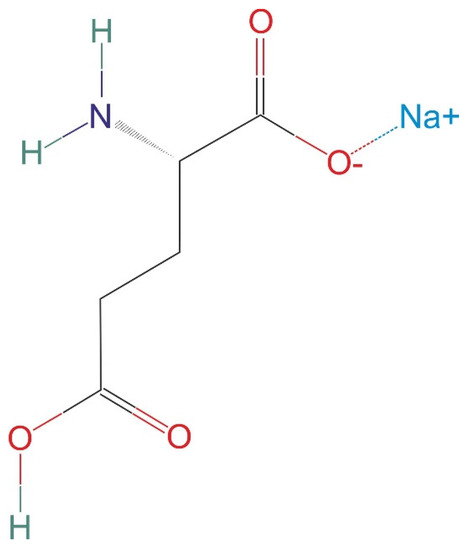
Figure 1.
Chemical structure of monosodium glutamate.
2.4. Organ and Blood Sample Collection
Swiss mice were subjected to deep narcosis by using the isoflurane substance. After that, the blood samples were taken from the orbital sinus using vacutainers with EDTA to determine the biochemical and hematology parameters. At the ending of the experiment, the Swiss mice were euthanized by prolonged narcosis until no heart or respiratory activity was recorded. The phenomenon was irreversible since cervical dislocation was realized. Liver and kidneys were collected and were stored in a freezer at −20 °C in order to realize histopathological examinations.
2.5. Biochemical Analysis of Blood
The biochemical analysis of blood was realized using the semi-automated screen-type biochemical analyzer STAT-FAX 1904 Plus, Global Medical Instrumentation, 6511 Bunker Lake Blvd., Ramsey, MN 55303 (USA) [46,47]. Creatinine (CRE), glucose (GLU), alanine aminotransferase (ALT), cholesterol (COL), triglyceride (TG), and aspartate aminotransferase (AST) were measured using special kits by following the producer’s instructions [46].
2.6. Histopathological Analysis
The histological analysis of the kidneys and liver were realized in order to investigate possible tissue injuries. The liver and kidney samples were maintained in a phosphate-buffered formalin substance for 24 h and then were embedded in paraffin wax [48]. Afterward, liver and kidney samples were cut into sections of 3 µm and stained with hematoxylin and eosin (H&E). Each section was individually analyzed with two veterinary pathologists (MT and CT) by using a light Olympus BX-41 microscope [49]. The photomicrographs were realized using an Olympus SP 350 digital camera and Stream Basic imaging software (Olympus Corporation, Tokyo, Japan) [50]. When the researchers had contrasting opinions, an additional diagnosis was made through simultaneous evaluation in a five-head microscope (Zeiss Axio Scope A1) [51] All samples were subjected to histological changes that included degenerative processes, necrosis, inflammation, fibrosis, and cell proliferation.
2.7. FTIR Measurements
The FTIR measurements were realized in the absorbance with a spectrophotometer FT-IR-4100 Jasco, using the KBr pellet technique previously reported by Andronie et al. [42]. The spectra were obtained in the wavenumber range 265 cm−1. The spectral resolution was set at 4 cm−1, and all spectra were acquired over 256 scans. The sample was acquired from 0.003 g of MSG and 0.003 g of spirulina powder (commercially available), which were used without further purification. Origin 6.0 software was used to analyze the spectral data.
2.8. Statistical Analysis
Data were analyzed using JMP 12 statistical software (SAS Institute, 2014) [52]. The normal distribution of the measured variables/parameters (AST, ALT, glucose, cholesterol, triglyceride, creatinine) was checked by Shapiro–Wilk tests. The average for each parameter is the arithmetic mean of five replications, and it was analyzed through an analysis of variance ANOVA model (JMP version 12, SAS Institute, Cary, NC, USA) that included terms for hematological parameters. Treatment means among groups (control, MSG 0.5, MSG1, and MSG1 + S) were determined using Tukey’s HSD test at the 0.05, 0.01, and 0.001 probability levels.
3. Results and Discussions
3.1. MSG’s Influence on Blood Biochemical Parameters in Swiss Mice
Table 1 and Figure 1 present the average values of blood serum parameters such as ALT, AST, COL, TG, GLU, and COL. All biomarkers have shown statistically significant differences within the experimental groups. Biochemical blood indices in Swiss mice from the experiment showed significant changes depending on the concentration of administered MSG (Table 1 and Figure 1). It can be observed that the administration of spirulina in combination with MSG has determined the decrease of biochemical parameters, regulating them closer to parameters of the control group. For the MSG1 group, AST level shows the statistically highest mean values of 357.20 ± 17.02 U/L, followed by a decrease in MSG0.5 group with an average of 227.4 ± 6.27 U/L. The group that received MSG in combination with spirulina presented AST average values of 152.20 ± 2.6 U/L, showing lower values compared to the control group (165.20 ± 2.84, U/L), respectively.

Table 1.
Average values for biochemical parameters: COL, TG, and CRE. The standard deviation of the mean (n = 5) is included.
The average means of AST values are consistent with those published by Seiva et al., [53] which tests the MSG toxicity combined with quercetin supplementation and presented average values of 233.36 ± 16.90, U/L [54]. According to the authors, significant changes in most of the biochemical blood parameters after treatment were observed. Moreover, the protective effects of quercetin supplementation ameliorated biochemical indices of the rodents treated with MSG.
The significant values of ALT were detected in the MSG1 group (265.80 ± 8.88), which was followed by the MSG 0.5 group (147 ± 3.96), U/L. For the control, the MSGS group showed ALT levels similar to those reported by other studies conducted for toxicity investigation of MSG. Interestingly, that group of Swiss mice treated with MSG and spirulina had significantly lower ALT values compared to the MSG1 and MSG0.5 with the average values of (80.60 ± 2.5), similar to the control group (91.4 ± 3.27), U/L, respectively. Generally, the increased concentrations of enzymes such as AST and ALT detected in the bloodstream are directly correlated with the injured tissues of the heart and liver [55]. Data obtained regarding biomarkers such as ALT and AST are in concordance with the results of Onaolapo et al. [13].
GLU levels behave similarly to all analyzed biochemical parameters, recording the highest numbers for the MSG1 group (230.20 ± 8.40), showing significantly higher average values compared to the other three groups. The lowest values have been recorded in the group of MSGS (spirulina) presenting a mean value of (114 ± 2.4) compared to the control group (124.40 ± 2.25), mg/dL. A reduction of GLU after the supplementation of rats with spirulina was observed in a similar study by Muthuraman et al. [56]. Similar observations were reported by other authors [13,57,58]. Moreover, GLU shows significantly decreased values for the experimental group of Swiss mice (MSGS), which received 1 mL of MSG and 1 mL of spirulina, compared to the group (MSG1) that received 1 mL of MSG, where the observed values are increased two-fold. Additionally, Grassiolli et al. revealed glucose disorders in newborn animals [59]. The hyperglycemia in laboratory animals is determined by the amount of glutamate that produces changes in adipocytes [60]. A study carried out by Seiva et al. shows that glucose concentrations have increased due to MSG treatment [53]. Therefore, MSG administration has increased food consumption and produced changes in glucose levels. Although the mechanisms that trigger metabolic changes are still unknown, it can be considered that the metabolic disorder is associated with the deterioration of pancreatic cells [61]. Hypersecretion of insulin induces changes in the nervous system and pancreas; these issues have been reported in laboratory animals [57]. Seiva et al. note that quercetin can ameliorate the nutrition parameters and metabolic functions produced by MSG treatment [53].
Furthermore, quercetin normalizes glucose levels and reduces the toxic effects of MSG on liver and kidney functions. These effects could be due to the strong antioxidant properties of quercetin. Laboratory Swiss mice fed diets supplemented with vitamins after receiving MSG showed a reduction of the negative effects of MSG and improvement in the regulation of metabolic functions [60,62].
COL presented concentrations in the group MSG1 with the values of 108.20 ± 3.43 mg/dL compared to the other three groups. Spirulina in combination with MSG revealed a decrease in cholesterol levels of blood (67.40 ± 3.39 mg/dL), and a diet supplemented with MSG0.5 (86.8 ± 6.47 mg/dL) resulted in increased COL levels compared to the initial control group (78.40 ± 1.23 mg/dL). The data are consistent with those reported by other researchers [13,49]. COL and TG are biochemical parameters showing the highest average values in the group treated with MSG1 and the lowest means in the group that received spirulina supplement. Spirulina was administered in order to reduce the potentially toxic effects of MSG. According to the Anwar and Mohamed study, flaxseed and canola oil may prevent the toxic effects of MSG on biochemical and physiological biomarkers of rodents [63].
The lowest TG levels of the Swiss mice were observed for the group MSGS with the values of 72.20 ± 2.84 mg/dL and the highest was recorded for the group MSG1 (106 ± 2.44, mg/dL). No statistically differences between the control and MSG0.5 group was observed. For the MSG1 Swiss mice group, CRE showed statistically elevated counts in the MSG1 group (0.12 ± 0.01 mg/dL), and similarities among the values of this parameter were recorded in the other three groups from the present study. From the obtained data, it can be concluded that spirulina, in combination with MSG, modifies the biochemical parameters providing, in the end, similar average values to the control group. During the experiment, behavioral changes were observed between experimental groups as follows: Swiss mice that belonged to the group supplemented with spirulina were much more active compared to the control group and the groups that consumed MSG in the two different concentrations. Interestingly that in the MSG-treated groups, Swiss mice were very apathetic and have experienced symptoms of drowsiness.
3.2. Histopathological Assay
In the control group, in the liver (Figure 2) and kidney (Figure 2), no important structural changes were found. In the tissues of individuals who received 0.5 mL, normal kidney and liver histological features were also noticed. In the tissues of Swiss mice that received 1 mL of sodium glutamate, minimal changes were identified only in the renal parenchyma, including interstitial congestion, hydropic degeneration, and necrosis of the proximal renal proximal tubules (Figure 2). In this group, the liver was normal (Figure 2). Tissue analyses from the group that received 1 mL of sodium glutamate and 1 mL of spirulina showed normal histological features (Figure 2).
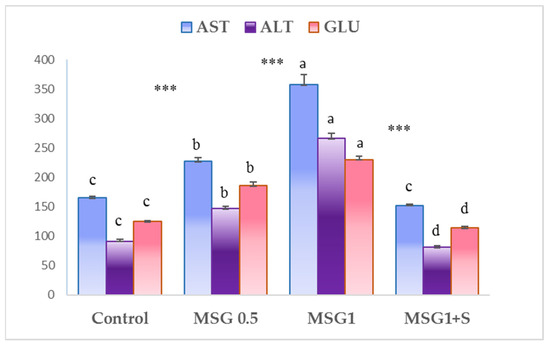
Figure 2.
Average values of blood biochemical parameters such as AST, ALT, and GLU for each experimental group. Mean values ± standard deviation (n = 5). Statistically significant differences among experimental groups for each biochemical parameter obtained with the Tukey HSD test are indicated by different lower-case letters. ***: significant at the 0.001 probability level.
According to Takasaki et al., no pathological changes were noticed on hypothalamic nuclei of rats and their fetuses nor in Swiss mice weaned after an MSG diet [64]. This may be attributed to the protection of the placenta because it is impermeable to glutamate [7]. A large dose of MSG produced neuronal necrosis in hypothalamic nuclei in newborn rats [65]. Other studies reported focal lesions in the hypothalamus areas of rodents [7]. The MSG has extensive negative effects, and these are not only limited to the hypothalamic area. MSG (4 mg/g, subcutaneously on postnatal days 1, 3, 5, and 7) led to changes in the prefrontal cerebral cortex, including fewer neurons, short dendritic processes, and less branched and loss of cortical cell counts compared to the control group [66]. Studies in neonates treated with MSG have shown neuronal cell death by reducing photoreceptor and glial cells [67,68,69,70]. Moreover, infant Swiss mice treated with MSG revealed selective loss of neurons [70]. In another study, treatment of the rats with spirulina resulted in the regeneration of pancreatic islets counteracting induced diabetes [56].
3.3. Spectroscopic Investigations
Figure 3 presents the FTIR spectrum of MSG (a) and spirulina powder (b) analyzed bands, which were compared to the similar molecule bands obtained by other authors [65,66,67]. Our result showed that the basic amino group of MSG could have joined to one of the two-carboxyl groups or formed an internal salt with the amine as in the case of amino acids.
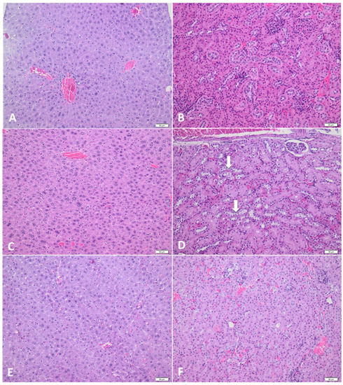
Figure 3.
Histological findings of the liver and kidney from experimental Swiss mice. (A,B) Normal features of liver and kidney in the control group; (C) Normal hepatic parenchyma found in group III—1 mL of sodium glutamate; (D) Mild to moderate and multifocal vacuolar degeneration of the proximal convoluted tubular epithelium (arrows) in group III—1 mL of sodium glutamate; (E,F) No histological changes of the liver and kidney are present in the individuals of group IV—1 mL of sodium glutamate + 1 mL of spirulina; hematoxylin–eosin (H&E) stain.
In the present study, the two weak peaks were registered (Figure 3). In detail, the first was found at 1687 cm−1, and it is due to the stretching mode of the C=O bond of the carboxyl. Meanwhile, the second weak peak was registered at values of 1571 cm−1, and it is attributed to the COO−Na+ stretching mode. In the first peak, the bending of the symmetrical mode of the NH3 group appeared at 1515 cm−1, while for the second peak, the symmetrical mode of the COO− stretching the medium-strong band represented it at 1399 cm−1. The nearby medium band at 1095 cm−1 corresponded to the CH2 rocking mode and C-C stretching mode that are remarked at 1001 cm−1 and 943 cm−1, which suggested the completely ionized form of MSG and also band positions [65].
The spirulina FTIR technique (Figure 3) was utilized to evaluate the type of organic and inorganic complexes in plants. The frequency range between 1300 and 1250 cm−1 demonstrated the presence of C-O asymmetrical C-O-C stretching with esters, while the peak range 1120–1030 cm−1 appeared in symmetric C-H stretching, indicating the presence of antioxidant enzymes. The infrared spectrum frequency ranged from 2925 to 2875 cm−1, and the peaks represented aliphatic C-H stretching vibrations. The peaks with values of 1650–1580 cm−1 occurred in the N-H bending vibration and presented the carbonyl β unsaturated ketone amide. The peaks in the frequency range of 1435–1405 cm−1 were presented in the CH2 bending vibration. The particular frequency that ranged from 1350 to 1260 cm−1 of C-O stretching and O-H bending vibration indicated the presence of alcohol.
Based on the systematic analysis, the spirulina contents included protein, lipid, carbohydrates, aliphatic (C-H), carbonyl (esters and acids), carbonyl β unsaturated ketone and amide (C=N), esters, symmetric C-H stretching vibration, halogen compounds (C-Cl), and lode compound (C-I). The FTIR spectrum demonstrated characteristic features. The presence of phytochemicals in this study could be responsible for the medicinal properties of the microalgae spirulina.
Figure 4 presented the comparison between FTIR spectra obtained from the pure MSG and MSG molecules combined with spirulina (using the same ratios) in order to highlight the positive effect that spirulina consumption might have on the body.
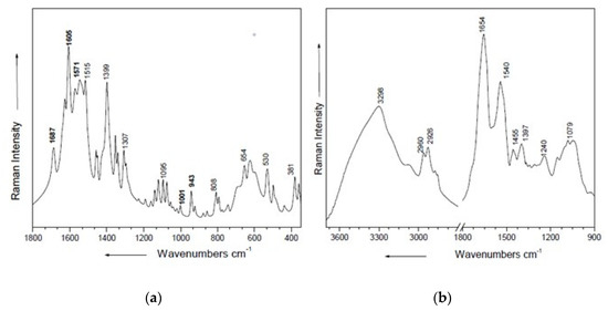
Figure 4.
The FTIR spectrum of MSG (a) and spirulina powder (b).
Comparing the FTIR spectra of MSG with the spectra of MSG in combination with spirulina (Figure 5), certain glutamate bands in the spectrum decrease in intensity in the obtained spectrum of spirulina and MSG. This is especially evident in the region 1800–1200 cm−1. This decrease in intensity might suggest that the MSG molecule can undergo some changes when mixed with spirulina, indicating that spirulina intake may provide the beneficial potential for the body and reduce the harmful effects of MSG.
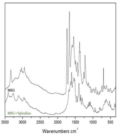
Figure 5.
Comparison of FTIR spectrum of MSG and MSG combined with spirulina.
Studies show that novel food products are most successful when they are developed based on consumer orientation [71]. In order to create potentially successful consumer-oriented products, novel product concepts incorporating spirulina and quercetin are recommended, as they have shown beneficial effects for consumers [64]. For this reason, the future research of our project will focus on (i) elucidating the beneficial effects of quercetin in Swiss mice treated with MSG, and (ii) based on the results of both studies, we will obtain a novel food product with beneficial effects for the consumer that could be marketed throughout all Europe.
4. Conclusions
Altogether, our findings evidenced that monosodium glutamate could produce histopathological changes in Swiss mice, which caused certain tissue damage of the kidney. In addition, this food additive induced important changes in all biochemical parameters of the blood serum. Noticeably, the potent ameliorating effect of spirulina was proved and was described by using the FTIR spectroscopy technique. Swiss mice treated with MSG supplemented with spirulina showed no histopathological changes of the organs, and biochemical parameters presented similar average values similar to the normal group of Swiss mice. Furthermore, intensive research is required to explore monosodium glutamate effects by longer trials investigating its usually harmful components and a better understanding of the related molecular and metabolic mechanisms.
Author Contributions
I.B. and A.C. conceived and designed the experiments; B.S., M.T. and C.T. completed the methodology; S.M. and L.A. analyzed the data using software; I.B. wrote the paper; A.C., M.T., I.B., B.S., O.M.T.-K. performed the experiments; Z.M. and A.L.L. provided supervision; A.C., L.A., I.P. and F.M. checked and improved the paper. All authors have read and agreed to the published version of the manuscript.
Funding
This research received no external funding.
Institutional Review Board Statement
The study was conducted according to Housing conditions, personal qualification, and the procedures conducted on laboratory animals agreed to Directive 2010/63/EU and national legislation, Law 43/2014, on the protection of animals used for scientific purposes. The project was approved by the Committee for Bioethics and Research Ethics of UASVM and the Regional State Veterinary Authority (project authorization No. 110/23.94.2018).
Informed Consent Statement
Not applicable.
Data Availability Statement
The data presented in this study are available on request from the corresponding author.
Acknowledgments
Funding for this work was provided by the University of Agricultural Sciences and Veterinary Medicine, Calea Mănăştur 3-5, Cluj-Napoca, 400372, Romania. We are thankful to Pere Rovira (Forest Sciences Centre of Catalonia, Crtr Sant Llorenç de Morunys, km 2, 25280, Solsona, Catalonia, Spain) for his useful contribution related to this review article.
Conflicts of Interest
The authors declare no conflict of interest.
References
- Burt, S. Essential oils: Their antibacterial properties and potential applications in foods-a review. Int. J. Food Microbiol. 2004, 94, 223–253. [Google Scholar] [CrossRef]
- Reis, J.A.; Paula, A.T.; Casarotti, S.N. Lactic Acid Bacteria Antimicrobial Compounds: Characteristics and Applications. Food Eng. Rev. 2012, 4, 124–140. [Google Scholar] [CrossRef]
- Balta, I.; Brinzan, L.; Stratakos, A. Geraniol and Linalool Loaded Nanoemulsions and Their Antimicrobial Activity. Bull. UASVM Anim. Sci. Biotechnol. 2017, 74, 157–161. [Google Scholar] [CrossRef]
- Pasca, C.; Coroian, A.; Socaci, S. Risks and Benefits of Food Additives-Review. Bull. UASVM Anim. Sci. Biotechnol. 2018, 75, 71–79. [Google Scholar] [CrossRef]
- Khorshidi, M.; Moini Ashraf Alipoor, E.; Rezvan, N.; Gorgani-Firuzjaee, S.; Yaseri, M.; Hosseinzadeh-Attar, M. The effects of quercetin supplementation on metabolic and hormonal parameters as well as plasma concentration and gene expression of resistin in overweight or obese women with polycystic ovary syndrome. Phytother. Res. 2018, 32, 2282–2289. [Google Scholar] [CrossRef] [PubMed]
- Onaolapo, A.Y.; Onaolapo, O.J.; Mosaku, T.J. A Histological Study of the Hepatic and Renal Effects of Subchronic Low Dose Oral Monosodium Glutamate in Swiss Albino Mice. Br. J. Med. Med Res. 2013, 3, 294–306. [Google Scholar] [CrossRef][Green Version]
- Campbell, A. Monosodium Glutamate (MSG). In Encyclopedia of Toxicology, Collection in Biomedical Sciences, 3rd ed.; Elsevier: Amsterdam, The Netherlands, 2014; pp. 391–392. [Google Scholar] [CrossRef]
- Alalwani, A.D. Monosodium glutamate induced testicular lesions in rats (histological study). Middle East Fertil. Soc. J. 2014, 19, 274–280. [Google Scholar] [CrossRef]
- Onaolapo, O.J.; Onaolapo, A.Y.; Akanmu, M. Changes in Spontaneous Working-memory, Memory-recall and Approach-avoidance following “Low Dose” Monosodium Glutamate in Mice. AIMS Neurosci. 2016, 3, 317–337. [Google Scholar] [CrossRef]
- Smriga, M. Food-added monosodium glutamate does not alter brain structure or antioxidant status. Pathophysiology 2016, 23, 303–305. [Google Scholar] [CrossRef]
- Henry-Unaeze, H.N. Update on food safety of monosodium l-glutamate (MSG). Pathophysiology 2017, 24, 243–249. [Google Scholar] [CrossRef]
- Onaolapo, O.J.; Onaolapo, A.Y.; Akanmu, M.A. Evidence of alterations in brain structure and antioxidant status following ‘low-dose’ monosodium glutamate ingestion. Pathophysiology 2016, 23, 147–156. [Google Scholar] [CrossRef]
- Onaolapo, A.Y.; Odetunde, I.; Akintola, A.S. Dietary composition modulates impact of food-added monosodium glutamate on behaviour, metabolic status and cerebral cortical morphology in mice. Biomed. Pharmacother. 2019, 109, 417–428. [Google Scholar] [CrossRef] [PubMed]
- Pallavi, R. The effect of monosodium glutamate on planarian memory retention. South Carol. Jr. Acad. Sci. 2018, 211. Available online: https://scholarexchange.furman.edu/scjas/2018/all/211 (accessed on 4 November 2021).
- Bhandari, U. Effect of Embelin in Monosodium Glutamate Induced Obesity in Male Neonatal Wistar Rats. Artheroscler. Suppl. 2018, 32, 138. [Google Scholar] [CrossRef]
- Jubaidi, F.F.; Mathialagan, R.D.; Noor, M.M. Monosodium glutamate daily oral supplementation: Study of its effects on male reproductive system on rat model. Syst. Biol. Reprod. Med. 2019, 65, 194–204. [Google Scholar] [CrossRef]
- Thanh, D.D.; Bossche, F.V.D.; Thanh, N.L. Synergistic effect of a monosodium glutamate/aspartame mixture on zebrafish larval neurobehavioral in comparison with the ADHD symptoms. In Proceedings of the Conference: Belgian Brain Congress 2018—Belgian Brain Council, Liege, Belgium, 19 October 2018. [Google Scholar] [CrossRef]
- Yousef, M.; El-Nassag, D.; Gasser, M. Potential Protective Effects of Propolis against Hepatotoxicity and Nephrotoxicity Induced by Monosodium Glutamate in Rabbits. Alex. Sci. Exch. J. 2019, 40, 30–42. [Google Scholar] [CrossRef]
- El-Shenawy, N.S.; Hamza, R.Z.; Al-Salmi, F.A. Evaluation of the Effect of Nanoparticles Zinc Oxide/Camellia sinensis Complex on the Kidney of Rats Treated with Monosodium Glutamate: Antioxidant and Histological Approaches. Curr. Pharm. Biotechnol. 2019, 20, 542–550. [Google Scholar] [CrossRef]
- Shimada, A.; Baad-Hansen, L.; Castrillon, E. Differential effects of repetitive oral administration of monosodium glutamate on interstitial glutamate concentration and muscle pain sensitivity. Nutrition 2015, 31, 315–323. [Google Scholar] [CrossRef]
- Morais, M.G.; Vaz, B.S.; Morais, E.G. Biological Effects of Spirulina (Arthrospira) Biopolymers and Biomass in the Development of Nanostructured Scaffolds. Biomed. Res. Int. 2014. [Google Scholar] [CrossRef]
- Chamorro, G.; Salazar, M.; Favila, L. Pharmacology and toxicology of Spirulina alga. Rev. Invest. Clin. 1966, 48, 389–399. [Google Scholar]
- Blé-Castillo, J.L.; Rodriguez-Hernandez, A.; Miranda-Zamora, R. Arthrospira maxima prevents the acute fatty liver induced by the administration of simvastatin, ethanol and a hypercholesterolemic diet to mice. Life Sci. 2002, 70, 2665–2673. [Google Scholar] [CrossRef]
- Tan, T.T. Filamentous tropical marine cyanobacteria: A rich source of natural products for anticancer drug discovery. J. Appl. Phycol. 2010, 22, 659–676. [Google Scholar] [CrossRef]
- Tobón-Velasco, J.C.; Palafox-Sanchez, V.; Mendieta, L. Antioxidant effect of Spirulina (Arthrospira) maxima in a neurotoxic model caused by 6-OHDA in the rat stratum. J. Neural Transm. 2013, 120, 1179–1189. [Google Scholar] [CrossRef]
- Ionov, V.A.; Basova, M.M. Use of blue-green micro-seaweed Spirulina platensis for the correction of lipid and hemostatic disturbances in patients with ischemic heart disease. Vopr. Pitan. 2003, 72, 28–31. [Google Scholar] [PubMed]
- Shalaby, E.A.; Shanab, M.M.; Singh, V. Salt stress enhancement of antioxidant and antiviral efficiency of Spirulina platensis. J. Med. Plants Res. 2010, 4, 2622–2632. [Google Scholar] [CrossRef]
- Ponce-Canchihuamán, J.C.; Pérez-Méndez, O.; Hernández-Muñoz, R. Protective effects of Spirulina maxima on hyperlipidemia and oxidative-stress induced by lead acetate in the liver and kidney. Lipids Health Dis. 2010, 9, 35. [Google Scholar] [CrossRef]
- Khan, Z.; Bhadouria, P.; Bisen, P.S. Nutritional and therapeutic potential of Spirulina. Curr. Pharm. Biotechnol. 2005, 6, 373–379. [Google Scholar] [CrossRef] [PubMed]
- Deng, R.; Chow, T.J. Hypolipidemic, Antioxidant and Anti-inflammatory Activities of Microalgae Spirulina. Cardiovasc. Ther. 2011, 28, 33–45. [Google Scholar] [CrossRef]
- Hirata, T.; Tanaka, M.; Ooike, M. Antioxidant activities of phycocyanobilin prepared from Spirulina platensis. J. Appl. Phycol. 2000, 12, 435–439. [Google Scholar] [CrossRef]
- Estrada, J.E.P.; Bescos, P.B.; Villar del Fresno, A.M. Antioxidant activity of different fractions of Spirulina platensis protean extract. II Farm. 2001, 56, 497–500. [Google Scholar] [CrossRef]
- Wang, L.; Pan, B.; Sheng, J. Antioxidant activity of Spirulina platensis extracts by supercritical carbon dioxide extraction. Food Chem. 2007, 105, 36–41. [Google Scholar] [CrossRef]
- Sinanoglu, O.; Yener, A.N.; Ekici, S. The protective effects of spirulina in cyclophosphamide induced nephrotoxicity and urotoxicity in rats. Urology 2012, 80, 1392.e1. [Google Scholar] [CrossRef] [PubMed]
- Shalaby, E.A.; Shanab, S.M.M. Comparison of DPPH and ABTS assays for determining antioxidant potential of water and methanol extracts of Spirulina platensis. Indian J. Geo-Mar. Sci. 2013, 42, 556–564. [Google Scholar]
- Ibrahim, A.E.; Abdel-Daim, M.M. Modulating Effects of Spirulina platensis against Tilmicosin-Induced Cardiotoxicity in Mice. Cell J. 2015, 17, 137–144. [Google Scholar] [CrossRef]
- Zhu, H.Z.; Zhang, Y.; Zhu, M.J. Protective effects of spirulina on hippocampal injury in exercise-fatigue mice and its mechanism. Chin. J. Appl. Physiol. 2018, 34, 562–567. [Google Scholar] [CrossRef]
- Abdel-Daim, M.; El-Bialy, B.E.; Rahman, H.G.A. Antagonistic effects of Spirulina platensis against sub-acute deltamethrin toxicity in mice: Biochemical and histopathological studies. Biomed. Pharmacother. 2016, 77, 79–85. [Google Scholar] [CrossRef]
- Fu, X.; Zhong, Z.; Hu, F. The protective effects of selenium-enriched Spirulina platensis on chronic alcohol-induced liver injury in mice. Food Funct. 2018, 9, 3155–3165. [Google Scholar] [CrossRef]
- Lee, H.Y.; Ryu, G.H.; Choi, W.Y. Protective Effect of Water Extracted Spirulina maxima on Glutamate-induced Neuronal Cell Death in Mouse Hippocampal HT22 Cell. Pharmacogn. Mag. 2018, 14, 242–247. [Google Scholar] [CrossRef]
- Eltantawy, F.M.; Sobh, M.A.A.; El-Waseef, A.M. Protective effect of Spirulina against cyclophosphamide-induced urotoxicity in mice. Egypt. J. Basic Appl. Sci. 2018, 5, 191–196. [Google Scholar] [CrossRef]
- Andronie, L.; Coroian, A.; Miresan, V. Results Obtained by Investigating Saffron Ussing FT-IR Spectroscopy. Bull. UASVM Anim. Sci. Biotechnol. 2016, 73, 238–239. [Google Scholar] [CrossRef][Green Version]
- Keseru, A.; Andronie, L.; Pop, I. Rotaru, A.; Maniutiu, D.; Coroian, A.; Raducu, C. Characterization of momordica charantia ussing FT-IR spectroscopy. Bull. UASVM Holticulture For. 2016, 73, 245–246. [Google Scholar]
- Directive 2010/63/EU of the European Parliament and of the Council of 22 September 2010 on the Protection of Animals Used for Scientific Purposes. Available online: https://eur-lex.europa.eu/legal-content/EN/TXT/?uri=celex%3A32010L0063 (accessed on 3 November 2021).
- Gonciarov, M.; Coman, C. General principles concerning the harmonization of Romanian Legislation with the European Union in the field of protection of animals used for scientific scope. Agric. Agric. Sci. Procedia 2015, 6, 336–341. [Google Scholar] [CrossRef][Green Version]
- Windmueller, H.G.; Spaeth, A.E. Intestinal Metabolism of Glutamine and Glutamate from the Lumen as Compared to Glutamine from Blood. Arch. Biochem. Biophys. 1975, 171, 662–672. [Google Scholar] [CrossRef]
- Windmueller, H.G.; Spaeth, A.E. Respiratory Fuels and Nitrogen Metabolism in Vivo in Small Intestine of Fed Rats. Quantitative Importance of Glutamine, Glutamate, and Aspartate. J. Biol. Chem. 1980, 255, 107–112. [Google Scholar] [CrossRef]
- Nehad, R.; Elyazji, I.; Osama, S.; Lubbad, A.M. Effects of Monosodium Glutamate on Some Biochemical and Hematological Parameters in Adult Rabbits and Potential Protective Effect of Soybean Oil. J. Biol. Chem. Res. 2014, 32, 131–141. [Google Scholar]
- Agnes, W.; Boots Guido, R.M.; Haenen, M.; Bast, A. Health effects of quercetin: From antioxidant to nutraceutical. Eur. J. Pharmacol. 2008, 585, 325–337. [Google Scholar] [CrossRef]
- McAnlis, G.T.; McEneny, J.; Pearce, J.; Young, I.S. Absorption and antioxidant effects of quercetin from onions in man. Eur. J. Clin. Nutr. 1999, 53, 92–96. [Google Scholar] [CrossRef] [PubMed]
- Moskowitz, H.; Hartmann, J. Consumer research: Creating a solid base for innovative strategies. Trends Food Sci. Technol. 2008, 19, 581–589. [Google Scholar] [CrossRef]
- SAS Institute JMP 12 Statistical Software; SAS Institute Inc., Cary: Lerida, Spain, 2014.
- Seiva, F.R.F.; Chuffa, L.G.A.; Braga, C.P. Quercetin ameliorates glucose and lipid metabolism and improves antioxidant status in postnatally monosodium glutamate-induced metabolic alterations. Food Chem. Toxicol. 2012, 50, 3556–3561. [Google Scholar] [CrossRef]
- Balta, I.; Sevastre, B.; Mireșan, V. Protective effect of blackthorn fruits (Prunus spinosa) against tartrazine toxicity development in albino Wistar rats. BMC Chem. 2019, 13, 1–11. [Google Scholar] [CrossRef]
- Hasan, K.M.; Tamanna, N.; Haque, A. Biochemical and histopathological profiling of Wistar rat treated with Brassica napus as a supplementary feed. Food Sci. Hum. Wellness 2018, 7, 77–82. [Google Scholar] [CrossRef]
- Muthuraman, P.; Senthikumar, R.; Srikumar, K. Alterations in beta-islets of Langerhans in alloxan-induced diabetic rats by marine Spirulina platensis. Enzym. Inhib. Med. Chem. 2009, 24, 1253–1256. [Google Scholar] [CrossRef]
- Balbo, S.L.; Bonfleur, M.L.; Carneiro, E.M. Parasympathetic activity changes insulin response to glucose and neurotransmitters. Diabetes Metab. 2002, 28, 3S13-7. [Google Scholar] [PubMed]
- Andreazzi, A.E.; Scomparin, D.X.; Mesquita, F.P. Swimming exercise at weaning improves glycemic control and inhibits the onset of monosodium L-glutamate-obesity in mice. J. Endocrinol. 2009, 201, 351–359. [Google Scholar] [CrossRef] [PubMed]
- Macho, L.; Ficková, M.; Jezova, Z.S. Late effects of postnatal administration of monosodium glutamate on insulin action in adult rats. Physiol. Res. 2000, 48, S79–S85. [Google Scholar]
- Prentki, M.; Nolan, C.J. Islet beta cell failure in type 2 diabetes. J. Clin. Investig. 2006, 116, 1802–1812. [Google Scholar] [CrossRef]
- Onyema, O.O.; Farombi, E.O.; Emerole, G.O. Effect of vitamin E on monosodium glutamate induced hepatotoxicity and oxidative stress in rats. Indian J. Biochem. Biophys. 2006, 43, 20–24. [Google Scholar]
- Anwar, M.M.; Mohamed, N.E. Impact of Flax Seed and Canola Oils Mixture Supplementation on The Physiological and Biochemical Changes Induced by Monosodium Glutamate in Rats. J. Rad. Res. Appl. Sci. 2010, 3, 943–964. [Google Scholar]
- Takasaki, Y. Studies on brain lesions after administration of monosodium L-glutamate to mice. II. Absence of brain damage following administration of monosodium L-glutamate in the diet. Toxicology 1978, 9, 307–318. [Google Scholar] [CrossRef]
- Peláez, B.; Blázquez, J.L.; Pastor, F.E. Lectinhistochemistry and ultrastructure of microglial response to monosodium glutamate-mediated neurotoxicity in the arcuate nucleus. Histol. Histopathol. 1999, 14, 165–174. [Google Scholar]
- Rivera-Cervantes, M.C.; Torres, J.S.; Feria-Velasco, A. NMDA and AMPA receptor expression and cortical neuronal death are associated with p38 in glutamate-induced excitotoxicity in vivo. Neurosci. Res. 2004, 76, 678–687. [Google Scholar] [CrossRef] [PubMed]
- Blank, M.L.; Lee, T.C.; Fitzgerald, V.; Snyder, F. A specific acetyl hydrolase for I-alkyl-2-acetyl-sn-glycero-3-phosphocholine (a hypotensive and platelet-activating lipid). J. Biol. Chem. 1981, 256, 175–178. [Google Scholar] [CrossRef]
- Reif-Lehrer, L.; Bergenthal, J.; Hanninen, L. Effects of monosodium glutamate on chick embryo retina in culture. Investig. Ophthalmol. 1975, 14, 114–124. [Google Scholar]
- Blanks, J.C.; Reif-Lehrer, L.; Casper, D. Effects of monosodium glutamate on the isolated retina of the chick embryo as a function of age: A morphological study. Exp. Eye Res. 1981, 32, 105–124. [Google Scholar] [CrossRef]
- Hyndman, A.G.; Adler, R. Analysis of glutamate uptake and monosodium glutamate toxicity in neural retina monolayer cultures. Dev. Brain Res. 1981, 2, 303–314. [Google Scholar] [CrossRef]
- Goldsmith, P.C. Neuroglial Responses to Elevated Glutamate in the Medial Basal Hypothalamus of the Infant Mouse. J. Nutr. 2000, 130, 1032–1038. [Google Scholar] [CrossRef]
- Karwowska, M.; Kononiuk, A. Nitrates/Nitrites in Food—Risk for Nitrosative Stress and Benefits. Antioxidants 2020, 9, 241. [Google Scholar] [CrossRef]


Publisher’s Note: MDPI stays neutral with regard to jurisdictional claims in published maps and institutional affiliations. |
© 2021 by the authors. Licensee MDPI, Basel, Switzerland. This article is an open access article distributed under the terms and conditions of the Creative Commons Attribution (CC BY) license (https://creativecommons.org/licenses/by/4.0/).