Article

GC-MS Method for Quantification and Pharmacokinetic Study of Four Volatile Compounds in Rat Plasma after Oral Administration of Commiphora myrrh (Nees) Engl. Resin and In Vitro Cytotoxic Evaluation

1
Department of Pharmacognosy, College of Pharmacy, King Saud University, Riyadh 11451, Saudi Arabia
2
Department of Pharmacology and Toxicology, College of Pharmacy, King Saud University, Riyadh 11451, Saudi Arabia
3
Department of Pharmaceutical Chemistry, College of Pharmacy, King Saud University, Riyadh 11451, Saudi Arabia
*
Authors to whom correspondence should be addressed.
Separations 2021, 8(12), 239; https://doi.org/10.3390/separations8120239
Submission received: 9 November 2021 / Revised: 24 November 2021 / Accepted: 2 December 2021 / Published: 7 December 2021
(This article belongs to the Special Issue Separations in Biomedical Analysis)

Abstract

:
A rapid, simple, and sensitive gas chromatography–tandem mass spectrometry (GC–MS) method was established and validated for simultaneous determination of four volatile compounds, namely curzerene, methoxyfuranodiene, β-elemene, and α-pinene in rat plasma samples after oral administration of the resin extract of Commiphora myrrh using limonene as an internal standard (IS). Liquid-liquid extraction using hexane and ethyl acetate (1:1) mixture as an extracting agent was used for the samples extraction procedure. The GC–MS system was operated under selective ion monitoring (SIM) mode using Perkin Elmer Elite 5MS column (30 m × 0.25 mm × 0.25 µm film thickness). Specificity, linearity, precision, accuracy, extraction recovery, and stability were used to validate the developed method. The assay showed good linearity (r2 ≥ 0.998), and the lowest limits of quantification (LLOQ) were 3.97–21.38 ng/mL for the four analytes. This assay was successfully applied to pharmacokinetic studies of the four volatile compounds in rat plasma. The antiproliferative activity of these volatile compounds was evaluated against lung carcinoma (A549) and colon (LoVo) cell lines, were each compound caused variable inhibition on cells proliferation and methoxyfuranodiene exerted the strong antiproliferative activity against both cell line according to IC50 values.

1. Introduction

Commiphoramyrrha (Nees) Engl. is a medicinal plant belonging to the genus Commiphora (Burseraceae family) and mainly grows in Yemen and the southern regions of Saudi Arabia [1,2]. C. myrrh is known to produce a natural compound (oleo-gum-resin), known as myrrh [3,4]. Myrrh, as traditional natural medicine, has been used to treat various diseases, such as amenorrhea, ache, dysmenorrhea, tumors, fever, stomach complaints, gall bladder, chest ailments, snake and scorpion bites, and skin infections in ancient India, China, Rome, Greece, and Babylon [5]. Previous investigations have revealed the myrrh can be used in the treatment of ulcers, Schistosoma, Fasciolopsis, respiratory catarrh, furunculosis, and diabetes [6,7,8]. Myrrh was also found to promote the permeability of the medicine from epidermis to dermal capillaries and also enhance the wound healing process [9].
The Myrrh’s Gum-resin-volatile oil are the major useful contents, where it contains (30–60%) gum including acidic polysaccharides, resin (25–40%), volatile oil (3–8%), eugenol, heerabolene and many furansesquiterpenes [4,10]. In the previous studies, the volatile constituents of myrrh were identified [11,12,13]. Among these volatile constituents, curzerene, methoxyfuranodiene, β-elemene and α-pinene are the key bioactive compounds [1,11,14,15,16,17], and primarily responsible for a wide range of reported pharmacological activities of myrrh. Curzerene has been verified that it has primarily responsible for the analgesic activity of myrrh [3]. methoxyfuranodiene was found that it has antioxidant and anti-angiogenic effects [4,18]. β-Elemene was widely used for removing blood stasis and it has an anti-tumor effect [19,20,21]. α-Pinene has a wide range of reported pharmacological activities, including antibacterial, anticoagulant, antitumor, antimicrobial, antimalarial, antioxidant, anti-inflammatory, anti-Leishmania, and analgesic effects [22].
Herbal medications have been utilized for preventing and treating diseases for hundreds of years. Recently, these drugs are becoming more popular globally. As being nature-derived is the essential attribute of herbal medicines, people believe that taking them for diseases treatment is safe enough and has no side effects. However, the efficacy of herbal resourced compounds depends on the constituents in the body and their pharmacokinetics [23], that is why we were encouraged to analyze the more predominant volatile constituent of C. myrrh resin extract in form of pharmacokinetic studies which could provide valuable references for determining the appropriate utilization of C. myrrh resin.
To our knowledge, there is no method about the determination of curzerene and methoxyfuranodiene (isolated in our lab) in plasma in the form of pharmacokinetic studies is available in the literatures. In comparison, the previous studies have discussed the pharmacokinetics of β-elemene and α-pinene in plasma (human or rat) samples using GC-MS method after administration of different herbal materials (e.g., Rhizoma curcumae and Mastered Oil) [24,25,26], while our study was conducted to determine β-elemene and α-pinene after oral administration of C. myrrh resin extract. It is worth mentioning here that multiple constituents in various medicinal plant extracts (including C. myrrh resin) could influence the bioavailability, metabolism, and half-life of β-elemene and α-pinene [23,27], therefore a better understanding of the pharmacokinetics parameters and bioavailability of those compounds would also provide a significant experimental basis for the proper clinical application of myrrh and predict the influence of pharmacokinetic interactions with the multiple constituents C. myrrh resin.
A simple, sensitive, and selective gas chromatography-mass spectrometry method (GC-MS) in selected ion mode (SIM) was established for the simultaneous quantification of curzerene, methoxyfuranodiene, β-elemene, and α-pinene in rat plasma. The antiproliferative activity of these volatile compounds was evaluated against lung and colon cancer cell lines. The demonstrated method can be applied successfully for pharmacokinetics studies of those bioactive compounds after oral administration of C. myrrh resin extract. We expect that the results of this study would provide some references for further pharmacological application of C. myrrh resin to enhance efficacy, decrease toxicity, and support therapeutic drug monitoring (TDM).

2. Materials and Methods

2.1. Plant Material

The C. myrrh resin (myrrh) is the thickened, bitter-tasting, yellowish-brown agreeably aromatic gum exudes as a fluid from resin ducts in the tree bark when the bark splits naturally or is cut in tapping. Upon exposure to the air, hardens and turns reddish-brown irregular chunks called tears, which are then collected from the tree. Myrrh sample was purchased from Alothim Market in Riyadh, Saudi Arabia. It was identified and verified by Prof. Ramzi mothana. A vouchered sample (no. 12486) was deposited in the herbarium of Medicinal, Aromatic, and Poisonous Plants Research Center, Department of Pharmacognosy King Saud University, Riyadh, Saudi Arabia.

2.2. Chemicals and Reagents

Standard of curzerene (purity ≥ 99%) was purchased from Biopurify phytochemicals Co., Ltd. (Chengdu, Sichuan, China). Standard of β-elemene (purity ≥ 99%) was purchased from Cayman Chemical Co., Ltd. (Ann Arbor, MI, USA). α-pinene (purity ≥ 98%) was obtained from Fluka Chemie GmbH (Buchs, Switzerland). A white crystalline methoxyfuranodiene was isolated and identified in previous work [4]. Methanol and acetonitrile (HPLC grade) were obtained from Sigma Co., Ltd. (St. Louis, MO, USA). Ultra-pure water was purified by Purelab Flex water purification system (Veolia Ltd., High Wycombe, UK).

2.3. Preparation of Calibration Standards and Quality Control Samples

Standard stock solutions of curzerene (200 µg/mL), methoxyfuranodiene (1.5 mg/mL), β-elemene (250 µg/mL), and α-pinene (2 mg/mL) were prepared in methanol. Then, gradient concentrations of the standard stock solutions were prepared by serial dilution with 90% methanol/water. The internal standard (IS) solution of limonene was prepared to the concentration of 250 ng/mL in methanol. The calibration solutions were prepared by serial dilution with blank rat plasma to obtain the concentrations ranging from 325.0 to 10.16 ng/mL for curzerene, 252.0–7.88 ng/mL for methoxyfuranodiene, 684.0–21.38 ng/mL for β-elemene, and 127.0–3.97 ng/mL for α-pinene. The Quality control (QC) samples concentration were 10.20, 81.30, 162 ng/mL for curzerene, 7.90, 63, 126 ng/mL for methoxyfuranodiene, 21.40, 171, 342 ng/mL for β-elemene and 8, 31.80, 127 ng/mL for α-pinene. All standard working solutions were stockpiled at 4 °C, then they were returned to room temperature before analysis.

2.4. GC-MS Apparatus and Analysis Conditions

GC-MS analysis was performed using a Perkin Elmer GC Calarus 600T combined with a Clarus 600T single quadrupole mass spectrometer. Separation was performed on a Perkin Elmer Elite 5MS column (30 m × 0.25 mm × 0.25 µm film thickness), with high-purity helium as the gas carrier, at a flow rate of 1 mL/min. The injector temperature was 280 °C and it was equipped with a splitless injector at 20:1. The temperature was set initially to 40 °C (held for 1 min), then was increased to 150 °C at 20 °C min−1 (held for 1 min), then increased further to 300 °C at 20 °C min−1 for 1 min. The MS ion source temperature was 220 °C and the inlet line temperature was set to 240 °C. The mass detector was operated at 70 eV ionization energy and the solvent delay of 3 min. The selective ion monitoring (SIM) was set for quantification and the total time required for analyzing a single sample was 16 min.

2.5. Assay of the Four Volatile Compounds in Myrrh Extract

The contents of curzerene, methoxyfuranodiene, β-elemene, and α-pinene in the C. myrrh resin extract were quantitatively measured by GC–MS. The crude C. myrrh resin was extracted according to our previously described method [2,4]. Briefly, the dried oleo-gum resin of C. myrrh (1 kg) was extracted with 2 L of ethanol (95%) for 3 days using cold maceration. The obtained alcoholic extract was filtered, dried using a rotatory evaporator, and then diluted. Finally, 1 µL of the filtrate was injected into the GC–MS system.

2.6. Animal Experiments and Drug Administration

Animal experiments were performed in line with the “guidelines of the animal care and use committee at King Saud University” and the protocol was authorized by approval from Research Ethics Committee (Reference no. KSU-SE-21-15, dated 3 February 2021). Male SPF Wistar albino rats weighing between 200–250 g were obtained from the “Animal care center, College of Pharmacy, King Saud University, Riyadh, Saudi Arabia”. Rats were housed on a standard chow diet in a room free from any source of chemical contamination, artificially illuminated (12 h dark/light cycle) under ambient temperature- and humidity environment (23 ± 2 °C, 50% relative humidity) at the animal facility. The single oral administration was prepared by constituting the extract with carboxyl methyl cellulose (0.5%, w/v) to acquire an oral formulation in form of suspension with a concentration equivalent to 500 mg/kg C. myrrh resin. Rats were administered orally the suspended extract using gastric gavage needle no. 24.

2.7. Extraction Procedure for Plasma Samples

After frozen plasma samples were thawed and allowed to equilibrate at room temperature, an aliquot of 200 µL plasma sample was mixed with 10 µL of IS solution (limonene, 250 ng/mL) in a 1.5-mL Eppendorf tube. The sample was shaken for 30 s and 300 µL of hexane and ethyl acetate (1:1) mixture was added and vortex-mixed for 60 s, then transferred for cold centrifugation (4 °C) at 10,000 rpm for 10 min. After, centrifugation the acquired supernatant was dried into a sample concentrator at 43 °C temperature. The residue was reconstituted in 100 µL acetonitrile and water (90:20, v/v) mixture and vortex-mixed for 2 min. Finally, the dissolved sample was centrifuged at 12,000 rpm for 10 min, and an aliquot (1 µL) of the supernatant was injected into the GC-MS/MS system for analysis.

2.8. Method Validation

The developed GC–MS assay was validated for selectivity, linearity, within-day and between-day precision, accuracy, recovery, and stability in accordance with US FDA bioanalytical method validation guidance [28].

2.8.1. Selectivity and Specificity

Blank plasma samples spiked with the IS were analyzed to investigate the interferences from endogenous substances or IS. Specificity and selectivity of the method were tested by comparing the chromatograms of six batches of blank plasma from six individual rats, corresponding plasma samples added with the 4 analytes and IS, and plasma samples from the rats 2 h after oral administration of C. myrrh resin extract.

2.8.2. Linearity and LLOQ

Standard plasma samples at series concentrations were analyzed to assess the linearity. The calibration curve was constructed by plotting the peak area ratio (y) of each analyte to IS versus the corresponding plasma concentrations (x) using a weighted least-squares linear regression model (1/x2). At the lower concentration on the standard analytical curve (LLOQ), the acceptable precision (relative standard deviation, RSD) and accuracy (relative error, RE) should be within ±20%.

2.8.3. Precision and Accuracy

The intra- and inter-day precisions were evaluated by RSD, and determined by analyzing three concentrations levels (low, medium, and high) of QC samples. Five replicates were measured on the same day and the three consecutive days, respectively. The precisions were expressed as the RSD, and the accuracy was determined by RE. The acceptable criteria for the precisions and accuracy were required to be within 15% for three concentrations levels.

2.8.4. Extraction Recovery and Matrix Effect

In order to investigate the extraction recoveries, the peak areas obtained from five plasma samples spiked with analytes and IS were compared before and after extraction at three QC levels. The matrix effect was assessed by comparing the peak areas obtained from the plasma matrix (analytes and IS were added) with the corresponding pure working solutions at identical concentrations.

2.9. Application of the Validated Assay in Pharmacokinetic Study

The developed method in plasma samples was successfully applied in pharmacokinetic studies of the four investigated analytes in rats. Animals were fasted overnight prior to the experiment to assure a more uniform drug absorption by preventing mixing test compounds with food. Blood samples (about 0.3 mL) were gathered into heparinized Eppendorf tube from the lateral saphenous vein at 0, 0.25, 0.5, 1.0, 2.0, 3.0, 5.0, 8.0, 12.0, and 24.0 h intervals after oral administration. Plasma was separated immediately by centrifugation at 10,000 rpm for 10 min and stored at −80 °C until analysis.
The pharmacokinetic parameters were measured by performing a non-compartment model using PKSolver software. The peak drug plasma concentration (Cmax), time to achieve Cmax (Tmax), area under plasma concentration-time curve zero to time and infinity (AUC0-t; AUC0-∞), elimination half-life (T½), elimination rate constant (Kel), and mean residence time were calculated. Statistical parameters including mean, standard deviation (SD), and % RSD were calculated using Excel 2013 (Microsoft Office Professional Plus 2013).

2.10. Cytotoxicity Assay

Cytotoxic activity was assessed using MTT assay as described by Nasr et al. [29]. In brief, A549 (lung) and LoVo (colon) cancer cells were seeded into 96-well plates at a density of 5 × 10⁴ cells in 100 µL medium per well and were allowed 24 h to attach to the bottom. After treatment with each compound, the plates were incubated at 37 °C (5% CO2, 95% humidity) for 48 h. Then, MTT solution (10 µL, 5 mg/mL in PBS) was added and the plates were incubated again for 2 h. Thereafter, isopropanol was added containing 0.01 M HCL to dissolve the formazan production in the cells. The plates were then allowed to sit for 10 min at plate shaker and absorbance measuring was taken at 570 nm using BioTek plate reader. The IC50 values were determined by OriginPro 8.5 software from a dose-response curve.

3. Results and Discussion

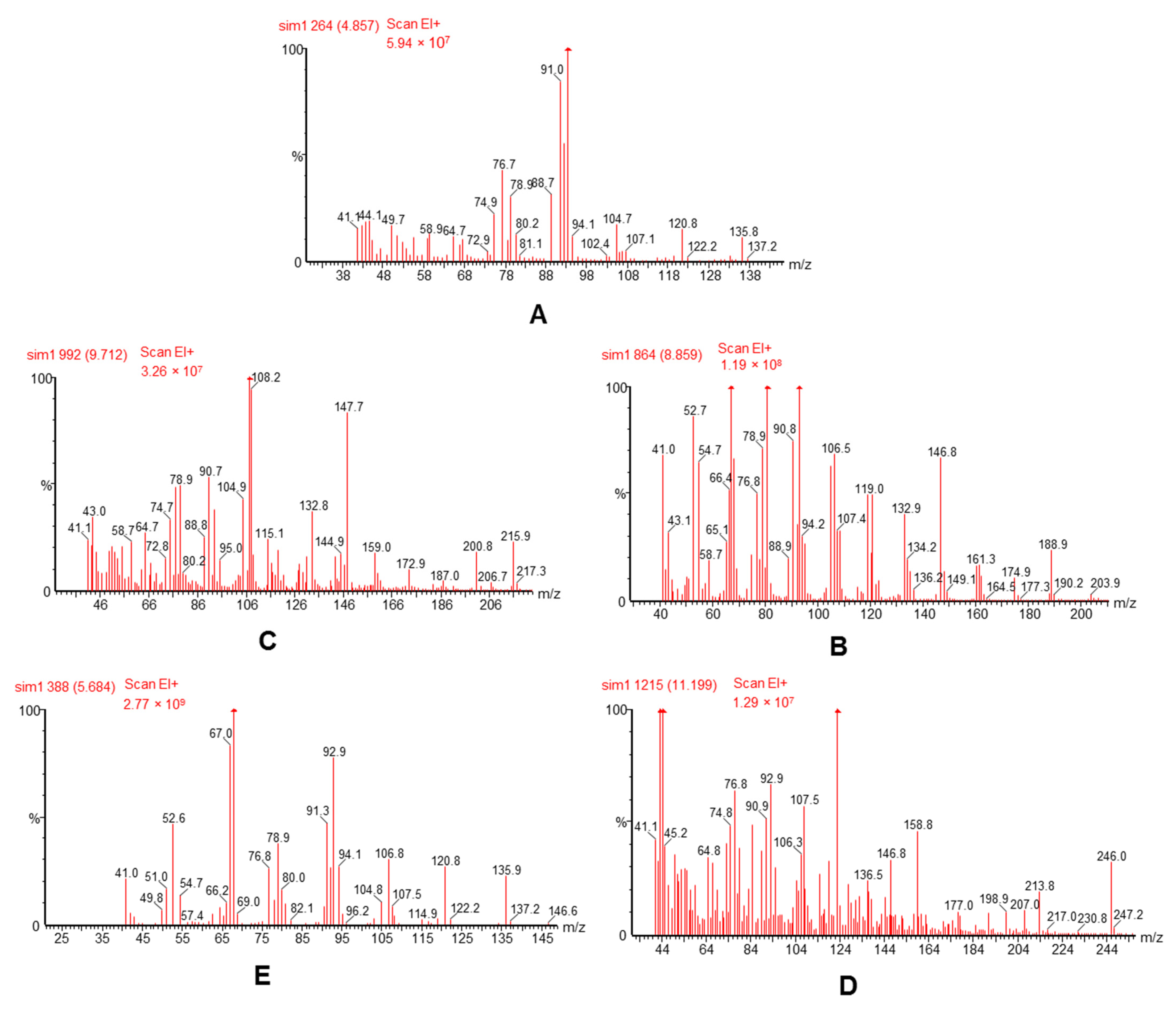
3.1. Optimization of GC–MS Conditions

To optimize the processing conditions of the mass spectrometer, the standard solution of each analyte and the IS were separately infused into the mass spectrometer. The selective ions for different analytes were 108/148 for curzerene, 123/246 for methoxyfuranodiene, 81/147 for β-elemene, 93/136 for α-pinene, and 67/121 for the IS. The MS spectra in scan mode were performed from 50–600 mass range and MS spectra of the four analytes and IS are presented in Figure 1. In order to obtain the best sensitivity, symmetrical peaks, and reduced run time, instrumental parameters such as injector temperature, detector temperature, initial column temperature, as well as the final column temperature, and split ratio were also optimized.

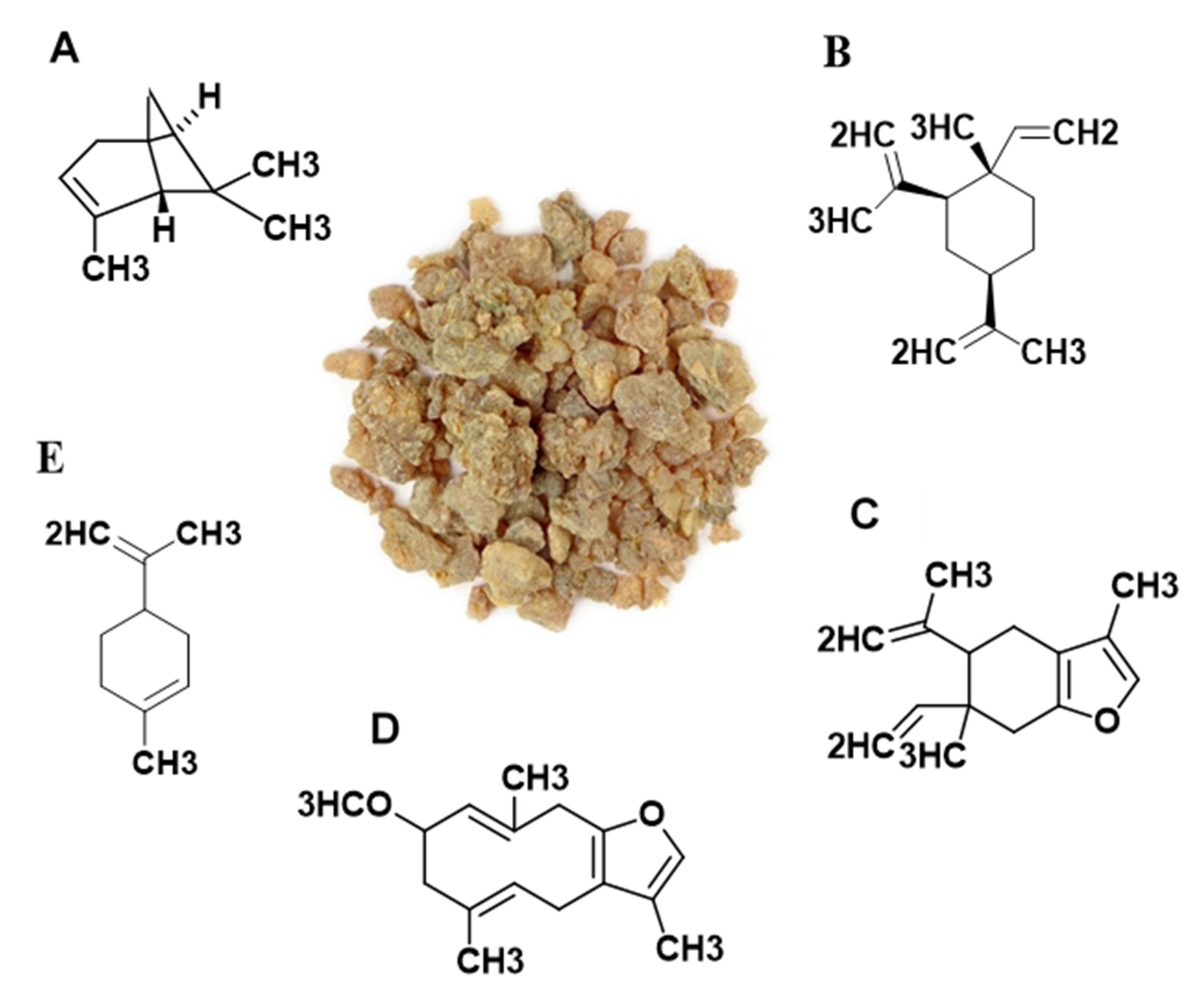
3.2. Contents of the Four Analytes in Myrrh Extract

The analytical data showed that the contents of curzerene, methoxyfuranodiene, β-elemene, and α-pinene (Figure 2) in C. myrrh resin extract were 0.153, 0.093, 0.046, and 0.023 mg/g, respectively. Quantitative results also indicated that curzerene, was the more predominant constituent in C. myrrh resin extract (Figure 3).

3.3. Optimization of Sample Extraction Method

Due to their different concentrations, dissolution properties, and pKa values as well as stabilities in the biological matrix, the extraction procedure for the analytes in plasma was optimized, and the extraction procedure should be simple, reproducible, minimize matrix effects, timesaving, and obtain a good recovery for all analytes. To obtain the desired recovery of all analytes, liquid-liquid extraction (LLE) was tried because this method could concentrate the analytes. For LLE, solvents including ethyl acetate, n-hexane, diethyl ether, and dichloromethane were investigated as extracting agents. We found that sample pretreatment using a mixture of hexane and ethyl acetate (1:1) could achieve good reproducibility and recovery for the four analytes.

3.4. Method Validation

3.4.1. Selectivity

The typical chromatograms acquired from blank plasma, blank plasma spiked with the 4 analytes and IS, and plasma sample from a rat at 2 h following oral administration of the C. myrrh resin extract are presented in Figure 4. From the typical chromatograms with good separation and excellent peak shapes, we saw that there are no interfering peaks from endogenous compounds at the retention times of the four analytes and IS. These results indicated that the developed method is selective and specific for these four analytes. The retention times of the four analytes and IS were as followed: α-pinene, β-elemene, curzerene, methoxyfuranodiene, and IS were 8.86, 12.86, 13.71, 15.20, and 9.70 min, respectively.

3.4.2. Assay Linearity and Sensitivity

The equation of the calibration curves, coefficient of determination (r2), linearity ranges, and LLOQs for the four analytes are listed in Table 1. Good linearity within the described ranges for the four analytes was indicated from the high coefficient of determination (r2) value (≥0.998). For pharmacokinetic studies, the LLOQs were sufficient enough for the quantitative determination of the four analytes in plasma.

3.4.3. Precision and Accuracy

Table 2 illustrates a summarize of precision and accuracy data for all four analytes at their three different QC levels (low, medium, and high). The within-day (intraday) and between-day (interday) precisions (RSD) for curzerene, methoxyfuranodiene, β-elemene, and α-pinene were found to be ≤7.23, ≤6.85, ≤4.92, and ≤14.58%, respectively. Within-day and between-day accuracy (RE) were ranged from −4.49 to 8.68% and from −5.08–5.15%, respectively. These results indicated that the developed method was accurate and precise for the determination of the tested analytes and therefore appropriate for routine pharmacokinetic analysis.

3.4.4. Recovery and Matrix Effects

The recovery and matrix effects of four analytes at level QC samples were listed in Table 3. The mean recoveries of the four analytes in the rat plasma sample were between 77.08% and 92.94%, and the mean matrix effects ranged from 83.10% to 101.15%. As for IS, the extraction recovery and matrix effects were 82.9% and 91.5%, respectively. Furthermore, all of the RSD values were less than 9.14%. Obviously, the sample pretreatment was suitable to obtain stable and high extraction recovery and no significant co-eluting endogenous interference existed in the plasma.

3.5. Application to Pharmacokinetic Study in Rats

The validated assay in this paper was sensitive enough to apply in the pharmacokinetic study of four volatile constituents in rat plasma after oral administration of C. myrrh resin extract at a reported therapeutic dose of 500 mg/kg [30]. The plasma concentration-time curve of the four analytes in rat plasma are shown in Figure 5, and the pharmacokinetic parameters result are summarized in Table 4. The results showed that curzerene and methoxyfuranodiene were absorbed easily and rapidly after oral administration of C. myrrh resin extract, and they achieved maximum plasma concentration (Cmax) after 1 h. However, the Tmax values were 2 h for β-elemene and α-pinene, which were absorbed slowly after oral administration. The Cmax value of methoxyfuranodiene was achieved to the relatively low level of 95.1 ± 6.10 ng/mL after oral administration, these results indicated that the absorbed methoxyfuranodiene might rapidly distribute into the tissues and organs because of its highly lipophilic or as a result of an extensive presystemic metabolism as many drugs may be metabolized before adequate plasma concentrations are reached [31]. On the other hand, the relatively low content of α-pinene in C. myrrh resin extract could be contributed to its low plasma concentration. The results also revealed that the elimination processes of the four compounds might be slow, as indicated by the mean residence time (MRT) from the time of dosing to the time of the last measurable concentration (9.10–34.28 h) moreover the T1/2 of the four compounds were more than 7.03 h, the slow elimination could be attributed to the high affinity binding to blood protein or because of the enterohepatic circulation. It is required to be proved by the hepatoenteral pharmacokinetic experiment further.
W. Li et al. [24] had studied the pharmacokinetics of β-elemene and α-pinene in rat plasma using GC-MS method after administration of oil of Rhizoma curcumae. According to their results, the Cmax of β-elemene and α-pinene were 248.98 ng/mL and 34.72 ng/mL, while the obtained results in this paper are 180.9 and 43.17 ng/mL for β-elemene and α-pinene, respectively. As far as we know, there is no method for the determination of curzerene and methoxyfuranodiene in plasma in the form of pharmacokinetic studies is available in the literature. The results about the pharmacokinetic profiles of β-elemen and α-pinene in C. myrrh resin in this paper were slightly different from that observed for their pure forms or other herbal prescriptions. These results indicated that the multiple constituents in the C. myrrh resin as well as the interactions among so many compounds in vivo could influence drugs bioavailability, metabolism, and half-life. This is the first report about a pharmacokinetic study of myrrh’s bioactive constitute and the results of this work can provide a significant experimental basis for the proper clinical application of C. myrrh resin.

3.6. Cytotoxicity Activity

In this study, the antiproliferative activity of these compounds was evaluated against lung carcinoma (A549) and colon (LoVo) cell lines. The results displayed in Figure 6 indicate that each compound caused a variable inhibition on tested cells proliferation and methoxyfuranodiene compound exerted the strong antiproliferative activity against both cell line in terms of IC50 values (Table 5). A similar cytotoxic effect of 2-methoxyfuranodiene was observed in our previous study against breast (MCF-7) and liver (HepG2) cancer cells [18]. In accordance with our results, curzerene from Curcuma longa also exerted antiproliferative effects in human lung cancer cells (SPC-A1) through enhancing apoptosis and cell cycle arrest. However, the reported IC50 in this study is slightly different which could be due to the type of cells and time incubation periods [32]. In addition, the cytotoxic activity of α-pinene and β-elemene is well documented against a variety of cell lines [22,33]. However, this is the first report on the activity of these two compounds isolated from C. myrrh resin against other cell lines indicating that they could be developed as anticancer therapeutics. Further research would be necessary in order to determine the possible mechanism responsible for the observed in vitro studies using detailed molecular mechanisms as well as in vivo anticancer models to give more insight on their biological effects.

4. Conclusions

We developed a simple, sensitive, accurate, and selective GC–MS method for the determination of myrrh’s bioactive constituents (curzerene, methoxyfuranodiene, β-elemene, and α-pinene) in rat plasma samples. The method was fully validated as per US FDA guidance for bioanalytical method validation, and all the evaluated parameters were within the acceptable limits. The implementation of liquid/liquid extraction techniques and the use of SIM for detection and quantitation resulted in better LOQ, linearity range, and recovery for all analyzed compounds. The matrix effect was evaluated and the analytical values confirmed no significant co-eluting endogenous interference existed in the plasma. For the first time, the method was successfully applied in a pharmacokinetic study of Myrrh’s bioactive constituents after the oral administration of the extract of C. myrrh resin in rats. The pharmacokinetic parameters of the studied compounds were evaluated including Cmax, Tmax, AUC0–t, AUC0–∞, T½, Kel, and mean residence time. The cytotoxicity of these volatile compounds was evaluated against different cell lines (colon (LoVo) and lung (A549) cancer cells), were each compound caused variable inhibition on cells proliferation and methoxyfuranodiene exerted the strong activity against both cell lines according to IC50 values. The obtained results can provide valuable experimental information for researchers and technicians for further clinical applications of C. myrrh resin and its prescriptions.

Author Contributions

Conceptualization, A.S.A.; data curation, F.A.; formal analysis, R.N.H., F.A. and S.R.A.; funding acquisition, A.S.A.; methodology, R.N.H., S.R.A. and F.A.N.; resources, A.S.A. and O.M.N.; software, F.A. and S.R.A.; supervision, A.S.A.; validation, R.N.H.; writing—original draft, R.N.H.; writing—review and editing, A.S.A. and R.N.H. All authors have read and agreed to the published version of the manuscript.

Funding

This research was funded by Researchers Supporting Project number (RSP-2021/132), King Saud University, Riyadh, Saudi Arabia.

Institutional Review Board Statement

The Study was conducted after approval of Research Ethics Committee, King Saud University (Reference no. KSU-SE-21-15, dated 3 February 2021).

Informed Consent Statement

Not applicable.

Data Availability Statement

Data is contained within the article.

Acknowledgments

Authors are thankful to the Researchers Supporting Project number (RSP-2021/132), King Saud University, Riyadh, Saudi Arabia for funding this research.

Conflicts of Interest

The authors declare no conflict of interest.

References

  1. Başer, K.; Demirci, B.; Dekebo, A.; Dagne, E. Essential oils of some Boswellia spp., myrrh and opopanax. Flavour Fragr. J. 2003, 18, 153–156. [Google Scholar] [CrossRef]
  2. Alqahtani, A.S.; Herqash, R.N.; Noman, O.M.; Tabish Rehman, M.; Shahat, A.A.; Alajmi, M.F.; Nasr, F.A. Impact of Different Extraction Methods on Furanosesquiterpenoids Content and Antibacterial Activity of Commiphora myrrha Resin. J. Anal. Methods Chem. 2021, 2021, 5525173. [Google Scholar] [CrossRef] [PubMed]
  3. Dolara, P.; Luceri, C.; Ghelardini, C.; Monserrat, C.; Aiolli, S.; Luceri, F.; Lodovici, M.; Menichetti, S.; Romanelli, M.N. Analgesic effects of myrrh. Nature 1996, 379, 29. [Google Scholar] [CrossRef] [PubMed]
  4. Alqahtani, A.S.; Noman, O.M.; Rehman, M.T.; Siddiqui, N.A.; Alajmi, M.F.; Nasr, F.A.; Shahat, A.A.; Alam, P. The influence of variations of furanosesquiterpenoids content of commercial samples of myrrh on their biological properties. Saudi Pharm. J. 2019, 27, 981–989. [Google Scholar] [CrossRef] [PubMed]
  5. Su, S.; Wang, T.; Duan, J.A.; Zhou, W.; Hua, Y.Q.; Tang, Y.P.; Yu, L.; Qian, D.W. Anti-inflammatory and analgesic activity of different extracts of Commiphora myrrha. J. Ethnopharmacol. 2011, 134, 251–258. [Google Scholar] [CrossRef] [PubMed]
  6. Lamichhane, R.; Lee, K.H.; Pandeya, P.R.; Sung, K.K.; Lee, S.; Kim, Y.K.; Jung, H.J. Subcutaneous Injection of Myrrh Essential Oil in Mice: Acute and Subacute Toxicity Study. Evid. Based Complement. Alternat. Med. 2019, 2019, 8497980. [Google Scholar] [CrossRef] [PubMed] [Green Version]
  7. Al-Rowais, N.A. Herbal medicine in the treatment of diabetes mellitus. Saudi Med. J. 2002, 23, 1327–1331. [Google Scholar]
  8. El Ashry, E.S.; Rashed, N.; Salama, O.M.; Saleh, A. Components, therapeutic value and uses of myrrh. Pharmazie 2003, 58, 163–168. [Google Scholar]
  9. Zhu, X.F.; Luo, J.; Guan, Y.M.; Yu, Y.T.; Jin, C.; Zhu, W.F.; Liu, H.N. Effects of Frankincense and Myrrh essential oil on transdermal absorption in vitro of Chuanxiong and penetration mechanism of skin blood flow. Zhongguo Zhong Yao Za Zhi 2017, 42, 680–685. [Google Scholar] [CrossRef]
  10. Chevallier, A. The Encyclopedia of Medicinal Plants; Dorling Kindersley limited: London, UK, 1996. [Google Scholar]
  11. Hanus, L.O.; Rezanka, T.; Dembitsky, V.M.; Moussaieff, A. Myrrh--Commiphora chemistry. Biomed. Pap. Med. Fac. Univ. Palacky Olomouc. Czech. Repub. 2005, 149, 3–27. [Google Scholar] [CrossRef] [Green Version]
  12. Chen, Y.; Zhou, C.; Ge, Z.; Liu, Y.; Liu, Y.; Feng, W.; Li, S.; Chen, G.; Wei, T. Composition and potential anticancer activities of essential oils obtained from myrrh and frankincense. Oncol. Lett. 2013, 6, 1140–1146. [Google Scholar] [CrossRef] [Green Version]
  13. Cao, B.; Wei, X.-C.; Xu, X.-R.; Zhang, H.-Z.; Luo, C.-H.; Feng, B.; Xu, R.-C.; Zhao, S.-Y.; Du, X.-J.; Han, L. Seeing the unseen of the combination of two natural resins, frankincense and myrrh: Changes in chemical constituents and pharmacological activities. Molecules 2019, 24, 3076. [Google Scholar] [CrossRef] [Green Version]
  14. Mahboubi, M.; Mohammad Taghizadeh Kashani, L. The anti-dermatophyte activity of Commiphora molmol. Pharm. Biol. 2016, 54, 720–725. [Google Scholar] [CrossRef] [Green Version]
  15. Hanuš, L.; Rosenthal, D.; Řezanka, T.; Dembitsky, V.; Moussaief, A. Fast and easy GC/MS identification of myrrh resins. Pharm. Chem. J. 2008, 42, 719–720. [Google Scholar] [CrossRef]
  16. Germano, A.; Occhipinti, A.; Barbero, F.; Maffei, M.E. A Pilot Study on Bioactive Constituents and Analgesic Effects of MyrLiq®, a Commiphora myrrha Extract with a High Furanodiene Content. Biomed Res. Int. 2017, 2017, 3804356. [Google Scholar] [CrossRef] [PubMed] [Green Version]
  17. Ahamad, S.R.; Al-Ghadeer, A.R.; Ali, R.; Qamar, W.; Aljarboa, S. Analysis of inorganic and organic constituents of myrrh resin by GC-MS and ICP-MS: An emphasis on medicinal assets. Saudi Pharm. J. 2017, 25, 788–794. [Google Scholar] [CrossRef]
  18. Alqahtani, A.S.; Nasr, F.A.; Noman, O.M.; Farooq, M.; Alhawassi, T.; Qamar, W.; El-Gamal, A. Cytotoxic Evaluation and Anti-Angiogenic Effects of Two Furano-Sesquiterpenoids from Commiphora myrrh Resin. Molecules 2020, 25, 1318. [Google Scholar] [CrossRef] [PubMed] [Green Version]
  19. Fang, M.; Mei, X.; Yao, H.; Zhang, T.; Zhang, T.; Lu, N.; Liu, Y.; Xu, W.; Wan, C. β-elemene enhances anticancer and anti-metastatic effects of osteosarcoma of ligustrazine in vitro and in vivo. Oncol. Lett. 2018, 15, 3957–3964. [Google Scholar] [CrossRef] [Green Version]
  20. Chen, W.; Lu, Y.; Wu, J.; Gao, M.; Wang, A.; Xu, B. Beta-elemene inhibits melanoma growth and metastasis via suppressing vascular endothelial growth factor-mediated angiogenesis. Cancer Chemother. Pharmacol. 2011, 67, 799–808. [Google Scholar] [CrossRef]
  21. Lim, C.B.; Ky, N.; Ng, H.M.; Hamza, M.S.; Zhao, Y. Curcuma wenyujin extract induces apoptosis and inhibits proliferation of human cervical cancer cells in vitro and in vivo. Integr. Cancer Ther. 2010, 9, 36–49. [Google Scholar] [CrossRef] [PubMed] [Green Version]
  22. Salehi, B.; Upadhyay, S.; Orhan, I.E.; Jugran, A.K.; Jayaweera, S.L.; Dias, D.A.; Sharopov, F.; Taheri, Y.; Martins, N.; Baghalpour, N.; et al. Therapeutic Potential of α- and β-Pinene: A Miracle Gift of Nature. Biomolecules 2019, 9, 738. [Google Scholar] [CrossRef] [Green Version]
  23. Sun, S.; Wang, Y.; Wu, A.; Ding, Z.; Liu, X. Influence Factors of the Pharmacokinetics of Herbal Resourced Compounds in Clinical Practice. Evid. Based Complement. Alternat. Med. 2019, 2019, 1983780. [Google Scholar] [CrossRef] [Green Version]
  24. Li, W.; Hong, B.; Li, Z.; Li, Q.; Bi, K. GC-MS method for determination and pharmacokinetic study of seven volatile constituents in rat plasma after oral administration of the essential oil of Rhizoma curcumae. J. Pharm. Biomed. Anal. 2018, 149, 577–585. [Google Scholar] [CrossRef] [PubMed]
  25. Papada, E.; Gioxari, A.; Amerikanou, C.; Galanis, N.; Kaliora, A.C. An Absorption and Plasma Kinetics Study of Monoterpenes Present in Mastiha Oil in Humans. Foods 2020, 9, 1019. [Google Scholar] [CrossRef]
  26. Chen, Z.; Song, Y.; Che, J.; Liu, X.; Ning, Y.; Shan, C.; Hou, Y.; Liu, Y.; Miao, X.; Cheng, Y. Validation of a sensitive gas chromatographic-mass spectrometric method for the simultaneous determination of beta-elemene and beta-elemenal in human plasma. J. Chromatogr. B Analyt. Technol. Biomed. Life Sci. 2009, 877, 408–414. [Google Scholar] [CrossRef] [PubMed]
  27. Bhattaram, V.A.; Graefe, U.; Kohlert, C.; Veit, M.; Derendorf, H. Pharmacokinetics and bioavailability of herbal medicinal products. Phytomedicine 2002, 9 (Suppl. 3), 1–33. [Google Scholar] [CrossRef] [PubMed]
  28. Zhang, X.; Zheng, W.; Xu, H.; Huang, X.; Ren, P.; Zou, H.; Liu, G.; Wang, J.; Ma, X. Pharmacokinetic study of representative anti-oxidative compounds from Denshen-Chuanxiong-Honghua following oral administration in rats. J. Chromatogr. B Analyt. Technol. Biomed. Life Sci. 2017, 1052, 82–90. [Google Scholar] [CrossRef] [PubMed]
  29. Nasr, F.A.; Shahat, A.A.; Alqahtani, A.S.; Ahmed, M.Z.; Qamar, W.; Al-Mishari, A.A.; Almoqbil, A.N. Centaurea bruguierana inhibits cell proliferation, causes cell cycle arrest, and induces apoptosis in human MCF-7 breast carcinoma cells. Mol. Biol. Rep. 2020, 47, 6043–6051. [Google Scholar] [CrossRef]
  30. Ahmad, A.; Raish, M.; Ganaie, M.A.; Ahmad, S.R.; Mohsin, K.; Al-Jenoobi, F.I.; Al-Mohizea, A.M.; Alkharfy, K.M. Hepatoprotective effect of Commiphora myrrha against d-GalN/LPS-induced hepatic injury in a rat model through attenuation of pro inflammatory cytokines and related genes. Pharm. Biol. 2015, 53, 1759–1767. [Google Scholar] [CrossRef] [Green Version]
  31. Version, M.M.P. Clinical Pharmacology (Pharmacokinetics): Drug Bioavailability. Available online: https://www.msdmanuals.com/professional/clinical-pharmacology/pharmacokinetics/drug-bioavailability (accessed on 27 October 2021).
  32. Wang, Y.; Li, J.; Guo, J.; Wang, Q.; Zhu, S.; Gao, S.; Yang, C.; Wei, M.; Pan, X.; Zhu, W.; et al. Cytotoxic and Antitumor Effects of Curzerene from Curcuma longa. Planta Med. 2017, 83, 23–29. [Google Scholar] [CrossRef] [Green Version]
  33. Jiang, Z.; Jacob, J.A.; Loganathachetti, D.S.; Nainangu, P.; Chen, B. β-Elemene: Mechanistic Studies on Cancer Cell Interaction and Its Chemosensitization Effect. Front. Pharmacol. 2017, 8, 105. [Google Scholar] [CrossRef] [PubMed] [Green Version]
Figure 1. Product ion mass spectrums of the four analytes and internal standard: α-pinene (A), β-elemene (B), curzerene (C), methoxyfuranodiene (D), and I.S (E).
Separations 08 00239 g001
Figure 2. The picture of C. myrrh resin and chemical structures of α-pinene (A), β-elemene (B), curzerene (C), methoxyfuranodiene (D), and I.S. (E).
Separations 08 00239 g002
Figure 3. GC/MS chromatograms of standard analyzed compounds (A), and C. myrrh resin extract (B).
Separations 08 00239 g003
Figure 4. Selected ion monitoring chromatograms of the four analytes and IS (A), blank rat plasma (B), blank rat plasma spiked with the four analytes at the LLOQ and IS (C), and rat plasma samples at 2 h after oral administration of C. myrrh resin extract (D). Peaks: (a) α-pinene, (b) β-elemene, (c) curzerene, (d) methoxyfuranodiene, and (e) limonene (IS).
Separations 08 00239 g004
Figure 5. Concentration-time curves of α-pinene (A), β-elemene (B), curzerene (C), and methoxyfuranodiene (D) in rat after oral administration of the C. myrrh resin extract (n = 5).
Separations 08 00239 g005
Figure 6. Antiproliferative effect of α-pinene (a), β-elemene (b), curzerene (c), and methoxyfuranodiene (d) on survival of A-549 and LoVo cells. Both Cells were treated with different concentration of each compound 48 h. Cell viability was determined using MTT assay as described in method section. Values are given as average ± standard error of three experiments.
Separations 08 00239 g006
Table 1. The regression data, linear ranges and LLOQs of the four analytes in rat plasma.
Analyte.Regression EquationRange (ng/mL)Correlation Coefficient (r2)LLOQs (ng/mL)
Curzereney = 0.0747x − 0.006410.16–325.00.998910.16
Methoxyfuranodieney = 0.0363x − 0.01277.88–252.00.99817.88
β-Elemeney = 0.2011x − 0.161321.38–684.00.998721.38
α-Pineney = 0.0502x − 0.0113.97–127.00.99843.97
Table 2. Intra- and inter-day precisions and accuracies of the four analytes from QC samples prepared in rat plasma (n = 5).
AnalyteNominal Conc (ng/mL)Intra-DayIntre-Day
ObservedPrecision (RSD, %)Accuracy (RE, %)ObservedPrecision (RSD, %)Accuracy (RE, %)
Curzerene10.2010.78 ± 0.363.375.689.80 ± 0.565.74−3.92
81.3083.06 ± 3.183.832.1683.51 ± 6.047.232.71
162.00156.89 ± 5.653.60−3.16166.63 ± 6.964.182.86
Methoxyfuranodiene7.908.32 ± 0.710.825.317.50 ± 0.415.50−5.08
63.0064.41 ± 2.974.612.2464.39 ± 1.402.182.20
126.00120.85 ± 4.833.99−4.08132.49 ± 9.086.855.15
β-Elemene21.4021.71 ± 0.874.001.4420.47 ± 0.512.50−4.34
171.00174.59 ± 7.584.342.10175.24 ± 2.801.602.48
342.00326.63 ± 9.893.03−4.49339.44 ± 16.444.92−0.75
α-Pinene8.008.69 ± 1.2714.588.687.70 ± 0.9011.70−3.77
31.8031.44 ± 1.374.36−1.1531.18 ± 0.772.48−1.95
Table 3. Extraction efficiency (recovery), and matrix effect of four analytes in rat plasma (n = 5).
AnalyteNominal Conc (ng/mL)RecoveryMatrix Effect
Mean ± SD (%)RSD (%)Mean ± SD (%)RSD (%)
Curzerene10.2077.08 ± 0.698.7593.35 ± 0.394.64
81.3083.46 ± 4.316.3596.86 ± 4.836.90
162.0089.60 ± 6.672.2991.40 ± 7.412.33
Methoxyfuranodiene7.9091.11 ± 0.669.1485.71 ± 0.465.45
63.0087.57 ± 3.806.8987.06 ± 4.727.45
126.0089.76 ± 7.173.1783.10 ± 6.752.48
β-Elemene21.4078.12 ± 1.106.56100.66 ± 1.408.44
171.0087.40 ± 7.935.3198.68 ± 9.766.44
342.0092.94 ± 8.481.33101.15 ± 6.521.04
α-Pinene8.0091.25 ± 0.236.3598.69 ± 0.205.52
31.8088.27 ± 1.906.7699.47 ± 1.304.60
127.0086.98 ± 3.663.32100.50 ± 7.727.02
Table 4. Pharmacokinetic parameters of the four investigated compounds in rats after oral administration of extract of C. myrrh resin (n = 5).
CompoundCmax (ng/mL)Tmax (h)T½ (h)AUC0–24
(ng·h/mL)
AUC0–∞
(ng·h/mL)
Kel (h−1)MRT (h)
Values (Mean ± SD)
Curzerene227 ± 21.9817.013 ± 3.081039 ± 80.321147 ± 117.500.148 ± 0.149.10 ± 2.23
Methoxyfuranodien95.10 ± 6.10111.26 ± 0.85495.5 ± 19.04640.4 ± 17.850.062 ± 0.0115.16 ± 1.55
β-Elemen180.90 ± 11.27226.96 ± 10.40982.1 ± 43.591845 ± 332.900.028 ± 0.0134.28 ± 13.36
α-Pinene43.17 ± 2.04629.379 ± 5.38263 ± 5.51314.2 ± 31.540.089 ± 0.0312.04 ± 4.75
Table 5. IC50 values of tested compounds against two different cancer cells.
CompoundCell Lines and IC50 (µM)
A549LoVo
Curzerene38.42 ± 0.1839.61 ± 0.41
Methoxyfuranodiene24.81 ± 0.4234.74 ± 0.62
β-Elemene80.62 ± 0.36107.53 ± 0.51
α-Pinene117.51 ± 0.51122.33 ± 0.82
Doxorubicin3.21 ± 0.165.00 ± 0.34
Publisher’s Note: MDPI stays neutral with regard to jurisdictional claims in published maps and institutional affiliations.

