Communication

Fluorometric Optimized Determination of Total Glutathione in Erythrocytes

1
Department of Biochemical Sciences “A. Rossi Fanelli”, Sapienza University of Rome, 00185 Rome, Italy
2
Department of Surgery “P. Valdoni”, Sapienza University of Rome, 00185 Rome, Italy
3
Department of Sciences, Roma Tre University, 00146 Rome, Italy
*
Author to whom correspondence should be addressed.
Separations 2021, 8(6), 83; https://doi.org/10.3390/separations8060083
Submission received: 29 April 2021 / Revised: 25 May 2021 / Accepted: 10 June 2021 / Published: 13 June 2021
(This article belongs to the Section Analysis of Natural Products and Pharmaceuticals)

Abstract

:
Glutathione is a tripeptide natural product characterized by a non-canonical peptide bond with an amide moiety linking the nitrogen of cysteine to the γ-carboxyl of glutamate, and is found ubiquitously in nature, in animals, plants and microorganisms. One of the most abundant biological matrices is represented by erythrocytes, being glutathione the only sulfur-containing mechanism for the red blood cell oxidative protection. Several analytical methods for glutathione determination from different samples are described in the literature and most of these methods are based on the use of high-performance liquid chromatography. HPLC equipment is not available in all the biochemical laboratories, and, moreover, displays lot of economic and ecological limitations, including organic solvent consumption and time-consuming analysis. Here, an organic-free high-throughput fluorometric methodology for the analysis of total glutathione in erythrocytes is reported, avoiding the use of time-consuming and not-sustainable techniques.

Graphical Abstract

1. Introduction

Erythrocytes are the blood cells with one of the most important roles for living beings, which is oxygen transport to all the districts of the organism. They are free of nuclei, and are unable to perform the transcription process and protein synthesis. The most important defense and chemical weapon of erythrocytes against oxidative damages and toxic compounds is glutathione (GSH). GSH is a tripeptide (γ-L-glutamyl-L-cysteinyl-glycine) characterized by a non-canonical peptide bond, with an amide moiety linking the nitrogen of cysteine to the carboxyl in the γ-position of the glutamic acid, and is found ubiquitously in nature, animals, plants and microorganisms. GSH in the red blood cells is present in the millimolar (mM) concentration range and plays a focal role in the thiol-based cell redox signalling and defence [1].
In human blood, glutathione levels are much higher in erythrocytes than in plasma and are often difficult to measure because of the presence of ferric ions (Fe3+), which accelerate the oxidation of GSH to GSSG (disulfide oxidized glutathione) [2]. There are several methodologies, mostly HPLC-based, for the determination of GSH in different biological samples (GSH, GSSG, total GSH) [3,4,5,6,7]. The most common methods for HPLC determination of GSH utilize pre-column derivatization of the molecule to produce a chromophore of fluorophore moieties. A lot of derivatizing agents, with different chemical, physical and spectroscopic features, are described in the literature [8,9,10].
Several methods utilize differential quantification of the reduced GSH and the oxidized GSSG to evaluate the molar ratio between the two forms [11,12]. This procedure implies the protection of SH moiety with alkylating agents such as N-ethymaleimide (NEM) or other SH-blocking agents.
The determination of total glutathione (reduced and oxidized) content in red blood cells (physiologically in the range between 0.4 and 3 mM) is very important data, which indicate the amount of synthetized and transported GSH by the erythrocytes, and it is indicative of their functionality and of particular oxidative stress conditions such as diabetes and other oxidative metabolic pathologies [13,14]. The analysis of total GSH content is based on previous reduction in all oxidized glutathione (GSSG) and all protein-bound GSH (GS-Sprotein).
In this work ortho-phthalaldehyde (OPA) was used as a derivatizing reagent, which forms a sensitive and stable fluorescent isoindole product with GSH, avoiding the addition of other co-reacting thiols for the reaction to occur (Figure 1) [15,16].
The aim of this work was to set up and optimize a rapid and simple microplate fluorometric method for total GSH determination in red blood cells, avoiding the use of HPLC chromatographic separation and allowing the analysis of a high number of samples simultaneously with minor costs, shorter analysis time and no extensive sample processing.

2. Results

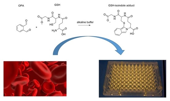
2.1. Optimized Fluorometric Method

After collection, whole-blood samples were centrifuged, and the plasma and the buffy coat layer of white blood cells was removed. The erythrocytes pellet was lysed in four volumes of water by freezing and thawing for three times, and treated with 1:5 volume of tributylphosphine (10% solution in DMF) for the reduction in GSSG and the protein-bound GSH. Free proteins were precipitated with two volumes (2:1) of 10% metaphosphoric acid at 4 °C for 30 min. Samples were centrifuged at 15,000× g for 30 min at 4 °C, and 20 µL of supernatant (sample or analytical standard) was diluted with 300 µL of 0.1 M phosphate buffer pH 8.0 (containing 0.1% EDTA) and derivatized with 20 μL of o-phthaldialdehyde (OPA) solution (1 mg/mL in methanol). After incubation in the dark at room temperature for 15 min, and subsequent fluorophore formation, 200 µL of each sample or standard were placed in a 96-well dark plate and analysed by a spectrofluorometric multiplate reader (Appliskan, Thermo Fischer, Waltham, Massachusetts, USA; ʎex: 340 nm, ʎem: 450 nm), and the remaining aliquot was used for HPLC determinations. GSH intracellular levels were expressed as millimolar concentrations (mM), which referred to the initial volume of red blood fraction.
The GSH calibration curves display a good linearity in the range 0.5–5 mM (R2 = 0.9919) (Figure 2). An aliquot of each sample after OPA derivatization was withdrawn and analysed via HPLC for comparative evaluation of GSH concentration with the chromatographic validated quantification. The limit of detection (LOD) and limit of quantitation (LOQ) were determined by calculating the signal-to-noise (S/N) ratio by the analysis of samples with decreasing concentrations of GSH. LOD, with this direct fluorometric method, corresponds to 0.15 mM, and LOQ to the lowest concentration values used in the calibration plot, which is 0.5 mM.

2.2. HPLC Glutathione Determination

The previously described OPA reaction mixture (50 µL) was analyzed by HPLC. The chromatographic system was equipped with a Waters 600F pump and controller, and a Waters 717 auto-sampler. The reverse phase column was an X-Bridge C18, 5 μm, 4.6 × 150 mm column associated with a C18, 3.9 × 20 mm guard column (Waters Corporation, Milford, MA, USA). The mobile phase consisted of 15% (v/v) methanol in 25 mM Na2HPO4, pH 6.0. Figure 3 shows the isocratic elution of the GSH-OPA derivative at 37 °C, with a flow rate of 0.6 mL/min (Figure 3). The excitation/emission wavelengths were set to 350/420 nm in a Shimadzu RF551 spectrofluorometric detector. A calibration curve was performed in the same conditions of the real samples, as previously described (Figure 4). The instrument control and data acquisition were carried out using the Waters Millennium32 software (Milford, MA, USA). As known from literature, OPA derivatization of GSH and HPLC chromatographic analysis are validated methods with total recovery and high stability of the indole fluorescent analyte [15,17].

2.3. Method Comparison

The comparative analyses of different RBC samples evidenced that the results obtained with the optimized fluorometric method are in agreement, and with a very good correlation, with the validated HPLC method. In Table 1, the GSH quantification results obtained by the two methods are shown, and in Figure 5 the correlation graph with an R2 value of 0.9809.

3. Discussion

The optimized fluorometric method reported here allows the determination of 96 samples per analysis, with respect to the well-established HPLC methods for total GSH determination in erythrocytes. In this work, the sample preparation and the derivatization were optimized for direct fluorescence detection of the GSH-OPA isoindole derivative. As reported in the literature, the advantage of OPA-GSH analytical determinations is the higher stability of the fluorescent derivative, with respect to other fluorophores [15,17]. HPLC still represents the best reliable technique that gives precise, accurate and validated results; however, the procedure has a lot of economical/ecological and time-consuming limitations, such as organic solvents consumption and larger chromatographic times. Erythrocytes are a rich biological matrix of GSH at the millimolar scale, and the method reported here compared to the HPLC validated method is demonstrated to be a suitable and convenient alternative for the analysis of total GSH in red blood cells.

Author Contributions

Conceptualization, A.F., S.F. and L.M.; methodology, A.F. and S.F.; software, A.F., R.M. and M.F.; validation, A.F., M.D., R.A.C. and M.F.; formal analysis, A.F., M.F., L.M. and R.A.C.; investigation, A.F. and S.F.; resources, A.F., L.M., M.D. and M.F.; data curation, R.A.C., L.M., M.D., R.M. and A.F.; writing—original draft preparation, A.F., L.M., R.A.C. and R.M.; writing—review and editing, R.A.C., R.M., L.M. and A.F.; visualization, A.F. and L.M.; supervision, M.F., M.D., A.F. and L.M.; project administration, L.M. and A.F.; funding acquisition, L.M., M.D., A.F. and M.F. All authors have read and agreed to the published version of the manuscript.

Funding

This work was supported by funds from Gnosis by Lesaffre; MIUR Ateneo 2018 and 2019 (Sapienza University of Rome, Italy); EMBO grant, FEBS grant, and Sapienza Ateneo 2019 and 2020 grant to A.F.

Institutional Review Board Statement

Ethical review and approval were waived for this study. The analyzed samples were provided by the same authors of this work and the remaining investigations were performed in leftover samples collected for purposes in the benefit of the patient.

Informed Consent Statement

Informed consent was obtained from all subjects involved in the study.

Conflicts of Interest

The authors declared no conflict of interest.

References

  1. Giustarini, D.; Milzani, A.; Dalle-Donne, I.; Rossi, R. Red blood cells as a physiological source of glutathione for extracellular fluids. Blood Cells Mol. Dis. 2008, 40, 174–179. [Google Scholar] [CrossRef] [PubMed]
  2. Ferreira, A.L.A.; Machado, P.E.A.; Matsubara, L.S. Lipid peroxidation, antioxidant enzymes and glutathione levels in human erythrocytes exposed to colloidal iron hydroxide in vitro. Braz. J. Med. Biol. Res. 1999, 32, 689–694. [Google Scholar] [CrossRef] [PubMed] [Green Version]
  3. Ates, B.; Ercal, B.C.; Manda, K.; Abraham, L.; Ercal, N. Determination of glutathione disulfide levels in biological samples using thiol-disulfide exchanging agent, dithiothreitol. Biomed. Chromatogr. 2009, 23, 119–123. [Google Scholar] [CrossRef] [PubMed]
  4. Ercal, N.; Yang, P.; Aykin, N. Determination of biological thiols by high-performance liquid chromatography following derivatization by ThioGlo maleimide reagents. J. Chromatogr. B Biomed. Sci. Appl. 2001, 753, 287–292. [Google Scholar] [CrossRef]
  5. Jones, D.P.; Carlson, J.L.; Samiec, P.S.; Sternberg, P.; Mody, V.C.; Reed, R.L.; Brown, L.A.S. Glutathione measurement in human plasma. Clin. Chim. Acta 1998, 275, 175–184. [Google Scholar] [CrossRef]
  6. Dinarelli, S.; Longo, G.; Dietler, G.; Francioso, A.; Mosca, L.; Pannitteri, G.; Boumis, G.; Bellelli, A.; Girasole, M. Erythrocyte’s aging in microgravity highlights how environmental stimuli shape metabolism and morphology. Sci. Rep. 2018, 8, 5277. [Google Scholar] [CrossRef] [PubMed] [Green Version]
  7. Francioso, A.; Fanelli, S.; Vigli, D.; Ricceri, L.; Cavallaro, R.A.; Baseggio Conrado, A.; Fontana, M.; D’Erme, M.; Mosca, L. HPLC Determination of Bioactive Sulfur Compounds, Amino Acids and Biogenic Amines in Biological Specimens. In Advances in Experimental Medicine and Biology; Springer Nature Switzerland AG: Cham, Switzerland, 2017; pp. 535–549. [Google Scholar]
  8. Haj-Yehia, A.I.; Benet, L.Z. 2-(4-N-Maleimidophenyl)-6-methoxybenzofuran: A superior derivatizing agent for fluorimetric determination of aliphatic thiols by high-performance liquid chromatography. J. Chromatogr. B Biomed. Sci. Appl. 1995, 666, 45–53. [Google Scholar] [CrossRef]
  9. Wang, X.; Chi, D.; Song, D.; Su, G.; Li, L.; Shao, L. Quantification of Glutathione in Plasma Samples by HPLC Using 4-Fluoro-7-nitrobenzofurazan as a Fluorescent Labeling Reagent. J. Chromatogr. Sci. 2012, 50, 119–122. [Google Scholar] [CrossRef] [PubMed]
  10. Di Pietra, A.M.; Gotti, R.; Bonazzi, D.; Andrisano, V.; Cavrini, V. HPLC determination of glutathione and l-cysteine in pharmaceuticals after derivatization with ethacrynic acid. J. Pharm. Biomed. Anal. 1994, 12, 91–98. [Google Scholar] [CrossRef]
  11. Sentellas, S.; Morales-Ibanez, O.; Zanuy, M.; Albertí, J.J. GSSG/GSH ratios in cryopreserved rat and human hepatocytes as a biomarker for drug induced oxidative stress. Toxicol. Vitr. 2014, 28, 1006–1015. [Google Scholar] [CrossRef] [PubMed]
  12. Giustarini, D.; Dalle-Donne, I.; Milzani, A.; Fanti, P.; Rossi, R. Analysis of GSH and GSSG after derivatization with N-ethylmaleimide. Nat. Protoc. 2013, 8, 1660–1669. [Google Scholar] [CrossRef] [PubMed]
  13. Whillier, S.; Raftos, J.E.; Kuchel, P.W. Glutathione synthesis by red blood cells in type 2 diabetes mellitus. Redox Rep. 2008, 13, 277–282. [Google Scholar] [CrossRef] [PubMed]
  14. Van ’T Erve, T.J.; Wagner, B.A.; Ryckman, K.K.; Raife, T.J.; Buettner, G.R. The concentration of glutathione in human erythrocytes is a heritable trait. Free Radic. Biol. Med. 2013, 65, 742–749. [Google Scholar] [CrossRef] [PubMed] [Green Version]
  15. Hou, Y.; Li, X.; Dai, Z.; Wu, Z.; Bazer, F.W.; Wu, G. Analysis of Glutathione in Biological Samples by HPLC Involving Pre-Column Derivatization with o-Phthalaldehyde. In Methods in Molecular Biology; Springer Nature Switzerland AG: Cham, Switzerland, 2018; pp. 105–115. [Google Scholar]
  16. Kand’ár, R.; Žáková, P.; Lotková, H.; Kučera, O.; Červinková, Z. Determination of reduced and oxidized glutathione in biological samples using liquid chromatography with fluorimetric detection. J. Pharm. Biomed. Anal. 2007, 43, 1382–1387. [Google Scholar] [CrossRef] [PubMed]
  17. Hamad, A.; Elshahawy, M.; Negm, A.; Mansour, F.R. Analytical methods for determination of glutathione and glutathione disulfide in pharmaceuticals and biological fluids. Rev. Anal. Chem. 2021. [Google Scholar] [CrossRef]
Figure 1. Glutathione (GSH) reaction with ortho-phthalaldehyde (OPA) (left) produces the stable isoindole detectable fluorophore (right).
Separations 08 00083 g001
Figure 2. Direct fluorometric method calibration curve in the range 0.5–5 mM.
Separations 08 00083 g002
Figure 3. HPLC chromatogram of GSH-OPA derivative.
Separations 08 00083 g003
Figure 4. HPLC calibration curve in the range 0.5–5 mM.
Separations 08 00083 g004
Figure 5. Correlation between optimized direct fluorometric method and validated HPLC analyses.
Separations 08 00083 g005
Table 1. GSH concentrations of different RBCs samples obtained by the direct optimized method and the validated HPLC one. RSD for all reported values was ≤5%.
SampleHPLC (mM)Fluorometric Method (mM)
10.580.55
21.831.96
31.451.59
40.780.86
50.840.91
60.910.98
71.021.08
81.151.22
91.231.24
101.481.53
111.611.71
121.721.66
Publisher’s Note: MDPI stays neutral with regard to jurisdictional claims in published maps and institutional affiliations.

