Article

Comparison of Phenolic Compounds in Olive Leaves by Different Drying and Storage Methods

1
College of Life Science, Sichuan Agricultural University, Ya’an 625014, China
2
College of Agricultural Science, Xichang University, Xichang 615000, China
3
College of Agronomy Science, Sichuan Agricultural University, Chengdu 611130, China
*
Author to whom correspondence should be addressed.
The author contributed equally to this manuscript.
Separations 2021, 8(9), 156; https://doi.org/10.3390/separations8090156
Submission received: 13 August 2021 / Revised: 4 September 2021 / Accepted: 8 September 2021 / Published: 17 September 2021

Abstract

:
Oleuropein, a bitter substance that exists in olive leaves, can be hydrolyzed to hydroxytyrosol. These are the main phenolic compounds, and they have beneficial properties to human bodies. In this study, we established a simple and new method to determine oleuropein and hydroxytyrosol quickly by HPLC. HPLC conditions were set as follows: water (A) acetonitrile (B) as mobile phase, gradient elution orders: 90%A–10%B for 0–10 min, 80%A–20%B for 14–30 min, and then change to 90%A–10%B for 30–33 min; detection wavelength: 280 nm. Compared with other detection methods, the method simplified the elution procedure and shortened the time. Additionally, we provided a better drying method and preservation of olive leaves in tea drinking production that were air-dried at room temperature of 25 °C.

1. Introduction

The olive (Olea europaea L.) is one of the most important economic trees in Mediterranean countries. Since the olive trees were introduced into China from Albania in 1956, they have rapidly become an emerging economic tree species. Recent advances have seen 80,000 hectares of olive trees being widely cultivated in several regions such as Gansu, Sichuan, Yunnan, and Hubei Province (China) by the end of 2017 [1,2].
The leaves were traditionally regarded as important by-products in olive oil production because they account for 10% of total weight of olive trees [3]. Accumulating evidence has demonstrated that the olive leaves are potential resources of various antioxidants, such as oleuropein and hydroxytyrosol. The leaf extracts are often mixed into oil to increase the flavor and resist deterioration [4,5]. In addition, the leaf extract is also commonly used as a safe natural additive in cosmetics, functional foods, and medicines [6]. Furthermore, due to the abundant antioxidant ingredients, Olive leaves have been used to develop novel beverages, such as olive leaf tea.
Oleuropein, hydroxytyrosol, flavonoids, and other phenolic compounds (rutin, caffeic acid and tyrosol) can contribute to many health promoting effects due to their free radical scavenging activity [7]. Oleuropein, the major component in olive fruits and leaves, exhibits strong preventive effects against oxidation [8,9]. Oleuropein consists of a molecular of hydroxytyrosol and elenolic acid glucoside [10]. It was well investigated that oleuropein possessed many beneficial pharmacological properties, such as antioxidant, anti-inflammatory, anti-microbial, anti-viral, neuro-protective effects, as well as anti-ageing [11], and it is widely used as a functional component in the food industry.
However, oleuropein is unstable and could degrade into hydroxytyrosol in fruits and leaves under certain conditions [12,13,14]. One of the products, hydroxytyrosol, also belongs to a strong anti-oxidative compound that has similar biological properties to oleuropein. The process of oleuropein degradation is also called de-astringent, and the hydroxytyrosol content is often evaluated as oleuropein degradation rate [15]. Therefore, a simple method to quickly detect these two components is necessary.
Additionally, processing methods significantly affect the nutrition and ingredients of many plant materials. For example, drying methods, storage conditions and processing had a varying degree of effect on the phenolic components of olive leaves after collection. Hence, it is an essential quality guarantee and cost-effectiveness issue to optimize these processes before commercial applications of olive leaves. Several common methods, such as vacuum drying, microwave, oven, and infrared heating were applied to the drying process. The content of most plant phenolic components was sensitive to the drying methods and preservation conditions. Therefore, it is of vital importance to optimize the dry conditions of olive leaves in food and animal feeding industries.
In this study, we established a simple chromatographic method to simultaneously detect oleuropein and hydroxytyrosol contents in olive leaves. Then, we investigated the effects of different drying methods on oleuropein, hydroxytyrosol and other phenolic compounds content in olive leaves in the drying process. Moreover, we also evaluated the effects of storage temperature and time on the content of oleuropein and hydroxytyrosol in dried leaf powder.

2. Materials and Methods

2.1. Reagents and Olive Leaves Sample

Hydroxytyrosol (≥98%), tyrosol (≥98%) and oleuropein (≥98%), which are used as standards, were obtained from Chengdu Biopurity Phytochemicals Ltd. China. Folin-Ciocalteau, rutin (≥99%), gallic acid (≥99%), HPLC grade acetonitrile and methanol were from Sigma-Aldrich (Shanghai, China).
Fresh olive leaves (Manzanilla variety) were harvested randomly from pruned branches and collected from Jintang, Sichuan province in October 2019. Collected leaves were divided into four parts, then dried in the oven at the temperature of 45 °C and 70 °C (DO45 and DO70) for 2 days. The other two parts were either air-dried at room temperature of 25 °C (ADRT) under ventilation for 10 days or dried in freezing vacuum dryer (DFVD) for 2 days. All groups were dried to constant weight. The olive leaves dried with different methods were powered and refined by a 60-mesh filter, then delivered to three sealed container and stored at the temperature of −20 °C, 4 °C, 25 °C in the dark, respectively.

2.2. Sample Preparation of Olive Leaves

A quantity of 0.1 g of dried leaf powder was mixed with 5 mL 78% v/v methanol-water solution, and incubated at 37 °C for 20 min for extraction. Then the extract was centrifuged at 4000 r/min for 4 min. The supernatant was collected for sebsequent determination and analysis.

2.3. Determination of Chemical Composition

2.3.1. Total Phenols Content of Olive Leaves

The total phenols content (TPC) determination of olive leaves under different drying methods was carried out as previously with some adjustments [16]. The experiment procedure was established in line with the Folin-Ciocalteau method. In brief, the olive leaf extracts (0.1 mL) were reacted with the 0.25 mL Folin-Ciocalteau for 2 min. Then we added 1 mL 7% (w/v) sodium carbonate solution and mixed uniformly. The mixture solution was diluted to 10 mL with ultra-pure water. The reaction solution in tubes was incubated at 25 °C for 1.5 h. The absorbance at 760 nm was measured by a micro-plate reader (Spectra max M2, Molecular Devices, Sunnyvale, CA, USA). The total phenols content was calculated with milligrams of Gallic acid equivalent (GAE) per gram dry weight of the sample (mg/g) as per the following equation: y = 0.5944x + 0.0376, R2 = 0.9915.

2.3.2. Total Flavonoids Content of Olive Leaves

Total flavonoids content (TFC) of the extract was estimated by NaNO2-Al (NO3)3 method that was previously described by [17,18] and with some modifications. Briefly, an 0.2 mL sample was diluted with 0.8 mL ethanol (70% w/v). The diluted sample solution was added with 0.2 mL NaNO2 (5% w/v) and Al (NO3)3 (10% w/v), then mixed to react for 5 min. Then we added 1 mL NaOH (4% w/v). The solution was adjusted to 3 mL with ethanol (70% w/v), and shaken to react for 15 min. The absorbance was measured at 510 nm. The TFC was expressed in terms of milligrams of rutin equivalent (RE) per gram dry weight of the sample (mg RE/g) with the following formula: y = 0.4172x − 0.0014, R2 = 0.9995.

2.4. HPLC Analysis of Phenolic Compounds

In our study, we have established a simple chromatographic method to analyze the two main phenols compounds. The extract was performed according to the method described in Section 2.2 and filtered through 0.22 µm filter. The sample analysis was conducted on the Agilent series 1260 HPLC instrument (Agilent Technologies, Shanghai, China) with a G1311B quaternary pump, a G1329B auto-sampler, a G1316A thermo-stated column compartment and equipped with a G1315D diode array detector (DAD). The chromatographic separation was performed on a reverse phase column ZORBAX SB-C18 (150 mm × 4.6 mm, 5.0 µm, Agilent). For hydroxytyrosol (HT), tyrosol (TYR), and oleuropein (OLE) determination, the mobile phase consisted of water (A) and acetonitrile (B) with a gradient elution of 90%A–10%B for 0~10 min, 80%A–20%B for 14~30 min, and then changed to 90%A–10%B for 30~33 min. The column temperature was set to 30 °C and the flow rate of mobile phase was 0.8 mL/min. The injection volume was 10 µL, and DAD recorded the ultraviolet light absorption value at 254, 260, and 280 nm. The other phenolic compounds analysis was performed as reported previously [16].
The data analyzed with the Agilent OpenLAB CDS ChemStation Edition software (revision C.01.05, Agilent Technologies, Shanghai, China). The extraction yield was calculated by the following equation: extraction yield (mg/g dw) = [(C × V)/W]/1000 where C is the concentration of analysis in each solvent calculated by calibration curves Table 1, V is the volume of the extract (mL), and W is the dried weight (dw) of sample (g).
Precision: In order to evaluate the reproducibility and stability of the detection method, the precision experiment was conducted as previously [19] with some adjustments. All calibration points were obtained in duplicate injection to set the confidence intervals. According to the chromatographic conditions, the mixed standards solution was continuously injected six times (n = 6) to obtained the precision data.
Recovery: Blank solution (methanol), appropriate standards, and olive leaf extract sample were analyzed under the analytical method conditions. The recovery rates were measured by spiking the olive leaf extract (0.6 mL) with a suitable volume of mixed phenolic standard stock solution. The mixed samples were continuously injected and analyzed five times (n = 5).
Repeatability: The olive leaves samples were prepared and analyzed in HPLC under the 280 nm for repeatability experiments. The sample detection was performed continuously, and each experiment was replicated five trials (n = 5).

2.5. Identified Reaction Monitoring Based on Quantitation of HPLC Analysis Condition

The standard stock solutions of HT, TYR and OLE were dissolved with methanol at the final concentration of 160 µg/mL, 180 µg/mL, 2723.84 µg/mL, respectively, and stored at 4 °C. HT working solutions at 1.25µg/mL, 2.50 µg/mL, 5.00 µg/mL, 10.00 µg/mL, 20.00 µg/mL, 40.00 µg/mL, 80.00 µg/mL, and 160.00 µg/mL were prepared by dilution of the stock solutions with methanol. TYR working solutions at 22.81 µg/mL, 5.63 µg/mL, 11.25 µg/mL, 22.50 µg/mL, 45.00 µg/mL, 90.00 µg/mL, and 180.00 µg/mL were prepared by dilution of the stock solutions with methanol. OLE working solutions at 10.64 µg/mL, 21.28 µg/mL, 42.56 µg/mL, 85.12 µg/mL, 170.24 µg/mL, 340.48 µg/mL, 680.96 µg/mL, 1361.92 µg/mL, and 2723.84 µg/mL by dilution of the stock solutions with methanol. The working standard solutions were used to prepare matrix-matched standards and spike samples in the validation studies.

2.6. HPLC Analysis of Olive Samples under Different Drying and Storage Condition

The fresh olive leaves were separated into four parts by using the four different drying methods. Then the materials were analyzed by HPLC of the contents of hydroxytyrosol, tyrosol and oleuropein at the 1st, 3rd, 5th, 7th and 9th week.

2.7. Statistical Analysis

The results were expressed as means ± standard deviation (SD) of replicate solvent extractions and triplicate of assays. One-way analysis of variance (ANOVA) was used to compare the means. The means were separated by Least Significant Difference (LSD). All statistical analysis was performed using the Statistical Package for the Social Sciences (SPSS) 19.0. The statistical graphs were drawn with GraphPad Prism software (version 6.0, San Diego, CA, USA).

3. Results and Discussion

3.1. Evaluation of HPLC Detection Method

Up to now, many researchers have developed a series of HPLC methods to determine the phenolic compounds. Different types of combined mobile phase, such as methanol, acetonitrile, isopropanol, phosphoric acid, formic acid or acetic acid, and sodium acetate, were applied in terms of various chromatographic columns [20,21,22,23,24]. In this study, we established a simple chromatographic method with water and acetonitrile with short detection time.
The target phenolic compounds were qualitatively and quantitatively analyzed in this study. In Figure 1, we chose the 254, 260 and 280 nm wavelengths that were commonly used to detect these compounds. The peak of the three compounds were completely detected in standard sample. However, in olive extract sample, the content of HT may be extremely low, and it can only be detected at 280 nm (Figure 1B). Thus, we chose 280 nm for subsequent experiments (Figure 1A).
As shown in Table 1, the retention times of HT, TYR and OLE standards were 4.8, 8.3 and 29.3 min, respectively. However, the TYR in olive sample failed to be detected. This might be due to the low content which exceeded the detection limit in the olive sample. The linear calibration ranges, calibration equations, regression coefficients, limit of detections (LOD), and quantitation (LOQ) of three compounds applied with this chromatographic method were shown in Table 1 in detail. The lowest detectable limitation for OLE of this method was lower than the study from Japón-Luján, which showed LOD values of 11.46 and 11.04 µg/mL for OLE using ultrasonic or microwave assisted extraction with HPLC tandem MS detection method in olive leaves [25,26]. The LOD value for HT was 1.25 µg/mL which was higher than other reported methods (LOD values for HT were 0.14 to 0.4 µg/mL) [27,28]. These results might be due to different plant materials, extraction methods, quantification conditions, and apparatus.

3.1.1. Precision

The Table 2 showed that the peak areas were extremely similar and the relative standard deviations (RSD) of HT, TYR and OLE were 0.822, 0.811 and 0.766%, respectively, indicated that the established method has high precision and strong repeatability.

3.1.2. Recovery

The recovery data were calculated by peak area under the 280 nm. The results were summarized in Table 3. The recovery rates of HT, TYR and OLE were in the range of 102.79–109.60, 97.14–102.28 and 89.43–95.24%, respectively. Similar recovery rates analyzed by HPLC were reported from an olive pomace extraction (recovery rates were 93–98%) [29]. The RSD values of HT, TYR and OLE were 2.208, 1.742 and 2.322%, respectively. The results indicated that using the new chromatographic method could obtain a stable, accurate, and reliable recycling result.

3.1.3. Repeatability

The results were showed in Table 2. The peak area values of HT and OLE are in the range of 41.82–44.63 mAU and 6026.10–6493.57 mAU, respectively. However, the tyrosol in olive leaves did not be observed, may be the content was below the detection limit. The RSD values of HT and OLE were 2.556 and 2.786%, respectively, which were better than the RSD value from a study employing dispersive liquid–liquid micro extraction coupled with HPLC analysis (RSD for OLE and HT were 4.12–5.63%) [29].

3.2. Effects of Different Drying Methods on Phenolic Compound Contents

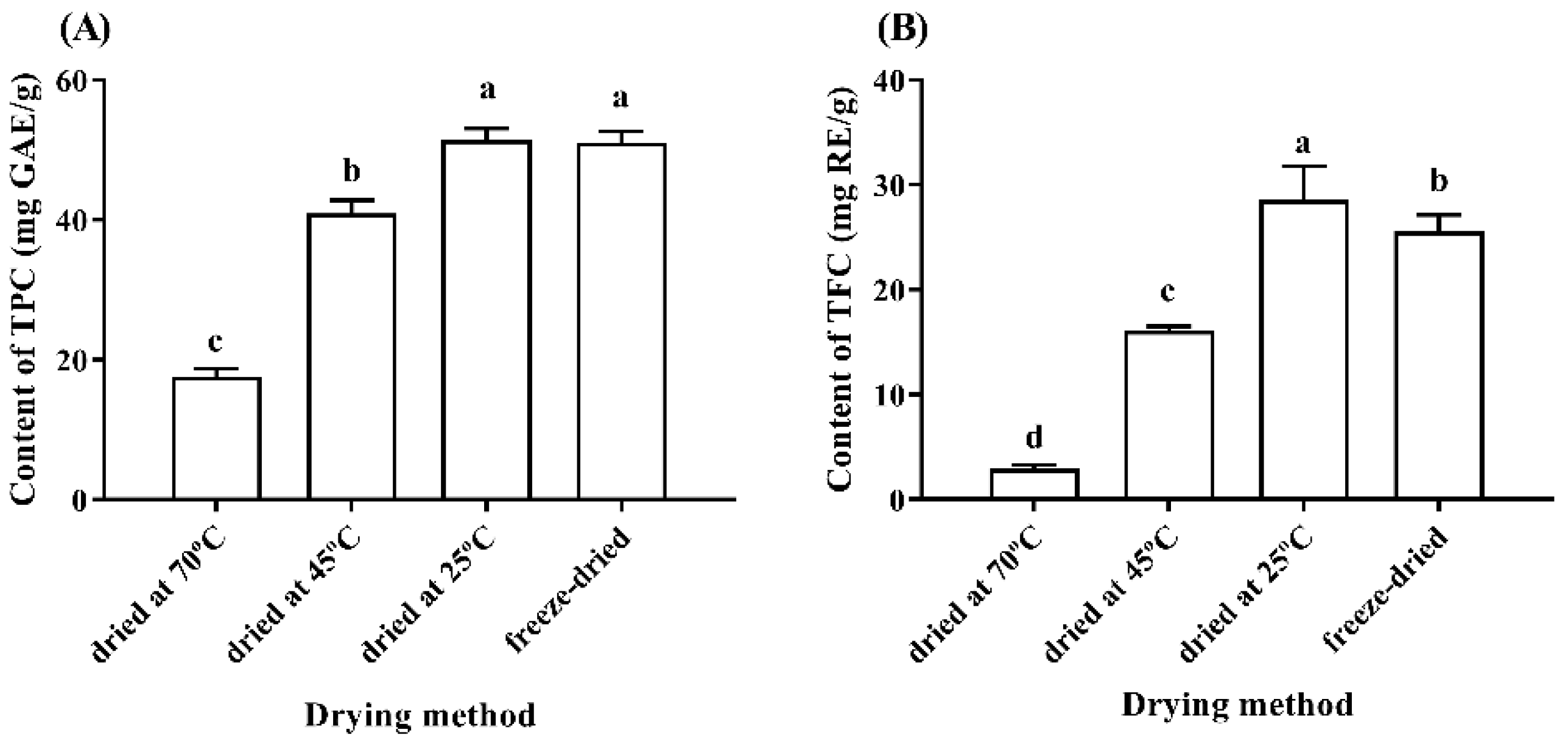
The olive leaves samples were extracted and determined according to the Section 2.2, respectively. The results showed that the TPC contents of DO70, DO45, ADRT and DFVD were 17.58 ± 1.14, 40.09 ± 1.91, 51.48 ± 1.58 and 51.07 ± 1.60 mg GAE/g, respectively (Figure 2A), indicated that plant materials processing with high temperature significantly reduced the total phenolic content. Similar results were reported in a green coffee roasting study [30]. The TFC contents of DO70, DO45, ADRT and DFVD were 2.91 ± 0.41, 16.12 ± 0.39 and 28.59 ± 3.20 and 25.59 ± 1.58 mg GE/g, respectively (Figure 2B). In other aqueous methanol extract of olive leaves, similar TFC content of 15.55 mg GE/g was determined [31]. The TPC contents of ADRT and DFVD had insignificant differences (p > 0.05), but obviously differed from DO70 and DO45 (Figure 2A) (p < 0.05). As shown in Figure 2B, the TFC contents of four groups of leaf powder exhibited significant differences (p < 0.05) with a trend that relates higher temperature to lower contents.
The contents of extracted ingredients from plants were often related to the material processing and extraction methods [18]. The polyphenol compounds containing multiple hydroxyl groups were unstable and more easily oxidized during the material processing, so there was an obvious difference in composition and content of active ingredients under different processing procedure or temperature [32], especially OLE and HT. Therefore, we investigated the OLE and HT contents of olive leaves materials under four drying methods. As can been seen in Figure 3A, the OLE contents of DO70, DO45, ADRT and DFVD were 1.32 ± 0.11, 40.75 ± 1.02, 82.72 ± 0.71, and 69.00 ± 0.61 mg/g dw, respectively. Similarly, 23 and 43.2 mg/g of OLE contents in olive leaves were shown in other studies with different extraction and drying methods [25,33]. However, these contents were lower than a previous study which reported that the content ranged from 87.2 to 136.1 mg/g dw in 73 olive leaf samples [34]. In Figure 3B, the HT contents of DO45, ADRT and DFVD were 0.15 ± 0.02, 0.36 ± 0.01 and 0.27 ± 0.01 mg/g dw, respectively. The HT content was lower than a hydrolyzed phenolic extract of olive leaves (0.2 g/100 g), which might be due to the different processing method [35].
In total, the OLE and HT contents in the experiments showed the same trend as TFC. Materials of ADRT contain the highest levels of OLE and HT. Secondly, the DO45 was followed by DFVD. The sample of DO70 contained a low OLE content and undetectable HT content. For the olive leaves treated with high temperature, the contents of OLE and HT decreased rapidly with increasing temperature. Normally, OLE could be hydrolyzed to HT. However, HT content did not increase with the OLE degradation under high temperature conditions, indicating that HT was sensitive to high temperature and might degrade quickly at higher temperatures. The findings showed that the OLE and HT contents were significantly different under the four common drying methods (i.e., different drying temperatures). The best drying method was air-dried at room temperature, followed by freeze-drying, and then drying at 45 °C. Air-dried at room temperature is the best drying method for olive leaves, because it maximizes OLE and HT contents in olive leaves.
Drying was the first procedure for olive leaves to apply in the process industry. As illustrated above, the bioactive components in olive leaves were influenced by different dehydrating methods. ADRT was the best method with the advantage that it is appropriate, simple, and low-cost. The olive leaves with different drying methods showed different contents of TP, TF, OLE, and HT. When treated with high temperature, the four main components were extremely reduced. Similar results were reflected in other phenolic compounds that are displayed in Table 4 (standard curve shown in Table 5). Low content compounds, such as rutin, luteolin-7-O-lucoside, and apigenin-7-O-glucoside, were also tested. The content of these compounds, except for luteolin, showed a similar trend to OLE and HT. Leaves dried at room temperature contained high phenolic compounds. Thus, high temperature was not suitable for olive leaves because it may cause a loss of bioactive components. Olive leaves should be protected from high temperatures during the drying process.

3.3. Effects of Different Storage Temperatures and Time on the OLE and HT Contents

Based on the results of the leaves treated with four drying methods, we found that the group dried at 25 °C had the highest contents of TP, TF, OLE and HT. The group in which leaves were air-dried in room temperature (ADRT) was selected for subsequent experiments. The ADRT leaf powder was stored at −20 °C, 4 °C and 25 °C for 9 weeks, and the OLE and HT contents were measured consecutively by HPLC. As can be seen in Table 6, there were no significant differences in the OLE and HT contents at three storage temperatures. As the HPLC chromatogram results show in Table 6, the OLE content decreased significantly from the beginning of the experiment to one week later. At −20, 4 and 25 °C, the OLE content was reduced from 82.72 to 78.28, 82.72 to 77.43 and 82.72 to 77.58 mg/g dw, respectively, in first week. However, from the 3rd to 9th week, it exhibited only a slight fluctuation. In general, the OLE contents maintained a stable state in long-term storage.
The HT content showed a significant increasing trend in the 1st week, which might be due to the ability for OLE to be converted to HT by β-glucosidase in the storage process. Then, there was a downward trend during the following 2 weeks. The HT contents were reduced from 0.45 to 0.33, 0.41 to 0.29, and 0.45 to 0.24 mg/g dw, respectively, at −20, 4 and 25 °C. Then, the HT content remained stable fluctuation after the 3rd week. In summary, the contents of OLE and HT in the powdered dry olive leaves were affected significantly in the early stage, then generally became unchanged under these three common storage conditions.
Therefore, we suggest that olive leaves are air-dried or dehydrated at room temperature before being made into food or quality additives. Olive leaves air-dried at room temperature preserve the leaf greenness of leaves, and their luminosity is enhanced. Considering industrial processing costs, material preservation and the nutritional contents and olive tea drinking habits, we recommend that fresh leaves are stored at room temperature for drying, and it is advisable to store dried olive leaves or olive tea at 4 °C.

4. Conclusions

In this study, a simple method was established for determining the contents of OLE and HT in olive leaves by HPLC. The results showed that OLE and HT could be completely detected within 33 min. The determination results had good repeatability, high accuracy, and good recovery under the chromatographic conditions.
The most suitable drying method for olive leaves was air-drying at room temperature. It attributed high phenolic compounds content. Moreover, it is recommended to prevent the high temperature baking olive leaves during the drying process. For storage conditions, there were insignificant differences between the OLE and HT contents stored at −20, 4 and 25 °C for several weeks. However, in industrial processing, the advisable storage condition for fresh or dried olive leaves could be 25 °C, because it is convenient, economical, and suitable for cosmetic applications.

Author Contributions

Writing-original draft preparation, writing-review and editing, S.F.; Methodology, writing—review and editing: S.F. and C.Z.; Conceptualization: Z.X.; Formal analysis, T.C.; Investigation, T.L.; Resources, M.Y.; Data curation, L.L.; Supervision, L.Z.; Project administration: C.D. All authors have read and agreed to the published version of the manuscript.

Funding

This work was supported by the National Natural Science Foundation of China [grant numbers: 31900294], Department of Sichuan Provincial Science and Technology [grant numbers: 20GJHZ0050], Chengdu Science and Technology Bureau [grant numbers: 2019-YF05-00075-SN], and Xichang Science and Technology Bureau [grant numbers: 19YYJS18].

Institutional Review Board Statement

This study did not involve humans or animals assays.

Informed Consent Statement

The study did not involve humans.

Data Availability Statement

The data presented in this study are available on request from the corresponding author.

Conflicts of Interest

The authors declare that they have no known competing financial interests or personal relationships that could have appeared to influence the work reported in this paper.

References

  1. Xiang, C.; Xu, Z.; Liu, J.; Li, T.; Yang, Z.; Ding, C. Quality, composition, and antioxidant activity of virgin olive oil from introduced varieties at Liangshan. LWT 2017, 78, 226–234. [Google Scholar] [CrossRef]
  2. Su, C.; Sun, J.; Zhu, W.; Peng, L. History, Distribution, and Potential of the Olive Industry in China: A Review. Sustainability 2018, 10, 1426. [Google Scholar] [CrossRef] [Green Version]
  3. Herrero, M.; Temirzoda, T.N.; Segura-Carretero, A.; Quirantes, R.; Plaza, M.; Ibañez, E. New possibilities for the valorization of olive oil by-products. J. Chromatogr. A 2011, 1218, 7511–7520. [Google Scholar] [CrossRef] [Green Version]
  4. Bouaziz, M.; Fki, I.; Jemai, H.; Ayadi, M.; Sayadi, S. Effect of storage on refined and husk olive oils composition: Stabilization by addition of natural antioxidants from Chemlali olive leaves. Food Chem. 2008, 108, 253–262. [Google Scholar] [CrossRef]
  5. Kiritsakis, K.; Kontominas, M.G.; Kontogiorgis, C.; Hadjipavlou-Litina, D.; Moustakas, A.; Kiritsakis, A. Composition and Antioxidant Activity of Olive Leaf Extracts from Greek Olive Cultivars. J. Am. Oil Chem. Soc. 2009, 87, 369–376. [Google Scholar] [CrossRef]
  6. Yuan, J.; Wang, C.; Ye, J.; Tao, R.; Zhang, Y. Enzymatic hydrolysis of oleuropein from olea europea (olive) leaf extract and antioxidant activities. Molecules 2015, 20, 2903–2921. [Google Scholar] [CrossRef] [PubMed] [Green Version]
  7. Rahmanian, N.; Jafari, S.M.; Wani, T.A. Bioactive profile, dehydration, extraction and application of the bioactive components of olive leaves. Trends Food Sci. Technol. 2015, 42, 150–172. [Google Scholar] [CrossRef]
  8. Lockyer, S.; Rowland, I.; Spencer, J.P.E.; Yaqoob, P.; Stonehouse, W. Impact of phenolic-rich olive leaf extract on blood pressure, plasma lipids and inflammatory markers: A randomised controlled trial. Eur. J. Nutr. 2016, 56, 1421–1432. [Google Scholar] [CrossRef] [PubMed] [Green Version]
  9. Shahdadi, F.; Mirzaei, H.O.; Daraei Garmakhany, A. Study of phenolic compound and antioxidant activity of date fruit as a function of ripening stages and drying process. J. Food Sci. Technol. 2013, 52, 1814–1819. [Google Scholar] [CrossRef] [PubMed] [Green Version]
  10. Nikolaivits, E.; Termentzi, A.; Skaltsounis, A.-L.; Fokialakis, N.; Topakas, E. Enzymatic tailoring of oleuropein from Olea europaea leaves and product identification by HRMS/MS spectrometry. J. Biotechnol. 2017, 253, 48–54. [Google Scholar] [CrossRef] [PubMed]
  11. Haris Omar, S. Oleuropein in olive and its pharmacological effects. Sci. Pharm. 2010, 78, 133–154. [Google Scholar] [CrossRef] [PubMed] [Green Version]
  12. De Leonardis, A.; Macciola, V.; Cuomo, F.; Lopez, F. Evidence of oleuropein degradation by olive leaf protein extract. Food Chem. 2015, 175, 568–574. [Google Scholar] [CrossRef] [PubMed]
  13. Ramírez, E.; Brenes, M.; García, P.; Medina, E.; Romero, C. Oleuropein hydrolysis in natural green olives: Importance of the endogenous enzymes. Food Chem. 2016, 206, 204–209. [Google Scholar] [CrossRef] [PubMed] [Green Version]
  14. Ramírez, E.; Brenes, M.; de Castro, A.; Romero, C.; Medina, E. Oleuropein hydrolysis by lactic acid bacteria in natural green olives. LWT 2017, 78, 165–171. [Google Scholar] [CrossRef]
  15. Ramírez, E.; Medina, E.; Brenes, M.; Romero, C. Endogenous enzymes involved in the transformation of oleuropein in Spanish table olive varieties. J. Agric. Food Chem. 2014, 62, 9569–9575. [Google Scholar] [CrossRef] [Green Version]
  16. Wang, B.; Qu, J.; Feng, S.; Chen, T.; Yuan, M.; Huang, Y.; Liao, J.; Yang, R.; Ding, C. Seasonal Variations in the Chemical Composition of Liangshan Olive Leaves and Their Antioxidant and Anticancer Activities. Foods 2019, 8, 657. [Google Scholar] [CrossRef] [PubMed] [Green Version]
  17. Chen, X.; Wu, X.; Chai, W.; Feng, H.; Shi, Y.; Zhou, H.; Chen, Q. Optimization of extraction of phenolics from leaves of Ficus virens. J. Zhejiang Univ. Sci. B 2013, 14, 903–915. [Google Scholar] [CrossRef] [Green Version]
  18. Jia Zhishen, T.M. Wu Jianming, The determination of flavonoid contents in mulberry and their scavenging effects on superoxide radicals. Food Chem. 1999, 64, 555–559. [Google Scholar] [CrossRef]
  19. Rivera-Mondragón, A.; Broeckx, G.; Bijttebier, S.; Naessens, T.; Fransen, E.; Kiekens, F.; Caballero-George, C.; Vander Heyden, Y.; Apers, S.; Pieters, L.; et al. Ultrasound-assisted extraction optimization and validation of an HPLC-DAD method for the quantification of polyphenols in leaf extracts of Cecropia species. Sci. Rep. 2019, 9, 1–16. [Google Scholar] [CrossRef]
  20. Ahmad-Qasem, M.H.; Barrajón-Catalán, E.; Micol, V.; Mulet, A.; García-Pérez, J.V. Influence of freezing and dehydration of olive leaves (var. Serrana) on extract composition and antioxidant potential. Food Res. Int. 2013, 50, 189–196. [Google Scholar] [CrossRef]
  21. Talhaoui, N.; Gómez-Caravaca, A.M.; León, L.; De la Rosa, R.; Segura-Carretero, A.; Fernández-Gutiérrez, A. Determination of phenolic compounds of ‘Sikitita’ olive leaves by HPLC-DAD-TOF-MS. Comparison with its parents ‘Arbequina’ and ‘Picual’ olive leaves. LWT-Food Sci. Technol. 2014, 58, 28–34. [Google Scholar] [CrossRef]
  22. Xynos, N.; Papaefstathiou, G.; Psychis, M.; Argyropoulou, A.; Aligiannis, N.; Skaltsounis, A.-L. Development of a green extraction procedure with super/subcritical fluids to produce extracts enriched in oleuropein from olive leaves. J. Supercrit. Fluids 2012, 67, 89–93. [Google Scholar] [CrossRef]
  23. Abaza, L.; Taamalli, A.; Nsir, H.; Zarrouk, M. Olive Tree (Olea europeae L.) Leaves: Importance and Advances in the Analysis of Phenolic Compounds. Antioxidants 2015, 4, 682–698. [Google Scholar] [CrossRef] [PubMed]
  24. Irakli, M.; Chatzopoulou, P.; Ekateriniadou, L. Optimization of ultrasound-assisted extraction of phenolic compounds: Oleuropein, phenolic acids, phenolic alcohols and flavonoids from olive leaves and evaluation of its antioxidant activities. Ind. Crop. Prod. 2018, 124, 382–388. [Google Scholar] [CrossRef]
  25. Japón-Luján, R.; Luque-Rodríguez, J.M.; Luque de Castro, M.D. Dynamic ultrasound-assisted extraction of oleuropein and related biophenols from olive leaves. J. Chromatogr. A 2006, 1108, 76–82. [Google Scholar] [CrossRef]
  26. Japón-Luján, R.; Luque-Rodríguez, J.M.; Luque de Castro, M.D. Multivariate optimisation of the microwave-assisted extraction of oleuropein and related biophenols from olive leaves. Anal. Bioanal. Chem. 2006, 385, 753–759. [Google Scholar] [CrossRef]
  27. Godoy-Caballero, M.P.; Acedo-Valenzuela, M.I.; Galeano-Díaz, T. New reversed phase dispersive liquid–liquid microextraction method for the determination of phenolic compounds in virgin olive oil by rapid resolution liquid chromatography with ultraviolet–visible and mass spectrometry detection. J. Chromatogr. A 2013, 1313, 291–301. [Google Scholar] [CrossRef]
  28. Reboredo-Rodríguez, P.; Rey-Salgueiro, L.; Regueiro, J.; González-Barreiro, C.; Cancho-Grande, B.; Simal-Gándara, J. Ultrasound-assisted emulsification–microextraction for the determination of phenolic compounds in olive oils. Food Chem. 2014, 150, 128–136. [Google Scholar] [CrossRef]
  29. Habibi, H.; Mohammadi, A.; Farhoodi, M.; Jazaeri, S. Application and optimization of microwave-assisted extraction and dispersive liquid–liquid microextraction followed by high-performance liquid chromatography for the determination of oleuropein and hydroxytyrosol in olive pomace. Food Anal. Methods 2018, 11, 3078–3088. [Google Scholar] [CrossRef]
  30. Alessia, P.; Rita, P.; Roberto, L.; Antonio, Z. UHPLC-PDA-ESI-TOF/MS metabolic profiling and antioxidant capacity of arabica and robusta coffee silverskin: Antioxidants vs phytotoxins. Food Res. Int. 2017, 99, 155–165. [Google Scholar]
  31. Zouhaier, B.; Junkuy, H.; Hiroko, I.; Sami, S. Hydroxytyrosol rich extract from olive leaves modulates cell cycle progression in MCF-7 human breast cancer cells. Food Chem. Toxicol. 2011, 49, 179–184. [Google Scholar]
  32. Boudhrioua, N.; Bahloul, N.; Ben Slimen, I.; Kechaou, N. Comparison on the total phenol contents and the color of fresh and infrared dried olive leaves. Ind. Crop. Prod. 2009, 29, 412–419. [Google Scholar] [CrossRef]
  33. Hedya, J.; Mohamed, B.; Ines, F.; Abdelfattah, E.F.; Sami, S. Hypolipidimic and antioxidant activities of oleuropein and its hydrolysis derivative-rich extracts from Chemlali olive leaves. Chem.-Biol. Interact. 2008, 176, 88–89. [Google Scholar]
  34. Fathia, A.; Nathalie, D.; Jacques, A.; Sevastianos, R.; Monji, M.; Isabelle, P.G.; Moktar, H. Rapid quantitative determination of oleuropein in olive leaves (Olea europaea) using mid-infrared spectroscopy combined with chemometric analyses. Ind. Crop. Prod. 2012, 37, 292–297. [Google Scholar]
  35. De Leonardis, A.; Aretini, A.; Alfano, G.; Macciola, V.; Ranalli, G. Isolation of a hydroxytyrosol-rich extract from olive leaves (Olea Europaea L.) and evaluation of its antioxidant properties and bioactivity. Eur. Food Res. Technol. 2008, 226, 653–659. [Google Scholar] [CrossRef]
Figure 1. HPLC chromatograms of standard sample at 280 (A) and olive leaves at 254, 260 and 280 nm (B). 1. Oleuropein; 2. Hydroxytyrosol; 3. Tyrosol.
Separations 08 00156 g001
Figure 2. Contents of TPC (A) and TFC (B) in olive leaves after drying in an oven at 70 and 45 °C, room temperature (25 °C) and freeze dryer, respectively. Values are the mean of triplicate experiments, and the error bar indicates the standard error of duplicate data. Significance data were obtained by analysis of variance (ANOVA). Different lowercase letters (a, b, c and d) on the top of columns represent significant difference (p < 0.05).
Separations 08 00156 g002
Figure 3. Contents of oleuropein (A) and hydroxytyrosol (B) in olive leaves after drying in an oven at 70 and 45 °C, room temperature (25 °C) and freeze dryer, respectively. Values are the mean of triplicate experiments and the error bar indicate the standard error of duplicate data. “nd” means “not detected”. Significance data were obtained by analysis of variance (ANOVA). Different lowercase letters (a, b, c and d) on the top of columns represent significant difference (p < 0.05).
Separations 08 00156 g003
Table 1. Analysis of phenolic compounds derived from olive leaves.
CompoundLinear Calibration Range (mg/mL)Retention Time (min)Calibration EquationRegression
Coefficient (R2)
LOD
(µg/mL)
LOQ (µg/mL)
Hydroxytyrosol1.25 × 10−3–0.164.8y = 9292.4x + 0.160.99991.254.47
Tyrosol2.81 × 10−3–0.188.3y = 6797.1x + 6.210.99992.817.41
Oleuropein0.01–2.7229.3y = 4252.1x + 39.220.99995.323.38
y: peak area; x: concentration as mg/mL; LOD: limit of detection; LOQ: limit of quantitation.
Table 2. Precision and repeatability analyses of phenolic compounds in standard and olive leaf sample at 280 nm.
SampleCompoundPeak Area/mAU (n = 6)RSD/%
123456
Standard sample Hydroxytyrosol1492.121485.621487.431489.811513.501510.780.822
Tyrosol1230.291234.901232.041235.021253.481250.830.811
Oleuropein11,622.7011,665.5011,653.6011,679.3011,834.8011,814.500.766
Olive leaf sampleHydroxytyrosol41.8142.7344.0744.6343.43nd2.556
Tyrosolndndndndndndnd
Oleuropein6352.986493.576383.466026.106372.61nd2.786
“nd” means “not detected”.
Table 3. Recovery of phenolic compounds standard sample at 280 nm.
Standard
Compound
Recovery/% (n = 6)RSD/%
123456
Hydroxytyrosol109.60106.46104.96107.63105.43102.792.208
Tyrosol101.07100.7899.54102.28100.1297.141.742
Oleuropein92.4192.8495.2489.8692.5689.432.322
Table 4. Contents of phenolic compounds (mg/g dw) affected by different drying methods.
Drying MethodDO70DO45ADRTDFVD
Rutin0.041 ± 0.003 d0.587 ± 0.007 b0.628 ± 0.009 a0.538 ± 0.027 c
Luteolin-7-O-lucoside0.57 ± 0.04 d4.13 ± 0.03 c6.27 ± 0.06 a4.41 ± 0.23 b
Apigenin-7-O-glucoside0.56 ± 0.03 c1.05 ± 0.01 a1.10 ± 0.01 a0.92 ± 0.05 b
Luteolin0.080 ± 0.011 c0.100 ± 0.006 b0.083 ± 0.005 c0.215 ± 0.007 a
Apigeninndndndnd
“nd” means “not detected”, different letter means different significance.
Table 5. Standard curve of other phenolic compounds.
CompoundLinear Calibration Range (mg/mL)Retention Time
(min)
Calibration EquationRegression
Coefficient (R2)
Rutin0.78 × 10−3–0.059.4y = 16,291x + 6.7710.9998
Luteolin-7-O-glucoside1.95 × 10−3–0.5010.8y = 28,031x + 100.760.9997
Apigenin-7-O-glucoside0.39 × 10−3–0.1014.1y = 48,123x + 24.0130.9999
Luteolin0.63 × 10−3–0.0221.8y = 38,673x − 7.17670.9994
Apigenin0.63 × 10−3–0.0426.8y = 31,968x − 1.08260.9997
Table 6. Contents of oleuropein and hydroxytyrosol in olive leaf powder after storage at −20, 4 and 25 °C for different time periods.
Storage Time (weeks)Oleuropein (mg/g dw)Hydroxytyrosol (mg/g dw)
25 °C4 °C−20 °C25 °C4 °C−20 °C
082.72 ± 0.54 Aa82.72 ± 0.54 Aa82.72 ± 0.54 Aa0.36 ± 0.02 Ab0.36 ± 0.02 Ab0.36 ± 0.02 Ab
177.58 ± 0.63 Ab77.43 ± 1.47 Ab78.28 ± 0.59 Ab0.45 ± 0.04 Aa0.41 ± 0.01 Ba0.45 ± 0.02 Aa
375.93 ± 1.34 Ab73.91 ± 0.65 Bc75.21 ± 1.02 Ab0.24 ± 0.02 Bc0.29 ± 0.03 Bc0.33 ± 0.02 Ab
574.61 ± 0.97 Ab75.40 ± 0.87 Ab73.90 ± 0.87 Bc0.24 ± 0.03 Ac0.28 ± 0.01 Ac0.26 ± 0.03 Ac
774.48 ± 0.90 Ab74.94 ± 0.85 Ab76.02 ± 0.45 Ab0.22 ± 0.02 Bc0.28 ± 0.01 Ac0.26 ± 0.01 Ac
974.91 ± 0.87 Ab74.51 ± 0.76 Ab76.17 ± 0.83 Ab0.25 ± 0.04 Ac0.27 ± 0.02 Ac0.28 ± 0.03 Ac
Note: Different capital letters mean significant difference of oleuropein and hydroxytyrosol content stored under 25 °C, 4 °C and −20 °C within the same time; different lowercase letters mean significant difference of oleuropein and hydroxytyrosol content stored for different time under the same temperature.
Publisher’s Note: MDPI stays neutral with regard to jurisdictional claims in published maps and institutional affiliations.