Abstract
Tecoma stans is an ornamental perennial tropical and subtropical plant belonging to the Bignoniaceae family with green leaves and yellow attractive fragrance flowers and commonly known as yellow trumpetbush or yellow bells. The plant originated in the tropical areas of South America and Mexico and has been cultivated in many countries such as Egypt, Iraq, and the United Arab Emirates (UAE). T. stans has been found in different parts of the UAE such as Ras Al Khaimah, Abu Dhabi, and Dubai, where it can be seen in public parks, side roads, and home gardens. The Flash Chromatography System is used in different aspects of drug discovery studies because of its ability to purify secondary metabolites from crude plant extracts. A method was developed using the Flash Chromatography System to isolate three components from the ethanolic extract of T. stans leaves that showed in vitro antioxidant activity. In vitro evaluation of the antioxidant activity of the isolated components of T. stans was conducted using 1,1-diphenyl-2-picryl hydrazyl and 2, 2′-azinobis-3-ethylbenzothiazoline-6-sulfonic acid methods. Isolated components A-4, A-3, and A-2 had antioxidant activity when compared to ascorbic acid. Component A-3 showed antioxidant activity using the DPPH and ABTS methods; antifungal activities when tested against Candida albicans; and more than 80% inhibitions in the third dilution when compared to itraconazole and nystatin as positive controls. This rapid and efficient method using flash chromatography was used for the isolation and purification of an isolated component A-3 that showed both antioxidant and antifungal activities.
1. Introduction
Natural products have historically been used for the treatment and prevention of many diseases; in particular, plants have been utilized either as a whole-plant extract or to isolate secondary metabolites that can be used in the treatment of various diseases []. Tecoma stans (L.) Juss. ex Kunth is an ornamental perennial tropical and subtropical plant belonging to the Bignoniaceae family with green leaves and yellow attractive fragrance flowers and commonly known as yellow trumpetbush or yellow bells []. The plant originated in the tropical areas of South America and Mexico and has been cultivated in many countries such as Egypt, Iraq, and the United Arab Emirates UAE [,,]. The presence of different types of secondary metabolites in T. stans, particularly alkaloids, has been reported since 1899, as cited by Jones et al. []. In 1958, Hernandez et al. first identified the hypoglycemic action of the whole plant extract of T. stans, and Hammouda et al. managed to isolate several different compounds including Tecostanin and indole oxidase [,]. In 1985, animal studies were conducted to evaluate the clinical importance of the plant; they showed that hepatic glycogenolysis is observed when extracts of T. stans are administered intraperitoneally to rats [].
Since the start of this century, scholars have emphasized the importance of the hypoglycemic action of the plant. For example, in 2003, Luca Costantino et al. isolated several compounds including tecomine, 5β-hydroxyskitanthine, and boschniakine from the leaves of the T. stans []. In 2009, Aguilar et al. demonstrated that the administration of an aqueous leaf extract of the plant reduced the intestinal-glucosidase and postprandial anti-hyperglycemic parameters []. In 2010, Alonso-Castro et al. illustrated the antidiabetic mechanism of action for the aqueous leaf extract of T. stans through stimulating glucose uptake in murine and human adipocytes []. In 2013, Mohamed et al. recorded that the plant extracts showed potential antioxidant activity []. Recently, Patriota et al. in 2016 demonstrated that T. stans leaf extracts have an antifungal activity through Trypsin inhibition of Candida albicans []. Moreover, in 2017, a study by Meelah noted that the effect of an isolated triterpenoid compound from the plant had a promising activity against different fungi species such as Aspergillus niger [].
The Flash Chromatography System (FCS) is used in different aspects of drug discovery studies because of its ability to purify secondary metabolites from crude plant extracts utilizing less time and solvents [,]. In 2004, Chen and Kong used the FCS to purify ferulic acid from the ether extract of Chuanxiong Rhizoma, and in 2006, Qiao and Han used the FCS to isolate diterpenoids from the ethanolic extract of the plant Tu-Jing-Pi [,]. In 2011, Weber et al. expressed the different effects on the chromatographic resolution by particle size in the packing material of the cartridges/column and type of introduction of the extracts, whether solid or liquid injection []. In 2021, Sandesh et al. in their review emphasized the advantages of FCS, including less time required and its acceptance of large quantities of sample, and considered FCS an economic technique used in drug discovery []. The aim of the present study was to isolate and purify secondary metabolite present in T. stans using FCS and to study the potential antioxidant and antimicrobial activity of the isolated component.
2. Materials and Methods
2.1. Ethical Approval
The research project was approved by the Research and Ethics Committee Ras Al-Khaimah Medical and Health and Sciences University, reference number REC-1-2016-PG-P in 2016.
2.2. The Plant
T. stans has been found in different parts of the UAE such as Ras Al Khaimah, Abu Dhabi, and Dubai, where it can be seen in public parks, side roads, and home gardens. The vivid UAE plant, mostly used for decoration, has characteristic features including dark brown striated bark and lance-shaped green leaves with a distinctive yellow trumpet-shaped flower, and grows to heights 1.5–5 m with biannual blossom flowers. For this study, T. stans was collected on two occasions: first in the month of October 2016, and then in December 2016 from Ras Al Khaimah Municipality Garden in the Al Khar-ran area, UAE. The plant material was authenticated by the agricultural engineer in the RAK Municipality.
2.3. Plant Preparation
The leaves of the plant were separated from the other parts and washed repeatedly; then they were air-dried at room temperature with continuous and careful flipping. The dried leaves were ground using an electric grinder to obtain a fine powder.
2.4. Chemicals Used
The chemicals used were all of analytical grade including methanol (Honeywell Riedel-de Haen, Charlotte, NC, United States of America), ethanol, ethyl acetate (Merck, Darmstadt, Germany), dichloromethane, toluene, acetic acid, N-hexane, acetone, ascorbic acid, 2,2′-Azino-bis (3-ethylbenzthiazoline-6-sulfonic acid) (ABTS), potassium persulfate, and 2,2-diphenyl-1-picrylhydrazyl (Sigma-Aldrich, Burlington, MA, United States of America).
2.5. Plant Ultrasonic Assisted Extraction
The ethanolic extract was prepared using 20 g of the fine-powdered plant leaves and mixed with 250 mL of ethanol in a 1000 mL beaker. The mixture was sonicated using an Ultrasonic Homogenizer (BioLogics, Derbyshire, United Kingdom) for 30 min in continuous mode and at a power of 80% []. The extract was filtered using a Buchner Funnel-Vacuum and dried under reduced pressure using a rotatory evaporator (BUCHI, Flawil, Switzerland) to obtain approximately 5 g of the dried extract.
2.6. Thin-Layer Chromatography (TLC)
Aluminum thin layer chromatography plates coated with silica gel and fluorescent indicator (20 × 20 cm, Sigma-Aldrich, United States of America) were used to determine the best mobile phase that could be used in FCS. Several trials using different solvent systems with different proportions are elaborated in Table 1. Finally, the solvent system that was used gave the best separation for flavonoids (N-butanol: 1% acetic acid: ethanol) and for alkaloids (Toluene: acetone: methanol) using Dragendorff’s reagent and 1% ethanolic solution of aluminum chloride for detection.

Table 1.
Solvent mixtures assayed in TLC.
2.7. Flash Chromatography System
The Flash Chromatography system (BUCHI, Switzerland) includes the following main components: solvent delivery system, sample injection system, cartridge assembly, detection system, and fraction collector. The delivery system accommodates four solvent revisors with pumps either delivering an isocratic mobile phase or a binary gradient mobile phase at a flow rate of 1–200 mL/min with a maximum pressure of 200 psi. We used an external nitrogen gas source with the pressure to of 40 psi and regulated by the instrument regulator to provide 2–2.5 L/min of gas flow to the evaporative light-scattering detector ELSD. The already prepared plant extract was injected on top of the cartridge using a 1–5 mL syringe while the flow was off. The cartridge/column that performs the separation of the compounds in the sample contains the stationary phase. The columns used contained Silica 12 g and reverse C18 12 g (Reveleris®, BUCHI, Switzerland). The columns used contained a stationary phase having a particle size of 40–63 µm silica and a pore size of 55–90 Å accepting a sample of 20 mL and a bed weight of 12 g. The UV detector provided variable wavelength detections between 200 and 500 nm and monitored three wave lengths simultaneously for triggering the fraction collector as well as the ELSD. The eluent was diverted to the fraction collector if the signal from any detector meets the selected criteria.
Several trials were conducted using FCS, and the selection of the solvent system that gave the best separation was based on the TLC trails. Additionally, other solvent systems were used in the trials with different concentrations and time intervals such as hexane: ethyl acetate, hexane: dichloromethane, premixed solvent of acetone and methanol (1:3): toluene, and methanol: chloroform: hexane. At the same time, different UV wavelengths were applied to best detect the isolated compounds, including 254, 265, 280, and 366 nm, and different columns were used, such as Silica 12 g and reverse C18 12 g.
2.8. In Vitro Antioxidant Activity
2.8.1. Using 1,1-diphenyl-2-picryl hydrazyl) DPPH Method
We prepared an Ascorbic Acid AA (Sigma-Aldrich, United States of America) stock solution of (0.5 mg/mL) concentration by dissolving 5 mg in 10 mL of methanol according to Blois’ protocol with some modifications [,]. The stock solution for 1, 1- diphenyl-2-picryl hydrazyl (DPPH) (Sigma-Aldrich, United States of America) was prepared by dissolving 4.5 mg in 100 mL of methanol. Eight dilutions were prepared using 10–80 μL from the AA diluted with the DPPH stock solution. The dilutions were immediately covered with aluminum foil to block out light and stored at room temperature in a dark place for 20 min. Using methanol as a blank, all readings were measured at 517 nm using a UV-spectrophotometer (UV-1800, Shimadzu, Kyoto, Japan). 5 mg of each of the isolated components (A-2/A-3/A-4) was dissolved in 10 mL of methanol separately to give a concentration of (0.5 mg/mL) and the same concentrations were prepared with DPPH. The experiment was repeated three times. The scavenging activity was calculated on the basis of the percentage of DPPH using the formula below []:
Scavenging effect (%) = control absorbance − sample absorbance/control absorbance × 100
2.8.2. Using ABTS Method (2,2′-azinobis-(3-ethylbenzothiazoline-6-sulfonic acid))
We prepared an AA stock solution of 0.5 mg/mL concentration by dissolving 5 mg in 10 mL methanol according to the protocol described by Arnao et al. with some modifications []. From the previously prepared solution 1 mL was diluted using a phosphate buffer up to 10 mL. Then we prepared 7 mM of a stock solution of 2,2′-azinobis-(3-ethylbenzothiazoline-6-sulfonic acid) (Sigma-Aldrich, United States of America) by dissolving 4 mg of the ABTS into 1 mL of distilled water. Next, a stock solution of potassium persulfate (Sigma-Aldrich, United States of America) was prepared by dissolving 7 mg into 10 mL of distilled water to give a 2.45 mM of potassium persulfate. We mixed 0.5 mL from the previous two stock solutions and kept for 12 h, and the mixtures were then diluted with a phosphate buffer system having a pH of 7.5 (PBS) to a volume of 50 mL. The absorbance for the control sample and the blank solution of PBS as reference were measured using 734 nm (UV-1800, Shimadzu, Japan). Volumes 10, 20, 30, 40, 50 μL of the previously prepared AA stock solution were added separately to a 10 mL volumetric flask, and then 2 mL of ABTS solution was added. Isolated components (A-2/A-3/A-4) were prepared by dissolving 25 mg in 10 mL of methanol (2.5 mg/mL). Dilutions were performed for each isolated component in the previous step using 2 mL of ABTS. The trial was repeated three times. The scavenging activity was calculated on the basis of the percentage of ABTS radical scavenged using the formula below:
Scavenging effect (%) = control absorbance − sample absorbance/control absorbance × 100
2.9. In Vitro Antimicrobial Activity
2.9.1. Using the Disc Diffusion Method
The Kirby-Bauer Disk Diffusion Susceptibility Test Protocol was used with microbial pathogens such as Staphylococcus aureus, Escherichia coli, and Candida albicans, which were swabbed (for lawn culture) on a Mueller-Hinton agar (MHA) [,,]. The isolated components were prepared by dissolving 25 mg of each component in 1 mL of methanol. The immersed sterile discs from the previous mixture of the isolated components were placed on the microbial inoculum. The blank discs were normal saline, methanol, and dichloromethane. The disc containing component A-3 was inoculated with other antibiotics discs such as amoxicillin, ceftriaxone, and ciprofloxacin to test for antibiotic susceptibility. The same was applied to determine the antifungal activity using nystatin and itraconazole as standards, along with component A-3 which was dissolved in dichloromethane and methanol and inoculated on Candida albicans. A control plate (by well inoculum method) for methanol, dichloromethane and component A-3 discs were also prepared. All the prepared inoculations were incubated for 18–24 h at 37 °C. The test was repeated three times.
2.9.2. Using the Minimum Inhibitory Concentration (MIC) & Minimum Bactericidal Concentration Methods
The method was adopted from the National Committee for Clinical Laboratory Standards with some modification, preparing 25 mg of isolated component A-3 in 1 mL of methanol []. First, 100 μL of the previous mixture was inserted into the first well, and 50 μL of Mueller Hinton Broth (MHB) was placed in each well starting from the 2nd up to the 10th well. Then 50 μL from the first well was mixed with each well from the 2nd up to the 10th using the microdilution method. Second, 50 μL of the inoculum of Candida albicans was added to each of the previous wells starting from the 1st up to 10th well, and 100 μL of the MHB was added in the 11th well, and 100 μL of component A-3 was placed only in the 12th well. The procedure was repeated for both Staphylococcus aureus and Escherichia coli. The same procedure was repeated for component A-3 alone, component A-3 and itraconazole, component A-3 and nystatin, itraconazole, and nystatin. The microtiter plates were incubated overnight, and the absorbance was measured at 530 nm (UV-1800, Shimadzu, Japan). Finally, a spot was applied to the Mueller-Hinton agar from the same wells, from each concentration of the mixture with Candida albicans to monitor the growth, and the test was repeated three times [,].
2.9.3. Using Biofilm Testing Method
The 96 wells were cultured with Candida albicans and prepared for the biofilm testing. The wells were stained with crystal violet, and the intensity of the violet color was visually monitored to observe the degree of inhibition of the biofilm. The test was repeated three times [].
2.10. Fourier Transform InfraRed Spectrophotometer
Analysis of the isolated component A3 was conducted using Fourier Transform InfraRed Spectrophotometer (Aglient technologies, Santa Clara, CA, United States of America) in the instrumentation laboratory at RAK College of Pharmacy, RAK Medical and Health Sciences University.
2.11. NMR Instrumentation
The NMR instrumentation for the isolated component A3 was conducted in the Centre for Spectroscopic Investigation of Complex Organic Molecules and Polymers, Department of Chemistry, University of Toronto.
2.12. Statistical Analysis
The data were analyzed by SPSS 24.0.0. Equations for the best fitted line to estimate IC50 values which were obtained by linear regression statistics based on the least squares method.
3. Results
3.1. Method Development for the Flash Chromatography
The method was developed using several runs, and each run was repeated several times as shown in Table S1, Supplementary Material 1. Run 8, the final concentrated fraction obtained after several runs, was dissolved in methanol and injected into the Colum (Reverse Silica C18 12 g) using a syringe, as shown in Table 2. The solvent system was composed of 1% acetic acid (A) and methanol (B). The run time was seven minutes with a flow rate of 10 mL/min and a gradient mode starting in the first minute with solvent (A) only, then increased to 60% of solvent (B). This was then increased to 65%, and with each one percent increment there was a hold for a one-minute interval. For detection, three UV wave lengths were used: 254, 265, 280 nm, along with the ELSD. From run 8, it was observed that the separation improved significantly and produced four subfractions isolated from the ethanolic extract of the T. stans leaves, as shown in Figure 1 and Table 2. This was repeated under the same conditions several times as shown in Table 3. Each of the obtained subfractions repeated from run 8 were collected into a signal vial. Due to the distinct peaks shown in the chromatogram for each of the subfractions collected, the individual vials were considered to contain isolated components that were numbered A2, A3, A4, and A5, respectively, as shown in Figure 2. The individual components were further identified using appropriate method of instrumental analysis and tested for their potential biological activity.
Table 2.
Demonstrates the gradient solvent system, UV detection, type of column, and flash chromatogram for run 8, and colors indicating the fractions separated.
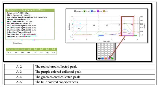
Figure 1.
Demonstrates the isolated subfractions in the flash chromatogram of run 8 with color indicating the fractions separated.
Table 3.
Demonstrates the number of repeats conducted for run 8.
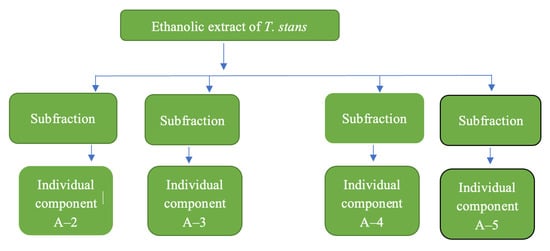
Figure 2.
Flow chart of the subfractions/individual components.
3.2. Repeatability
The method developed was validated by the test of repeatability by conducting three independent separations. In the three independent separations for run 8, all conditions and parameters—such as flow rate, wavelength, solvent systems, column, and the gradient elution—were maintained. All the replications had similar retention times for the separated peaks as shown in Table 3. The average amounts of the isolated components were 90, 93, and 95 mg with a standard deviation of 3.5% for component A2; and 100, 105, and 108 mg for components A3 with a standard deviation of 4%. For component A4, the isolated amount was approximately 86, 90, and 93 mg with a standard deviation of 3.5%. This suggests that the developed method can be repeated for the isolation of the components on a larger scale.
3.3. Antioxidant Activity Screening
3.3.1. DPPH Method
The average IC50 (μg/mL) and the R2 readings for three tests of the standard solution of ascorbic acid and the isolated components A-2, A-3, A-4 were recorded, and the isolated components showed in vitro antioxidant activity when compared to the standard-solution of ascorbic acid, as shown in Table 4:
Table 4.
UV spectrophotometer observations of the scavenging effect of AA and isolated components A-2, A-3, and A-4 using the DPPH method (average of three tests).
3.3.2. ABTS Method
The average IC50 (μg/mL) and the R2 readings for the standard solution of ascorbic acid and for the isolated components A-2, A-3, A-4 were recorded, and the isolated components showed in vitro antioxidant activity when compared to the standard-solution ascorbic acid, as shown in Table 5.
Table 5.
UV spectrophotometer observations of the scavenging effect of AA and isolated components A-2, A-3, and A-4 using the ABTS method (average readings of the three tests).
3.4. Antimicrobial Activity Screening
The antimicrobial activity for the isolated components A-2, A-3, and A-4 were evaluated through the disc diffusion method, where the isolated component A-3 showed an inhibition zone of 2.5 mm against Candida albicans, as shown in Table 6. Additionally, we observed that the isolated component A-3 inhibited the growth of the Candida albicans until the third dilution; in comparison, itraconazole inhibited the growth of the Candida albicans until the second dilution, and nystatin inhibited the growth of the Candida albicans until the fourth dilution. For the MIC and MBC, we observed through the 96 wells that the isolated component A-3 demonstrated the MIC to be of up to four folds dilution and the MBC to be up to two folds dilution on Candida albicans when compared to nystatin and itraconazole. For the bio film method, we observed that the isolated component A-3 inhibited the Candida albicans up to the second dilution.
Table 6.
Antimicrobial activity of isolated components A-2, A-3, and A-4 using the disc diffusion method.
3.5. The Spectral Data for Isolated Component A3
Isolated component A3 was analyzed using a Fourier transform infrared spectrophotometer (Agilent technologies), which showed the presence of characteristic chemical structures and functional groups such as OH at the 3372 cm−1, alkane at the 2932 cm−1, and CO at the 1622 cm−1, as shown in Figure 3. In addition, the isolated component A3 was analyzed using NMR instrumentation, where the 1H NMR included (500 MHz, methanol-d4) δ9.24 (d, J = 3.0 Hz, 1H), 7.59 (d, J = 15.9 Hz, 1H), 7.39 (s, OH), 7.34 (s, H), 7.06 (d, J = 2.0 Hz, 1H), 6.95 (dd, J = 8.3, 2.0 Hz, 1H), 6.78 (d, J = 8.1 Hz, 1H), 6.71, 6.65 (m, 2H), 6.56 (dd, J 8.0, 2.0 Hz, 1H), 6.28 (d, J = 15.9 Hz, IH), 5.84 (d, J = 1.7 Hz, OH), 5.69 (d, J = 2.7 Hz, 1H), 5.18 (d, J = 1.7 Hz, 1H), 4.63 (d, J = 7.9 Hz, 1H), 4.60 (d, J = 10.8 Hz, OH), 4.38 (d, J = 7.9 Hz, 1H), 4.04 (ddd, J = 9.7, 8.0, 6.5 Hz, 1H), 3.95, 3.91 (m, 1H), 3.90 (d, J = 2.0 Hz, 1H), 3.82 (t, J = 9.2 Hz, 1H), 3.75, 3.70 (m, 1H), 3.68 (dd, J = 11.9, 5.7 Hz, 1H), 3.64, 3.59 (m, 1H), 3.58, 3.57 (m, OH), 3.56 (d, J = 3.1 Hz, OH), 3.40 (ddd, J = 9.3, 8.2, 1.5 Hz, 2H), 3.34 (s, 4H), 3.20 (ddd, J = 10.5, 8.1, 2.6 Hz, 1H) 2.81, 2.75 (m, 1H), 2.46, 2.41 (m, OH), 2.29, 2.23 (m, OH), 2.17, 2.08 (m, 2H), 1.99 (sm2H), 1.88 (dd, J = 9.0, 2.7 Hz, 1H), 1.70 (ddd, J = 17.2, 15.2, 7.8, 6.2 Hz, 2H), 1.43, 1.35 (m, 1H), 1.17, 1.15 (m, OH), 1.12 (d, J = 6.3 Hz, 3H), 1.09 (d, J = 6.2 Hz, 2H), 0.93 (d, J = 6.8 Hz, 1H), 0.89 (ddd, J = 6.6, 2.3, 1.3 Hz, OH), 0.79 (d, J = 7.0 Hz, OH), as shown in Figure 4. The 2D NMR—such as heteronuclear multiple-bond correlation (HMBC), heteronuclear single quantum correlation (HSQC), homonuclear correlation spectroscopy (COSY), and total correlated spectroscopy (TOCSY)—is shown in Figures S1–S4 in Supplementary Material 2.
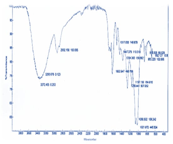
Figure 3.
Fourier transform infrared spectroscopy for the isolated component A3.
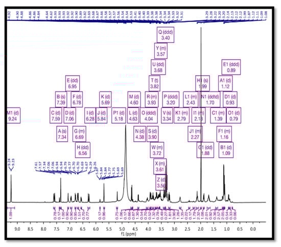
Figure 4.
Nuclear magnetic resonance (NMR) for the isolated component A-3.
4. Discussion
The use of herbal medicines to treat various health challenges in different healthcare settings continues to expand rapidly across the world []. Natural products are used as a rich source to isolate active ingredients that tackle different medical conditions, particularly fungal infection []. Secondary metabolites that have antioxidant activity may play a vital role in the prevention of several cardiovascular diseases such as heart attack and strokes [,,]. In 2019, Larbie et al. emphasized the safety of the hydroethanolic extract of T. stans by measuring the median acute toxicity of the extract on mice []. Our work used FCS to isolate active ingredients that had antifungal and antioxidant activity. In the preparation of the extract, we used the ultrasonic homogenizer to disturb the cells in the extract, which enabled the release of the secondary metabolites by inducing an electric frequency which was converted to a mechanical energy. This was carried out to improve the extraction process more effectively than with traditional methods such as maceration and percolation []. Additionally, frequency and time were adjusted to ensure maximum extraction; this was in line with the study by Annegowda, which concluded that the changing time of the sonication process will affect the antioxidant activity of the plant []. TLC was used with different solvents based on polarity to investigate the best solvent system that can be used in FCS to isolate secondary metabolites such as alkaloids and flavonoids [].
FCS has been widely used in the isolation and purification of naturally occurring products with potential medicinal actions, for example, the identification of large molecular toxins found in food products, and laboratory synthesized chemical compounds []. FCS improves the separation and purification of secondary metabolites by using a mixture of solvents and reduced time durations. The use of specific wavelengths in FCS was intended to detect phenolic metabolites. FCS has a UV detector that can monitor three wavelengths simultaneously in a range of 200 and 500 nm and can offer adjustable sensitivity settings to detect and collect small and large quantities of compounds. Additionally, FCS has an evaporative light-scattering detector (ELSD) that provides the system with the ability to monitor all non-volatile compounds and detect compounds that do not absorb UV radiation. For example, FCS can detect chromophores (a compound with a covalently bonded group) that show a characteristic absorption in the UV/Visible region. In our study, we selected the columns, the UV, and the solvents system to obtain better separation and larger quantities of the separated subfractions, and consequently produced isolated components. For example, the use of columns such as the C18 flash cartridges with the reversed-phase gradient elution provided good separation with faster elution of the subfractions with maximum purity and better resolution of the isolated components []. The method developed using flash chromatography enabled the collection of larger quantities of purified compounds with less time and solvent when compared to other analytical techniques such as column chromatography and preparative high-performance liquid chromatography (HPLC) [,]. This was in line with the findings of Jarvis et al., which indicated that the use of flash chromatography can produce large quantities of the purified components with high purity and less time consumption [,].
The in vitro antioxidant activity of the isolated components A2, A3, and A4 was significant compared to ascorbic acid as a control. In the in vitro antioxidant activity with DPPH, the three isolated components A2, A3, and A4 gave a very good response, and component A-4 had an IC50 of 9.64 compared to ascorbic acid at IC50 of 9.40. In contrast, component A3 had an IC50 of 20.16 to give approximately the same effect as the ascorbic acid. As for the ABTS in vitro antioxidant activity, the three isolated components A2, A3, and A4 gave a good response, and component A-4 showed antioxidant activity with IC50 of 56.56 compared to ascorbic acid. Isolated component A-3 showed consistent antioxidant activity in both methods of DPPH and ABTS with an IC50 of 20.16 and 27.52, respectively. These results confirm the outcomes from the study conducted by Marzouk et al., which demonstrated the potential presence of antioxidant components in T. stans []. As for the antibacterial activity, the three isolated components A2, A3, and A4 gave a negative response when tested against Staphylococcus aureus and E. coli representing gram-positive and gram-negative species, respectively, and compared to amoxicillin, ciprofloxacin, and ceftriaxone as positive controls. However, Gonçalves et al. 2022 demonstrated that ethanol, dichloromethane, and ethyl acetate fractions from the flowers of T. stans interacted synergistically with amoxicillin and tetracycline, decreasing the MIC of antibiotics by 2–5 folds when compared with the MIC of antibiotics used alone []. For antifungal activity, component A3 gave a positive response when tested against Candida albicans; however, both isolated components A-2 and A-4 showed no activity against Candida albicans when compared to nystatin and itraconazole as positive controls. The isolated component A-3 showed an antifungal activity with the second dilution and more than 80% inhibitions in the third dilution compared to itraconazole and nystatin as positive controls. Isolated component A-3 also inhibited biofilm in the second dilution. The isolated component A-3 showed both antioxidant and antifungal activities when compared to other fractions. The potential antifungal activity demonstrated by the isolated component is in line with the study conducted by Patriota et al. demonstrating the potential antifungal activity of the plant T. stans. The instrumental data showcased the purity of the isolated component and the presence of characteristic chemical structures and functional groups. The isolated component A-3 can be formulated in pharmaceutical formulation and in vivo studies for the development of antifungal ointment, shampoo, or soap.
5. Conclusions
This paper demonstrates a rapid and efficient method for the isolation and purification of isolated components using flash chromatography. This method utilizes less time and solvent with a high yield of isolated components, and the isolated components can be assessed for their antimicrobial and antioxidant activity.
Supplementary Materials
The following supporting information can be downloaded at: https://www.mdpi.com/article/10.3390/separations9100317/s1, Figure S1: Heteronuclear Multiple bond correlation (HMBC) Spectrum, Figure S2: Heteronuclear single quantum Correlation (HSQC) Spectrum for Fraction A-3, Figure S3: Homonuclear correlation spectroscopy (COSY) Spectrum for Fraction A-3, Figure S4: Total correlated spectroscopy (TOCSY) Spectrum. Table S1: Method development for the ethanolic extract.
Author Contributions
Conceptualization, A.A.-A. and A.A.D.; methodology, A.A.-A.; software, A.A.D.; validation, A.A.-A., S.T. and A.F.W.; formal analysis, A.A.-A.; investigation, A.A.-A. and A.A.D.; resources, A.A.-A.; data curation, A.A.-A., A.A.D., S.T., A.F.W. and O.S.; writing—original draft preparation, A.A.-A. and A.A.D.; writing—review and editing, A.A.-A., A.A.D., S.T., A.F.W. and O.S.; visualization, A.A.-A., A.A.D., S.T., A.F.W. and O.S.; supervision, A.A.-A. and S.T. All authors have read and agreed to the published version of the manuscript.
Funding
This research received no external funding.
Acknowledgments
The authors would like to thank S. Gurumadhva Rao, president of Ras Al Khaimah Medical and Health Sciences University, and Padma GM Rao, dean of RAK College of Pharmacy for their encouragement and support, and Ashfaque Hossain, Godfred A. Menezes, and Michael V. Magaogao of the microbiology department of RAKMHSU for their help and guidance. The authors would like to extend their gratitude to Darcy Burns, manager of the Centre for Spectroscopic Investigation of Complex Organic Molecules and Polymers, Department of Chemistry, University of Toronto for his help in the NMR instrumentation.
Conflicts of Interest
The authors declare no conflict of interest.
References
- Dias, D.A.; Urban, S.; Roessner, U. A historical overview of natural products in drug discovery. Metabolites 2012, 2, 303–336. [Google Scholar] [CrossRef]
- Pelton, J. A Survey of the Ecology of Tecoma stans. Butl. Univ. Bot. Stud. 1964, 14, 11. [Google Scholar]
- Hammouda, Y.; Khalafallah, N. Stability of tecomine, the major antidiabetic factor of Tecoma stans (Juss.) f. bignoniaceae. J. Pharm. Sci. 1971, 60, 1142–1145. [Google Scholar] [CrossRef] [PubMed]
- Al-Azzawi, A.; Al-Khateeb, E.; Al-Sameraei, K.; Al-Juboori, A. Antibacterial activity and the histopathological study of crude extracts and isolated tecomine from Tecoma stans Bignoniaceae in Iraq. J. Pharmacogn. Res. 2012, 4, 37–43. [Google Scholar] [CrossRef] [PubMed]
- Aguilar, L.C.; Macias, S.; Chagoya, A.; Cardenas, A.; Diaz, P.; Cantu, J.M. Antidiabetic activity of Tecoma stans in rats. Fitoterapia 1993, 64, 304–305. [Google Scholar]
- Jones, G.H.; Fales, M.; Wildman, W.C. The structure of tecomanine. Tetrahedron Lett. 1963, 6, 397–400. [Google Scholar]
- Hernandez, J.R.; Garcia, C.G. Research on the hypoglycemic action of the active fraction of Tecoma stans. Medicina 1958, 38, 25–34. [Google Scholar]
- Hammouda, Y.; Plat, M.; Lemen, J. Isolation of Tecostanin. Alkaloids of Tecoma stans (Juss) Bignoniaceae. Ann. Pharm. Fr. 1963, 21, 699–702. [Google Scholar]
- Meckes, M.; Mellado Campos, V. Is The Tecoma Stans infusion an ant diabetic remedy? J. Ethnopharmacol. 1985, 14, 1–9. [Google Scholar] [CrossRef]
- Costantino, L.; Raimondi, L.; Pirisino, R.; Brunetti, T.; Pessotto, P.; Giannessi, F.; Lins, A.P.; Barlocco, D.; Antolini, L.; El-Abady, S.A. Isolation and pharmacological activities of the Tecoma stans alkaloids. Il Farm. 2003, 58, 781–785. [Google Scholar] [CrossRef]
- Aguilar-Santamaría, L.; Ramírez, G.; Nicasio, P.; Alegría-Reyes, C.; Herrera-Arellano, A. Antidiabetic activities of Tecoma stans (L.) Juss. ex Kunth. J. Ethnopharmacol. 2009, 124, 284–288. [Google Scholar] [CrossRef] [PubMed]
- Alonso-Castro, A.J.; Zapata-Bustos, R.; Romo-Yañez, J.; Camarillo-Ledesma, P.; Gómez-Sánchez, M.; Salazar-Olivo, L.A. The antidiabetic plants Tecoma stans (L.) Juss. ex Kunth (Bignoniaceae) and Teucrium cubense Jacq (Lamiaceae) induce the incorporation of glucose in insulin-sensitive and insulin-resistant murine and human adipocytes. J. Ethnopharmacol. 2010, 127, 1–6. [Google Scholar] [CrossRef] [PubMed]
- Salem, M.Z.M.; Gohar, Y.M.; Camacho, L.M.; El-Shanhorey, N.A.; Salem, A.Z.M. Antioxidant and antibacterial activities of leaves and branches extracts of Tecoma stans (L.) Juss. ex Kunth against nine species of pathogenic bacteria. Afr. J. Microbiol. Res. 2013, 7, 418–426. [Google Scholar]
- Patriota, L.L.; Procópio, T.F.; De Souza, M.F.; De Oliveira, A.P.S.; Carvalho, L.V.; Pitta, M.G.; Rego, M.J.; Paiva, P.M.; Pontual, E.V.; Napoleão, T.H. A Trypsin Inhibitor from Tecoma stans Leaves Inhibits Growth and Promotes ATP Depletion and Lipid Peroxidation in Candida albicans and Candida krusei. Front. Microbiol. J. 2016, 7, 611. [Google Scholar] [CrossRef]
- Meelah, M.M.; Mdee, L.K.; Eloff, J.N. Tecoma stans (Bignoniaceae), leaf extracts, fractions and isolated compound have promising activity against fungal phytopathogens. Suid-Afrik. Tydskr. Vir Nat. Tegnol. 2017, 36, a1489. [Google Scholar] [CrossRef]
- Product Identification: Operation Manual (Original), Reveleris® X2, Published on 2016 May, BÜCHI Labortechnik AG. Available online: https://www.buchi.com/en/products/instruments/pure-chromatography-systems (accessed on 20 May 2022).
- Compton, D.L.; Appell, M.; Kenar, J.A.; Evans, K.O. Enzymatic Synthesis and Flash Chromatography Separation of 1,3-Diferuloyl-sn-Glycerol and 1-Feruloyl-sn-Glycerol. Methods Protoc. 2020, 3, 8. [Google Scholar] [CrossRef]
- Chen, X.; Kong, L.; Su, X.; Fu, H.; Ni, J.; Zhao, R.; Zou, H. Separation and identification of compounds in Rhizoma chuanxiong by comprehensive coupled to mass spectrometry. J. Chromatogr. A 2004, 1040, 169–178. [Google Scholar] [CrossRef]
- Qiao, C.; Han, Q.; Song, J.; Mo, S.; Tai, C.; Xu, H. HPLC analysis of Bioactive diterpenoids from the root bark of pseudolarix kaempferi. J. Food Drug Anal. 2006, 14, 353–356. [Google Scholar] [CrossRef]
- Weber, P.; Hamburger, M.; Schafroth, N.; Potterat, O. Flash chromatography on cartridges for the separation of plant extracts: Rules for the selection of chromatographic conditions and comparison with medium pressure liquid chromatography. Fitoterapia 2011, 82, 155–161. [Google Scholar] [CrossRef]
- Sandesh, J.S.S.; Shyamala, S.K.; Balaiah, S.; Sharma, J.V.C. A Review on Flash Chromatography and Its Pharmaceutical Applications. J. Biomed. Pharm. Res. 2021, 10, 120–124. [Google Scholar] [CrossRef]
- Biswas, T.; Ajayakumar, P.V.; Mathur, A.K.; Mathur, A. Solvent-based extraction optimisation for efficient ultrasonication-assisted ginsenoside recovery from Panax quinquefolius and P. sikkimensis cell suspension lines. Nat. Prod. Res. 2015, 29, 1256–1263 . [Google Scholar] [CrossRef] [PubMed]
- Blois, M. Antioxidant Determinations by the Use of a Stable Free Radical. Nature 1958, 181, 1199–1200. [Google Scholar] [CrossRef]
- Kedare, S.B.; Singh, R.P. Genesis and development of DPPH method of antioxidant assay. J. Food Sci. Technol. 2011, 48, 412–422. [Google Scholar] [CrossRef]
- Biswas, M.; Haldar, P.K.; Ghosh, A.K. Antioxidant and free-radical-scavenging effects of fruits of Dregea volubilis. J. Nat. Sci. Biol. Med. 2010, 1, 29–34. [Google Scholar] [CrossRef] [PubMed]
- Arnao, M.B.; Cano, A.; Hernández-Ruiz, J.; García-Cánovas, F.; Acosta, M. Inhibition by L-ascorbic acid and other antioxidants of the 2.2′-azino-bis(3-ethylbenzthiazoline-6-sulfonic acid) oxidation catalyzed by peroxidase: A new approach for determining total antioxidant status of foods. Anal. Biochem. 1996, 236, 255–261. [Google Scholar] [CrossRef]
- Bauer, A.W.; Kirby, W.M.M.; Sherris, J.C.; Turk, M. Antibiotic susceptibility testing by a standardized single disk method. Am. J. Clin. Pathol. 1966, 45, 493–496. [Google Scholar] [CrossRef] [PubMed]
- Clinical and Laboratory Standards Institute. Performance Standards for Antimicrobial Disk Susceptibility Tests. Approved Standard M2–A10; Clinical and Laboratory Standards Institute: Wayne, PA, USA, 2009. [Google Scholar]
- Reller, L.B.; Weinstein, M.; Jorgensen, J.H.; Ferraro, M.J. Antimicrobial Susceptibility Testing: A Review of General Principles and Contemporary Practices. Clin. Infect. Dis. 2009, 49, 1749–1755. [Google Scholar] [CrossRef]
- Prakash, V.; Lewis, J.S.I.I.; Jorgensen, J.H. Vancomycin MICs with methicillin-resistant Staphylococcus aureus (MRSA) isolates differ based upon the susceptibility test method used. Antimicrob Agents Chemother 2008, 52, 4528. [Google Scholar] [CrossRef]
- O’Toole, G.A. Microtiter dish biofilm formation assay. J. Vis. Exp. 2011, 47, e2437. [Google Scholar] [CrossRef]
- Samadi, F.M.; Suhail, S.; Sonam, M.; Sharma, N.; Singh, S.; Gupta, S.; Dobhal, A.; Pradhan, H. Antifungal efficacy of herbs. J. Oral Biol. Craniofac. Res. 2019, 9, 28–32. [Google Scholar] [CrossRef]
- Anand, M.; Basavaraju, R. A Review on Phytochemistry and Pharmacological Uses of Tecoma Stans (L.) Juss. Ex Kunth. J. Ethnopharmacol. 2021, 265, 113270. [Google Scholar] [CrossRef] [PubMed]
- Halliwell, B. Free radicals, antioxidants, and human disease: Curiosity, cause, or consequence? Lancet (Br. Ed.) 1994, 344, 721–724. [Google Scholar] [CrossRef]
- Leopold, J.A. Antioxidants and coronary artery disease: From pathophysiology to preventive therapy. Coron Artery Dis. 2015, 26, 176–183. [Google Scholar] [CrossRef] [PubMed]
- Mangge, H.; Becker, K.; Fuchs, D.; Gostner, J.M. Antioxidants, inflammation and cardiovascular disease. World J. Cardiol. 2014, 6, 462–477. [Google Scholar] [CrossRef] [PubMed]
- Larbie, C.; Owusu Nyarkoh, C.; Owusu Adjei, C. Phytochemical and Safety Evaluation of Hydroethanolic Leaf Extract of Tecoma stans (L.) Juss. ex Kunth. Evid. Based Complement. Alternat. Med. 2019, 2019, 7417624. [Google Scholar] [CrossRef]
- Annegowda, H.V.; Bhat, R.; Min-Tze, L.; Karim, A.A.; Mansor, S.M. Influence of sonication treatments and extraction solvents on the phenolics and antioxidants in star fruits. J. Food Sci. Technol. 2012, 49, 510–514. [Google Scholar] [CrossRef]
- Fair, J.D.; Kormos, C.M. Flash column chromatograms estimated from thin-layer chromatography data. J. Chromatogr. A 2008, 1211, 49–54. [Google Scholar] [CrossRef]
- Goossens, E.; Wijnants, M.; Packet, D.; Lemière, F. Enhanced separation, and analysis procedure reveals production of tri-acylated mannosylerythritol lipids by Pseudozyma aphidis. J. Ind. Microbiol. Biotechnol. 2016, 43, 1537–1550. [Google Scholar] [CrossRef]
- Uckoo, R.M.; Jayaprakasha, G.K.; Patil, B.S. Rapid separation method of polymethoxyflavones from citrus using flash chromatography. Sep. Purif. Technol. 2011, 81, 151–158. [Google Scholar] [CrossRef]
- Kou, P.; Kang, Y.F.; Wang, L.T.; Niu, L.J.; Xiao, Y.; Guo, N.; Cui, Q.; Li, Y.Y.; Fu, Y.J. An integrated strategy for production of four anthocyanin compounds from Ribes nigrum L. by deep eutectic solvents and flash chromatography. J. Ind. Eng. Chem. 2019, 80, 614–625. [Google Scholar] [CrossRef]
- Jarvis, A.P.; Morgan, E.D.; Edwards, C. Rapid separation of triterpenoids from Neem seed extracts. Phytochem. Anal. 1999, 10, 39–43. [Google Scholar] [CrossRef]
- Moosmann, B.; Kneisel, S.; Wohlfarth, A.; Brecht, V.; Auwärter, V. A fast and inexpensive procedure for the isolation of synthetic cannabinoids from ‘Spice’ products using a flash chromatography system. Anal. Bioanal. Chem. 2013, 405, 3929–3935. [Google Scholar] [CrossRef] [PubMed]
- Marzouk, M.; Gamal-Eldeen, A.; Mohamed, M.; El-Sayed, M. Anti-proliferative and antioxidant constituents from Tecoma stans. Z. Nat. C J. Biosci. 2006, 61, 783–791. [Google Scholar]
- Gonçalves, T.; Parreira, A.; Zanuncio, V.; Farias, K.; Silva, D.; Lima, L. Antibacterial and antioxidant properties of flowers from Tecoma stans (L.) Juss. ex Kunth (Bignoniaceae). S. Afr. J. Bot. 2022, 144, 156–165. [Google Scholar] [CrossRef]


Publisher’s Note: MDPI stays neutral with regard to jurisdictional claims in published maps and institutional affiliations. |
© 2022 by the authors. Licensee MDPI, Basel, Switzerland. This article is an open access article distributed under the terms and conditions of the Creative Commons Attribution (CC BY) license (https://creativecommons.org/licenses/by/4.0/).