Article

Purification, Identification and Neuroprotective Effects of Proteins from Bombyx batryticatus in Glu-Stimulated PC12 Cells

1
Institute of Pharmaceutical & Food Engineering, Shanxi University of Chinese Medicine, Jinzhong 030619, China
2
Key Laboratory of Traditional Chinese Medicine Processing of Shanxi Province, Shanxi University of Chinese Medicine, Jinzhong 030619, China
3
School of Pharmacy, Chengdu University of Traditional Chinese Medicine, Chengdu 611137, China
*
Author to whom correspondence should be addressed.
Separations 2022, 9(9), 236; https://doi.org/10.3390/separations9090236
Submission received: 29 July 2022 / Revised: 16 August 2022 / Accepted: 29 August 2022 / Published: 1 September 2022
(This article belongs to the Special Issue Separation and Quantitative Analysis of Natural Product Extracts)

Abstract

:
Bombyx batryticatus (BB) is one of the most commonly used Traditional Chinese Medicines (TCMs) in the treatment of convulsions and epilepsy. The antiepileptic effects of total proteins from BB (BBPs) have been proven in our previous research. In this study, BBPs were further purified, the neuroprotective effects were evaluated in Glu-stimulated PC12 cells, and the structure was identified by Matrix-assisted laser desorption ionization time-of-flight mass spectrometry (MALDI-TOF-MS). Six subfractions (PF-1 to PF-6) were obtained by DEAE-52 Sepharose FF ion-exchange chromatography. It was found that PF-1, PF-2, and PF-3, with similar protein compositions, possessed neuroprotective effects in Glu-stimulated PC12 cells by significantly increasing the GABA level, and decreasing the levels of IL-1β and TNF-α. The most active fraction (PF-2) was further separated by Sephadex G-75 gel filtration chromatography, and an effective protein component named PF-2-2 was obtained. Fluorescein isothiocyanate-labeled PF-2-2 (FITC-PF-2-2) was prepared, and the binding of FITC-F-2-2 to the PC12 cells was directly observed with a confocal microscope. PF-2-2 was found to first bind to the surface of PC12 cells and then internalize into the cells. The main band of PF-2-2 was then analyzed by MALDI-TOF/TOF-MS and searched in the MASCOT database; finally a protein named Low molecular mass 30 kDa lipoprotein 21G1 was identified. In conclusion, PF-2-2 and purified proteins isolated from BBPs have potential application prospects in the treatment of epilepsy.

1. Introduction

Epilepsy is one of the most common and most disabling chronic neurological disorders, affecting more than 70 million people worldwide [1]. It is characterized by abnormal or excessive brain neuron activity leading to brain activity interruption and rapid seizures [2]. Epilepsy is caused by the abnormal synchronous firing of neurons, due to an imbalance of the excitatory and inhibitory neurotransmission [3]. It is usually associated with anxiety, depression, autism, schizophrenia, cognitive decline and Alzheimer’s disease (AD), among others [4]. Although epilepsy and seizures have been extensively studied, the underlying pathological mechanisms of epilepsy remain unclear [5]. However, the role of mutations, oxidative stress and inflammatory brain damage in the pathogenesis of epilepsy has been widely recognized [5,6,7,8].
At present, more than 25 antiepileptic drugs (AEDs) are used as the primary therapeutic strategy for treating epileptic seizures [9]. However, these AEDs can only alleviate symptoms and cannot completely prevent disease progression [10]. Due to drug resistance, about one-third of epileptic patients cannot completely control their seizures, even if a variety of AEDs are used, either alone or in combination [1]. In addition to reducing seizures, AEDs themselves can also lead to cognitive dysfunction [3]. Therefore, due to the complexity of epilepsy pathogenesis, it is necessary to develop improved treatment strategies for epilepsy, not only to prevent disease progression, but to also improve the neurobehavioral comorbidity related to epilepsy.
In TCM, animal-derived drugs are widely used because of their high activity and unique curative effects [11]. Compared with small molecular compounds derived from natural plants, most bioactive ingredients derived from animals are proteins or peptides, such as antithrombotic peptides, antihypertensive peptides, antibacterial peptides and various animal toxins [12,13,14,15]. Bombyx batryticatus (BB) is the dried larva of Bombyx mori Linnaeus (commonly known as the silkworm) which are naturally or artificially infected with Beauveria bassiana (Bals.) Vuillant [16]. It is one of the most commonly used TCMs, called “Jiangcan” in Chinese, and was first recorded in “Sheng Nong’s Herbal Classic” [16,17]. BB is also widely used in Japanese Kampo and Korean folk medicine [16]. In the theory of TCM, BB can dispel wind to relieve convulsion, resolve masses and reduce phlegm. It is traditionally used to treat stroke-related aphasia, seizure, intermittent headache, asthma, facial paralysis, hemiplegia, and other diseases [18]. In TCM, BB is mainly used for the treatment of epilepsy and convulsions. Animal or cell experiments have shown that the extracts or compounds from BB have significant anticonvulsant and antiepileptic effects [19,20,21].
The traditional uses, origin, chemical constituents, pharmacology, and toxicity of BB have been reviewed in our previous study [22]. BB contains various biological ingredients, such as protein and peptides, polysaccharides, flavonoids, fatty acids, steroids, nucleosides and coumarins, [23], etc. The principal constituents of BB are proteins, accounting for 40–70% of total components [22]. Multiple pharmacological effects of BB have been found, including neuroprotective, anticancer, antibacterial, anticoagulation, and hypoglycemic lipid-lowering effects, [22,24,25,26], etc. In our previous studies, protein-rich extracts from BB (BBPs) were proven to have a significant protective effect in PC12 cells injured by Glu and H2O2 via different signaling pathways, and the antiepileptic effects have also been proven in mice [19,20]. In this study, proteins with antiepileptic effects were further isolated and purified, the neuroprotective effects were studied in PC12 cells, and the structures were identified by MALDI-TOF-MS.

2. Materials and Methods

2.1. Materials and Chemicals

BBs were obtained from Chengdu Min-Jiang-Yuan Pharmaceutical Co., Ltd. (Chengdu, China), and identified by Prof. Chun-Jie Wu (The fifth author of this paper). A voucher specimen (No. Y170628) was deposited in the laboratory of the School of Pharmacy, Chengdu University of Traditional Chinese Medicine (Chengdu, China). Fetal bovine serum (FBS), and horse serum were obtained from Hyclone (Logan, UT, USA). The RPMI-1640 medium was a Gibco product (Burlington, ON, Canada). Glu and fluorescein isothiocyanate (FITC) were from Sigma-Aldrich Co. (St. Louis, MO, USA). The 3-(4,5-dimethylthiazol-2-yl)-2,5-diphenyltetrazolium bromide (MTT) was a product of the Beyotime Institute of Biotechnology (Shanghai, China). Sephadex G-75 and DEAE Sepharose FF were purchased from H&E Co., Ltd. (Beijing, China). GABA, IL-4 and TNF-α ELISA kits were obtained from Multisciences (LIANKE) Biotech Co., Ltd. (Hangzhou, China). All other reagents used in this study were of an analytical grade.

2.2. Preparation of Total Proteins from BB

Total proteins from BB (BBPs) were extracted as per our previous report, with some modifications [20]. Briefly, BB powder was defatted with petroleum ether (1:5, w/v) and then extracted with a phosphate buffer (PBS, pH 8.0, 30 mM) at a ratio of 1:4 (w/v) for 1 h with ultrasonic assisted extraction. Subsequently, the extractions were centrifuged (5000 rpm, 30 min). Proteins were precipitated with 80% saturated ammonium sulfate ((NH4)2SO4) overnight at 4 °C. After centrifugation (5000 rpm, 30 min), the precipitated proteins were redissolved in PBS and dialyzed with distilled water (4 °C, 24 h) using dialysis membranes (3 kDa). BBPs were obtained by freeze-drying and the yield was 2.16% (the protein content was 71.98%).

2.3. Purification of Neuroprotective Proteins from BBPs

A BBPs solution (20 mg/mL) was prepared with Tris-HCl (0.05 M, pH = 8.0), and loaded onto a DEAE-52 Sepharose FF column (2.5 × 40 cm). Samples were eluted with Tris-HCl (0.05 M, pH = 8.0) and 0.10, 0.20, 0.30, 0.40, 0.50 M NaCl solution (prepared with 0.05 M Tris-HCl, pH = 8.0), respectively. The flow rate was controlled to 1.0 mL/min and the absorbance was monitored at 280 nm. Six fractions (PF-1 to PF-6) were collected according to the chromatogram map. After being dialyzed with distilled water using 3 kDa dialysis membranes (4 °C for 24 h) and concentrated by polyethylene glycol (PEG 20 000), dried samples of PF-1 to PF-6 were obtained by freeze-drying.
The obtained PF-2 was redissolved in Tris-HCl (0.05 M, pH = 8.0). A solution of 30.0 mg/mL was loaded onto the Sephadex G-75 column (2.5 cm × 40 cm) and eluted with 0.05 M Tris-HCl (pH = 8.0). The eluent was set at 0.5 mL/min and a component named PF-2-2 was isolated from PF-2, based on the chromatographic curve at 280 nm. A dried sample of PF-2-2 was obtained as above.

2.4. Protein Patterns of PF-1 to PF-6 Analyzed by SDS-PAGE

SDS-PAGE analysis was performed as per the method in our previous report [20]. 12% acrylamide separating gel and 4% stacking gel (Mini-PROTEAN 3 Cell, Bio-Rad Laboratories Inc., Hercules, CA, USA) were used for protein separation. Image Lab software (Bio-Rad, 6.0.1, Hercules, CA, USA) was used to analyze the molecular weight (MW) and the relative quantification of proteins from SDS-PAGE patterns (Relative to Marker 9, 15 kDa). Pre-stained protein markers (Thermo Fisher, Waltham, MA, USA) with MWs of 10–180 kDa were used as a standard control.

2.5. Cell Culture

Rat pheochromocytoma-derived cell line PC12 cells were obtained from Wuhan Pu-nuo-sai Life Technology Co., Ltd. (Wuhan, China). Cells were cultured with RPMI-1640 medium containing 5% horse serum, 5% FBS and 1% penicillin/streptomycin in a humidified atmosphere (37 °C, 5% CO2).

2.6. Cell Viability Assay

The cell viability was determined by the MTT assay [27]. Briefly, cells (3000 cells/well) were seeded into 96-well plates and incubated for 24 h. Protein samples (the final concentrations of 50 to 800 μg/mL) were added and cultured for another 24 h. The culture medium was then discarded and MTT solution was added for 4 h. The optical density (OD) values at 490 nm were measured using a microplate reader (Multlskan Mk3, Thermo Fisher, Waltham, MA, USA). Cell viability was expressed as an OD percentage value of the control cells (without any treatment).
The effect of proteins on the viability of Glu-stimulated PC12 cells was then determined. Protein samples at the final concentrations based on the above results were added to the cells and cultured for 24 h. Subsequently, Glu (the final concentration of 20 mM) was added and treated for another 24 h. The cell viability was assayed as above.

2.7. Determination of GABA, IL-1β, and TNF-α Contents in PC12 Cells

Cells were seeded into a 24-well plate and incubated for 24 h. Cells were treated with PF-1 to PF-3 (at the final concentrations of 200, 400, and 800 μg/mL) or PF-2-2 (at the final concentrations of 200, 300, 400 μg/mL), based on pre-experiment results, for another 24 h. Glu (the final concentration of 20 mM) was then added and incubated for 24 h. The contents of GABA, IL-1β, and TNF-α in-cell in the culture medium were determined using commercial ELISA kits from Multisciences (LIANKE) Biotech, Co., Ltd. (Hangzhou, China), according to the manufacturer’s instructions.

2.8. Laser Scanning Confocal Microscopy Analysis

To determine whether PF-2-2 can bind to PC12 cells and the binding site, laser scanning confocal microscopy was employed. FITC-labeled F-2-2 was synthesized and prepared by the method reported previously [28]. PC12 cells were preincubated with FITC-labeled F-2-2 (400 μg/mL) and rinsed with PBS before observation. The stained cells were observed after 6 and 24 h using TCS SP8 fluorescence confocal laser scanning microscopy (Leica Microsystems, Wetzlar, Germany) with a 488 nm excitation wavelength. The experiment was carried out at the Chengdu University of Traditional Chinese Medicine (Chengdu, China).

2.9. MALDI-TOF/TOF-MS Analysis

A main protein band of PF-2-2 was cut from the staining SDS-PAGE gel and in-gel digested as previously reported [29]. MS and MS/MS of peptides were carried out on an ABI 5800 MALDI-TOF/TOF Plus mass spectrometer (Applied Biosystems, Foster City, CA, USA). The data obtained from MALDI-TOF/TOF were integrated and processed by using the GPS Explorer V3.6 software (Applied Biosystems, Foster City, CA, USA) with default parameters. Proteins were identified by using the MASCOT V2.3 search engine (Matrix Science Ltd., London, UK), using the following parameters: taxonomy of Metazoa (animals) (15843440 sequences), database of NCBIprot 20180429 (152462470 sequences; 55858910152 residues), trypsin as digestion enzyme, one max missed cleavage, peptide mass tolerance of 100 ppm, fragment mass tolerance of 0.3 Da, Dioxidation (W), Deamidated (NQ), Acetyl (Protein N-term), Oxidation (M) as variable modification. A confidence interval of protein score > 95% indicated successful identification.

2.10. Statistical Analysis

Data were presented as mean ± standard deviations (SD). Student’s t-test or one-way analysis of variance (ANOVA) were used for statistical comparisons between groups carried out by GraphPad Prism 7 software (GraphPad Software Inc., La Jolla, CA, USA). p values < 0.05 were considered statistically significant.

3. Results

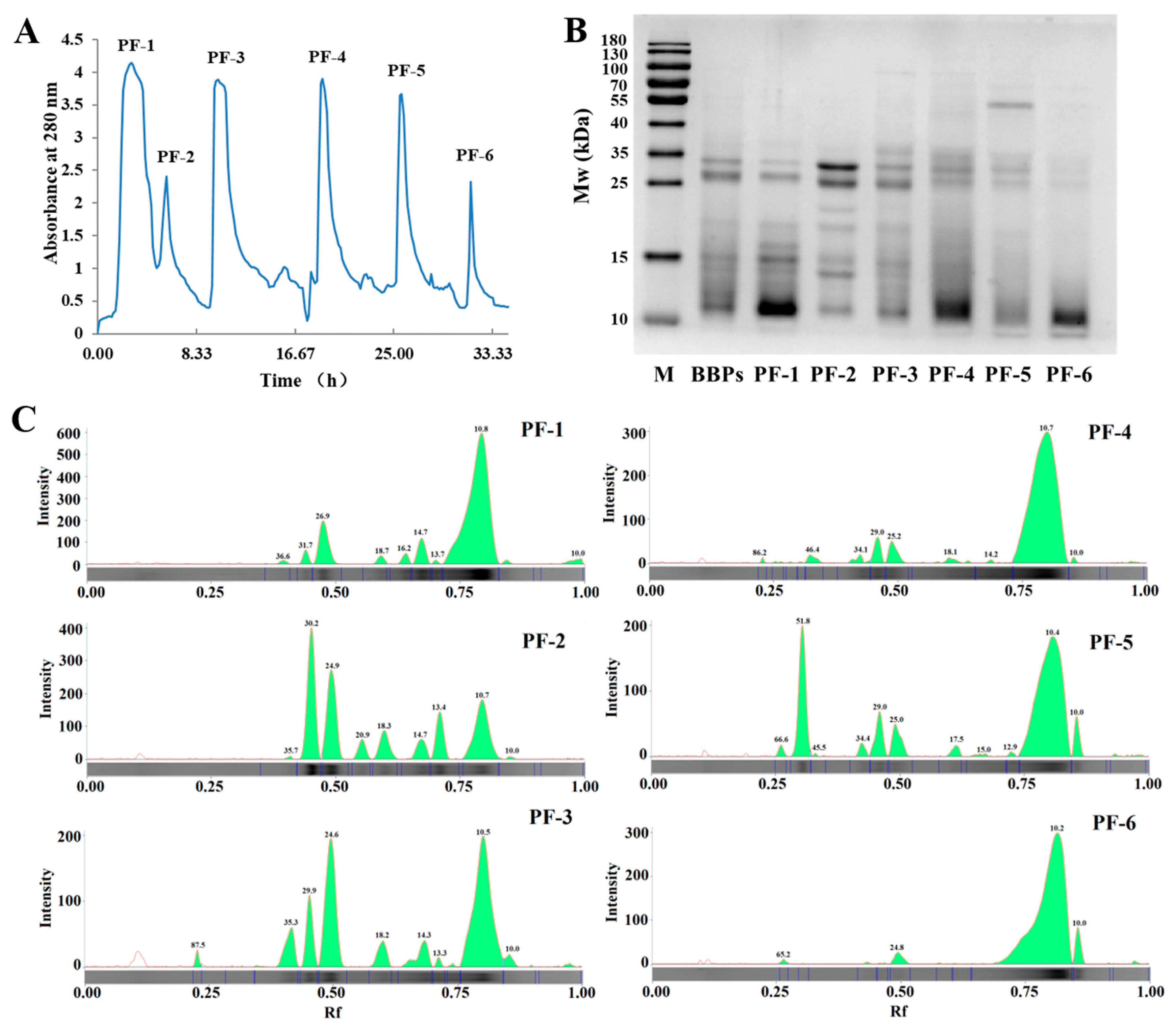
3.1. BBPs Fractionated by Ion-Exchange Chromatography

As shown in Figure 1A, to obtain subfractions with higher neuroprotective effects, BBPs were first fractionated by DEAE-52 Sepharose FF ion-exchange chromatography, and six subfractions named PF-1 to PF-6 were obtained. The subfractions were then analyzed by SDS-PAGE and the results are shown in Figure 1B,C. The molecular weight (kDa) and relative quantification (relative to Marker 9 of MW 15 kDa) of protein bands are presented in Table 1. It can be seen that the molecular weight information of PF-1, PF-2 and PF-3 was relatively similar. The main different bands were between 10 to 35 kDa, and the bands were clear. The color of the freeze-dried powder of these three fractions is light white or yellow. Fewer bands with a molecular weight between 10–25 kDa were found in PF-4, PF-5 and PF-6, mostly distributed around 10 kDa and 25–35 kDa, but the content was lower than that of PF-1, PF-2 and PF-3. The color of the freeze-dried powder is darker than PF-1 to PF-3, which is a light yellowish brown.

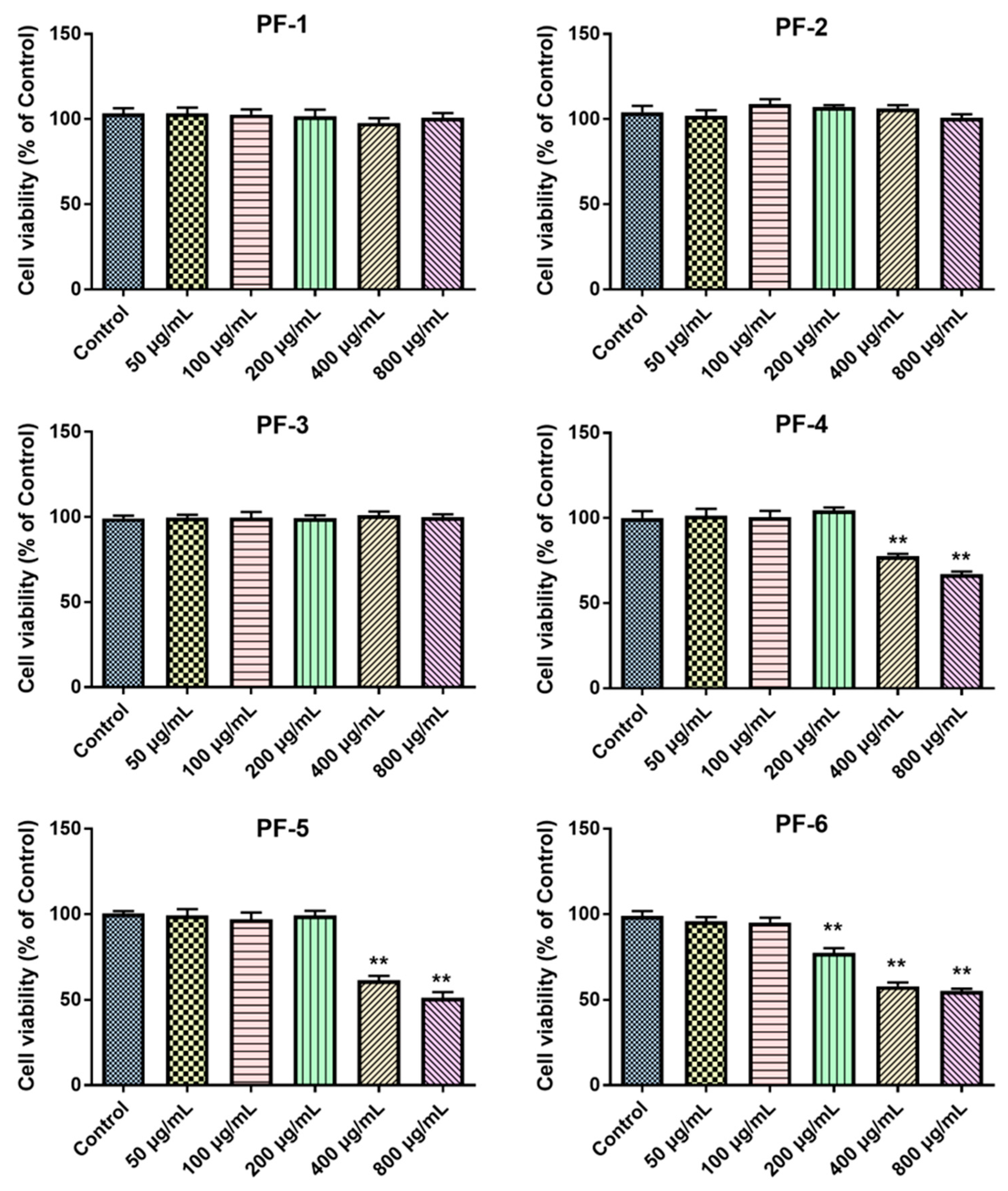
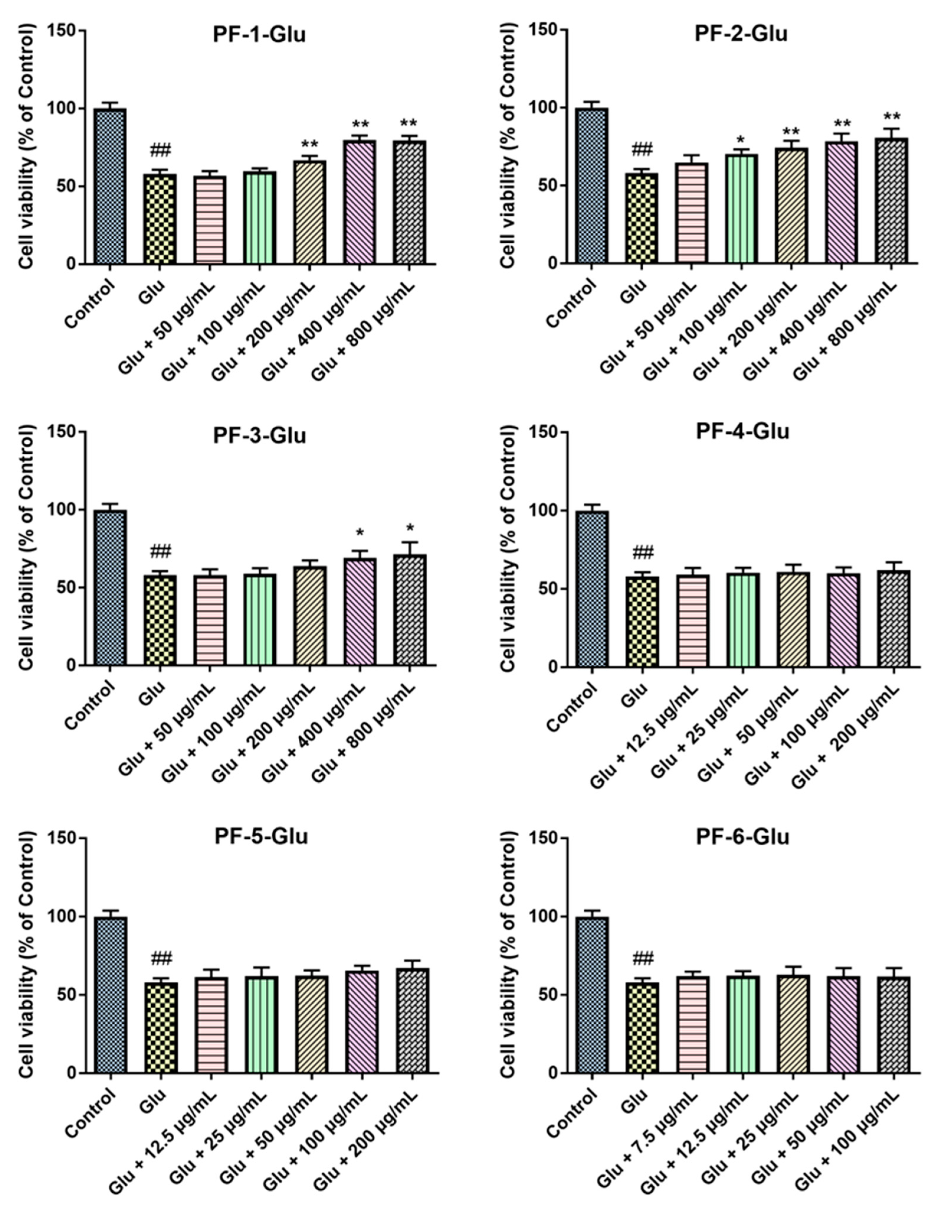
3.2. Effects of PF-1 to PF-6 on the Cell Viability of Normal and Glu-Injured PC12 Cells

The effects of PF-1 to PF-6 on the cell viability of the normal and Glu-stimulated PC12 cells were determined by MTT. As shown in Figure 2, PF-1, PF-2 and PF-3 did not show any inhibitory effects in normal PC12 cells up to a concentration of 800 μg/mL, compared to the control group. PF-4, PF-5, and PF-6 also had no inhibitory effect on the cell growth of PC12 cells at low concentrations. However, PF-4 and PF-5 showed significant inhibitory effects on the cell viability of PC12 cells at the higher concentrations of 400 and 800 μg/mL, while PF-6 affected the cell growth of PC12 cells at the concentrations of 200, 400 and 800 μg/mL.
Based on the performed experiments above, the results of the effects of the fractions on the cell viability of Glu-injured PC12 cells are shown in Figure 3. The cell viability decreased significantly after Glu treatment (p < 0.01). Importantly, PF-1 (200–800 μg/mL), PF-2 (100–800 μg/mL) and PF-3 (400 and 800 μg/mL) significantly increased the cell viability of Glu-injured PC12 cells, compared to cells treated with Glu alone (p < 0.05). PF-4, PF-5 and PF-6 did not show any protective effect on Glu-injured PC12 cells within the investigated concentration range (p > 0.05). Therefore, PF-1, PF-2 and PF-3 were selected for further experiments.

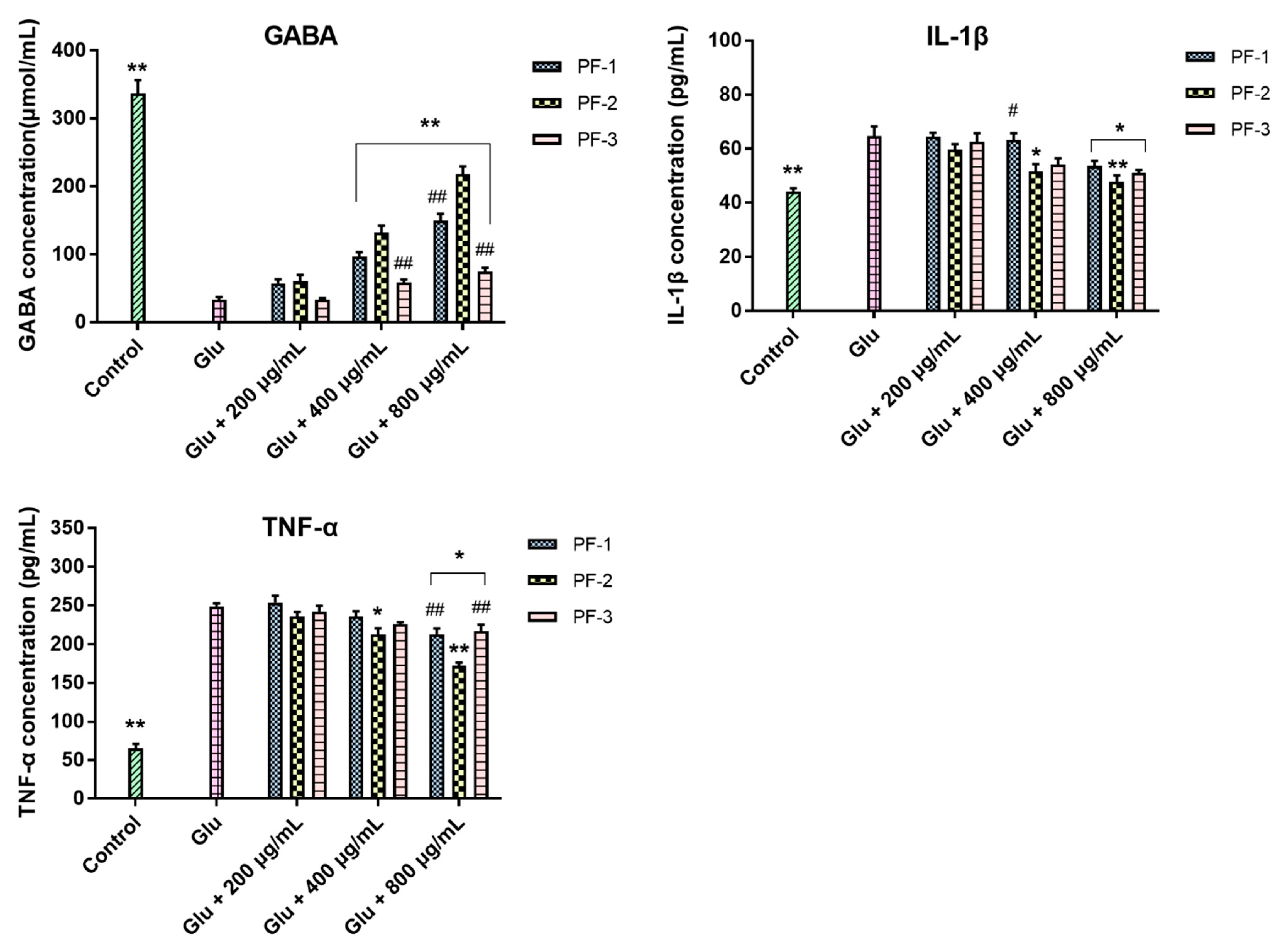
3.3. Effects of PF-1, PF-2 and PF-3 on the Levels of GABA, IL-1β, and TNF-α in Glu-Injured PC12 Cells

The effects of PF-1, PF-2 and PF-3 on the levels of GABA, IL-1β, and TNF-α in Glu-stimulated PC12 cells were further investigated. As can be seen from Figure 4, the content of GABA in the cell culture medium of Glu-stimulated PC12 cells significantly decreased, when compared with the control cells (p < 0.01). However, GABA content increased significantly with PF-1, PF-2 and PF-3 (400 and 800 μg/mL), compared to the Glu-treated group (p < 0.01). At the low concentration of 200 μg/mL, no significant difference was found among the effects of PF-1, PF-2 and PF-3 (p > 0.05). However, at the higher concentrations of 400 and 800 μg/mL, the effect of PF-2 on GABA content was significantly stronger than that of PF-1 and PF-3 at the same concentration (p < 0.01). The IL-1β and TNF-α contents in the Glu-treated group were significantly higher than that of the control group (p < 0.01). Compared with the Glu-treated group, the content of IL-1β and TNF-α in the PF-1 (800 μg/mL), PF-2 (400 and 800 μg/mL) and PF-3 (800 μg/mL) groups significantly decreased (p < 0.05). Interestingly, the effect of PF-2 on TNF-α level was also significantly stronger than that of PF-1 and PF-2 at the concentration of 800 μg/mL.
The above experimental results showed that PF-1, PF-2 and PF-3 significantly protected PC12 cells from Glu injury, which is consistent with the molecular weight distribution information of the three fractions. PF-1, PF-2 and PF-3 had similar protein compositions and showed a significant protective effect on Glu-injured PC12 cells, but the protective effect of PF-2 was stronger than that of PF-1 and PF-3. Therefore, PF-2 was selected for further experimental research.

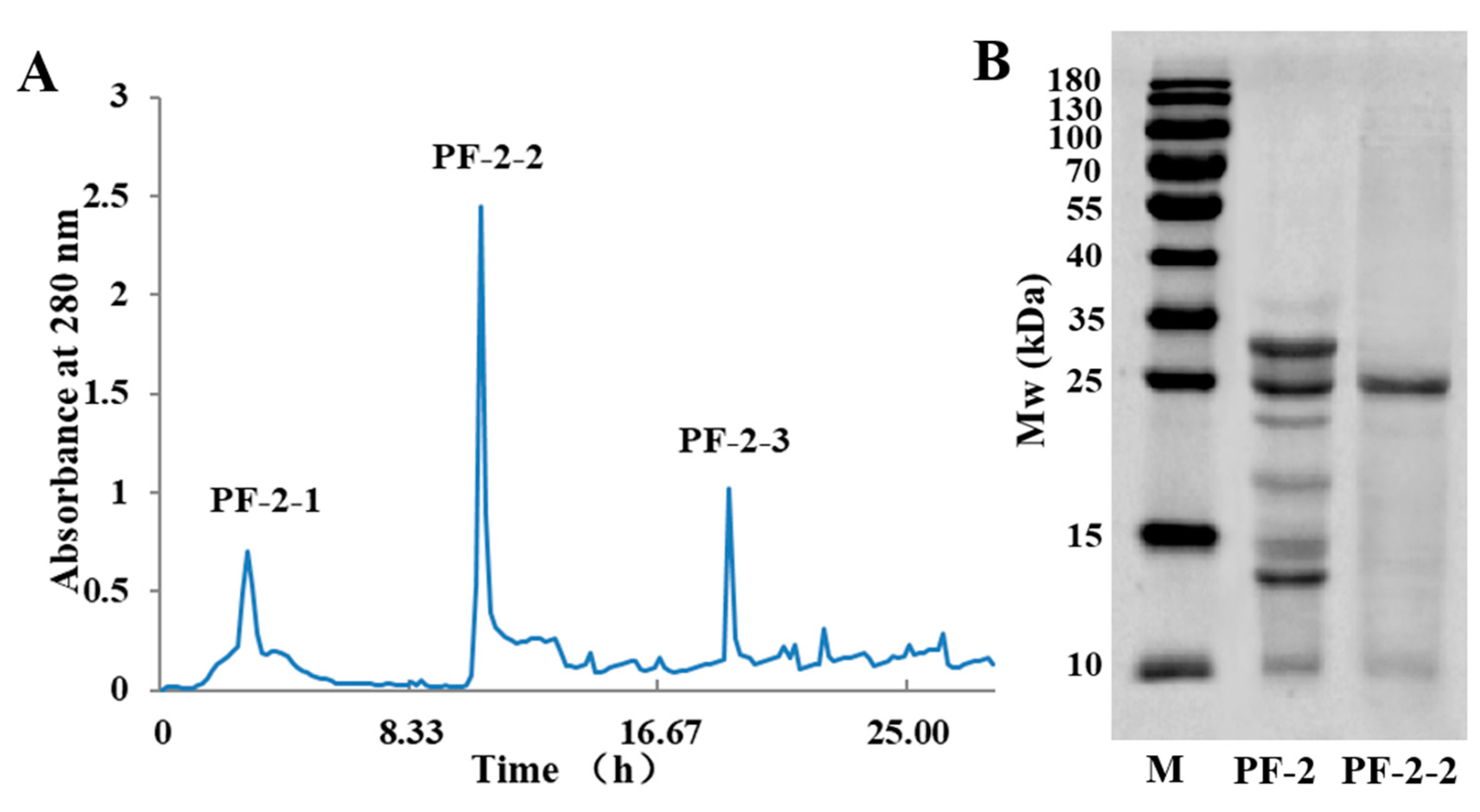
3.4. Size-Exclusion Chromatography of PF-2-2

The approximate molecular weight range of PF-2 was determined according to the results of the molecular weight determination. PF-2 was purified with Sephadex G-75 gel exclusion chromatography. Three protein fractions (PF-2-1, PF-2-2 and PF-2-3) were found, PF-2-1 and PF-2-3 were not obtained due to the small amounts present; PF-2-2 was finally prepared (Figure 5A). The result of SDS-PAGE analysis is shown in Figure 5B; a main band was found in PF-2-2 with a molecular weight of around 25 kDa. The electrophoresis lane profile, molecular weight and relative quantification are shown in Figure S1 and Table S1 (Supplemental Materials). The main band of PF-2-2 was then analyzed by MALDI-TOF/TOF-MS to identify the protein structure.

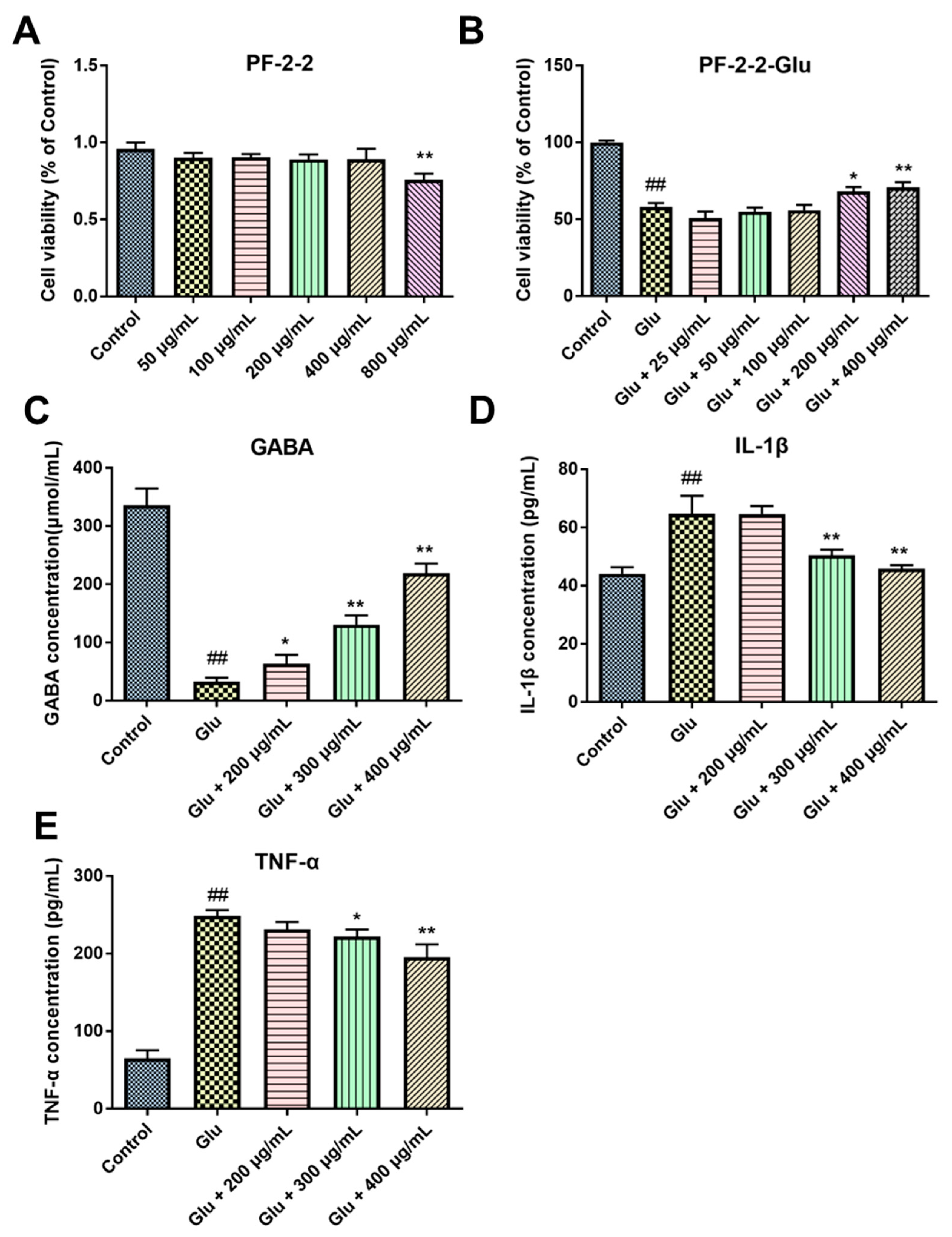
3.5. The Neuroprotective Effects of PF-2-2 in Glu-Stimulated PC12 Cells

The effect of PF-2-2 on the viability of normal PC12 cells is shown in Figure 6A. PF-2-2 inhibited the growth of PC12 cells at the concentration of 800 μg/mL, but did not show any inhibitory effect at a concentration below 400 μg/mL. The concentrations of 25 to 400 μg/mL were selected for studying the effects of PF-2-2 on the viability of PC12 cells injured by Glu. The cell growth was significantly inhibited by Glu treatment (p < 0.01), but PF-2-2 at 200 and 400 μg/mL (p < 0.05, p < 0.01) significantly increased the cell viability of Glu-treated cells (Figure 6B).
The effects of PF-2-2 (200, 300 and 400 μg/mL) on GABA, IL-1β, and TNF-α in Glu-injured PC12 cells were determined (Figure 6C–E). Compared with the control cells, the GABA content of Glu-injured PC12 cells significantly decreased (p < 0.01), while the content of IL-1β and TNF-α significantly increased (p < 0.01). Compared with the Glu-treated group, PF-2-2 significantly increased the GABA content at 200, 300 and 400 μg/mL, and decreased the content of IL-1β and TNF-α at 300 and 400 μg/mL.

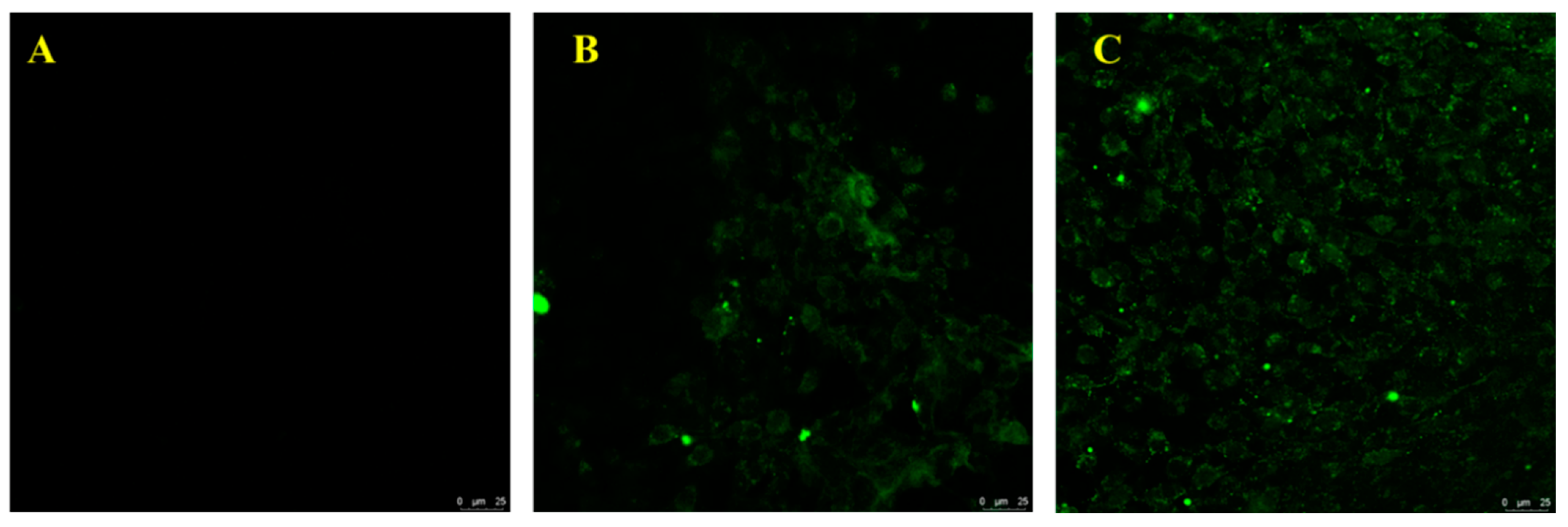
3.6. Interaction Evaluation between PF-2-2 and PC12 Cells

To confirm the binding of PF-2-2 to PC12 cells, fluorescein isothiocyanate-labeled PF-2-2 (FITC-PF-2-2) was prepared. Figure 7 shows that the binding of FITC-F-2-2 to the cells could be directly observed with the confocal microscope. The green color was the FITC-protein sites. After 6 h of incubation, the PC12 cells were labeled with the green fluorescence of FITC-PF-2-2, indicating the binding of the protein to the PC12 cell surface (Figure 7B). To assess whether PF-2-2 could be internalized into the PC12 cells, cells were observed after 24 h of incubation. As shown in Figure 7C, FITC-PF-2-2 was not only presented on the cell surface, but also internalized into the cell after 24 h. The above results indicated that PF-2-2 first bound to the surface of PC12 cells and then internalized into the cells to exert neuroprotective effects through regulating the expression of GABA, IL-1β, and TNF-α.

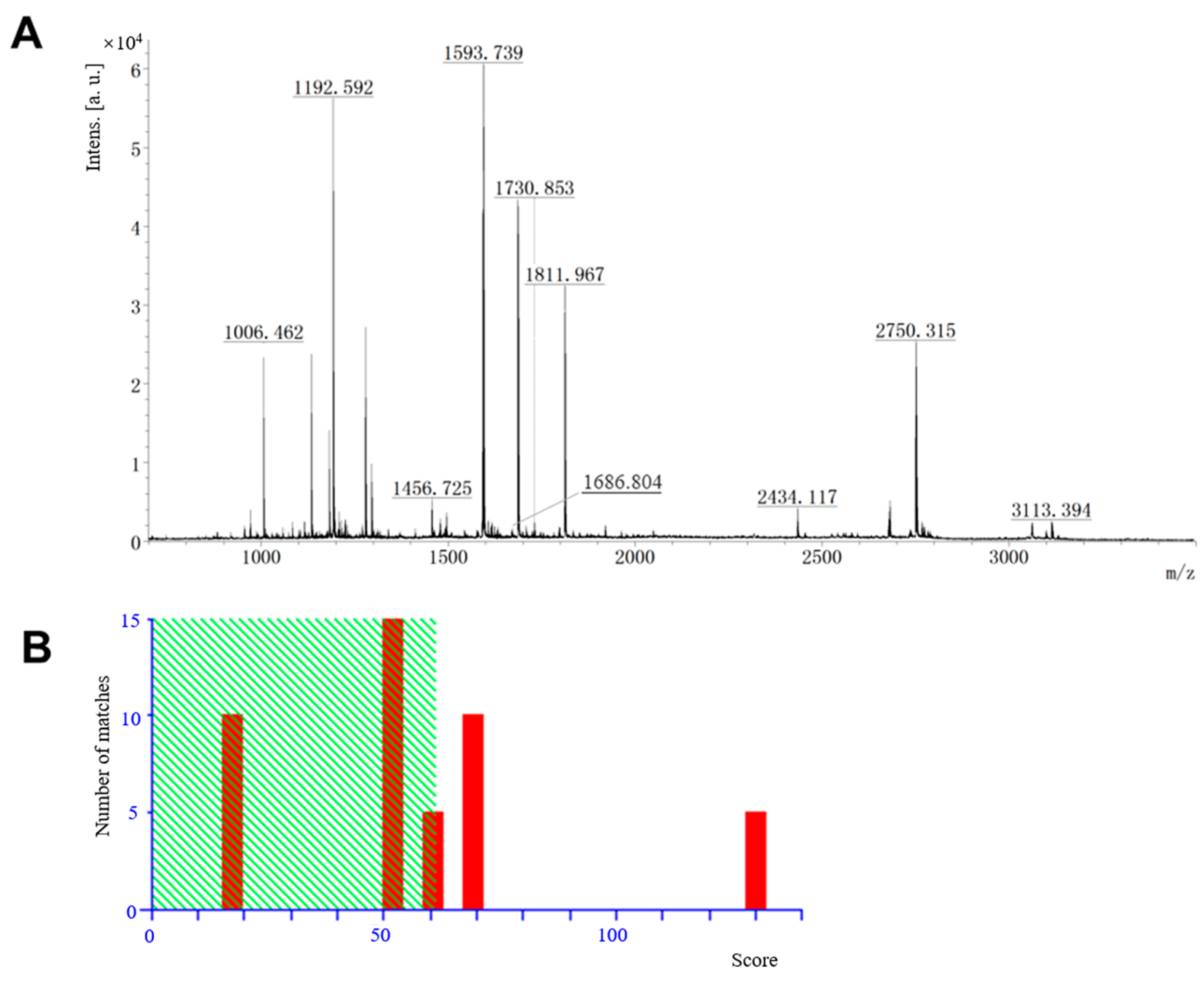
3.7. Protein Identification by MALDI-TOF/TOF-MS

A main band (around 25 kDa in Figure 5) of PF-2-2 in the gel was identified using in-gel trypsinolysis and then further analyzed using MALDI-TOF/TOF-MS. As shown in Figure 8A, 10 ions with the highest intensities were selected for sequencing by MS/MS. The analysis results were compared with previous literature using the MASCOT database. As shown in Figure 8B, there are three peptides (K.FITLWENNR.V, K.LYNSILTGDYDSAVR.Q and K.GSIIQNVVNNLIIDGSR.N) with the m/z of 1192.592, 1686.804, and 1811.967, respectively, and matches above an identity threshold for 10 queries. On average, individual ion scores > 61 (beyond green shading) indicate identity or extensive homology (p < 0.05). The individual ion scores for the three matched peptides were 68, 71, and 130, respectively. Based on the three matched peptides, a matching protein with the molecular mass of 30 335, named Low molecular mass 30 kDa lipoprotein 21G1 (UniProtKB/Swiss-Prot: Q00801.2), was identified. The amino acid sequence of the Low molecular mass 30 kDa lipoprotein 21G1 (L21G1) is shown in Table 2. The protein consisted of 263 amino acids, and was a member of the 30 kDa protein family. Other members of this protein family have been reported to have a similar amino acid composition and anti-apoptotic activity [30,31]. Our study found that L21GA was the main protein in PF-2-2, indicating a close relationship between the neuroprotective effects in the Glu-stimulated PC12 cells of PF-2-2 and the protein L21G1.

4. Discussion

Protein and peptides isolated from animals and plants have been reported to possess significant activities, and it is an important way to discover new drugs. There are many fractionation methods available for peptides and proteins, usually using multidimensional methods or system combinations. A variety of chromatographic separation methods can be used for proteins, including ion exchange, size exclusion, reversed-phase chromatography, [32], etc. Anion-exchange chromatography is a method for separating compounds according to their charges using ion-exchange resins containing positively charged groups [33]. Gel filtration chromatography is a widely used method for the separation of proteins and peptides based upon molecular size. In gel filtration chromatography, separation is based on molecular weight (MW). Substances with a larger MW elute at an early stage, while components with a smaller MW elute at a later stage [34]. In this study, anion-exchange chromatography (DEAE-52 Sepharose FF) and gel filtration chromatography (Sephadex G-75) were used for the separation of proteins from BB. As a result, six subfractions were obtained by anion-exchange chromatography, and the most active fraction (PF-2) was further separated by gel filtration chromatography to obtain an effective protein named PF-2-2.
Glutamate (Glu) is a neurotransmitter which releases synaptic information in the brain [35]. Pathophysiological researches have shown that Glu can induce brain neurotoxicity and then cause cerebral ischemic injury [36]. Glu could significantly induce brain cell damage by reducing cell viability, accelerating autophagy and apoptosis [37]. The PC12 cell model by Glu damage has been widely used in the research of nervous system diseases such as epilepsy [19,37]. In the central nervous system (CNS) of mammals, GABA is the main brain inhibitory neurotransmitter, which should be balanced with Glu under normal circumstances [38]. These two neurotransmitters can ensure normal neural circuit function, and the interruption of the neurotransmitter GABA/Glu balance is related to the pathophysiology of epilepsy [39]. Furthermore, neuroinflammation is related to changes in neuronal injury and the electronic encephalography of epileptic activity. Proinflammatory cytokines, including IL-1β and TNF-α, can regulate the pathophysiological processes of epilepsy [40]. Studies have shown that during seizure in rats, IL-1β and TNF-α increased in the hippocampus, amygdala, and parietal cortex [41]. In the present study, a Glu-injured PC12 cell model was employed to evaluate the neuroprotective effect of isolated proteins. The results showed that PF-1, PF-2, PF-3 (separated by ion-exchange chromatography) and PF-2-2 (separated by size-exclusion chromatography) possessed potential neuroprotective effects by significantly increasing the GABA level, and decreasing the levels of IL-1β and TNF-α.
With the development of mass spectrometry technology, coupled with the availability of a wide range of protein sequence databases and software tools for data mining, rapid and sensitive protein identification based on mass spectrometry has become possible [42]. There are two main ionization technologies for biomolecular mass spectrometry, including MALDI and electro spray ionization (ESI) [43]. Although these two techniques have successfully analyzed large biopolymers, peptide mass fingerprinting (PMF) using MALDI-TOF-MS is still the main method of protein identification [44]. The principle is that the digestion of a protein with a specific protease will produce a peptide mixture specific to the protein. The MW of these peptides is measured, and then a characteristic data set (PMF) is obtained. The PMF data can then be compared with the theoretical peptide MW, which is generated by digesting each protein in the sequence database with the same protease, in order to find the best match. If the database used for the search includes the protein being analyzed, and the data quality is good enough, then the correct protein can be matched. In order to evaluate the effectiveness of this method in identifying proteins, some methods to score the matching quality should be used [42,45]. In this study, the main band of PF-2-2 was analyzed by MALDI-TOF/TOF-MS, and the data were searched in the MASCOT database. Peptide ion scores greater than 61 were used to match proteins, and a protein named Low molecular mass 30 kDa lipoprotein 21G1 was finally identified.
Low molecular weight 30 kDa lipoprotein 21G1 belongs to the structure related to “30 kDa proteins” and constitutes the main protein component of the fifth (and last) instar larvae and pupae [46]. The 30 kDa protein family has a similar immunological activity and amino acid composition. So far, many proteins of this family have been found [31,47,48]. Previous studies have shown that the 30 kDa lipoprotein binds to glucose and glucan, through β-glucan binding, and causes the invading fungi to gather and trigger an immune response in the blood, indicating that they participate in the insect’s self-defense system [49]. In 2001, Kim et al. [50] isolated four 30 kDa anti-apoptotic components from the silkworm hemolymph and the component (28 kDa) with the strongest anti-apoptotic activity was purified. Recombinant 30 kDa proteins had comparable anti-apoptotic activity with the whole hemolymph of the silkworm [30]. The isolated protein PF-2-2, which was identified to be low molecular weight 30 kDa lipoprotein 21G1, showed a protective effect in Glu-injured PC12 cells, which may be related to its anti-apoptotic activity, but further research is needed.
In the present study, neuroprotective proteins from BB were separated and purified, and the main band of PF-2-2 fraction with the strongest activity was identified by MALDI-TOF/TOF-MS. However, further investigations on BBPs can be carried out in the future: Firstly, more proteins can be isolated and purified in the future, especially from the components which have been proven to be active (PF-1, PF-2 and PF-3), and the structures can be identified. Two-dimensional difference gel electrophoresis (2D-DIGE), semi-preparative RP-HPLC separation and protein synthesis, etc., can be used for further research. Secondly, the neuroprotective effects of the obtained protein were preliminarily studied in Glu-stimulated PC12 cells, but its in-depth mechanisms need to be further explored by using various cell and animal models, and the signaling pathways can be clarified from the aspect of anti-apoptosis based on the results of this study. Finally, the binding of the obtained protein to PC12 cells was preliminarily proven in this research; the binding site and action pathway need to be further studied to clarify the mechanisms.

5. Conclusions

In summary, ion-exchange chromatography (DEAE-52 Sepharose FF) and gel filtration chromatography (Sephadex G-75) were used to purify proteins from BBPs. PF-1, PF-2, PF-3, and PF-2-2 (from PF-2) were found to possess neuroprotective effects by significantly increasing the GABA level, and decreasing the levels of IL-1β and TNF-α. FITC-labeled F-2-2 was found to be successfully bound to the PC12 cells under a confocal microscope; PF-2-2 was found to first bind to the surface of PC12 cells and then internalize into the cells. The main band of PF-2-2 was identified to be Low molecular mass 30 kDa lipoprotein 21G1 by MALDI-TOF/TOF-MS. PF-2-2 and purified proteins isolated from BBPs have potential application prospects in the field of epilepsy treatment.

Supplementary Materials

The following supporting information can be downloaded at: https://www.mdpi.com/article/10.3390/separations9090236/s1, Figure S1: Electrophoresis lane profile for Markers, PF-2, and PF-2-2.; Table S1: The molecular weight and relative quantification of Markers, PF-2, and PF-2-2.

Author Contributions

Writing—original draft preparation, M.-B.H. and Y.-J.L.; methodology, S.-S.Z.; software, X.-L.M.; validation, C.-J.W.; investigation, M.-B.H., X.-L.M., P.W. and Y.-J.L.; writing—review and editing, Y.-J.L.; funding acquisition, M.-B.H. All authors have read and agreed to the published version of the manuscript.

Funding

This research was funded by the National Natural Science Foundation of China (No. 82104397), Basic Research Planned Project of Shanxi Provincial Department of Science and Technology (No. 20210302124630), Science and Technology Innovation Planned Project of Colleges and Universities in Shanxi Province (No. 2021L357), Doctoral Research Startup Fund Project of Shanxi University of Chinese Medicine (No. 2022BK07), and Scientific Research Awarding and Start-up Fund Project of Outstanding Doctoral Graduates to work in Shanxi of Shanxi University of Chinese medicine (No. 2022BKS04).

Institutional Review Board Statement

Not applicable.

Informed Consent Statement

Not applicable.

Data Availability Statement

Not applicable.

Acknowledgments

We thank the XueYouTang Laboratory in School of Pharmacy, Chengdu University of Traditional Chinese Medicine for the help in the experiments.

Conflicts of Interest

The authors declare no conflict of interest.

References

  1. Loscher, W.; Potschka, H.; Sisodiya, S.M.; Vezzani, A. Drug Resistance in Epilepsy: Clinical Impact, Potential Mechanisms, and New Innovative Treatment Options. Pharmacol. Rev. 2020, 72, 606–638. [Google Scholar] [CrossRef] [PubMed]
  2. Patel, D.C.; Tewari, B.P.; Chaunsali, L.; Sontheimer, H. Neuron-glia interactions in the pathophysiology of epilepsy. Nat. Rev. Neurosci. 2019, 20, 282–297. [Google Scholar] [CrossRef] [PubMed]
  3. Akyuz, E.; Paudel, Y.N.; Polat, A.K.; Dundar, H.E.; Angelopoulou, E. Enlightening the neuroprotective effect of quercetin in epilepsy: From mechanism to therapeutic opportunities. Epilepsy Behav. 2021, 115, 107701. [Google Scholar] [CrossRef] [PubMed]
  4. Paudel, Y.N.; Shaikh, M.F.; Shah, S.; Kumari, Y.; Othman, I. Role of inflammation in epilepsy and neurobehavioral comorbidities: Implication for therapy. Eur. J. Pharmacol. 2018, 837, 145–155. [Google Scholar] [CrossRef] [PubMed]
  5. Nandini, H.S.; Paudel, Y.N.; Krishna, K.L. Envisioning the neuroprotective effect of Metformin in experimental epilepsy: A portrait of molecular crosstalk. Life Sci. 2019, 233, 116686. [Google Scholar]
  6. Yang, N.; Guan, Q.W.; Chen, F.H.; Xia, Q.X.; Yin, X.X.; Zhou, H.H.; Mao, X.Y. Antioxidants Targeting Mitochondrial Oxidative Stress: Promising Neuroprotectants for Epilepsy. Oxid. Med. Cell Longev. 2020, 2020, 6687185. [Google Scholar] [CrossRef]
  7. Sanz, P.; Garcia-Gimeno, M.A. Reactive Glia Inflammatory Signaling Pathways and Epilepsy. Int. J. Mol. Sci. 2020, 21, 4096. [Google Scholar] [CrossRef]
  8. Perucca, P.; Perucca, E. Identifying mutations in epilepsy genes: Impact on treatment selection. Epilepsy Res. 2019, 152, 18–30. [Google Scholar] [CrossRef]
  9. Steriade, C.; French, J.; Devinsky, O. Epilepsy: Key experimental therapeutics in early clinical development. Expert Opin. Investig. Drugs 2020, 29, 373–383. [Google Scholar] [CrossRef]
  10. Beghi, E.; Beghi, M. Epilepsy, antiepileptic drugs and dementia. Curr. Opin. Neurol. 2020, 33, 191–197. [Google Scholar] [CrossRef]
  11. Bai, Y.; Zhao, Q.; He, M.; Ye, X.; Zhang, X. Extensive characterization and differential analysis of endogenous peptides from Bombyx batryticatus using mass spectrometric approach. J. Pharm. Biomed. Anal. 2019, 163, 78–87. [Google Scholar] [CrossRef] [PubMed]
  12. Bhat, Z.F.; Kumar, S.; Bhat, H.F. Antihypertensive peptides of animal origin: A review. Crit. Rev. Food Sci. Nutr. 2017, 57, 566–578. [Google Scholar] [CrossRef] [PubMed]
  13. Chen, H.; Cheng, S.; Fan, F.; Tu, M.; Xu, Z.; Du, M. Identification and molecular mechanism of antithrombotic peptides from oyster proteins released in simulated gastro-intestinal digestion. Food Funct. 2019, 10, 5426–5435. [Google Scholar] [CrossRef] [PubMed]
  14. Bi, J.; Tian, C.; Jiang, J.; Zhang, G.L.; Hao, H.; Hou, H.M. Antibacterial Activity and Potential Application in Food Packaging of Peptides Derived from Turbot Viscera Hydrolysate. J. Agric. Food Chem. 2020, 68, 9968–9977. [Google Scholar] [CrossRef]
  15. Kachel, H.S.; Buckingham, S.D.; Sattelle, D.B. Insect toxins—Selective pharmacological tools and drug/chemical leads. Curr. Opin. Insect Sci. 2018, 30, 93–98. [Google Scholar] [CrossRef]
  16. Lin, L.; Zhang, Y.; Li, Y.; Fu, H.; Hu, J.; Zhou, Y.; Xu, Y.; Xia, G.; Sun, X.; Yang, H.; et al. Identification of signature proteins of processed Bombyx batryticatus by comparative proteomic analysis. Int. J. Biol. Macromol. 2020, 153, 289–296. [Google Scholar] [CrossRef]
  17. Song, H.Y.; Han, J.M.; Byun, E.H.; Kim, W.S.; Seo, H.S.; Byun, E.B. Bombyx batryticatus Protein-Rich Extract Induces Maturation of Dendritic Cells and Th1 Polarization: A Potential Immunological Adjuvant for Cancer Vaccine. Molecules 2021, 26, 476. [Google Scholar] [CrossRef] [PubMed]
  18. Wu, J.Y.; Sheikho, A.; Ma, H.; Li, T.C.; Zhao, Y.Q.; Zhang, Y.L.; Wang, D. Molecular mechanisms of Bombyx batryticatus ethanol extract inducing gastric cancer SGC-7901 cells apoptosis. Cytotechnology 2017, 69, 875–883. [Google Scholar] [CrossRef]
  19. He, L.Y.; Hu, M.B.; Li, R.L.; Zhao, R.; Fan, L.H.; Wang, L.; Peng, W.; Liu, Y.J.; Wu, C.J. The Effect of Protein-Rich Extract from Bombyx batryticatus against Glutamate-Damaged PC12 Cells Via Regulating gamma-Aminobutyric Acid Signaling Pathway. Molecules 2020, 25, 553. [Google Scholar] [CrossRef]
  20. Hu, M.; Liu, Y.; He, L.; Yuan, X.; Peng, W.; Wu, C. Antiepileptic Effects of Protein-Rich Extract from Bombyx batryticatus on Mice and Its Protective Effects against H2O2-Induced Oxidative Damage in PC12 Cells via Regulating PI3K/Akt Signaling Pathways. Oxid. Med. Cell Longev. 2019, 2019, 7897584. [Google Scholar] [CrossRef]
  21. Xing, D.; Shen, G.; Li, Q.; Xiao, Y.; Yang, Q.; Xia, Q. Quality Formation Mechanism of Stiff Silkworm, Bombyx batryticatus Using UPLC-Q-TOF-MS-Based Metabolomics. Molecules 2019, 24, 3780. [Google Scholar] [CrossRef] [PubMed] [Green Version]
  22. Hu, M.; Yu, Z.; Wang, J.; Fan, W.; Liu, Y.; Li, J.; Xiao, H.; Li, Y.; Peng, W.; Wu, C. Traditional Uses, Origins, Chemistry and Pharmacology of Bombyx batryticatus: A Review. Molecules 2017, 22, 1779. [Google Scholar] [CrossRef]
  23. Wang, H.; Xu, L.; Dou, Y.; Wang, J. Bombyx batryticatus Cocoonase Inhibitor Separation, Purification, and Inhibitory Effect on the Proliferation of SMCC-7721 HeLa-Derived Cells. Evid. Based Complement. Alternat. Med. 2022, 2022, 4064829. [Google Scholar] [CrossRef] [PubMed]
  24. Wu, W.P.; Cao, J.; Wu, J.Y.; Chen, H.; Wang, D. Anticancer activity of Bombyx batryticatus ethanol extract against the human tumor cell line HeLa. Genet. Mol. Res. 2015, 14, 79–88. [Google Scholar] [CrossRef]
  25. Lim, H.S.; Kim, J.S.; Moon, B.C.; Ryu, S.M.; Lee, J.; Park, G. Batryticatus Bombyx Protects Dopaminergic Neurons Against MPTP-Induced Neurotoxicity by Inhibiting Oxidative Damage. Antioxidants 2019, 8, 574. [Google Scholar] [CrossRef] [PubMed]
  26. Wang, P.; Shang, E.; Fan, X. Effect of San’ao decoction with scorpio and Bombyx batryticatus on CVA mice model via airway inflammation and regulation of TRPA1/TRPV1/TRPV5 channels. J. Ethnopharmacol. 2021, 264, 113342. [Google Scholar] [CrossRef]
  27. Liu, Z.; Shi, Y.; Liu, H.; Jia, Q.; Liu, Q.; Tu, J. Purification and Identification of Pine Nut (Pinus yunnanensis Franch.) Protein Hydrolysate and Its Antioxidant Activity in Vitro and in Vivo. Chem. Biodivers. 2021, 18, e2000710. [Google Scholar] [CrossRef]
  28. Zhao, H.; Jin, H.; Li, Q.; Gu, Q.; Zheng, Z.; Wu, H.; Ye, S.; Sun, X.; Xu, X.; Ho, P.C. Inhibition of pathologic retinal neovascularization by a small peptide derived from human apolipoprotein(a). Investig. Ophthalmol. Vis. Sci. 2009, 50, 5384–5395. [Google Scholar] [CrossRef]
  29. Katayama, H.; Nagasu, T.; Oda, Y. Improvement of in-gel digestion protocol for peptide mass fingerprinting by matrix-assisted laser desorption/ionization time-of-flight mass spectrometry. Rapid Commun. Mass Spectrom 2001, 15, 1416–1421. [Google Scholar] [CrossRef]
  30. Pakkianathan, B.C.; Singh, N.K.; Konig, S.; Krishnan, M. Antiapoptotic activity of 30 kDa lipoproteins family from fat body tissue of silkworm, Bombyx mori. Insect Sci. 2015, 22, 629–638. [Google Scholar] [CrossRef]
  31. Pietrzyk, A.J.; Bujacz, A.; Lochynska, M.; Jaskolski, M.; Bujacz, G. Crystal structure of Bombyx mori lipoprotein 6: Comparative structural analysis of the 30-kDa lipoprotein family. PLoS ONE 2014, 9, e108761. [Google Scholar] [CrossRef] [PubMed] [Green Version]
  32. Wang, J.; Shimada, M.; Nagaoka, S. Identification of the active protein in rice bran protein having an inhibitory activity of cholesterol micellar solubility. Biosci. Biotechnol. Biochem. 2017, 81, 1216–1219. [Google Scholar] [CrossRef] [PubMed]
  33. Zhang, S.Y.; Zhao, G.X.; Suo, S.K.; Wang, Y.M.; Chi, C.F.; Wang, B. Purification, Identification, Activity Evaluation, and Stability of Antioxidant Peptides from Alcalase Hydrolysate of Antarctic Krill (Euphausia superba) Proteins. Mar. Drugs 2021, 19, 347. [Google Scholar] [CrossRef] [PubMed]
  34. Xia, J.; Song, H.; Huang, K.; Li, S.; Guan, X. Purification and characterization of antioxidant peptides from enzymatic hydrolysate of mungbean protein. J. Food Sci. 2020, 85, 1735–1741. [Google Scholar] [CrossRef]
  35. Yu, L.J.; Wall, B.A.; Wangari-Talbot, J.; Chen, S. Metabotropic glutamate receptors in cancer. Neuropharmacology 2017, 115, 193–202. [Google Scholar] [CrossRef]
  36. Mayor, D.; Tymianski, M. Neurotransmitters in the mediation of cerebral ischemic injury. Neuropharmacology 2018, 134, 178–188. [Google Scholar] [CrossRef]
  37. Yu, S.; Xin, W.; Jiang, Q.; Li, A. Propofol exerts neuroprotective functions by down-regulating microRNA-19a in glutamic acid-induced PC12 cells. Biofactors 2020, 46, 934–942. [Google Scholar] [CrossRef]
  38. Sarlo, G.L.; Holton, K.F. Brain concentrations of glutamate and GABA in human epilepsy: A review. Seizure 2021, 91, 213–227. [Google Scholar] [CrossRef]
  39. Guo, M.; Cui, C.; Song, X.; Jia, L.; Li, D.; Wang, X.; Dong, H.; Ma, Y.; Liu, Y.; Cui, Z.; et al. Deletion of FGF9 in GABAergic neurons causes epilepsy. Cell Death Dis. 2021, 12, 196. [Google Scholar] [CrossRef]
  40. Zhang, W.; Wang, L.; Pang, X.; Zhang, J.; Guan, Y. Role of microRNA-155 in modifying neuroinflammation and gamma-aminobutyric acid transporters in specific central regions after post-ischaemic seizures. J. Cell Mol. Med. 2019, 23, 5017–5024. [Google Scholar] [CrossRef]
  41. Su, J.; Yin, J.; Qin, W.; Sha, S.; Xu, J.; Jiang, C. Role for pro-inflammatory cytokines in regulating expression of GABA transporter type 1 and 3 in specific brain regions of kainic acid-induced status epilepticus. Neurochem. Res. 2015, 40, 621–627. [Google Scholar] [CrossRef] [PubMed]
  42. Webster, J.; Oxley, D. Protein identification by MALDI-TOF mass spectrometry. Methods Mol. Biol. 2012, 800, 227–240. [Google Scholar] [PubMed]
  43. Gogichaeva, N.V.; Alterman, M.A. Amino Acid Analysis by Means of MALDI TOF Mass Spectrometry or MALDI TOF/TOF Tandem Mass Spectrometry. Methods Mol. Biol. 2019, 2030, 17–31. [Google Scholar] [PubMed]
  44. Cakir, B.; Gulseren, I. Identification of Novel Proteins from Black Cumin Seed Meals Based on 2D Gel Electrophoresis and MALDI-TOF/TOF-MS Analysis. Plant Foods Hum. Nutr. 2019, 74, 414–420. [Google Scholar] [CrossRef]
  45. Weng, C.Y.; Wang, C.E.; Xie, W.B.; Xu, S.Y.; Wang, Y.J.; Zheng, Y.G. Comparative proteome analysis of Actinoplanes utahensis grown on various saccharides based on 2D-DIGE and MALDI-TOF/TOF-MS. J. Proteom. 2021, 239, 104193. [Google Scholar] [CrossRef]
  46. Shi, X.F.; Li, Y.N.; Yi, Y.Z.; Xiao, X.G.; Zhang, Z.F. Identification and Characterization of 30 K Protein Genes Found in Bombyx mori (Lepidoptera: Bombycidae) Transcriptome. J. Insect Sci. 2015, 15, 71. [Google Scholar] [CrossRef]
  47. Pakkianathan, B.C.; Singh, N.K.; Krishnan, M.; Konig, S. A proteomic view on the developmental transfer of homologous 30 kDa lipoproteins from peripheral fat body to perivisceral fat body via hemolymph in silkworm, Bombyx mori. BMC. Biochem. 2012, 13, 5. [Google Scholar] [CrossRef]
  48. Yang, J.P.; Ma, X.X.; He, Y.X.; Li, W.F.; Kang, Y.; Bao, R.; Chen, Y.; Zhou, C.Z. Crystal structure of the 30 K protein from the silkworm Bombyx mori reveals a new member of the beta-trefoil superfamily. J. Struct. Biol. 2011, 175, 97–103. [Google Scholar] [CrossRef]
  49. Ujita, M.; Kimura, A.; Nishino, D.; Yokoyama, E.; Banno, Y.; Fujii, H.; Hara, A. Specific binding of silkworm Bombyx mori 30-kDa lipoproteins to carbohydrates containing glucose. Biosci. Biotechnol. Biochem. 2002, 66, 2264–2266. [Google Scholar] [CrossRef]
  50. Kim, E.J.; Rhee, W.J.; Park, T.H. Isolation and characterization of an apoptosis-inhibiting component from the hemolymph of Bombyx mori. Biochem. Biophys. Res. Commun. 2001, 285, 224–228. [Google Scholar] [CrossRef]
Figure 1. DEAE-52 Sepharose FF chromatographic diagram (A), SDS-PAGE analysis of six subfractions (B), and electrophoresis lane profile for each subfraction (C). BBPs: Total proteins from Bombyx batryticatus; M: Protein Markers.
Separations 09 00236 g001
Figure 2. Effects of PF-1 to PF-6 on the cell viability of normal PC12 cells. Cells were treated with different fractions at concentrations of 0 to 800 μg/mL for 24 h. The cell viability was determined by MTT. ** p < 0.01 vs. the control group.
Separations 09 00236 g002
Figure 3. Effects of PF-1 to PF-6 on the cell viability of Glu-stimulated PC12 cells. Cells were treated with different fractions at corresponding concentrations for 24 h, and then Glu (the final concentration of 20 mM) was added for 24 h. The cell viability was determined by MTT. ## p < 0.01 vs. the control group, * p < 0.05 vs. the Glu-treated group, ** p < 0.01 vs. the Glu-treated group.
Separations 09 00236 g003
Figure 4. Effects of PF-1, PF-2 and PF-3 on the GABA, IL-1β, and TNF-α level in Glu-injured PC12 Cells. Cells were treated with corresponding fractions at concentrations of 200, 400 and 800 μg/mL for 24 h, subsequently with Glu (the final concentration of 20 mM) for another 24 h. # p < 0.05 vs. the PF-2 group at the same concentration, ## p < 0.01 vs. the PF-2 group at the same concentration, * p < 0.05 vs. the Glu-treated group, ** p < 0.01 vs. the Glu-treated group.
Separations 09 00236 g004
Figure 5. Sephadex G-75 chromatographic diagram (A) and SDS-PAGE analysis of PF-2-2 (B). M: Protein Markers.
Separations 09 00236 g005
Figure 6. Effects of PF-2-2 on the cell viability of normal (A) and Glu-injured (B) PC12 cells. Effects of PF-2-2 on levels of GABA (C), IL-1β (D), and TNF-α (E) in Glu-injured PC12 cells. ## p < 0.01 vs. the control group, * p < 0.05 vs. the Glu-treated group, ** p < 0.01 vs. the Glu-treated group.
Separations 09 00236 g006
Figure 7. Determination of binding in PC12 cells with PF-2-2 at 0 h (A), 6 h (B), and 24 h (C). The binding site was observed using a confocal microscope at the magnification, the scale is 25 μm.
Separations 09 00236 g007
Figure 8. Mass spectrogram of PF-2-2 by MALDI-TOF/TOF-MS (A) and peptide score distribution (B). Ion score is −10log(P), where P is the probability of the observed match (a random event). There are 3 peptide matches above identity threshold and 5 matches above homology threshold for 10 queries. Individual ions scores of peptides > 61 (beyond green shading) indicate identity or extensive homology (p < 0.05).
Separations 09 00236 g008
Table 1. The molecular weight and relative quantification of Markers, BBPs, PF-1, PF-2, PF-3, PF-4, PF-5 and PF-6.
Band No.12345678910
MarkerMW (kDa)18013010070554035251510
RQ0.220.370.680.880.990.620.820.661.001.04
BBPsMW (kDa)95.64 57.83 36.51 32.24 27.14 19.1715.0013.88 10.8110.00
RQ0.01 0.01 0.01 0.14 0.43 0.010.070.02 0.290.19
PF-1MW (kDa)36.6331.7226.9218.7016.1714.7313.6910.7610.00
RQ0.020.070.340.050.060.170.022.190.05
PF-2MW (kDa)35.6930.1924.8820.8818.3314.7313.3910.71
RQ0.010.570.470.090.150.110.210.47
PF-3MW (kDa)87.4835.3429.9524.6318.2414.3313.3210.5110.00
RQ0.020.100.130.370.070.080.010.610.03
PF-4MW (kDa)86.1946.4434.1528.9825.2118.0510.6610.00
RQ0.010.030.030.090.090.031.640.01
PF-5MW (kDa)66.5751.8134.4328.9825.0017.5215.0010.3710.00
RQ0.020.260.020.090.080.020.010.830.06
PF-6MW (kDa)65.2524.7510.1810.00
RQ0.010.051.670.10
MW: Molecular weight; RQ: Relative quantification. The RQ was performed relative to Marker 9 (15 KDa) using Image Lab software (Bio-Rad, 6.0.1, Hercules, CA, USA).
Table 2. Amino acid sequence of Low molecular mass 30 kDa lipoprotein 21G1.
Protein NameAmino Acid Sequence
Low molecular mass 30 kDa lipoprotein 21G11 MKFLVVFASC VLAVSAGVTE MSAGSMSSSN KELEEKLYNS ILTGDYDSAV RQSLEYENQG
61 KGSIIQNVVN NLIIDKSRNT MEYCYKLWVG NGQHIVRKYF PYNFRLIMAG NFVKLIYRNY
121 NLALKLGPTL DPANERLAYG DGKEKNSDLI SWKFITLWEN NRVYFKIHNT KYNQYLKLSS
181 TTDCNTQDRV IFGTNTADTT REQWFLQPTK YENDVLFFIY NREYNDALKL GRIVDASGDR
241 MAFGHDGEVA GLPDIFSWFV TPF
Publisher’s Note: MDPI stays neutral with regard to jurisdictional claims in published maps and institutional affiliations.

